Next Issue
Volume 10, March
Previous Issue
Volume 10, January


Nanomaterials, Volume 10, Issue 2 (February 2020) – 219 articles

Cover Story (view full-size image): 
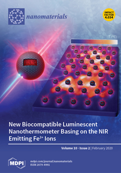
Luminescent thermometry is a contactless and noninvasive method of thermal imaging, basing on thermally affected spectroscopic properties of phosphors. A new approach, aiming to enhance the accuracy of temperature readouts relies on the implementation of strong thermal susceptibility of transition metal ions luminescence. In this paper, for the first time Fe3+-based nanocrystalline luminescent thermometer is shown. The biocompatible LiAl5O8:Fe3+ and LiAl5O8:Fe3+, Nd3+ nanocrystals exhibit intense red emissions of Fe3+ ions, located in the spectral range of 1st biological optical window, which undergo the thermal quenching in -150oC-300oC temperature range. View this paper.
  • Issues are regarded as officially published after their release is announced to the table of contents alert mailing list.
  • You may sign up for e-mail alerts to receive table of contents of newly released issues.
  • PDF is the official format for papers published in both, html and pdf forms. To view the papers in pdf format, click on the "PDF Full-text" link, and use the free Adobe Reader to open them.
Order results
Result details
Section
Select all
Export citation of selected articles as:
15 pages, 57106 KiB  
Article
Safe Administration of Carbon Nanotubes by Intravenous Pathway in BALB/c Mice
by José Jesús Guzmán-Mendoza, Silvia Lorena Montes-Fonseca, Ernesto Ramos-Martínez, Carmen González-Horta, Pilar del Carmen Hernández-Rodríguez, Erasmo Orrantia-Borunda, David Chávez-Flores and Blanca Sánchez-Ramírez
Nanomaterials 2020, 10(2), 400; https://doi.org/10.3390/nano10020400 - 24 Feb 2020
Cited by 8 | Viewed by 3441
Abstract
Carbon nanotubes (CNTs) are nanomaterials with multiple possible uses as drug carriers or in nanovaccine development. However, the toxicity of CNTs administered intravenously in in vivo models has not been fully described to date. This work aimed to evaluate the toxic effect of [...] Read more.
Carbon nanotubes (CNTs) are nanomaterials with multiple possible uses as drug carriers or in nanovaccine development. However, the toxicity of CNTs administered intravenously in in vivo models has not been fully described to date. This work aimed to evaluate the toxic effect of pristine multi-walled CNTs (UP-CNTs), purified (P-CNTs), or CNTs functionalized with fluorescein isothiocyanate (FITC-CNTs) administered by intravenous injection in BALB/c mice. Biochemical and histopathological parameters were analyzed at 1, 14, 29, and 60 days post-exposure. Pristine CNTs were the most toxic nanoparticles in comparison with P-CNTs or FITC-CNTs, increasing serum AST (≈ 180%), ALT (≈ 300%), and LDH (≈ 200%) levels at one day post-exposure. The urea/creatinine ratio suggested pre-renal injury at the 14th day accompanied of extensive lesions in kidneys, lungs, and liver. Biochemical and histological findings in mice exposed to P-CNTs had not significant differences compared to the controls. A lower toxic effect was detected in animals exposed to FITC-CNTs which was attributable to FITC toxicity. These results demonstrate that the purification process of CNTs reduces in vivo toxicity, and that toxicity in functionalized CNTs is dependent on the functionalized compound. Therefore, P-CNTs are postulated as potential candidates for safe biomedical applications using an intravenous pathway. Full article
(This article belongs to the Special Issue Sustainable Design for Safer Nanotechnology)
Show Figures

Graphical abstract

11 pages, 4463 KiB  
Article
Development of Crystalline Cu2S Nanowires via a Direct Synthesis Process and Its Potential Applications
by Chih-Yen Chen, Jian-Ru Jiang, Wen-Shuo Chuang, Ming-Song Liu and Sheng-Wei Lee
Nanomaterials 2020, 10(2), 399; https://doi.org/10.3390/nano10020399 - 24 Feb 2020
Cited by 6 | Viewed by 3754
Abstract
Large-scale and uniform copper(I) sulfide (Cu2S) nanowires have been successfully synthesized via a cheap, fast, easily handled, and environmentally friendly approach. In addition to the reductive properties of the biomolecule-assisted method, they also have a strong shape- or size-directing functionality in [...] Read more.
Large-scale and uniform copper(I) sulfide (Cu2S) nanowires have been successfully synthesized via a cheap, fast, easily handled, and environmentally friendly approach. In addition to the reductive properties of the biomolecule-assisted method, they also have a strong shape- or size-directing functionality in the reaction process. The field-emission properties of the Cu2S nanowires in a vacuum were studied by the Folwer–Nordheim (F–N) theory. The Cu2S nanowires have a low turn-on field at 1.19 V/μm and a high enhancement factor (β) of 19,381. The photocatalytic degradation of Cu2S nanowires was investigated by the change in the concentrations of rhodamine B (RhB) under UV illumination. These outstanding results of Cu2S nanowires indicate that they will be developed as good candidates as electron field emitters and chemical photocatalysts in future nanoelectronic devices. Full article
Show Figures

Figure 1

11 pages, 2285 KiB  
Article
Enhanced Photo-Assisted Acetone Gas Sensor and Efficient Photocatalytic Degradation Using Fe-Doped Hexagonal and Monoclinic WO3 Phase−Junction
by Ji-Chao Wang, Weina Shi, Xue-Qin Sun, Fang-Yan Wu, Yu Li and Yuxia Hou
Nanomaterials 2020, 10(2), 398; https://doi.org/10.3390/nano10020398 - 24 Feb 2020
Cited by 18 | Viewed by 3401
Abstract
The development of WO3-based gas sensors for analysis of acetone in exhaled breath is significant for noninvasive diagnosis of diabetes. A series of Fe-doped hexagonal and monoclinic WO3 phase−junction (Fe−h/m−WO3) sensors were synthesized by the hydrothermal [...] Read more.
The development of WO3-based gas sensors for analysis of acetone in exhaled breath is significant for noninvasive diagnosis of diabetes. A series of Fe-doped hexagonal and monoclinic WO3 phase−junction (Fe−h/m−WO3) sensors were synthesized by the hydrothermal calcination method, and the influences of operating temperature and light irradiation on the response were studied. Under light emitting diode (LED) illumination, Fe−h/m−WO3 exhibited higher responses to acetone than those of the undoped WO3-based sensors at an operating temperature of 260 °C with 90% relative humidity, and good linearity between response and acetone concentration (0.5 to 2.5 ppm) was achieved under the 90% relative humidity condition. Meanwhile, the optimal Fe−h/m−WO3 sensor exhibited high selectivity and stability for a duration of three months. The excellent sensing performance of Fe−h/m−WO3 was attributed to the formation of phase−junction and Fe doping, and these were beneficial for the separation of photon−generated carriers and oxygen adsorption on the WO3 surface, promoting the generation of superoxide radicals, which was demonstrated by electron paramagnetic resonance and photocurrent tests. Additionally, the Fe−doped WO3 phase−junction sample also showed good photocatalytic performance for rhodamine B degradation. This study may provide some insights into rational design of new types of gas sensors and offer an alternative for noninvasive diagnosis of diabetes. Full article
Show Figures

Graphical abstract

9 pages, 2466 KiB  
Article
3D Nanoparticle Tracking Inside the Silver Nanofluid
by Saeid Vafaei
Nanomaterials 2020, 10(2), 397; https://doi.org/10.3390/nano10020397 - 24 Feb 2020
Cited by 2 | Viewed by 3104
Abstract
Movement of nanoparticle was investigated at the vicinity of silver nanofluid by using a microscope equipped with 100X lens. It was observed that silver nanoparticles were constantly moving inside the nanofluid for the first time. To explore the silver nanoparticle movement, the silver [...] Read more.
Movement of nanoparticle was investigated at the vicinity of silver nanofluid by using a microscope equipped with 100X lens. It was observed that silver nanoparticles were constantly moving inside the nanofluid for the first time. To explore the silver nanoparticle movement, the silver nanofluid was mixed with fluorescent nanoparticles. The coated nanoparticles were tracked three-dimensionally using a Delta Vision Elite inverted optical microscope. It was found that Marangoni flow was a possible reason of the nanoparticle movement which was generated by a gradient of the surface tension at the vicinity of the triple line. A gradient of the surface tension was formed by the segregation of the surfactant from the base liquid at the vicinity of the triple line. The surfactant was separated from the base liquid inside the triple region, since they have different affinities for the substrate. It was also shown that ring phenomenon took place when nanoparticle movement was weak or negligible. Full article
(This article belongs to the Special Issue Future and Prospects in Nanofluids Research)
Show Figures

Figure 1

17 pages, 5207 KiB  
Article
Porous Si Partially Filled with Water Molecules—Crystal Structure, Energy Bands and Optical Properties from First Principles
by Ya. Shchur, O. Pavlyuk, A.S. Andrushchak, S. Vitusevich and A.V. Kityk
Nanomaterials 2020, 10(2), 396; https://doi.org/10.3390/nano10020396 - 24 Feb 2020
Cited by 10 | Viewed by 2332
Abstract
The paper reports the results on first-principles investigation of energy band spectrum and optical properties of bulk and nanoporous silicon. We present the evolution of energy band-gap, refractive indices and extinction coefficients going from the bulk Si of cubic symmetry to porous Si [...] Read more.
The paper reports the results on first-principles investigation of energy band spectrum and optical properties of bulk and nanoporous silicon. We present the evolution of energy band-gap, refractive indices and extinction coefficients going from the bulk Si of cubic symmetry to porous Si with periodically ordered square-shaped pores of 7.34, 11.26 and 15.40 Å width. We consider two natural processes observed in practice, the hydroxylation of Si pores (introduction of OH groups into pores) and the penetration of water molecules into Si pores, as well as their impact on the electronic spectrum and optical properties of Si superstructures. The penetration of OH groups into the pores of the smallest 7.34 Å width causes a disintegration of hydroxyl groups and forms non-bonded protons which might be a reason for proton conductivity of porous Si. The porosity of silicon increases the extinction coefficient, k, in the visible range of the spectrum. The water structuring in pores of various diameters is analysed in detail. By using the bond valence sum approach we demonstrate that the types and geometry of most of hydrogen bonds created within the pores manifest a structural evolution from distorted hydrogen bonds inherent to small pores (∼7 Å) to typical hydrogen bonds observed by us in larger pores (∼15 Å) which are consistent with those observed in a wide database of inorganic crystals. Full article
(This article belongs to the Section Nanophotonics Materials and Devices)
Show Figures

Figure 1

16 pages, 5701 KiB  
Article
High-Performance Ultraviolet Photodetector Based on a Zinc Oxide Nanoparticle@Single-Walled Carbon Nanotube Heterojunction Hybrid Film
by Myung-Soo Choi, Taehyun Park, Woo-Jae Kim and Jaehyun Hur
Nanomaterials 2020, 10(2), 395; https://doi.org/10.3390/nano10020395 - 24 Feb 2020
Cited by 31 | Viewed by 4728
Abstract
A hybrid film consisting of zinc oxide nanoparticles (ZnO NPs) and carbon nanotubes (CNTs) is formed on a glass substrate using a simple and swift spin coating process for the use in ultraviolet photodetectors (UV PDs). The incorporation of various types of CNTs [...] Read more.
A hybrid film consisting of zinc oxide nanoparticles (ZnO NPs) and carbon nanotubes (CNTs) is formed on a glass substrate using a simple and swift spin coating process for the use in ultraviolet photodetectors (UV PDs). The incorporation of various types of CNTs into ZnO NPs (ZnO@CNT) enhances the performance of UV PDs with respect to sensitivity, photoresponse, and long-term operation stability when compared with pristine ZnO NP films. In particular, the introduction of single-walled CNTs (SWNTs) exhibits a superior performance when compared with the multiwalled CNTs (MWNTs) because SWNTs can not only facilitate the stability of free electrons generated by the O2 desorption on ZnO under UV irradiation owing to the built-in potential between ZnO and SWNT heterojunctions, but also allow facile and efficient transport pathways for electrons through SWNTs with high aspect ratio and low defect density. Furthermore, among the various SWNTs (arc-discharged (A-SWNT), Hipco (H-SWNT), and CoMoCat (C-SWNT) SWNTs), we demonstrate the ZnO@A-SWNT hybrid film exhibits the best performance because of higher conductivity and aspect ratio in A-SWNTs when compared with those of other types of SWNTs. At the optimized conditions for the ZnO@A-SWNT film (ratio of A-SWNTs and ZnO NPs and electrode distance), ZnO@A-SWNT displays a sensitivity of 4.9 × 10³ % with an on/off current ratio of ~104 at the bias of 2 V under the UV wavelength of 365 nm (0.47 mW/cm2). In addition, the stability in long-term operation and photoresponse time are significantly improved by the introduction of A-SWNTs into the ZnO NP film when compared with the bare ZnO NPs film. Full article
Show Figures

Graphical abstract

18 pages, 7683 KiB  
Article
MRI Relaxivity Changes of the Magnetic Nanoparticles Induced by Different Amino Acid Coatings
by Iryna Antal, Oliver Strbak, Iryna Khmara, Martina Koneracka, Martina Kubovcikova, Vlasta Zavisova, Martina Kmetova, Eva Baranovicova and Dusan Dobrota
Nanomaterials 2020, 10(2), 394; https://doi.org/10.3390/nano10020394 - 24 Feb 2020
Cited by 16 | Viewed by 3251
Abstract
In this study, we analysed the physico-chemical properties of positively charged magnetic fluids consisting of magnetic nanoparticles (MNPs) functionalised by different amino acids (AAs): glycine (Gly), lysine (Lys) and tryptophan (Trp), and the influence of AA–MNP complexes on the MRI relaxivity. We found [...] Read more.
In this study, we analysed the physico-chemical properties of positively charged magnetic fluids consisting of magnetic nanoparticles (MNPs) functionalised by different amino acids (AAs): glycine (Gly), lysine (Lys) and tryptophan (Trp), and the influence of AA–MNP complexes on the MRI relaxivity. We found that the AA coating affects the size of dispersed particles and isoelectric point, as well as the zeta potential of AA–MNPs differently, depending on the AA selected. Moreover, we showed that a change in hydrodynamic diameter results in a change to the relaxivity of AA–MNP complexes. On the one hand, we observed a decrease in the relaxivity values, r1 and r2, with an increase in hydrodynamic diameter (the relaxivity of r1 and r2 were comparable with commercially available contrast agents); on the other hand, we observed an increase in r2* value with an increase in hydrodynamic size. These findings provide an interesting preliminary look at the impact of AA coating on the relaxivity properties of AA–MNP complexes, with a specific application in molecular contrast imaging originating from magnetic nanoparticles and magnetic resonance techniques. Full article
Show Figures

Figure 1

17 pages, 5608 KiB  
Article
Polyol-Made Luminescent and Superparamagnetic β-NaY0.8Eu0.2F4@γ-Fe2O3 Core-Satellites Nanoparticles for Dual Magnetic Resonance and Optical Imaging
by Walid Mnasri, Lotfi Ben Tahar, Patricia Beaunier, Darine Abi Haidar, Michel Boissière, Olivier Sandre and Souad Ammar
Nanomaterials 2020, 10(2), 393; https://doi.org/10.3390/nano10020393 - 23 Feb 2020
Cited by 7 | Viewed by 3042
Abstract
Red luminescent and superparamagnetic β-NaY0.8Eu0.2F4@γ-Fe2O3 nanoparticles, made of a 70 nm-sized β-NaY0.8Eu0.2F4 single crystal core decorated by a 10 nm-thick polycrystalline and discontinuous γ-Fe2O3 shell, have [...] Read more.
Red luminescent and superparamagnetic β-NaY0.8Eu0.2F4@γ-Fe2O3 nanoparticles, made of a 70 nm-sized β-NaY0.8Eu0.2F4 single crystal core decorated by a 10 nm-thick polycrystalline and discontinuous γ-Fe2O3 shell, have been synthesized by the polyol process. Functionalized with citrate ligands they show a good colloidal stability in water making them valuable for dual magnetic resonance and optical imaging or image-guided therapy. They exhibit a relatively high transverse relaxivity r2 = 42.3 mM−1·s−1 in water at 37 °C, for an applied static magnetic field of 1.41 T, close to the field of 1.5 T applied in clinics, as they exhibit a red emission by two-photon excited fluorescence microscopy. Finally, when brought into contact with healthy human foreskin fibroblast cells (BJH), for doses as high as 50 µg·mL−1 and incubation time as long as 72 h, they do not show evidence of any accurate cytotoxicity, highlighting their biomedical applicative potential. Full article
Show Figures

Graphical abstract

14 pages, 3669 KiB  
Article
Nanofiber NiMoO4/g-C3N4 Composite Electrode Materials for Redox Supercapacitor Applications
by Kannadasan Thiagarajan, Thirugnanam Bavani, Prabhakarn Arunachalam, Seung Jun Lee, Jayaraman Theerthagiri, Jaganathan Madhavan, Bruno Georges Pollet and Myong Yong Choi
Nanomaterials 2020, 10(2), 392; https://doi.org/10.3390/nano10020392 - 23 Feb 2020
Cited by 60 | Viewed by 5064
Abstract
NiMoO4/g-C3N4 was fabricated by a hydrothermal method and used as an electrode material in a supercapacitor. The samples were characterized by XRD, FTIR, scanning electron microscopy (SEM) and transmission electron microscopy (TEM) to study the physical and structural [...] Read more.
NiMoO4/g-C3N4 was fabricated by a hydrothermal method and used as an electrode material in a supercapacitor. The samples were characterized by XRD, FTIR, scanning electron microscopy (SEM) and transmission electron microscopy (TEM) to study the physical and structural properties of the as-prepared NiMoO4/g-C3N4 material. The electrochemical responses of pristine NiMoO4 and the NiMoO4/g-C3N4 nanocomposite material were investigated by cyclic voltammetry (CV), galvanostatic charge-discharge (GCD) and electrochemical impedance spectroscopy (EIS). From the CD studies, the NiMoO4/g-C3N4 nanocomposite revealed a higher maximum specific capacitance (510 Fg−1) in comparison to pristine NiMoO4 (203 Fg−1). In addition, the NiMoO4/g-C3N4 composite electrode material exhibited high stability, which maintained up to 91.8% capacity even after 2000 charge-discharge cycles. Finally, NiMoO4/g-C3N4 was found to exhibit an energy density value of 11.3 Whkg−1. These findings clearly suggested that NiMoO4/g-C3N4 could be a suitable electrode material for electrochemical capacitors. Full article
(This article belongs to the Section Energy and Catalysis)
Show Figures

Figure 1

9 pages, 2230 KiB  
Article
Multiwalled Carbon Nanotubes-CeO2 Nanorods: A “Nanonetwork” Modified Electrode for Detecting Trace Rifampicin
by Na Zhang, Mariela Brites Helu, Keying Zhang, Xia Fang, Hu Yin, Jinmin Chen, Shangshang Ma, Aidong Fang and Cong Wang
Nanomaterials 2020, 10(2), 391; https://doi.org/10.3390/nano10020391 - 23 Feb 2020
Cited by 8 | Viewed by 2567
Abstract
Herein, a “nanonetwork” modified electrode was fabricated based on multiwalled carbon nanotubes and CeO2 nanorods. Scanning electron microscopy, X-ray powder diffraction and zeta potential were employed to characterize this electrode. Multiwalled carbon nanotubes negatively charged and CeO2 nanorods positively charged form [...] Read more.
Herein, a “nanonetwork” modified electrode was fabricated based on multiwalled carbon nanotubes and CeO2 nanorods. Scanning electron microscopy, X-ray powder diffraction and zeta potential were employed to characterize this electrode. Multiwalled carbon nanotubes negatively charged and CeO2 nanorods positively charged form “nanonetwork” via electrostatic interaction. The performance of the CeO2 nanorods-based electrode remarkably improved due to the introduction of multiwalled carbon nanotubes. The detection of rifampicin (RIF) was used as a model system to probe this novel electrode. The results showed a significant electrocatalytic activity for the redox reaction of RIF. Differential pulse voltammetry was used to detect rifampicin, the reduction peak current of rifampicin linear with the logarithm of their concentrations in the range of 1.0 × 10⁻¹³–1.0 × 10⁻⁶ mol/L, The linear equation is ip = 6.72 + 0. 46lgc, the detect limit is 3.4 × 10⁻¹⁴ mol/L (S/N = 3). Additionally, the modified electrode exhibits enduring stability, excellent reproducibility, and high selectivity. This strategy can be successfully used to detect trace rifampicin in samples with satisfactory results. Full article
Show Figures

Figure 1

16 pages, 8131 KiB  
Article
Silver Nanoparticles-Composing Alginate/Gelatine Hydrogel Improves Wound Healing In Vivo
by Flavia Resende Diniz, Romerito Cesar A. P. Maia, Lucas Rannier M. de Andrade, Luciana Nalone Andrade, Marco Vinicius Chaud, Classius Ferreira da Silva, Cristiane Bani Corrêa, Ricardo Luiz C. de Albuquerque Junior, Luiz Pereira da Costa, Su Ryon Shin, Shabir Hassan, Elena Sanchez-Lopez, Eliana Barbosa Souto and Patricia Severino
Nanomaterials 2020, 10(2), 390; https://doi.org/10.3390/nano10020390 - 23 Feb 2020
Cited by 141 | Viewed by 9379 | Correction
Abstract
Polymer hydrogels have been suggested as dressing materials for the treatment of cutaneous wounds and tissue revitalization. In this work, we report the development of a hydrogel composed of natural polymers (sodium alginate and gelatin) and silver nanoparticles (AgNPs) with recognized antimicrobial activity [...] Read more.
Polymer hydrogels have been suggested as dressing materials for the treatment of cutaneous wounds and tissue revitalization. In this work, we report the development of a hydrogel composed of natural polymers (sodium alginate and gelatin) and silver nanoparticles (AgNPs) with recognized antimicrobial activity for healing cutaneous lesions. For the development of the hydrogel, different ratios of sodium alginate and gelatin have been tested, while different concentrations of AgNO3 precursor (1.0, 2.0, and 4.0 mM) were assayed for the production of AgNPs. The obtained AgNPs exhibited a characteristic peak between 430–450 nm in the ultraviolet-visible (UV–Vis) spectrum suggesting a spheroidal form, which was confirmed by Transmission Electron Microscopy (TEM). Fourier Transform Infra-red (FT–IR) analysis suggested the formation of strong intermolecular interactions as hydrogen bonds and electrostatic attractions between polymers, showing bands at 2920, 2852, 1500, and 1640 cm−1. Significant bactericidal activity was observed for the hydrogel, with a Minimum Inhibitory Concentration (MIC) of 0.50 µg/mL against Pseudomonas aeruginosa and 53.0 µg/mL against Staphylococcus aureus. AgNPs were shown to be non-cytotoxic against fibroblast cells. The in vivo studies in female Wister rats confirmed the capacity of the AgNP-loaded hydrogels to reduce the wound size compared to uncoated injuries promoting histological changes in the healing tissue over the time course of wound healing, as in earlier development and maturation of granulation tissue. The developed hydrogel with AgNPs has healing potential for clinical applications. Full article
(This article belongs to the Special Issue Synthesis and Application of Antimicrobial Nanomaterials)
Show Figures

Figure 1

11 pages, 2261 KiB  
Article
Formation of Nanostructured Carbon from [Ni(NH3)6]3[Fe(CN)6]2
by Denis P. Domonov, Sophiya I. Pechenyuk, Alexander T. Belyaevskii and Kirill V. Yusenko
Nanomaterials 2020, 10(2), 389; https://doi.org/10.3390/nano10020389 - 23 Feb 2020
Cited by 7 | Viewed by 2667
Abstract
The products of thermal decomposition in an argon atmosphere of [Ni(NH3)6]3[Fe(CN)6]2 as a precursor has been studied. Decomposition products were studied up to 800 °C. Above 600 °C, all coordination bonds in the residues [...] Read more.
The products of thermal decomposition in an argon atmosphere of [Ni(NH3)6]3[Fe(CN)6]2 as a precursor has been studied. Decomposition products were studied up to 800 °C. Above 600 °C, all coordination bonds in the residues are broken with a formation of Ni3Fe, Fe, and free carbon with a small admixture of nitrogen. Elementary carbon can be easily separated from metals by treatment with a water solution of hydrochloric acid. Only carbon is responsible for the specific surface of the composite products. The released carbon has a high degree of graphitization and begins to oxidize in air above 500 °C and is completely oxidized above 700 °C. Full article
(This article belongs to the Special Issue Bimetallic Nanoparticles)
Show Figures

Graphical abstract

19 pages, 11884 KiB  
Article
Deagglomeration of Ultrafine Hydrophilic Nanopowder Using Low-Frequency Pulsed Fluidization
by Ebrahim H. Al-Ghurabi, Mohammed Shahabuddin, Nadavala Siva Kumar and Mohammad Asif
Nanomaterials 2020, 10(2), 388; https://doi.org/10.3390/nano10020388 - 23 Feb 2020
Cited by 15 | Viewed by 2612
Abstract
Low-frequency flow pulsations were utilized to improve the hydrodynamics of the fluidized bed of hydrophilic ultrafine nanosilica powder with strong agglomeration behavior. A gradual fluidization of unassisted fluidized bed through stepwise velocity change was carried out over a wide range of velocities followed [...] Read more.
Low-frequency flow pulsations were utilized to improve the hydrodynamics of the fluidized bed of hydrophilic ultrafine nanosilica powder with strong agglomeration behavior. A gradual fluidization of unassisted fluidized bed through stepwise velocity change was carried out over a wide range of velocities followed by a gradual defluidization process. Bed dynamics in different regions of the fluidized bed were carefully monitored using fast and sensitive pressure transducers. Next, 0.05-Hz square-wave flow pulsation was introduced, and the fluidization behavior of the pulsed fluidized bed was rigorously characterized to delineate its effect on the bed hydrodynamics by comparing it with one of the unassisted fluidized bed. Flow pulsations caused a substantial decrease in minimum fluidization velocity and effective agglomerate diameter. The frequencies and amplitudes of various events in different fluidized bed regions were determined by performing frequency domain analysis on real-time bed transient data. The pulsations and their effects promoted deagglomeration and improved homogeneity of the pulsed fluidized bed. Full article
Show Figures

Figure 1

31 pages, 2810 KiB  
Review
Titanium Dioxide Nanoparticles: Prospects and Applications in Medicine
by Daniel Ziental, Beata Czarczynska-Goslinska, Dariusz T. Mlynarczyk, Arleta Glowacka-Sobotta, Beata Stanisz, Tomasz Goslinski and Lukasz Sobotta
Nanomaterials 2020, 10(2), 387; https://doi.org/10.3390/nano10020387 - 23 Feb 2020
Cited by 368 | Viewed by 27145
Abstract
Metallic and metal oxide nanoparticles (NPs), including titanium dioxide NPs, among polymeric NPs, liposomes, micelles, quantum dots, dendrimers, or fullerenes, are becoming more and more important due to their potential use in novel medical therapies. Titanium dioxide (titanium(IV) oxide, titania, TiO2) [...] Read more.
Metallic and metal oxide nanoparticles (NPs), including titanium dioxide NPs, among polymeric NPs, liposomes, micelles, quantum dots, dendrimers, or fullerenes, are becoming more and more important due to their potential use in novel medical therapies. Titanium dioxide (titanium(IV) oxide, titania, TiO2) is an inorganic compound that owes its recent rise in scientific interest to photoactivity. After the illumination in aqueous media with UV light, TiO2 produces an array of reactive oxygen species (ROS). The capability to produce ROS and thus induce cell death has found application in the photodynamic therapy (PDT) for the treatment of a wide range of maladies, from psoriasis to cancer. Titanium dioxide NPs were studied as photosensitizing agents in the treatment of malignant tumors as well as in photodynamic inactivation of antibiotic-resistant bacteria. Both TiO2 NPs themselves, as well as their composites and combinations with other molecules or biomolecules, can be successfully used as photosensitizers in PDT. Moreover, various organic compounds can be grafted on TiO2 nanoparticles, leading to hybrid materials. These nanostructures can reveal increased light absorption, allowing their further use in targeted therapy in medicine. In order to improve efficient anticancer and antimicrobial therapies, many approaches utilizing titanium dioxide were tested. Results of selected studies presenting the scope of potential uses are discussed in this review. Full article
(This article belongs to the Section Biology and Medicines)
Show Figures

Figure 1

11 pages, 10269 KiB  
Article
Enhanced Acetone Sensing Property of a Sacrificial Template Based on Cubic-Like MOF-5 Doped by Ni Nanoparticles
by Ning Zhang, Huijun Li, Zhouqing Xu, Rui Yuan, Yongkun Xu and Yanyu Cui
Nanomaterials 2020, 10(2), 386; https://doi.org/10.3390/nano10020386 - 22 Feb 2020
Cited by 19 | Viewed by 3668
Abstract
Studying an acetone sensor with prominent sensitivity and selectivity is of great significance for the development of portable diabetes monitoring system. In this paper, cubic-like NiO/ZnO composites with different contents of Ni2+ were successfully synthesized by modifying MOF-5 with Ni2+-doped. [...] Read more.
Studying an acetone sensor with prominent sensitivity and selectivity is of great significance for the development of portable diabetes monitoring system. In this paper, cubic-like NiO/ZnO composites with different contents of Ni2+ were successfully synthesized by modifying MOF-5 with Ni2+-doped. The structure and morphology of the prepared composites were characterized by XRD, XPS, and SEM. The experimental results show that the NiO/ZnO composite showed an enhanced gas sensing property to acetone compared to pure ZnO, and the composites showed the maximum response value when Ni2+ loading amount was 5 at%. The response value of the 5% NiO/ZnO composite to acetone (500 ppm) at the optimum operating temperature (340 °C) is 7.3 times as that of pure ZnO. At the same time, the 5% NiO/ZnO composite has excellent selectivity and reproducibility for acetone. The gas sensing mechanism of the heterojunction sensor was described. Full article
(This article belongs to the Special Issue Bimetallic Nanoparticles)
Show Figures

Figure 1

18 pages, 6983 KiB  
Article
Functional Bioglass—Biopolymer Double Nanostructure for Natural Antimicrobial Drug Extracts Delivery
by Irina Negut, Laura Floroian, Carmen Ristoscu, Cristian N. Mihailescu, Julia Claudia Mirza Rosca, Tatiana Tozar, Mihaela Badea, Valentina Grumezescu, Claudiu Hapenciuc and Ion N. Mihailescu
Nanomaterials 2020, 10(2), 385; https://doi.org/10.3390/nano10020385 - 22 Feb 2020
Cited by 17 | Viewed by 3660
Abstract
Aseptic loosening and periprosthetic infections are the main causes of implant failure. Strategies to mitigate this drawback are therefore mandatory to avoid primary and revision replacement surgeries. A functional bioapatite–biopolymer double nanostructure fabricated by matrix-assisted pulsed laser evaporation to prevent infection of orthopedic [...] Read more.
Aseptic loosening and periprosthetic infections are the main causes of implant failure. Strategies to mitigate this drawback are therefore mandatory to avoid primary and revision replacement surgeries. A functional bioapatite–biopolymer double nanostructure fabricated by matrix-assisted pulsed laser evaporation to prevent infection of orthopedic and dental implants could promote osseointegration and ensure controlled delivery of natural antimicrobial drugs. The synthesized nanostructure consists of two overlapping layers, the lower from a biocompatible polymer for anticorrosive protection, and the upper of bioactive glass incorporating antimicrobial plant extract, acting as a potential drug delivery system. Morphology, composition, adherence, ability for drug delivery and biological properties (cytotoxicity and antimicrobial effect) were studied. Structures proved compact and stable, conserving a remarkable drug delivery ability for more than 21 days, i.e., enough to ensure long-term microbes’ eradication. Full article
(This article belongs to the Section Synthesis, Interfaces and Nanostructures)
Show Figures

Figure 1

11 pages, 3078 KiB  
Article
Structural and Electrical Comparison of Si and Zr Doped Hafnium Oxide Thin Films and Integrated FeFETs Utilizing Transmission Kikuchi Diffraction
by Maximilian Lederer, Thomas Kämpfe, Norman Vogel, Dirk Utess, Beate Volkmann, Tarek Ali, Ricardo Olivo, Johannes Müller, Sven Beyer, Martin Trentzsch, Konrad Seidel and Lukas M. Eng
Nanomaterials 2020, 10(2), 384; https://doi.org/10.3390/nano10020384 - 22 Feb 2020
Cited by 48 | Viewed by 5418
Abstract
The microstructure of ferroelectric hafnium oxide plays a vital role for its application, e.g., non-volatile memories. In this study, transmission Kikuchi diffraction and scanning transmission electron microscopy STEM techniques are used to compare the crystallographic phase and orientation of Si and Zr doped [...] Read more.
The microstructure of ferroelectric hafnium oxide plays a vital role for its application, e.g., non-volatile memories. In this study, transmission Kikuchi diffraction and scanning transmission electron microscopy STEM techniques are used to compare the crystallographic phase and orientation of Si and Zr doped HfO2 thin films as well as integrated in a 22 nm fully-depleted silicon-on-insulator (FDSOI) ferroelectric field effect transistor (FeFET). Both HfO2 films showed a predominately orthorhombic phase in accordance with electrical measurements and X-ray diffraction XRD data. Furthermore, a stronger texture is found for the microstructure of the Si doped HfO2 (HSO) thin film, which is attributed to stress conditions inside the film stack during crystallization. For the HSO thin film fabricated in a metal-oxide-semiconductor (MOS) like structure, a different microstructure, with no apparent texture as well as a different fraction of orthorhombic phase is observed. The 22 nm FDSOI FeFET showed an orthorhombic phase for the HSO layer, as well as an out-of-plane texture of the [111]-axis, which is preferable for the application as non-volatile memory. Full article
(This article belongs to the Special Issue Characterization of Nanomaterials)
Show Figures

Graphical abstract

28 pages, 3886 KiB  
Review
Nano Meets Micro-Translational Nanotechnology in Medicine: Nano-Based Applications for Early Tumor Detection and Therapy
by Svenja Siemer, Désirée Wünsch, Aya Khamis, Qiang Lu, Arnaud Scherberich, Miriam Filippi, Marie Pierre Krafft, Jan Hagemann, Carsten Weiss, Guo-Bin Ding, Roland H. Stauber and Alena Gribko
Nanomaterials 2020, 10(2), 383; https://doi.org/10.3390/nano10020383 - 22 Feb 2020
Cited by 31 | Viewed by 6324
Abstract
Nanomaterials have great potential for the prevention and treatment of cancer. Circulating tumor cells (CTCs) are cancer cells of solid tumor origin entering the peripheral blood after detachment from a primary tumor. The occurrence and circulation of CTCs are accepted as a prerequisite [...] Read more.
Nanomaterials have great potential for the prevention and treatment of cancer. Circulating tumor cells (CTCs) are cancer cells of solid tumor origin entering the peripheral blood after detachment from a primary tumor. The occurrence and circulation of CTCs are accepted as a prerequisite for the formation of metastases, which is the major cause of cancer-associated deaths. Due to their clinical significance CTCs are intensively discussed to be used as liquid biopsy for early diagnosis and prognosis of cancer. However, there are substantial challenges for the clinical use of CTCs based on their extreme rarity and heterogeneous biology. Therefore, methods for effective isolation and detection of CTCs are urgently needed. With the rapid development of nanotechnology and its wide applications in the biomedical field, researchers have designed various nano-sized systems with the capability of CTCs detection, isolation, and CTCs-targeted cancer therapy. In the present review, we summarize the underlying mechanisms of CTC-associated tumor metastasis, and give detailed information about the unique properties of CTCs that can be harnessed for their effective analytical detection and enrichment. Furthermore, we want to give an overview of representative nano-systems for CTC isolation, and highlight recent achievements in microfluidics and lab-on-a-chip technologies. We also emphasize the recent advances in nano-based CTCs-targeted cancer therapy. We conclude by critically discussing recent CTC-based nano-systems with high therapeutic and diagnostic potential as well as their biocompatibility as a practical example of applied nanotechnology. Full article
(This article belongs to the Special Issue Toxicology and Biocompatibility of Nanomaterials)
Show Figures

Graphical abstract

15 pages, 9654 KiB  
Article
Tailoring the Anodic Hafnium Oxide Morphology Using Different Organic Solvent Electrolytes
by Arlete Apolinário, Célia T. Sousa, Gonçalo N. P. Oliveira, Armandina M. L. Lopes, João Ventura, Luísa Andrade, Adélio Mendes and João P. Araújo
Nanomaterials 2020, 10(2), 382; https://doi.org/10.3390/nano10020382 - 22 Feb 2020
Cited by 5 | Viewed by 2638
Abstract
Highly ordered anodic hafnium oxide (AHO) nanoporous or nanotubes were synthesized by electrochemical anodization of Hf foils. The growth of self-ordered AHO was investigated by optimizing a key electrochemical anodization parameter, the solvent-based electrolyte using: Ethylene glycol, dimethyl sulfoxide, formamide and N-methylformamide organic [...] Read more.
Highly ordered anodic hafnium oxide (AHO) nanoporous or nanotubes were synthesized by electrochemical anodization of Hf foils. The growth of self-ordered AHO was investigated by optimizing a key electrochemical anodization parameter, the solvent-based electrolyte using: Ethylene glycol, dimethyl sulfoxide, formamide and N-methylformamide organic solvents. The electrolyte solvent is here shown to highly affect the morphological properties of the AHO, namely the self-ordering, growth rate and length. As a result, AHO nanoporous and nanotubes arrays were obtained, as well as other different shapes and morphologies, such as nanoneedles, nanoflakes and nanowires-agglomerations. The intrinsic chemical-physical properties of the electrolyte solvents (solvent type, dielectric constant and viscosity) are at the base of the properties that mainly affect the AHO morphology shape, growth rate, final thickness and porosity, for the same anodization voltage and time. We found that the interplay between the dielectric and viscosity constants of the solvent electrolyte is able to tailor the anodic oxide growth from continuous-to-nanoporous-to-nanotubes. Full article
(This article belongs to the Section Synthesis, Interfaces and Nanostructures)
Show Figures

Figure 1

10 pages, 10974 KiB  
Article
Template-Free Fabrication of Refractive Index Tunable Polysiloxane Coating Using Homogeneous Embedding Strategy: Application in High-Power Laser System
by Xue-Ran Deng, Xiang-Yang Lei, Wei Yang, Hao-Hao Hui, Tian-Yu Wang, Jin-Ju Chen, Ji-Liang Zhu and Qing-Hua Zhang
Nanomaterials 2020, 10(2), 381; https://doi.org/10.3390/nano10020381 - 22 Feb 2020
Cited by 7 | Viewed by 2976
Abstract
A refractive index (RI) tunable polysiloxane coating was fabricated based on the cross-linked network structure embedded with mesoporous silica nanoparticles (MSNs), in which the MSNs were utilized to modulate the RI as well as to support the interior structure of the polysiloxane coating. [...] Read more.
A refractive index (RI) tunable polysiloxane coating was fabricated based on the cross-linked network structure embedded with mesoporous silica nanoparticles (MSNs), in which the MSNs were utilized to modulate the RI as well as to support the interior structure of the polysiloxane coating. The Si–O–Si inorganic backbone structure in combination with characteristics from the photopolymerization of active bonds produced the main cross-linked network structure, and controllable embedding of MSNs constructed the network-sphere structure. This approach eliminated the high-temperature post-treatment that was needed to remove the template, which ensures the safe application for temperature-sensitive laser crystal substrates and avoids coating structure collapse. In addition, degradation of the resulting coating can be minimized due to the similar chemical formation between MSN and polysiloxane coating. Hereby, a polysiloxane coating with expected spectral and laser damage-resistant properties can be obtained. This will facilitate the fabrication and application of a laser component with both high-transmission and high-flux capability for a high-power laser system. Full article
(This article belongs to the Special Issue Applications of Mesoporous Silica Nanostructures)
Show Figures

Graphical abstract

11 pages, 1916 KiB  
Article
Polarity Control of ZnO Films Grown on Ferroelectric (0001) LiNbO3 Substrates without Buffer Layers by Pulsed-Laser Deposition
by Im Taek Yoon, Juwon Lee, Ngoc Cuong Tran and Woochul Yang
Nanomaterials 2020, 10(2), 380; https://doi.org/10.3390/nano10020380 - 22 Feb 2020
Cited by 2 | Viewed by 2839
Abstract
For this study, polarity-controlled ZnO films were grown on lithium niobate (LiNbO3) substrates without buffer layers using the pulsed-laser deposition technique. The interfacial structure between the ZnO films and the LiNbO3 was inspected using high-resolution transmission electron microscopy (HR-TEM) measurements, [...] Read more.
For this study, polarity-controlled ZnO films were grown on lithium niobate (LiNbO3) substrates without buffer layers using the pulsed-laser deposition technique. The interfacial structure between the ZnO films and the LiNbO3 was inspected using high-resolution transmission electron microscopy (HR-TEM) measurements, and X-ray diffraction (XRD) measurements were performed to support these HR-TEM results. The polarity determination of the ZnO films was investigated using piezoresponse force microscopy (PFM) and a chemical-etching analysis. It was verified from the PFM and chemical-etching analyses that the ZnO film grown on the (+z) LiNbO3 was Zn-polar ZnO, while the O-polar ZnO occurred on the (-z) LiNbO3. Further, a possible mechanism of the interfacial atomic configuration between the ZnO on the (+z) LiNbO3 and that on the (-z) LiNbO3 was suggested. It appears that the electrostatic stability at the substrate surface determines the initial nucleation of the ZnO films, leading to the different polarities in the ZnO systems. Full article
Show Figures

Graphical abstract

15 pages, 3933 KiB  
Article
Potential Role of Soluble Metal Impurities in the Acute Lung Inflammogenicity of Multi-Walled Carbon Nanotubes
by Dong-Keun Lee, Soyeon Jeon, Jiyoung Jeong, Il Je Yu, Kyung Seuk Song, Aeyeon Kang, Wan Soo Yun, Jong Sung Kim and Wan-Seob Cho
Nanomaterials 2020, 10(2), 379; https://doi.org/10.3390/nano10020379 - 21 Feb 2020
Cited by 11 | Viewed by 2870
Abstract
Multi-walled carbon nanotubes (MWCNTs) have variable metal impurities, but little is known about the impact of soluble metal impurities on the toxicity of MWCNTs. Here, we evaluated the role of soluble metal impurities to the acute inflammogenic potential of MWCNTs, using five types [...] Read more.
Multi-walled carbon nanotubes (MWCNTs) have variable metal impurities, but little is known about the impact of soluble metal impurities on the toxicity of MWCNTs. Here, we evaluated the role of soluble metal impurities to the acute inflammogenic potential of MWCNTs, using five types of high purity MWCNTs (>95%). MWCNTs and their soluble fractions collected at 24 h after incubation in phosphate-buffered saline showed diverse metal impurities with variable concentrations. The fiber-free soluble fractions produced variable levels of reactive oxygen species (ROS), and the iron level was the key determinant for ROS production. The acute inflammation at 24 h after intratracheal instillation of MWCNTs to rats at 0.19, 0.63, and 1.91 mg MWCNT/kg body weight (bw) or fiber-free supernatants from MWCNT suspensions at 1.91 and 7.64 mg MWCNT/kg bw showed that the number of granulocytes, a marker for acute inflammation, was significantly increased with a good dose-dependency. The correlation study showed that neither the levels of iron nor the ROS generation potential of the soluble fractions showed any correlations with the inflammogenic potential. However, the total concentration of transition metals in the soluble fractions showed a good correlation with the acute lung inflammogenic potential. These results implied that metal impurities, especially transitional metals, can contribute to the acute inflammogenic potential of MWCNTs, although the major parameter for the toxicity of MWCNTs is size and shape. Full article
(This article belongs to the Special Issue Impact on Human Health of Engineered Nanomaterials)
Show Figures

Graphical abstract

32 pages, 12406 KiB  
Review
Carbon Sphere Template Derived Hollow Nanostructure for Photocatalysis and Gas Sensing
by Zirui Lou, Yichen Wang, Yingchen Yang, Yanwen Wang, Chao Qin, Rong Liang, Xuehua Chen, Zhizhen Ye and Liping Zhu
Nanomaterials 2020, 10(2), 378; https://doi.org/10.3390/nano10020378 - 21 Feb 2020
Cited by 13 | Viewed by 6165
Abstract
As a green and preferred technology for energy crisis and environmental issues, continuous research on photocatalysis and gas sensing has come forth at an explosive rate. Thus far, promising synthetic methods have enabled various designs and preparations of semiconductor-based nanostructure which have shown [...] Read more.
As a green and preferred technology for energy crisis and environmental issues, continuous research on photocatalysis and gas sensing has come forth at an explosive rate. Thus far, promising synthetic methods have enabled various designs and preparations of semiconductor-based nanostructure which have shown superior activity. This review summarized various synthetic routines toward carbon sphere template derived hollow nanostructures and their successful attempts in synthesize doping, solid solution, heterostructure, and surface modified nanostructures for heterogeneous photocatalysis and gas sensing. Moreover, the challenges and future prospects are briefly discussed. It is eagerly anticipated that this review may broaden the view and in-depth understanding of carbon sphere template derived hollow nanostructures while expected to have further progresses in heterogeneous photocatalysis, gas sensing and other related fields which will make great contributions to their application. Full article
(This article belongs to the Special Issue Hybrid Nanosystems for Artificial Photosynthesis)
Show Figures

Graphical abstract

3 pages, 195 KiB  
Editorial
Supramolecular Gold Chemistry: From Atomically Precise Thiolate-Protected Gold Nanoclusters to Gold-Thiolate Nanostructures
by Rodolphe Antoine
Nanomaterials 2020, 10(2), 377; https://doi.org/10.3390/nano10020377 - 21 Feb 2020
Cited by 16 | Viewed by 2695
Abstract
Supramolecular chemistry is defined as chemistry beyond the molecule [...] Full article
22 pages, 3632 KiB  
Article
Graphene Oxide–Silver Nanoparticle Nanohybrids: Synthesis, Characterization, and Antimicrobial Properties
by Mónica Cobos, Iker De-La-Pinta, Guillermo Quindós, M. Jesús Fernández and M. Dolores Fernández
Nanomaterials 2020, 10(2), 376; https://doi.org/10.3390/nano10020376 - 21 Feb 2020
Cited by 126 | Viewed by 8292
Abstract
Drug resistance of pathogenic microorganisms has become a global public health problem, which has prompted the development of new materials with antimicrobial properties. In this context, antimicrobial nanohybrids are an alternative due to their synergistic properties. In this study, we used an environmentally [...] Read more.
Drug resistance of pathogenic microorganisms has become a global public health problem, which has prompted the development of new materials with antimicrobial properties. In this context, antimicrobial nanohybrids are an alternative due to their synergistic properties. In this study, we used an environmentally friendly one-step approach to synthesize graphene oxide (GO) decorated with silver nanoparticles (GO–AgNPs). By this process, spherical AgNPs of average size less than 4 nm homogeneously distributed on the surface of the partially reduced GO can be generated in the absence of any stabilizing agent, only with ascorbic acid (L-AA) as a reducing agent and AgNO3 as a metal precursor. The size of the AgNPs can be controlled by the AgNO3 concentration and temperature. Smaller AgNPs are obtained at lower concentrations of the silver precursor and lower temperatures. The antimicrobial properties of nanohybrids against Gram-negative bacteria Escherichia coli and Pseudomonas aeruginosa, Gram-positive Staphylococcus aureus, and the yeast Candida albicans were found to be concentration- and time-dependent. C. albicans and S. aureus showed the highest susceptibility to GO–AgNPs. These nanohybrids can be used as nanofillers in polymer nanocomposites to develop materials with antimicrobial activity for applications in different areas, and another potential application could be cancer therapeutic agents. Full article
(This article belongs to the Special Issue Nano-Hybrids: Synthesis, Characterization and Applications)
Show Figures

Graphical abstract

11 pages, 3768 KiB  
Article
Graphene Quantum Dots with High Yield and High Quality Synthesized from Low Cost Precursor of Aphanitic Graphite
by Shuling Shen, Junjie Wang, Zhujun Wu, Zheng Du, Zhihong Tang and Junhe Yang
Nanomaterials 2020, 10(2), 375; https://doi.org/10.3390/nano10020375 - 21 Feb 2020
Cited by 41 | Viewed by 4069
Abstract
It is difficult to keep the balance of high quality and high yield for graphene quantum dots (GQDs). Because the quality is uncontrollable during cutting large 2D nanosheets to small 0D nanodots by top-down methods and the yield is low for GQDs with [...] Read more.
It is difficult to keep the balance of high quality and high yield for graphene quantum dots (GQDs). Because the quality is uncontrollable during cutting large 2D nanosheets to small 0D nanodots by top-down methods and the yield is low for GQDs with high quality obtained from bottom-up strategy. Here, aphanitic graphite (AG), a low-cost graphite contains a large amount of small graphite nanocrystals with size of about 10 nm is used as the precursor of graphene oxide quantum dots (GO-QDs) for the first time. GO-QDs with high yield and high quality were successfully obtained directly by liquid phase exfoliating AG without high strength cutting. The yield of these GO-QDs can reach up to 40 wt. %, much higher than that obtained from flake graphite (FG) precursor (less than 10 wt. %). The size of GO-QDs can be controlled in 2–10 nm. The average thickness of GO-QDs is about 3 nm, less than 3 layer of graphene sheet. Graphene quantum dots (GQDs) with different surface properties can be easily obtained by simple hydrothermal treatment of GO-QDs, which can be used as highly efficient fluorescent probe. Developing AG as precursor for GQDs offers a way to produce GQDs in a low-cost, highly effective and scalable manner. Full article
Show Figures

Figure 1

22 pages, 3903 KiB  
Article
The Structure and Chemical Composition of the Cr and Fe Pyrolytic Coatings on the MWCNTs’ Surface According to NEXAFS and XPS Spectroscopy
by Danil Sivkov, Olga Petrova, Alena Mingaleva, Anatoly Ob’edkov, Boris Kaverin, Sergey Gusev, Ilya Vilkov, Sergey Isaenko, Dmitriy Bogachuk, Roman Skandakov, Viktor Sivkov and Sergey Nekipelov
Nanomaterials 2020, 10(2), 374; https://doi.org/10.3390/nano10020374 - 21 Feb 2020
Cited by 25 | Viewed by 4007
Abstract
The paper is devoted to the structure and properties of the composite material based on multi-walled carbon nanotubes (MWCNTs) covered with pyrolytic iron and chromium. Fe/MWCNTs and Cr/MWCNTs nanocomposites have been prepared by the metal organic chemical vapor deposition (MOCVD) growth technique using [...] Read more.
The paper is devoted to the structure and properties of the composite material based on multi-walled carbon nanotubes (MWCNTs) covered with pyrolytic iron and chromium. Fe/MWCNTs and Cr/MWCNTs nanocomposites have been prepared by the metal organic chemical vapor deposition (MOCVD) growth technique using iron pentacarbonyl and bis(arene)chromium compounds, respectively. Composites structures and morphologies preliminary study were performed using X-ray diffraction, scanning and transmission electron microscopy and Raman scattering. The atomic and chemical composition of the MWCNTs’ surface, Fe-coating and Cr-coating and interface—(MWCNTs surface)/(metal coating) were studied by total electron yield method in the region of near-edge X-ray absorption fine structure (NEXAFS) C1s, Fe2p and Cr2p absorption edges using synchrotron radiation of the Russian-German dipole beamline (RGBL) at BESSY-II and the X-ray photoelectron spectroscopy (XPS) method using the ESCALAB 250 Xi spectrometer and charge compensation system. The absorption cross sections in the NEXAFS C1s edge of the nanocomposites and MWCNTs were measured using the developed approach of suppressing and estimating the contributions of the non-monochromatic background and multiple reflection orders radiation from the diffraction grating. The efficiency of the method was demonstrated by the example of the Cr/MWCNT nanocomposite, since its Cr2p NEXAFS spectra contain additional C1s NEXAFS in the second diffraction order. The study has shown that the MWCNTs’ top layers in composite have no significant destruction; the MWCNTs’ metal coatings are continuous and consist of Fe3O4 and Cr2O3. It is shown that the interface between the MWCNTs and pyrolytic Fe and Cr coatings has a multilayer structure: a layer in which carbon atoms along with epoxy –C–O–C– bonds form bonds with oxygen and metal atoms from the coating layer is formed on the outer surface of the MWCNT, a monolayer of metal carbide above it and an oxide layer on top. The iron oxide and chromium oxide adhesion is provided by single, double and epoxy chemical binding formation between carbon atoms of the MWCNT top layer and the oxygen atoms of the coating, as well as the formation of bonds with metal atoms. Full article
Show Figures

Graphical abstract

9 pages, 6305 KiB  
Article
Towards Exciton-Polaritons in an Individual MoS2 Nanotube
by Dmitrii Kazanov, Maxim Rakhlin, Alexander Poshakinskiy and Tatiana Shubina
Nanomaterials 2020, 10(2), 373; https://doi.org/10.3390/nano10020373 - 20 Feb 2020
Cited by 8 | Viewed by 3101
Abstract
We measure low-temperature micro-photoluminescence spectra along a MoS 2 nanotube, which exhibit the peaks of the optical whispering gallery modes below the exciton resonance. The energy fluctuation and width of these peaks are determined by the changes of the nanotube wall thickness and [...] Read more.
We measure low-temperature micro-photoluminescence spectra along a MoS 2 nanotube, which exhibit the peaks of the optical whispering gallery modes below the exciton resonance. The energy fluctuation and width of these peaks are determined by the changes of the nanotube wall thickness and propagation of the optical modes along the nanotube axis, respectively. We demonstrate the potential of the high-quality nanotubes for realization of the strong coupling between exciton and optical modes when the Rabi splitting can reach 400 meV. We show how the formation of exciton-polaritons in such structures will be manifested in the micro-photoluminescence spectra and analyze the conditions needed to realize that. Full article
(This article belongs to the Section Nanophotonics Materials and Devices)
Show Figures

Figure 1

13 pages, 2300 KiB  
Article
Evaluation of the Oxidative Stress Response of Aging Yeast Cells in Response to Internalization of Fluorescent Nanodiamond Biosensors
by Kiran J. van der Laan, Aryan Morita, Felipe P. Perona-Martinez and Romana Schirhagl
Nanomaterials 2020, 10(2), 372; https://doi.org/10.3390/nano10020372 - 20 Feb 2020
Cited by 16 | Viewed by 2879
Abstract
Fluorescent nanodiamonds (FNDs) are proposed to be used as free radical biosensors, as they function as magnetic sensors, changing their optical properties depending on their magnetic surroundings. Free radicals are produced during natural cell metabolism, but when the natural balance is disturbed, they [...] Read more.
Fluorescent nanodiamonds (FNDs) are proposed to be used as free radical biosensors, as they function as magnetic sensors, changing their optical properties depending on their magnetic surroundings. Free radicals are produced during natural cell metabolism, but when the natural balance is disturbed, they are also associated with diseases and aging. Sensitive methods to detect free radicals are challenging, due to their high reactivity and transiency, providing the need for new biosensors such as FNDs. Here we have studied in detail the stress response of an aging model system, yeast cells, upon FND internalization to assess whether one can safely use this biosensor in the desired model. This was done by measuring metabolic activity, the activity of genes involved in different steps and the locations of the oxidative stress defense systems and general free radical activity. Only minimal, transient FND-related stress effects were observed, highlighting excellent biocompatibility in the long term. This is a crucial milestone towards the applicability of FNDs as biosensors in free radical research. Full article
(This article belongs to the Section Biology and Medicines)
Show Figures

Graphical abstract

10 pages, 2734 KiB  
Article
Pushing of Magnetic Microdroplet Using Electromagnetic Actuation System
by Georgios Banis, Konstantinos Tyrovolas, Spyridon Angelopoulos, Angelo Ferraro and Evangelos Hristoforou
Nanomaterials 2020, 10(2), 371; https://doi.org/10.3390/nano10020371 - 20 Feb 2020
Cited by 8 | Viewed by 3211
Abstract
Treatment of certain diseases requires the administration of drugs at specific areas of tissues and/or organs to increase therapy effectiveness and avoid side effects that may harm the rest of the body. Drug targeting is a research field that uses various techniques to [...] Read more.
Treatment of certain diseases requires the administration of drugs at specific areas of tissues and/or organs to increase therapy effectiveness and avoid side effects that may harm the rest of the body. Drug targeting is a research field that uses various techniques to administrate therapies at specific areas of the body, including magnetic systems able to drive nano “vehicles”, as well as magnetically labeled molecules, in human body fluids and tissues. Most available actuation systems can only attract magnetic elements in a relatively small workspace, limiting drug target applications to superficial tissues, and leaving no alternative cases where deep targeting is necessary. In this paper, we propose an electromagnetic actuation system able to push and deflect magnetic particles at distance of ~10 cm, enabling the manipulation of magnetic nano- and microparticles, as well as administration of drugs in tissues, which are not eligible for localized drug targeting with state-of-the-art systems. Laboratory experiments and modeling were conducted to prove the effectiveness of the proposed system. By further implementing our device, areas of the human body that previously were impossible to treat with magnetically labeled materials such as drugs, cells, and small molecules can now be accessible using the described system. Full article
(This article belongs to the Special Issue Nanomaterials for Biomedical and Biotechnological Applications)
Show Figures

Figure 1

Previous Issue
Next Issue
Back to TopTop