Article

Evaluation of Antimicrobial Properties, Cell Viability, and Metalloproteinase Activity of Bioceramic Endodontic Materials Used in Vital Pulp Therapy

by
Felipe Immich
1,
Durvalino de Oliveira
2,
Juliana Silva Ribeiro de Andrade
1,
Andressa da Silva Barboza
1,
Carlos Enrique Cuevas-Suárez
3,
Adriana Fernandes da Silva
1,
Wellington Luiz de Oliveira da Rosa
1,
Álvaro Henrique Borges
2,
Neftali Lenin Villarreal Carreno
4,
Evandro Piva
1,4 and
Rafael Guerra Lund
1,4,*
1
Graduate Program in Dentistry, School of Dentistry, Federal University of Pelotas (UFPEL), Pelotas 96015-560, Brazil
2
Graduate Program in Dentistry, School of Dentistry, University of Cuiabá (UNIC), Cuiabá 78000-000, Brazil
3
Graduate Program in Dentistry, School on Dentistry, Autonomous University of State of Hidalgo, San Agustín Tlaxiaca, Pachuca de Soto 42080, Mexico
4
Graduate Program in Materials Science and Engineering, Technological Development Center, Federal University of Pelotas (UFPEL), Pelotas 96010-610, Brazil
*
Author to whom correspondence should be addressed.
J. Funct. Biomater. 2024, 15(3), 70; https://doi.org/10.3390/jfb15030070
Submission received: 23 January 2024 / Revised: 27 February 2024 / Accepted: 7 March 2024 / Published: 14 March 2024
(This article belongs to the Section Dental Biomaterials)

Abstract

:
This study aimed to evaluate the antimicrobial properties, cell viability, and matrix metalloproteinase (MMP) inhibition capacity of several endodontic materials aimed at vital pulp therapy: Pro Root MTA®, EndoSequence®, Biodentine®, MTA Angelus®, TheraCal LC®, and BioC Repair®. The materials were prepared according to the manufacturer’s instructions. Antimicrobial tests were conducted using a microcosm biofilm model, cell viability was assessed using murine fibroblasts (L929), and MMP activity was analyzed through electrophoresis. The results showed that BioC Repair®, Biodentine®, and EndoSequence® exhibited similar antimicrobial properties, while MTA Angelus® and ProRoot MTA® had inferior results but were comparable to each other. In terms of cell viability, no significant differences were observed among the materials. EndoSequence® demonstrated the highest MMP inhibition capacity. In conclusion, BioC Repair®, Biodentine®, EndoSequence®, and TheraCal® showed better antimicrobial properties among the tested materials. The materials did not exhibit significant differences in terms of cytotoxicity. However, EndoSequence® displayed superior MMP inhibition capacity.

1. Introduction

Vital pulp therapy (VPT) refers to a dental procedure designed to address teeth with compromised dental pulps, with the goal of preserving the pulp tissue and maintaining its health [1]. VPT involves the protection of the pulp by applying capping agents directly or indirectly on pulp tissue [2]. The advent of bioceramic materials marked a significant advancement in this emerging paradigm of endodontic therapy [3]. In the field of endodontics, bioceramics commonly employed are typically bioactive, with calcium silicate-based cements (CSCs) being the most prevalent [4]. Applied in vital pulp therapy, bioceramic materials can serve as direct pulp cappers in instances of pulp exposure due to trauma, caries, or other mechanical causes [5].
Being the first bioactive ceramic material applied in endodontics, mineral trioxide aggregate (MTA) stands as the most extensively researched bioceramic to date [6]. MTA was introduced into endodontics in 1993 by Torabinejad as a root-end filling material for endodontic surgery [7]. MTA is currently recommended in diverse clinical situations [8,9]. It is commercially available as ProRoot MTA® (Dentsply Tulsa Dental, Tulsa, OK, USA). It was followed by the release of the commercially labeled MTA Angelus® (Angelus Soluções Odontológicas, Londrina, Brazil). Nevertheless, potential drawbacks such as extended setting time, tooth discoloration, elevated cost, and challenging handling characteristics have emerged [10,11,12,13].
To address the limitations of MTA, various bioceramic materials have been created, with manufacturers claiming comparable characteristics to MTA but without its shortcomings [14]. Brasseler USA, (Savannah, GA, USA), introduced EndoSequence® (Brasseler USA, Savannah, GA, USA), employing bioceramic technology to assert certain inconsistencies associated with conventional MTA [15]. More recently, Bio-C Repair® (Angelus, Londrina, PR, Brazil) (BCR), a new silicate-based hydraulic cement, conveniently provided in a ready-for-use form, was introduced [16]. As per the manufacturer, it demonstrates outstanding consistency for easy application, serves as a barrier against microorganisms, promotes tissue healing, and does not induce discoloration [17].
Other materials have relied on polymer technology to overcome these drawbacks of MTA. Biodentine® (Septodont Ltd., Saint Maur des Faussés, France) emerged as an inorganic restorative commercial cement based on tricalcium silicate (Ca3SiO5) and marketed as a ‘bioactive dentine substitute’ [18]. It was claimed as having superior physical and biological properties when compared to other tricalcium silicate cements such as MTA [18]. The enhanced handling of Biodentine is attributed to a water-soluble polymer that is incorporated to reduce the water/cement ratio, thus improving the physical properties of the cement [18,19]. Theracal LC® (ThLC; Bisco Inc, Schaumburg, IL, USA) was introduced as a light activated polymerizable material, blending the favorable properties of the silicate-based component with the excellent handling characteristics of resin [20,21].
In dentistry, bioceramic materials intended for application in vital pulp therapy are anticipated to exhibit properties such as biocompatibility and bioactivity [22]. The interactions of bioceramics with surrounding tissues primarily manifest through these properties. These materials influence the proliferation, differentiation, migration, and apoptosis of various cells, including stem cells, osteoblasts/osteoclasts, dental pulp cells (DPCs)/periodontal ligament cells (PDLCs), and immune cells [5]. The outcome of wound healing and tissue repair is determined by the cellular response to bioceramics [4]. It is imperative that the material does not inflict harm upon the surrounding tissues and facilitates adequate healing. Also, evaluating cell viability can help determine whether bioceramics are safe to use in the human body [23]. Not least, bacteria are the main cause of endodontic diseases. Antimicrobial properties are an important prerequisite for the application of bioceramics in endodontics [4].
MMPs are enzymes that break down components of the extracellular matrix [24]. Gelatinases (MMP-2 and MMP-9) are associated with basement membrane degradation [25]. MMP-2 is found in normal cells, while MMP-9 is expressed in inflammatory cells [25]. Increased MMP-2 and MMP-9 levels are observed in pulpal and periapical lesions [26,27]. These enzymes contribute to dentin matrix degradation and inflammation [24]. Inhibiting MMPs could be crucial for proper healing in endodontic treatment. Assessing endodontic bioceramics’ ability to inhibit MMPs determines their effectiveness in promoting healing and preventing damage [28]. These materials are expected to have a role in tissue healing and regeneration, thereby contributing to the success of endodontic treatment [29].
Assessing the antimicrobial properties, cell viability, and metalloproteinase inhibitory potential of endodontic bioceramic materials is crucial to ensure the success of vital pulp therapy. Thus, the objective of this study is to evaluate Pro Root MTA, EndoSequence, Biodentine, MTA Angelus, TheraCal LC, and BioC Repair regarding their antimicrobial action in microcosm biofilm, cell viability in mouse fibroblasts (L929), and MMP2 and MMP9 inhibitory capacity by electrophoresis (Zymography).

2. Materials and Methods

The manuscript of this laboratory study has been written according to Preferred Reporting Items for Laboratory studies in Endodontology (PRILE) 2021 guidelines (Supplementary Material Figure S1). The compositions of the tested materials are described in Table 1.

2.1. Antimicrobial Assay

This methodology followed a previously proposed protocol [30] with adaptations. For each material tested, discs of 10 mm diameter and 1.5 mm thickness were fabricated (n = 6 per group) with the aid of a silicone matrix; all materials were manipulated following the manufacturers’ guidelines. After total curing of the specimens, they were sterilized with gamma radiation (Cobalt 60) for 2 h in a laminar flow cabinet [31].
Supragingival plaque was collected from a healthy adult volunteer, previously instructed not to perform oral hygiene for 24 h before collection, and without food consumption for 2 h. The dental plaque sample was suspended in brain heart infusion broth (BHI-Becton Dickinson, Sparks, MD, USA). Then, the discs of the tested materials were incubated in the wells of culture plates containing 1.8 mL of sterile BHI broth and 0.2 mL of inoculum, for 24 h in a microaerophilic environment (AnaeroGen; OXOID, Hampshire, UK) at 37 °C.
In the spectrophotometer (Sp22-325 at 1000 nM, Bioespectro, Curitiba, PR, Brazil), the cell density of the inoculum was adjusted, at a wavelength of 405 nM, to approximately 7.5 × 10⁷ colony-forming units per milliliter (CFU/mL) in BHI broth. Once a week, the BHI medium was changed, without replacing the microbial inoculum, until the end of the 21-day test, as biofilms with three weeks of growth or more are considered mature and may be resistant to antimicrobial agents because they have more extracellular matrix in its composition, which increases intercellular communications enabling gene exchange and the occurrence of mutations [32].
In this evaluation of the biovolume removed from the BHI medium, the disks were washed with 2 mL of saline solution, and sonicated (Sonicator UNIQUE, Indaiatuba, SP, Brazil) with a power of 30 W and amplitude of 5%, to obtain the biofilm in homogeneous suspension by 30 s.
After being sonicated, serial dilution of the bacterial suspension was performed. Then, in disposable plates containing BHI agar, the dilutions were plated, and each plate received two drops of 20 μL per dilution. Then, the plates were incubated at 37 °C for 24 h. After the incubation period, the colony-forming units (CFU/mL) were counted and the bacterial count was calculated, multiplied by the inverse of the dilution, resulting in the number of colony-forming units (CFU) per mL. The test was performed in triplicate [33]. The evaluator was blinded to the experimental groups.
The evaluation of microbiological activity was analyzed statistically by one-way analysis of variance post hoc test (ANOVA) and Tukey using SPSS software 22.9 (SPSS Inc., Chicago, IL, USA) with the significance level p < 0.05.

2.2. Cell Viability Assay/Cytotoxicity

The samples were prepared according to the manufacturer’s instructions in a laminar flow hood, previously sterilized with gamma radiation (Cobalt 60) [31]. Three disks of the tested materials (n = 3 per group) were prepared using silicone disks with 5 mm in diameter and 1 mm in thickness. These specimens were conditioned for 24 h in an oven at 37 °C, to wait for their setting, enabling the removal of the silicone molds, and subsequent removal with a sterile scalpel of the excess material.
Immediately, the specimens were stored in Dulbecco’s Modified Eagle’s Medium (DMEM, PAA, Cölbe, Germany) with 10% fetal bovine serum, penicillin, and streptomycin at 37 °C in a CO2 oven (5% CO2) for 24 h. Elution was performed using a ratio between cement surface and medium volume of approximately 150 mm2/mL.
For the cytotoxicity assays, the cell line of fibroblasts from L929 rats, from the Cell Culture Laboratory of the Faculty of Dentistry, Federal University of Pelotas (NCT-BIO/FOUFPel, Pelotas, Brazil), were used. The 4.5 mL graduated cryogenic tube (Techno Plastic Products, Trasadingen, Switzerland) containing the L929 cell line was brought to a temperature of 37 °C by means of partial immersion in a water bath (Biopar, Mod BM 03, Porto Alegre, Brazil) for 5 min. Then, inside a vertical laminar flow cabinet, the thawed content was added to a cell culture bottle with a surface area of 75 cm2 (Techno Plastic Products, Lonza, Basel, Switzerland), previously prepared with 8 mL of DMEM (Lonza, Basel, Switzerland).
The culture bottle was placed in a 5% CO2 oven at 37 °C and 95% humidity for a period of 3 h to promote cell adhesion to the bottom of the bottle. After this period, the culture medium containing the cryogenic protector DMSO (dimethylsulfoxide) was removed from the bottle with a sterile Pauster pipette coupled to the vacuum pump (AspiraMax Indústria de Equipamentos Médicos Ltd.a. São Paulo, Brazil). Periodically, the culture medium (DMEM) was changed from the cell line that remained stored in an incubator with a humid atmosphere, at 37 °C and 5% CO2, until obtaining the confluence of approximately 70% of the bottle’s cultivable surface.
Before carrying out the experiments, the number of cells in the culture bottles was determined. To determine the number (cell count), the cells were washed once in PBS and resuspended from their substrate using 4 mL of trypsin-EDTA solution (Gibco/Invitrogen, Waltham, MA, USA) for 75 cm2 culture bottles, for 5 min.
To neutralize the action of trypsin, 10 mL DMEM supplemented with 10% FBS and 1% antibiotic was added. Then, the total contents of the bottle were placed in a 15 mL Falcon. The cells were then centrifuged at 1200 rpm for 5 min.
After this process, the supernatant was removed, and the cells that remained at the bottom of the Falcon were diluted in 10 mL DMEM. A drop of the medium containing the cells was placed in a Neubauer chamber for cell counting in the inverted phase microscope (model AAKER). Cells with a shiny appearance and rounded shape were considered viable, and the four quadrants at the ends were not counted. The calculation was performed based on the formula where the total number of viable cells counted in the outer quadrants is multiplied by 104 (correction factor). This value is divided by the number of squares counted (in this case 4) and multiplied by the dilution factor, which is the amount of culture medium added to the cells after centrifugation. From this formula, the approximate number of cells present in each bottle was obtained. The total number of cells (TN) present in the bottle was obtained through the equation below, where DF is the dilution factor.
T N = t o t a l   n u m b e r   o f   v i a b l e   c e l l s × D F × 10 4 4 ( n u m b e r   o f   s q u a r e s   c o u n t e d )
According to the amount of existing viable cells, the number of cells needed for cell plating was calculated to perform the cytotoxicity assays. For this test, 2 × 10⁴ cells/well are required. The cell suspension was diluted according to the number of wells required for the assay. For cytotoxicity, 96-well cell culture plates were used. Through a rule of three, the amount of cell suspension necessary for dilution in a culture medium was obtained to obtain the necessary volume in each experiment.
The cytotoxicity assay was carried out by incubating the cells with the elution of the material tested according to the international standard (ISO 10993-5, 2009) [23]. Mitochondrial activity was quantified using the MTT Assay Kit (tetrazolium salt [3-(4,5-dimethiazol-2-yl)-2,5–diphenyltetrazolium bromide (Sigma/Aldrich, St. Louis, MO, USA). A 1:1 standard ratio between cement area/medium volume of 150 mm2/mL was established. After a period of 24 h, for the material to set the elution was left in contact with the cells cultivated for 24 h, under 37 °C, in a humidified oven under 5% CO2 (Thermo Electron Corporation, Waltham, MA, USA), and then, the MTT assay for cell viability was performed. Negative control samples were treated under the same conditions; however, they were exposed to a normal culture medium.
After this exposure, the medium was aspirated and the cells washed twice with 1 mL of sterile FBS (Fetal Bovine Serum). An amount of 1 mL of MTT solution (tetrazolium salt [3-(4,5-dimethylthiazol-2-yl) 2,5 diphenyltetrazolium bromide) (Sigma/Aldrich, St. Louis, MO, USA) (0.5 mg/mL) was added to each well. After incubation for 2 h, all the MTT solution was removed, and 500 μL of sterile DMSO (Synth, São Paulo, Brazil) was added to the wells. The plates were shaken on a vortex (Biomixer, TS-2000A VDRL Shaker, Curitiba, Brazil) for 10 min and then remained immobile for another 10 min for color stabilization, protected from external light interference. After that, 100 μL of each sample was removed and transferred to a 96-well plate for subsequent reading in a microplate reader. Absorbance was determined by spectrophotometer (Thermoplate, TP-Reader, Waltham, MA, USA) and maintained at 540 nM. The test was performed in triplicate.

2.3. Evaluation of the Activity of Metalloproteinases

Three specimens of 5 mm in diameter and 1 mm in thickness (n = 3 per group) were made of each material to be evaluated, which were suspended in a culture medium appropriate for each cell type to obtain the eluate. All evaluations were performed in vitro.
First, the separation gel was made (Table S1), which was placed between two 10 × 10 glass plates with the aid of a serological pipette, waiting 30 min for complete polymerization, at room temperature. This gel was made with the addition of gelatin to serve as a substrate digestible by gelatinases. For the preparation of the starting gel (Table S1), it was placed on top of the first one, together with the comb for the formation of the insertion wells of the samples and stored at 4 °C overnight [34].
MMP-2 and MMP-9 were extracted from human saliva samples following stimulation. The samples underwent centrifugation at 1000 RPM for 3 min, and the supernatant was subsequently collected to isolate MMPs. The obtained samples were stored at −20 °C for future utilization. This study was approved by the Research Ethical Committee of the Pelotas Dental School (Federal University of Pelotas) on 26 October 2016, and was conducted in accordance with the principles of the 2016 Declaration of Helsinki (CAAE/UFPEL no. 57403816.2.0000.5318).
Aliquots containing enough for 11 wells of a gel were removed from the freezer (Freezer Indrel/IULT 335D, Londrina, Brazil) a few min before electrophoresis. Samples containing the proteins were mixed with sample buffer (4×) (100 mM Tris-HCl, pH 6.8 (Vetec, Rio de Janeiro, RJ, Brazil); 4% SDS, (Vetec); 20% glycerol (Vetec); 200 μg/mL of bromophenol blue (Vetec) in a 1:5 ratio. The samples/buffer were incubated at 36 °C for 10 min and then immediately added to the wells of the starting gel with the aid of a micropipette, 15 μL/well.
The samples were submitted to electrophoresis under non-reducing conditions (Sodium Dodecyl Sulfate, SDS), and the run was performed with the amperage fixed at 0.02 A, taking an average of 4 h (Table S2). Right after the end of the run, the gel was carefully removed from the glasses and transferred to a reservoir containing 2% Triton (Table S3), under agitation for 30 min. This procedure was performed twice [34].
Finally, the gel was cut into strips of approximately 1 cm, and each strip was incubated in an incubation solution (Table S4) containing EDTA (Synth, Diadema, SP, Brazil), an MMP inhibitor, NEM (Fluka Biochemika, Buchs, Switzerland), a serine protease inhibitor, and the eluates of the tested materials. The buffer containing the gel strips had the pH adjusted to 7.4, and each gel was incubated at 37 °C for 48 h to determine the gelanolytic activity of MMP [34].
As a negative control of gelanolytic activity, one strip of each polyacrylamide gel was incubated only in Tris-CaCl2 incubation buffer for 48 h. One control was used for each assay. As a positive control, EDTA was used. Then, in one of the wells of the polyacrylamide gel, 5 μL of a molecular weight standard was added, so that it could be compared with the molecular weight of the studied enzyme. As a standard, the molecular weight marker BenchMark Protein LadderTM, Cat 10747-012 (Invitrogen®, Carlsbad, CA, USA) was used.
To identify the enzymes present in the conditioned medium, parallel inhibition experiments were performed. Two strips of each gel containing gelatin were incubated in 45 mL Falcon tubes, in Tris-CaCl2 buffer at 37 °C for 48 h. In a specific tube, 0.05 mL of EDTA was added, as well as 0.05 mm of NEM (N-ethylmaleimide—Fluka Biochemika, Buchs, Switzerland).
To examine the effect of different types of solvents on enzymatic activity, after electrophoresis, the gels were cut into strips of approximately 1 cm each and each strip incubated at 37 °C for 48 h in Tris-CaCl2 buffer (Table S4), each containing a solvent tested. After adding the solvents to the incubation buffer, the pH was adjusted to 7.4, and the gels were incubated at 37 °C for 48 h. After the incubation period, the buffer was replaced in each Falcon by 40 mL of 0.05% Coomassie blue (Vetec), for approximately 8 h (Table S5) [34].
After that, they were bleached with a methanol/acetic acid bleaching solution (Table S6) for 60 min. After addition of the decolorizer, proteins with gelanolytic activity were visualized as negative bands. Assays were performed in triplicate [34].
To quantify the relative inhibition of MMP by different types of solvents, the bands were scanned, digitized, and analyzed with the Image J program (NIH, Bethesda, MD, USA). The quantification was carried out through the percentage of enzymatic digestion. The image was converted to a grayscale, and the band density was calculated with the background removal, to prevent influence on the density of each pixel of the band. The percentage of inhibition of enzymatic activity was related to the type of solvent.

2.4. Statistical Analysis

In the zymography test, the densitometric analysis of the area and pixels corresponding to the studied bands utilized the Image J software (NIH, version 1,28, Bethesda, MD, USA). Statistical analysis of the data obtained for each studied metalloproteinase (MMP-2 and MMP-9) was conducted using Sigma Stat software (v3.0, SysStat Software GmbH, Erkrath, Germany), employing the one-way analysis of variance (ANOVA) test.
For the cytotoxicity test, absorbance data were subjected to comparison through the ANOVA test, followed by post hoc Tukey analysis based on the sample distribution. The significance level was set at 5%. Statistical analysis for this test was carried out using the IBM SPSS software version 22.0 statistics software (SPSS Inc., New York, NY, USA).
In the antimicrobial test, data analysis was performed using the SigmaPlot12 program (Systat Inc., San Jose, CA, USA), employing one-way analysis of variance (ANOVA). The significance level for this test was established at p < 0.05.

3. Results

3.1. Antimicrobial Assay

Following the microbiological test involving a 21-day microcosm biofilm and the analysis of materials based on colony-forming units (UFCs), the results reveal similar values (p > 0.05) for BioC Repair, Biodentine, and EndoSequence (Figure 1). MTA Ângelus and ProRoot MTA exhibit comparable values at p > 0.5; however, these values are lower (p < 0.05) when contrasted with BioC Repair, Biodentine, and EndoSequence. TheraCal demonstrates lower values (p < 0.05) in comparison to BioC Repair, Biodentine, and EndoSequence, yet higher (p < 0.05) than the values of MTA Ângelus and ProRoot MTA.

3.2. Cell Viability/Cytotoxicity

Regarding the cytotoxicity test, (Figure 2) depicts the percentage of cell viability assessed after 24 h. The untreated group (cell control without eluate) was regarded as 100%. All groups exhibited statistical similarity to the cellular control. TheraCal demonstrated the highest cell viability at 154.27%, followed by Biodentine at 150.64%, ProRoot MTA at 145.12%, MTA Angelus at 133.84%, BioC Repair at 133.18%, and EndoSequence at 129.61%.

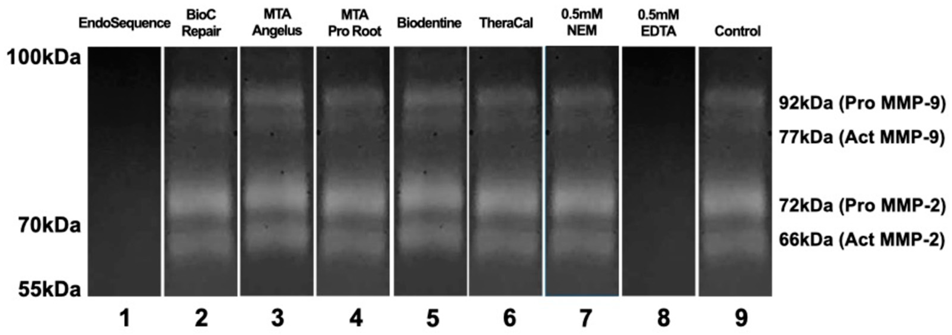
3.3. Evaluation of the Activity of Metalloproteinases

Assessing the inhibition of metalloproteinases’ enzymatic activity of MMP-2 and MMP-9, as determined by the electrophoresis assay, the results indicate that only EndoSequence exhibited inhibitory capabilities (Figure 3).

4. Discussion

Bioceramics have been shown to exhibit remarkable bioactivity and biocompatibility, making them extensively utilized in clinical endodontic practice. Nevertheless, no bioceramic material is entirely ideal, as each has its specific limitations in practical applications [14]. There are important characteristics that materials used in endodontics need to have to promote healing. In this study, we employed the microcosm biofilm assay to evaluate the antimicrobial properties of these materials. Additionally, we assessed cell viability by conducting cytotoxicity tests using rat fibroblasts (L929), and the inhibitory effects on metalloproteinases (MMP2 and MMP9) were evaluated through electrophoresis assay (Zymography).
During the setting process, the antimicrobial and antibiofilm properties are activated by increasing the pH and the release of ions from the material [35]. The attainment of both a high-quality seal and antimicrobial properties is crucial for the success of endodontic treatment [36]. In this study, ProRoot MTA and MTA Angelus demonstrated superior antimicrobial action with lower colony-forming unit (CFU) values. Previous studies have also highlighted the antimicrobial potential of ProRoot MTA [37,38,39].
The results of our study are similar to those found in several other studies which found that MTA Angelus and ProRoot MTA exhibited greater antibacterial activity compared to Biodentine [40,41,42,43]. On the contrary, it deviates from a previous study, which indicated that Biodentine exhibited greater antimicrobial activity than ProRoot MTA [44]. Nevertheless, this assessment exclusively focused on the antimicrobial efficacy against E. fecaelis and did not encompass a polymicrobial culture, as in our study.
A large number of microorganisms are involved in persistent endodontic infections [45], with over 500 strains identified [46]. Certain species can have a crucial impact on a microbial community, ensuring its stability and contributing to the virulence of persistent infections [47]. Different bacterial strains vary in susceptibility to antimicrobial materials, highlighting the need for testing against multiple strains [37]. Our study focused on evaluating antimicrobial activity using biofilm microcosm, which mimics natural dental plaque in a lab setting, maintaining its complexity and biodiversity [48].
Besides antimicrobial activity, the biological compatibility of materials for vital pulp therapy is of utmost importance [49]. In the present study, all the materials achieved the minimum required recommendation of a 70% relative cell viability rate, as recommended by the ISO 10993-5 standard [50]. Moreover, all the materials showed cell viability values above 100%, indicating cell proliferation.
In terms of cell viability, TheraCal LC showed the highest cell viability, followed by Biodentine and MTA ProRoot, with no significant differences. However, other studies reported conflicting results, with TheraCal LC exhibiting higher cytotoxicity compared to MTA Angelus and Biodentine in some cases [51,52] One possible explanation for the contrasting results between these studies is that cell viability is significantly influenced by the concentration of the eluate used [53]. Different concentrations may have affected the outcomes of the studies.
Bacterial products can stimulate MMP production [25,54]. MMPs were expressed in inflamed pulps and periapical pathosis, which suggests that MMPs play an important role in pulpal and periapical inflammation and destruction of acute inflamed pulp, especially in symptomatic pulpitis [27]. Notably, increased concentrations of MMP2 and MMP9 are detected in such lesions. [26,27]. Excessive MMP production leads to tissue destruction [46]. Only EndoSequence showed the ability to inhibit MMP-2 and MMP-9 activity in the electrophoresis-based evaluation using Zymography. This inhibition is significant as it can reduce inflammation and improve pathological processes.
After dismissing the infection, promoting an anti-inflammatory environment that is conducive to tissue regeneration at the dentine-pulp interface is crucial for vital pulp therapy [55]. The key to enhancing the success of pulp vital therapy lies in the thorough assessment of pulp inflammation and the selection of suitable materials before initiating treatment [56]. In this context, bioceramic materials designed for Vital Pulp Therapy, which exhibit the inhibition of or reduction in MMPs activity, may offer advantages in terms of treatment prognosis.
While the findings indicate the suppression of MMP-2 and MMP-9 activity by EndoSequence, further investigations are required to elucidate the underlying mechanism of this inhibition. Additionally, it is essential to conduct studies specifically focused on evaluating the inhibition of MMP-9 in inflammatory cells, considering its predominant expression in these cell types.

5. Conclusions

This study evaluated bioceramic materials aimed at vital pulp therapy and concluded the following:
(a)
All materials showed antimicrobial effectiveness, with Endosequence being the most effective, followed by Bio-C Repair, Biodentine, TheraCal LC, MTA Angelus, and MTA ProRoot.
(b)
All materials promoted cell proliferation, with TheraCal showing the highest cell growth, followed by Biodentine, MTA ProRoot, MTA Angelus, BioC Repair, and EndoSequence.
(c)
In the electrophoresis assay, EndoSequence successfully inhibited MMP2 and MMP9, while the other materials did not inhibit MMPs.

Supplementary Materials

The following supporting information can be downloaded at: https://www.mdpi.com/article/10.3390/jfb15030070/s1. Figure S1: Flowchart according to the Preferred Reporting Items for Laboratory studies in Endodontology (PRILE) 2021 Guidelines; Table S1: Description of polyacrylamide gel reagents; Table S2: Running buffer; Table S3: 2% Triton Solution; Table S4: Incubation Buffer; Table S5: Dye Solution; Table S6: Bleaching Solution.

Author Contributions

Conceptualization, F.I., D.d.O., J.S.R.d.A., A.d.S.B., A.F.d.S., W.L.d.O.d.R., C.E.C.-S., Á.H.B., E.P. and R.G.L.; Formal Analyses, W.L.d.O.d.R., J.S.R.d.A., Á.H.B., N.L.V.C., E.P. and R.G.L.; Funding Acquisition E.P., Á.H.B. and R.G.L.; Investigation, F.I., D.d.O., J.S.R.d.A., A.d.S.B. and C.E.C.-S. Methodology, F.I., D.d.O., J.S.R.d.A., A.d.S.B., A.F.d.S., W.L.d.O.d.R., C.E.C.-S., Á.H.B., E.P. and R.G.L.; Resources, A.F.d.S., W.L.d.O.d.R., Á.H.B., N.L.V.C., E.P. and R.G.L.; Software, W.L.d.O.d.R., J.S.R.d.A., C.E.C.-S. and R.G.L.; Supervision, R.G.L., Á.H.B. and E.P.; Validation, F.I., D.d.O., R.G.L. and Á.H.B.; Visualization, F.I., D.d.O., J.S.R.d.A., A.d.S.B., A.F.d.S., W.L.d.O.d.R., C.E.C.-S., Á.H.B., E.P. and R.G.L.; Writing—Original draft, F.I., D.d.O., E.P., R.G.L. and Á.H.B. Writing—Review and editing, F.I., D.d.O., E.P., R.G.L. and Á.H.B. All authors have read and agreed to the published version of the manuscript.

Funding

This study was financed by government research grants (#432127/2018-6 and #306225/2021-2) of Brazilian National Council of Scientific and Technological Development, CNPq/DAI grant #142536/2019-9, FAPERGS, Grant #21/2551-0000691-9, and UFPel #23110.018613/2019-70) for financial support.

Institutional Review Board Statement

This study was approved by the Research Ethical Committee and was carried out in accordance with the principles of the Declaration of Helsinki (CAAE/UFPEL No. 57403816.2.0000.5318, 26 October 2016).

Informed Consent Statement

Informed consent was obtained from all subjects involved in the study.

Data Availability Statement

The original contributions presented in the study are included in the article and Supplementary Material, further inquiries can be directed to the corresponding authors.

Conflicts of Interest

The authors declare no conflict of interest.

References

  1. Almeida, L.H.S.; Moraes, R.R.; Morgental, R.D.; Cava, S.S.; Rosa WL, O.; Rodrigues, P.; Ribeiro, A.S.; Só, M.; Pappen, F.G. Synthesis of silver-containing calcium aluminate particles and their effects on a MTA-based endodontic sealer. Dent. Mater. 2018, 34, e214–e223. [Google Scholar] [CrossRef]
  2. Aminoshariae, A.; Primus, C.; Kulild, J.C. Tricalcium silicate cement sealers. J. Am. Dent. Assoc. 2022, 153, 750–760. [Google Scholar] [CrossRef]
  3. Anshida, V.; Kumari Ra Murthy, C.; Samuel, A. Extracellular matrix degradation by host matrix metalloproteinases in restorative dentistry and endodontics: An overview. J. Oral Maxillofac. Pathol. 2020, 24, 352. [Google Scholar] [CrossRef]
  4. Benetti, F.; Queiroz, Í.O.A.; Cosme-Silva, L.; Conti, L.C.; Oliveira, S.H.P.; Cintra, L.T.A. Cytotoxicity, Biocompatibility and Biomineralization of a New Ready-for-Use Bioceramic Repair Material. Braz. Dent. J. 2019, 30, 325–332. [Google Scholar] [CrossRef]
  5. Bhavana, V.; Chaitanya, K.; Dola, B.; Gandi, P.; Patil, J.; Reddy, R. Evaluation of antibacterial and antifungal activity of new calcium-based cement (Biodentine) compared to MTA and glass ionomer cement. J. Conserv. Dent. 2015, 18, 44. [Google Scholar] [CrossRef]
  6. Bortoluzzi, E.A.; Niu, L.N.; Palani, C.D.; El-Awady, A.R.; Hammond, B.D.; Pei, D.D.; Tian, F.C.; Cutler, C.W.; Pashley, D.H.; Tay, F.R. Cytotoxicity and osteogenic potential of silicate calcium cements as potential protective materials for pulpal revascularization. Dent. Mater. 2015, 31, 1510–1522. [Google Scholar] [CrossRef]
  7. Camilleri, J.; Sorrentino, F.; Damidot, D. Investigation of the hydration and bioactivity of radiopacified tricalcium silicate cement, Biodentine and MTA Angelus. Dent. Mater. 2013, 29, 580–593. [Google Scholar] [CrossRef] [PubMed]
  8. Chang, S.W.; Gaudin, A.; Tolar, M.; Oh, S.; Moon, S.-Y.; Peters, O.A. Physicochemical and biological properties of four calcium silicate-based endodontic cements. J. Dent. Sci. 2022, 17, 1586–1594. [Google Scholar] [CrossRef]
  9. Chang, Y.H.; Chiang, C.Y.; Fu, E.; Chiu, H.C. Staphylococcus aureus enhances gelatinase activities in monocytic U937 cells and in human gingival fibroblasts. J. Dent. Sci. 2022, 17, 1321–1328. [Google Scholar] [CrossRef] [PubMed]
  10. Collado-González, M.; García-Bernal, D.; Oñate-Sánchez, R.E.; Ortolani-Seltenerich, P.S.; Álvarez-Muro, T.; Lozano, A.; Forner, L.; Llena, C.; Moraleda, J.M.; Rodríguez-Lozano, F.J. Cytotoxicity and bioactivity of various pulpotomy materials on stem cells from human exfoliated primary teeth. Int. Endod. J. 2017, 50, e19–e30. [Google Scholar] [CrossRef] [PubMed]
  11. da Rosa, W.L.O.; Cocco, A.R.; daSilva, T.M.; Mesquita, L.C.; Galarça, A.D.; da Silva, A.F.; Piva, E. Current trends and future perspectives of dental pulp capping materials: A systematic review. J. Biomed. Mater. Res. Part B Appl. Biomater. 2018, 106, 1358–1368. [Google Scholar] [CrossRef] [PubMed]
  12. Dal’Agnol, C.Z.; Stefenon, L.; van de Sande, F.H.; della Bona, Á.; Cenci, M.S.; Webber, B.; Rodrigues, L.B.; dos Santos, L.R. Microcosm Biofilm Formation on Titanium Surfaces. Mater. Res. 2015, 18, 677–682. [Google Scholar] [CrossRef]
  13. Damas, B.A.; Wheater, M.A.; Bringas, J.S.; Hoen, M.M. Cytotoxicity Comparison of Mineral Trioxide Aggregates and EndoSequence Bioceramic Root Repair Materials. J. Endod. 2011, 37, 372–375. [Google Scholar] [CrossRef] [PubMed]
  14. Dong, X.; Xu, X. Bioceramics in Endodontics: Updates and Future Perspectives. Bioengineering 2023, 10, 354. [Google Scholar] [CrossRef]
  15. Eldeniz, A.U.; Hadimli, H.H.; Ataoglu, H.; Ørstavik, D. Antibacterial effect of selected root-end filling materials. J. Endod. 2006, 32, 345–349. [Google Scholar] [CrossRef]
  16. Gandolfi, M.G.; Siboni, F.; Botero, T.; Bossù, M.; Riccitiello, F.; Prati, C. Calcium Silicate and Calcium Hydroxide Materials for Pulp Capping: Biointeractivity, Porosity, Solubility and Bioactivity of Current Formulations. J. Appl. Biomater. Funct. Mater. 2015, 13, 43–60. [Google Scholar] [CrossRef]
  17. Gandolfi, M.G.; Siboni, F.; Prati, C. Chemical–physical properties of TheraCal, a novel light-curable MTA-like material for pulp capping. Int. Endod. J. 2012, 45, 571–579. [Google Scholar] [CrossRef]
  18. Ghilotti, J.; Sanz, J.L.; López-García, S.; Guerrero-Gironés, J.; Pecci-Lloret, M.P.; Lozano, A.; Llena, C.; Rodríguez-Lozano, F.J.; Forner, L.; Spagnuolo, G. Comparative Surface Morphology, Chemical Composition, and Cytocompatibility of Bio-C Repair, Biodentine, and ProRoot MTA on hDPCs. Materials 2020, 13, 2189. [Google Scholar] [CrossRef]
  19. de Almeida Gomes, B.P.F.; Herrera, D.R. Etiologic role of root canal infection in apical periodontitis and its relationship with clinical symptomatology. Braz. Oral Res. 2018, 32, 82–110. [Google Scholar] [CrossRef]
  20. Gusman, H.; Santana, R.B.; Zehnder, M. Matrix metalloproteinase levels and gelatinolytic activity in clinically healthy and inflamed human dental pulps. Eur. J. Oral Sci. 2002, 110, 353–357. [Google Scholar] [CrossRef]
  21. Haapasalo, M.; Parhar, M.; Huang, X.; Wei, X.; Lin, J.; Shen, Y. Clinical use of bioceramic materials. Endod. Top. 2015, 32, 97–117. [Google Scholar] [CrossRef]
  22. Hiremath, G.; Kulkarni, R.; Naik, B. Evaluation of minimal inhibitory concentration of two new materials using tube dilution method: An in vitro study. J. Conserv. Dent. 2015, 18, 159. [Google Scholar] [CrossRef]
  23. ISO 10993-5; 2009; Biological evaluation of medical devices–part 5: In vitro cytotoxicity testing. International Organization for Standardization: Geneva, Switzerland, 2020.
  24. Kahler, B.; Rossi-Fedele, G. A Review of Tooth Discoloration after Regenerative Endodontic Therapy. J. Endod. 2016, 42, 563–569. [Google Scholar] [CrossRef] [PubMed]
  25. Kang, T.Y.; Choi, J.W.; Seo, K.J.; Kim, K.M.; Kwon, J.S. Physical, chemical, mechanical, and biological properties of four different commercial root-end filling materials: A comparative study. Materials 2021, 14, 1693. [Google Scholar] [CrossRef]
  26. Khan, A.S.; Ur Rehman, S.; Ahmad, S.; AlMaimouni, Y.K.; Alzamil MA, S.; Dummer PM, H. Five decades of the International Endodontic Journal: Bibliometric overview 1967–2020. Int. Endod. J. 2021, 54, 1819–1839. [Google Scholar] [CrossRef]
  27. Kim RJ, Y.; Kim, M.O.; Lee, K.S.; Lee, D.Y.; Shin, J.H. An in vitro evaluation of the antibacterial properties of three mineral trioxide aggregate (MTA) against five oral bacteria. Arch. Oral Biol. 2015, 60, 1497–1502. [Google Scholar] [CrossRef] [PubMed]
  28. Kodonas, K.; Fardi, A.; Gogos, C.; Economides, N. Scientometric analysis of vital pulp therapy studies. Int. Endod. J. 2021, 54, 220–230. [Google Scholar] [CrossRef]
  29. Koruyucu, M.; Topcuoglu, N.; Tuna, E.B.; Ozel, S.; Gencay, K.; Kulekci, G.; Seymen, F. An assessment of antibacterial activity of three pulp capping materials on Enterococcus faecalis by a direct contact test: An in vitro study. Eur. J. Dent. 2015, 9, 240–245. [Google Scholar] [CrossRef] [PubMed]
  30. Lee, B.-N.; Lee, B.-G.; Chang, H.-S.; Hwang, Y.-C.; Hwang, I.-N.; Oh, W.-M. Effects of a novel light-curable material on odontoblastic differentiation of human dental pulp cells. Int. Endod. J. 2017, 50, 464–471. [Google Scholar] [CrossRef]
  31. Li, Z.; Zhang, H.; Ge, S.; Gu, X.; Gao, G.; Luo, J. Expression pattern divergence of duplicated genes in rice. BMC Bioinform. 2009, 10 (Suppl. S6), S8. [Google Scholar] [CrossRef]
  32. Lovato, K.F.; Sedgley, C.M. Antibacterial activity of EndoSequence root repair material and ProRoot MTA against clinical isolates of enterococcus faecalis. J. Endod. 2011, 37, 1542–1546. [Google Scholar] [CrossRef]
  33. Mazzoni, A.; Pashley, D.H.; Tay, F.R.; Gobbi, P.; Orsini, G.; Ruggeri, A.; Carrilho, M.; Tjäderhane, L.; di Lenarda, R.; Breschi, L. Immunohistochemical identification of MMP-2 and MMP-9 in human dentin: Correlative FEI-SEM/TEM analysis. J. Biomed. Mater. Res. Part A 2009, 88, 697–703. [Google Scholar] [CrossRef]
  34. Oliveira, D. Assessment of antimicrobial properties, cell viability and metalloproteinase activities of retrofilling materials. Doctoral Thesis, University of Cuiabá, Cuiabá, 2019. (In Portuguese). [Google Scholar]
  35. Parirokh, M.; Torabinejad, M. Mineral Trioxide Aggregate: A Comprehensive Literature Review—Part III: Clinical Applications, Drawbacks, and Mechanism of Action. J. Endod. 2010, 36, 400–413. [Google Scholar] [CrossRef] [PubMed]
  36. Parirokh, M.; Torabinejad, M.; Dummer PM, H. Mineral trioxide aggregate and other bioactive endodontic cements: An updated overview—Part I: Vital pulp therapy. Int. Endod. J. 2018, 51, 177–205. [Google Scholar] [CrossRef] [PubMed]
  37. Pinto, K.P.; Barbosa AF, A.; Silva EJ, N.L.; Santos AP, P.; Sassone, L.M. What Is the Microbial Profile in Persistent Endodontic Infections? A Scoping Review. J. Endod. 2023, 49, 786–798.e7. [Google Scholar] [CrossRef] [PubMed]
  38. Poggio, C.; Arciola, C.R.; Beltrami, R.; Monaco, A.; Dagna, A.; Lombardini, M.; Visai, L. Cytocompatibility and Antibacterial Properties of Capping Materials. Sci. World J. 2014, 2014, 181945. [Google Scholar] [CrossRef] [PubMed]
  39. Poggio, C.; Ceci, M.; Beltrami, R.; Chiesa, M.; Colombo, M. Biological and chemical-physical properties of root-end filling materials: A comparative study. J. Conserv. Dent. 2015, 18, 94. [Google Scholar] [CrossRef] [PubMed]
  40. Qureshi, A. Recent Advances in Pulp Capping Materials: An Overview. J. Clin. Diagn. Res. 2014, 8, 316–321. [Google Scholar] [CrossRef] [PubMed]
  41. Rajasekharan, S.; Martens, L.C.; Cauwels RG, E.C.; Verbeeck RM, H. BiodentineTM material characteristics and clinical applications: A review of the literature. Eur. Arch. Paediatr. Dent. 2014, 15, 147–158. [Google Scholar] [CrossRef] [PubMed]
  42. Sanz, J.L.; Soler-Doria, A.; López-García, S.; García-Bernal, D.; Rodríguez-Lozano, F.J.; Lozano, A.; Llena, C.; Forner, L.; Guerrero-Gironés, J.; Melo, M. Comparative Biological Properties and Mineralization Potential of 3 Endodontic Materials for Vital Pulp Therapy: Theracal PT, Theracal LC, and Biodentine on Human Dental Pulp Stem Cells. J. Endod. 2021, 47, 1896–1906. [Google Scholar] [CrossRef]
  43. Shen, Y.; Stojicic, S.; Haapasalo, M. Bacterial viability in starved and revitalized biofilms: Comparison of viability staining and direct culture. J. Endod. 2010, 36, 1820–1823. [Google Scholar] [CrossRef]
  44. Shin, S.; Lee, J.; Baek, S.; Lim, S. Tissue Levels of Matrix Metalloproteinases in Pulps and Periapical Lesions. J. Endod. 2002, 28, 313–315. [Google Scholar] [CrossRef]
  45. Siqueira, J.F.; Rôças, I.N. Community as the unit of pathogenicity: An emerging concept as to the microbial pathogenesis of apical periodontitis. Oral Surg. Oral Med. Oral Pathol. Oral Radiol. Endodontology 2009, 107, 870–878. [Google Scholar] [CrossRef]
  46. Song, W.; Li, S.; Tang, Q.; Chen, L.; Yuan, Z. In vitro biocompatibility and bioactivity of calcium silicate-based bioceramics in endodontics (Review). Int. J. Mol. Med. 2021, 48, 128. [Google Scholar] [CrossRef]
  47. Surya Raghavendra, S.; Jadhav, G.R.; Gathani, K.M.; Kotadia, P. Bioceramics In Endodontics—A Review. J. Istanb. Univ. Fac. Dent. 2017, 51, S128–S137. [Google Scholar] [CrossRef]
  48. Tanabe, S.I.; Grenier, D. Macrophage tolerance response to Aggregatibacter actinomycetemcomitans lipopolysaccharide induces differential regulation of tumor necrosis factor-α, interleukin-1β and matrix metalloproteinase 9 secretion. J. Periodontal Res. 2008, 43, 372–377. [Google Scholar] [CrossRef]
  49. Tanalp, J.; Karapinar-Kazandag, M.; Ersev, H.; Bayirli, G. The Status of Mineral Trioxide Aggregate in Endodontics Education in Dental Schools in Turkey. J. Dent. Educ. 2012, 76, 752–758. [Google Scholar] [CrossRef] [PubMed]
  50. Torabinejad, M.; Parirokh, M. Mineral Trioxide Aggregate: A Comprehensive Literature Review—Part II: Leakage and Biocompatibility Investigations. J. Endod. 2010, 36, 190–202. [Google Scholar] [CrossRef] [PubMed]
  51. Torabinejad, M.; Ung, B.; Kettering, J.D. In vitro bacterial penetration of coronally unsealed endodontically treated teeth. J. Endod. 1990, 16, 566–569. [Google Scholar] [CrossRef] [PubMed]
  52. Torabinejad, M.; Watson, T.F.; Pitt Ford, T.R. Sealing ability of a mineral trioxide aggregate when used as a root end filling material. J. Endod. 1993, 19, 591–595. [Google Scholar] [CrossRef] [PubMed]
  53. von Arx, T. Apical surgery: A review of current techniques and outcome. Saudi Dent. J. 2011, 23, 9–15. [Google Scholar] [CrossRef] [PubMed]
  54. Zaky, S.; Shehabeldin, M.; Ray, H.; Sfeir, C. The role of inflammation modulation in dental pulp regeneration. Eur. Cells Mater. 2021, 41, 184–193. [Google Scholar] [CrossRef] [PubMed]
  55. Zanini, M.; Meyer, E.; Simon, S. Pulp Inflammation Diagnosis from Clinical to Inflammatory Mediators: A Systematic Review. J. Endod. 2017, 43, 1033–1051. [Google Scholar] [CrossRef] [PubMed]
  56. Zanini, M.; Sautier, J.M.; Berdal, A.; Simon, S. Biodentine Induces Immortalized Murine Pulp Cell Differentiation into Odontoblast-like Cells and Stimulates Biomineralization. J. Endod. 2012, 38, 1220–1226. [Google Scholar] [CrossRef]
Figure 1. Antimicrobial action (CFU) according to each material (n = 6 per group). Mean and standard deviation. Different lowercase letters indicate significant differences (p ˂ 0.05).
Jfb 15 00070 g001
Figure 2. Cell viability (%) after 24 h according to each material (n = 3 per group). Mean and standard deviation. Different lowercase letters indicate significant differences (p ˂ 0.05).
Jfb 15 00070 g002
Figure 3. Results of the electrophoresis according to each material (n = 6 per group). Results of the assay demonstrate that only EndoSequence showed inhibitory capacity of the gelatinolytic activity of MMP-2 and MMP-9.
Jfb 15 00070 g003
Table 1. Composition of materials according to the manufacturer .
MaterialManufacturerComposition
ProRoot MTADentsply Tulsa Dental,
Johnson City, TN, USA
Tricalcium silicate, Dicalcium silicate, Tricalcium aluminate, Bismuth oxide, Gypsum.
EndoSequenceBrasseler USA, Savannah, GA, USATricalcium Silicate, Dicalcium Silicate, Zirconium Oxide, Tantalum Oxide, Calcium Phosphate Monobasic, Fillers.
BiodentineSeptodont, Saint Maur des Fossés, FranceTricalcium silicate Zirconium oxide Calcium carbonate, Calcium chloride, polymer, Calcium chloride aqueous solution and excipients.
MTA AngelusAngelus, Londrina, BrazilTricalcium silicate, Tricalcium aluminate, Calcium oxide, Calcium tungstate.
TheraCal LCBISCO, Schaumburg, IL, USAPortland cement, Polyethylene glycol Di methacrylate, Barium zirconate.
Bio-C RepairAngelus, Londrina, BrazilCalcium Silicate, Calcium Aluminate, Calcium Oxide, Zirconium Oxide, Iron Oxide, Silicon Dioxide and Dispersing Agent
Composition according to manufacturers’ information (MSDS-Material Safety Data Sheet).
Disclaimer/Publisher’s Note: The statements, opinions and data contained in all publications are solely those of the individual author(s) and contributor(s) and not of MDPI and/or the editor(s). MDPI and/or the editor(s) disclaim responsibility for any injury to people or property resulting from any ideas, methods, instructions or products referred to in the content.

