

Green Synthesis of Controlled Shape Silver Nanostructures and Their Peroxidase, Catalytic Degradation, and Antibacterial Activity


Abstract
:1. Introduction
2. Materials and Methods
2.1. Synthesis of Silver Nanoparticles
2.2. Peroxidase Activity
2.3. Catalytic Activity
2.4. Antibacterial Activity
2.5. Characterization of Silver Nanostructures
3. Results and Discussion
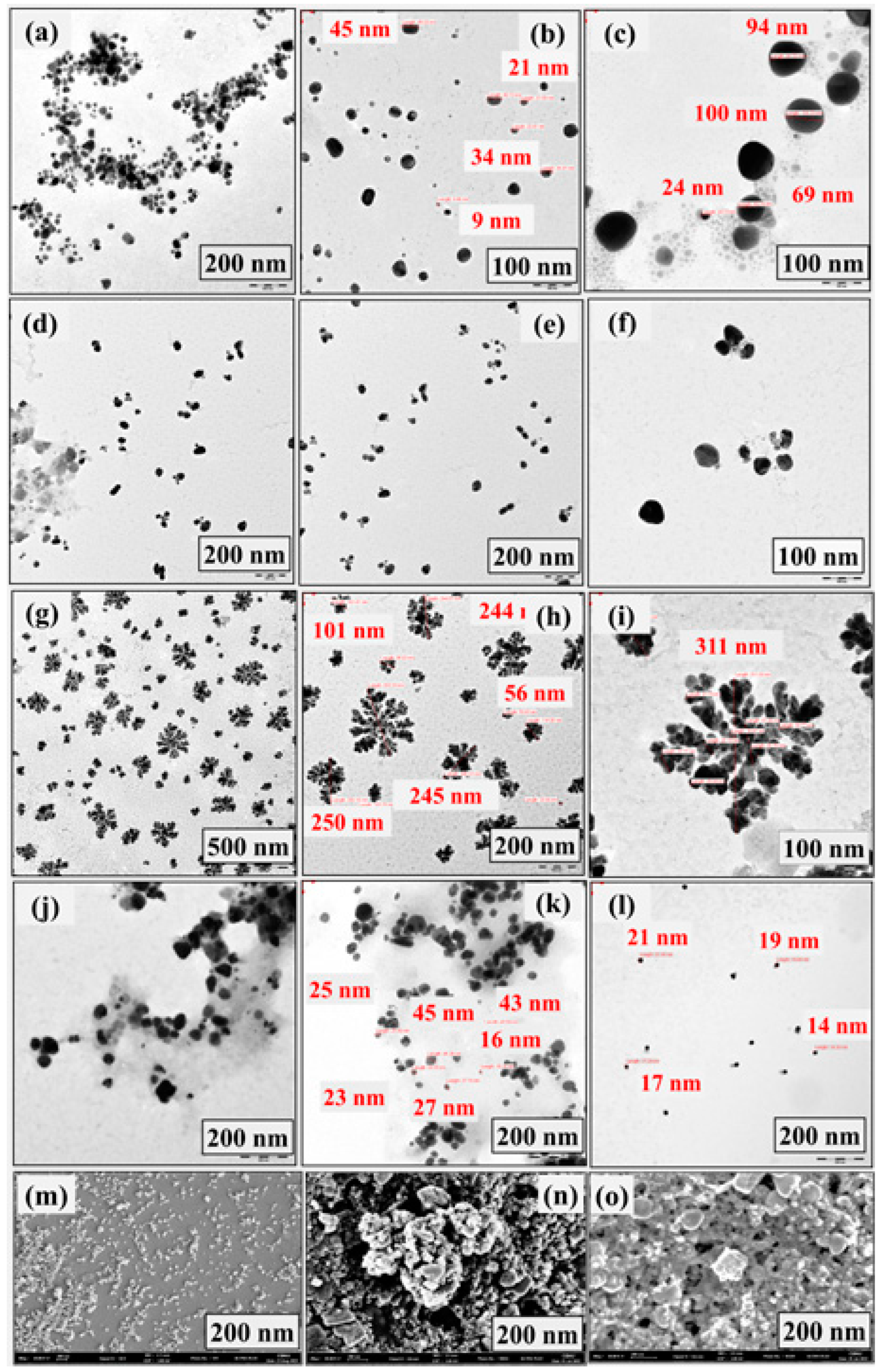
3.1. Microscopic Observation of Silver Nanostructures
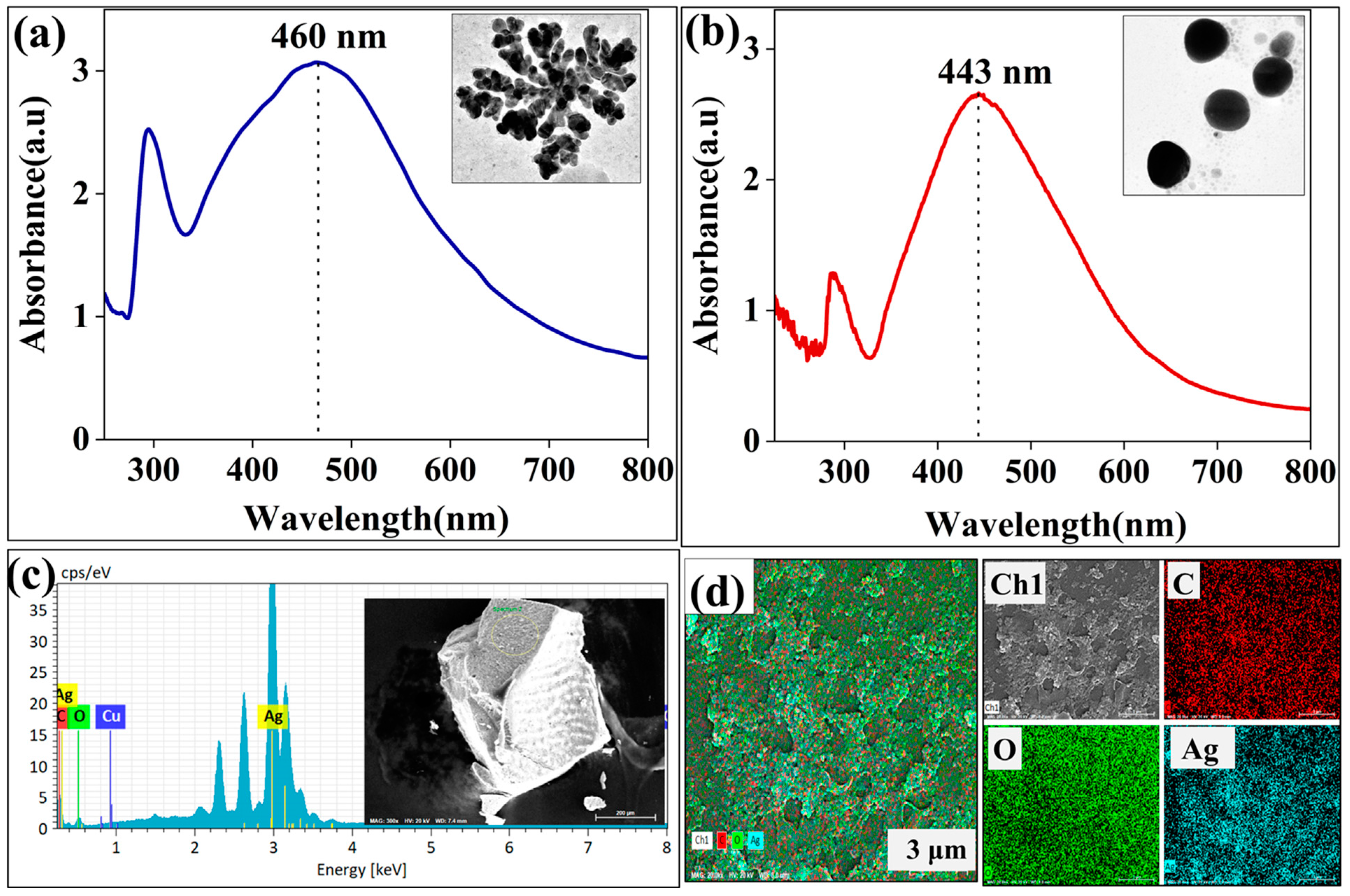
3.2. UV–Vis Spectroscopy and Elemental Analysis
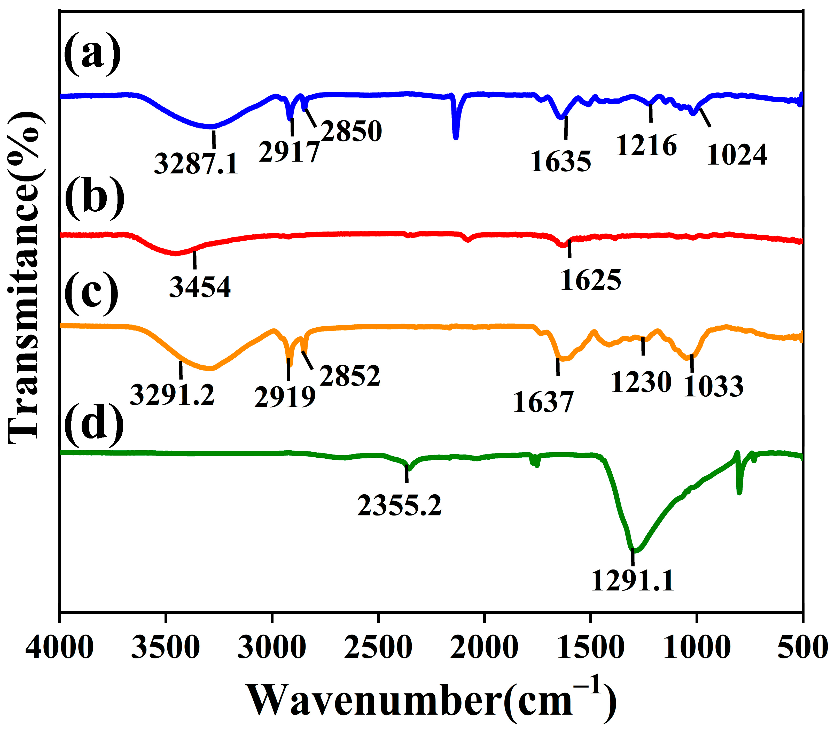
3.3. FTIR Analysis
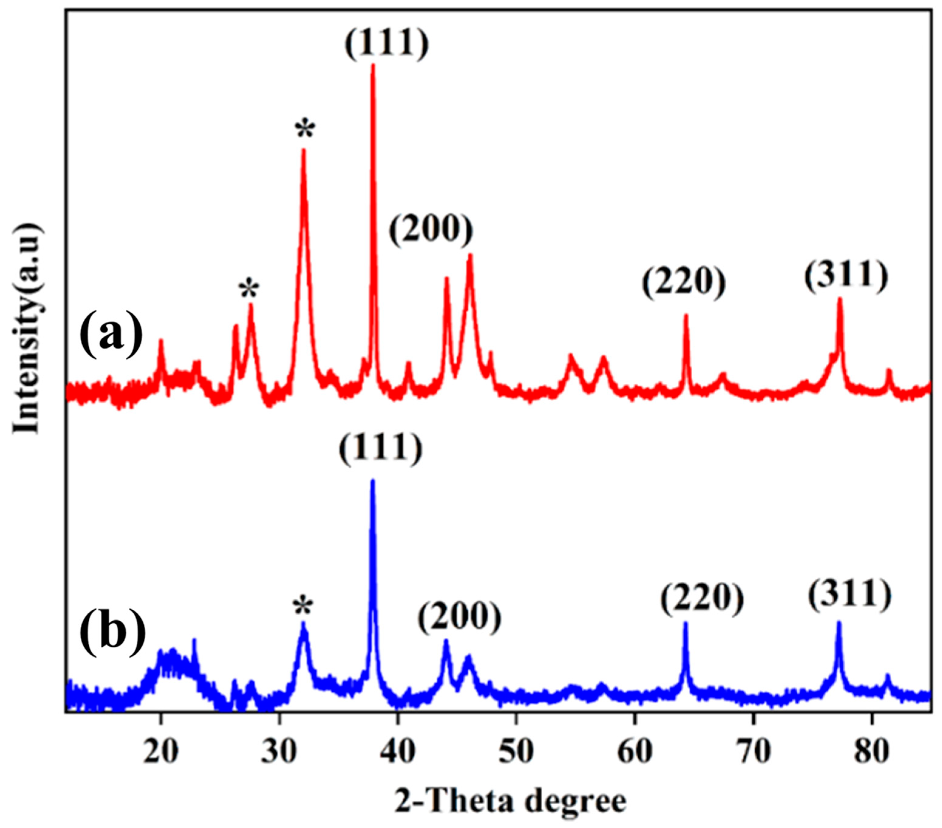
3.4. XRD Analysis
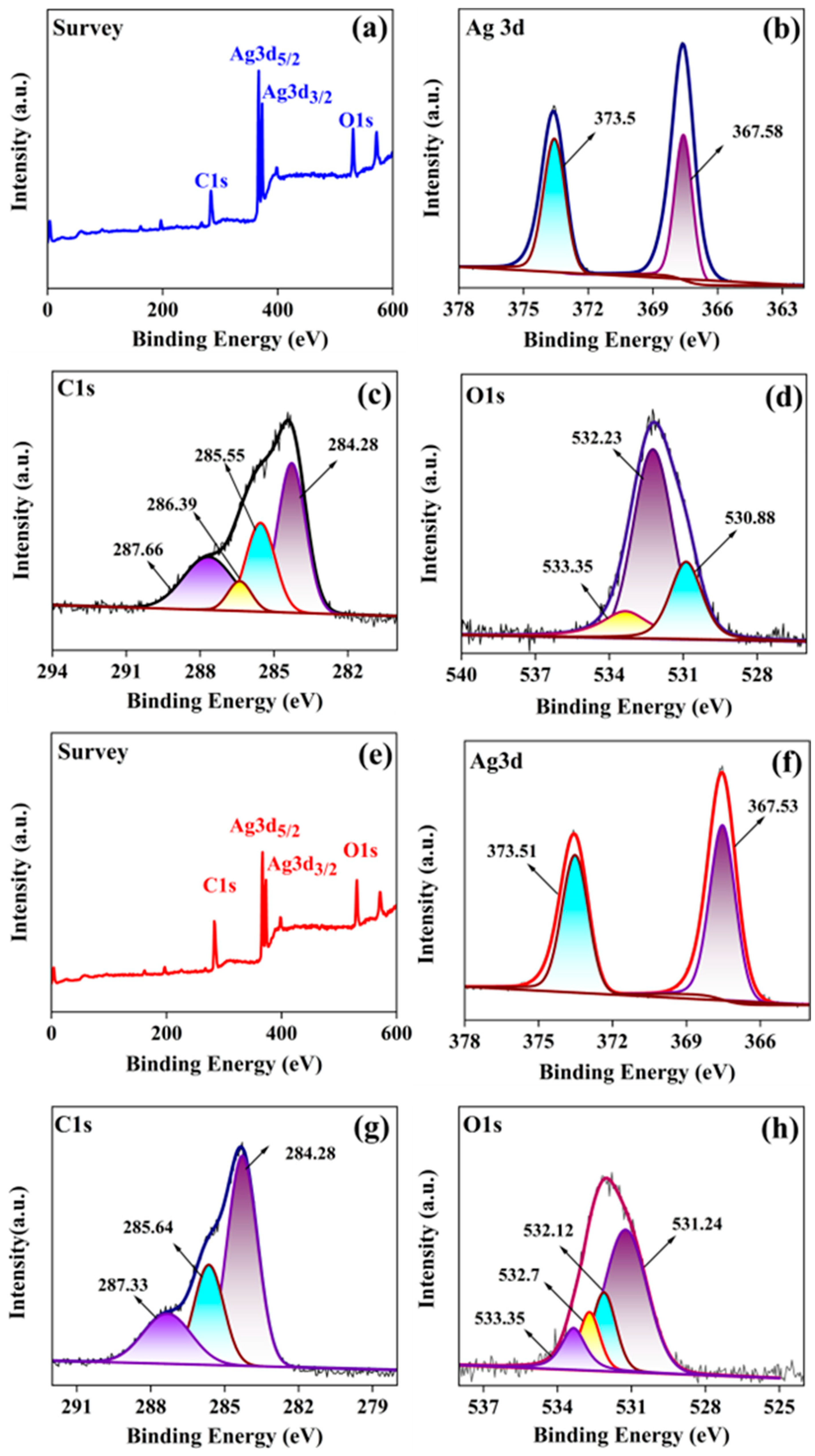
3.5. XPS Analysis
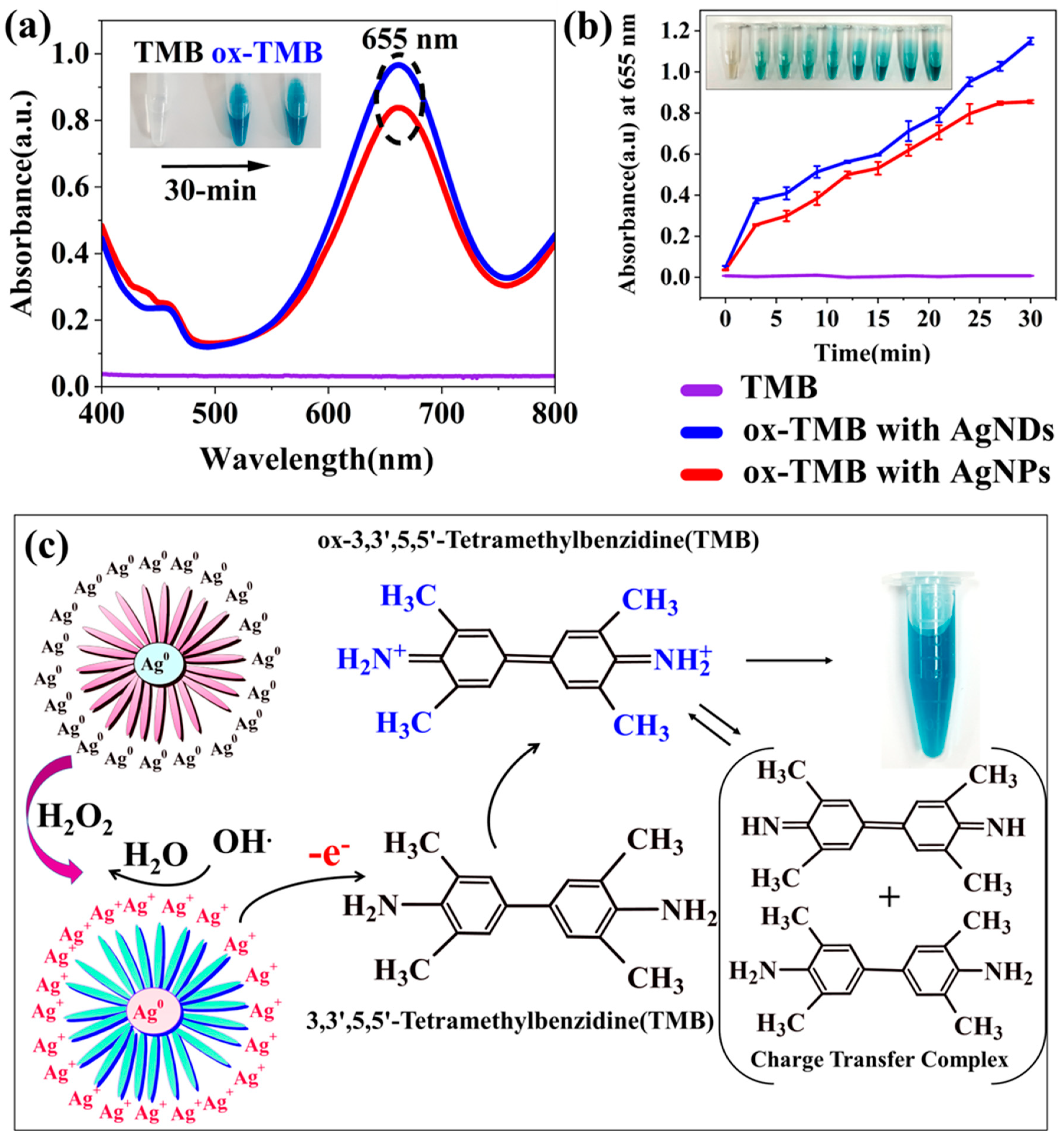
3.6. Peroxidase Activity
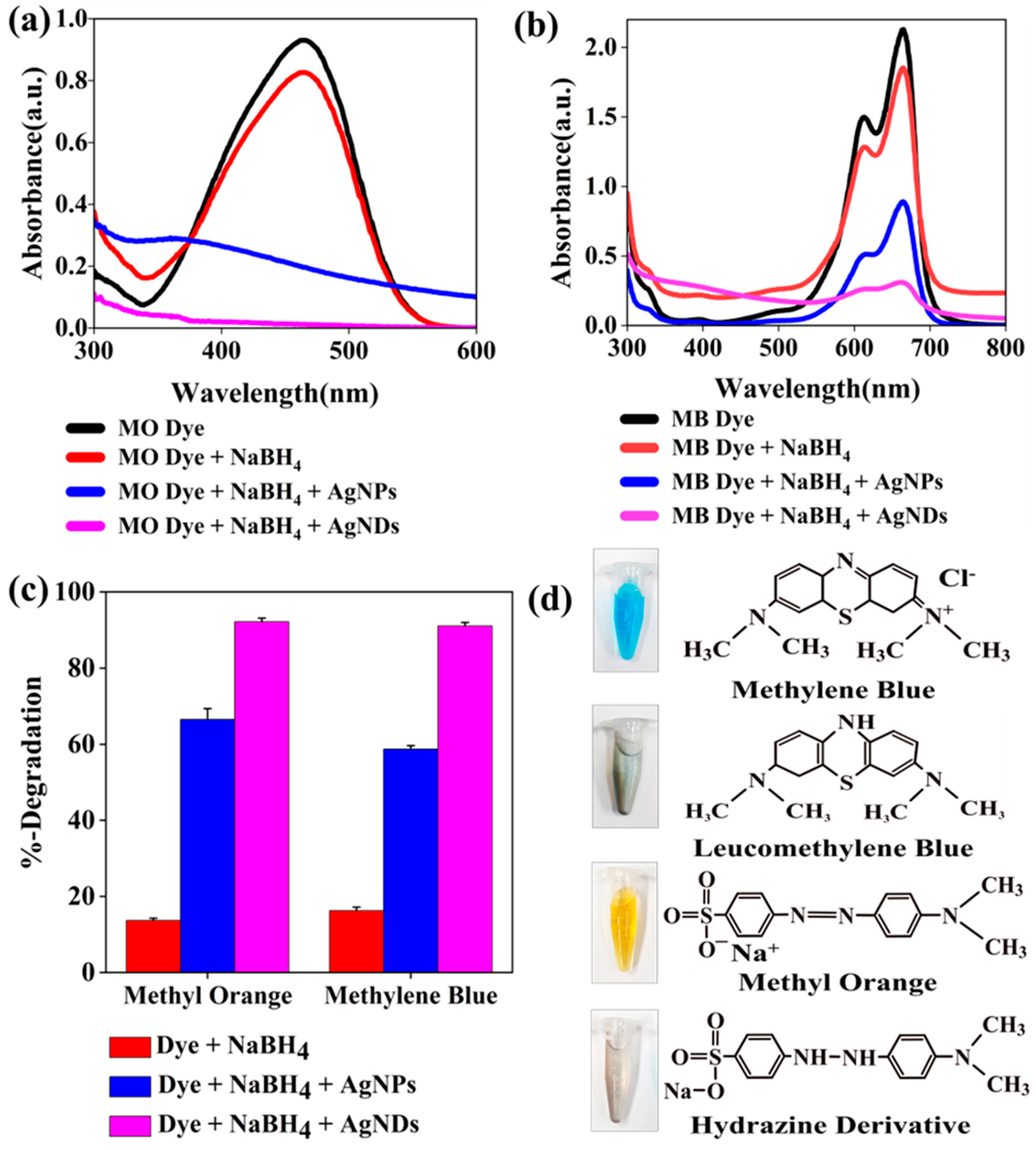
3.7. Catalytic Degradation
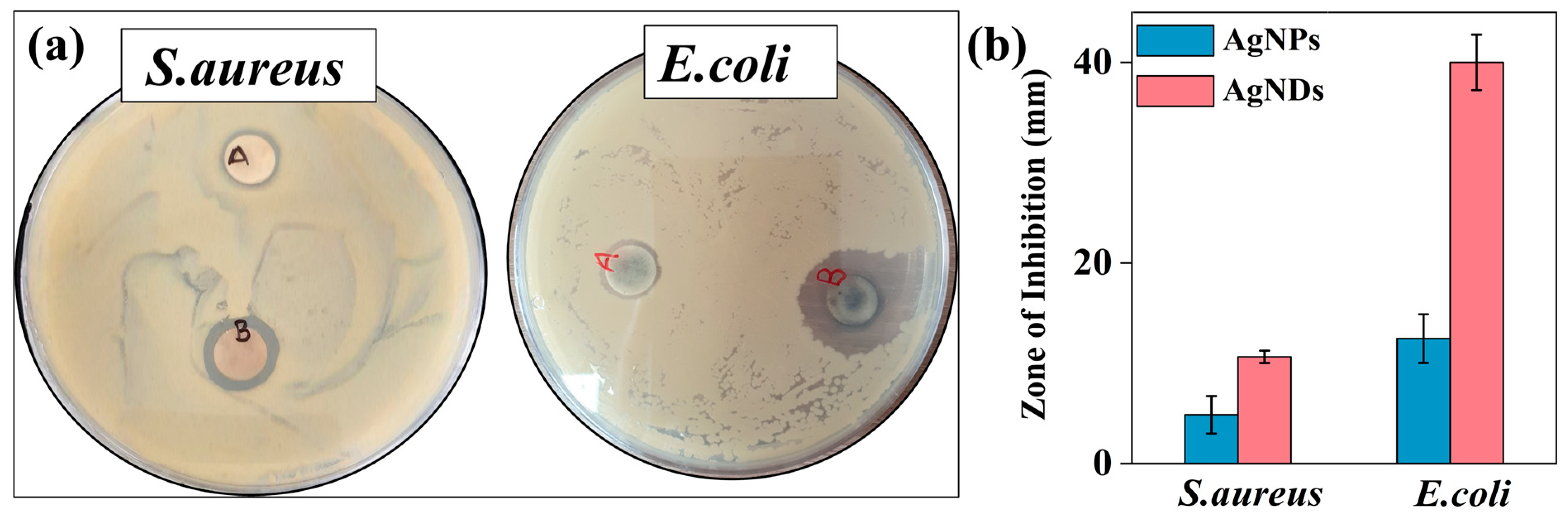
3.8. Antibacterial Activity
4. Conclusions
Author Contributions
Funding
Data Availability Statement
Conflicts of Interest
References
- Qin, L.; Zeng, G.; Lai, C.; Huang, D.; Xu, P.; Zhang, C.; Cheng, M.; Liu, X.; Liu, S.; Li, B.; et al. “Gold Rush” in Modern Science: Fabrication Strategies and Typical Advanced Applications of Gold Nanoparticles in Sensing. Coord. Chem. Rev. 2018, 359, 1–31. [Google Scholar] [CrossRef]
- Dimapilis, E.A.S.; Hsu, C.S.; Mendoza, R.M.O.; Lu, M.C. Zinc Oxide Nanoparticles for Water Disinfection. Sustain. Environ. Res. 2018, 28, 47–56. [Google Scholar] [CrossRef]
- AbouAitah, K.; Piotrowska, U.; Wojnarowicz, J.; Swiderska-Sroda, A.; El-Desoky, A.H.H.; Lojkowski, W. Enhanced Activity and Sustained Release of Protocatechuic Acid, a Natural Antibacterial Agent, from Hybrid Nanoformulations with Zinc Oxide Nanoparticles. Int. J. Mol. Sci. 2021, 22, 5287. [Google Scholar] [CrossRef]
- Xu, M.; Luo, H.; Rong, H.; Wu, S.; Zheng, Z.; Chen, B. Calcium Alginate Gels-Functionalized Polyurethane Foam Decorated with Silver Nanoparticles as an Antibacterial Agent for Point-of-Use Water Disinfection. Int. J. Biol. Macromol. 2023, 231, 123289. [Google Scholar] [CrossRef] [PubMed]
- Islam, M.A.; Jacob, M.V.; Antunes, E. A Critical Review on Silver Nanoparticles: From Synthesis and Applications to Its Mitigation through Low-Cost Adsorption by Biochar. J. Environ. Manag. 2021, 281, 111918. [Google Scholar] [CrossRef] [PubMed]
- Calderón-Jiménez, B.; Johnson, M.E.; Montoro Bustos, A.R.; Murphy, K.E.; Winchester, M.R.; Baudrit, J.R.V. Silver Nanoparticles: Technological Advances, Societal Impacts, and Metrological Challenges. Front. Chem. 2017, 5, 6. [Google Scholar] [CrossRef] [Green Version]
- Navaladian, S.; Viswanathan, B.; Viswanath, R.P.; Varadarajan, T.K. Thermal Decomposition as Route for Silver Nanoparticles. Nanoscale Res. Lett. 2007, 2, 44–48. [Google Scholar] [CrossRef] [Green Version]
- Pathrose, B.; Nampoori, V.P.N.; Radhakrishnan, P.; Sahira, H.; Mujeeb, A. Effect of Femtosecond Laser Ablated Silver Nanoparticles in the Thermo-Optic Properties of Basic Fuchsin Dye. Optik 2016, 127, 3684–3687. [Google Scholar] [CrossRef]
- Iravani, S.; Korbekandi, H.; Mirmohammadi, S.V.; Zolfaghari, B. Synthesis of Silver Nanoparticles: Chemical, Physical and Biological Methods. Res. Pharm. Sci. 2014, 9, 385–406. [Google Scholar]
- Alabdallah, N.M.; Hasan, M.M. Plant-Based Green Synthesis of Silver Nanoparticles and Its Effective Role in Abiotic Stress Tolerance in Crop Plants. Saudi J. Biol. Sci. 2021, 28, 5631–5639. [Google Scholar] [CrossRef] [PubMed]
- Islam, Z.; Islam, S.M.R.; Hossen, F.; Mahtab-Ul-Islam, K.; Hasan, M.R.; Karim, R. Moringa oleifera is a Prominent Source of Nutrients with Potential Health Benefits. Int. J. Food Sci. 2021, 2021, 6627265. [Google Scholar] [CrossRef]
- Fahey, J.W.D. Moringa oleifera: A Review of the Medical Evidence for Its Nutritional, Therapeutic, and Prophylactic Properties. Part 1. Trees Life J. 2005, 1, 5. [Google Scholar]
- Shousha, W.G.; Aboulthana, W.M.; Salama, A.H.; Saleh, M.H.; Essawy, E.A. Evaluation of the Biological Activity of Moringa oleifera Leaves Extract after Incorporating Silver Nanoparticles, in Vitro Study. Bull. Natl. Res. Cent. 2019, 43, 212. [Google Scholar] [CrossRef] [Green Version]
- Saini, R.K.; Shetty, N.P.; Giridhar, P. GC-FID/MS Analysis of Fatty Acids in Indian Cultivars of Moringa oleifera: Potential Sources of PUFA. J. Am. Oil Chem. Soc. 2014, 91, 1029–1034. [Google Scholar] [CrossRef]
- Abdulkadir, A.R.; Zawawi, D.D.; Jahan, M.S. Proximate and Phytochemical Screening of Different Parts of Moringa Oleifera. Russ. Agric. Sci. 2016, 42, 34–36. [Google Scholar] [CrossRef]
- Cuellar-Nuñez, M.L.; Luzardo-Ocampo, I.; Campos-Vega, R.; Gallegos-Corona, M.A.; González de Mejía, E.; Loarca-Piña, G. Physicochemical and Nutraceutical Properties of Moringa (Moringa Oleifera) Leaves and Their Effects in an in Vivo AOM/DSS-Induced Colorectal Carcinogenesis Model. Food Res. Int. 2018, 105, 159–168. [Google Scholar] [CrossRef]
- Leone, A.; Spada, A.; Battezzati, A.; Schiraldi, A.; Aristil, J.; Bertoli, S. Moringa oleifera Seeds and Oil: Characteristics and Uses for Human Health. Int. J. Mol. Sci. 2016, 17, 2141. [Google Scholar] [CrossRef] [PubMed] [Green Version]
- Mumtaz, M.Z.; Kausar, F.; Hassan, M.; Javaid, S.; Malik, A. Anticancer Activities of Phenolic Compounds from Moringa oleifera Leaves: In Vitro and in Silico Mechanistic Study. Beni. Suef. Univ. J. Basic Appl. Sci. 2021, 10, 12. [Google Scholar] [CrossRef]
- Maheshwaran, G.; Malai Selvi, M.; Selva Muneeswari, R.; Nivedhitha Bharathi, A.; Krishna Kumar, M.; Sudhahar, S. Green Synthesis of Lanthanum Oxide Nanoparticles Using Moringa oleifera Leaves Extract and Its Biological Activities. Adv. Powder Technol. 2021, 32, 1963–1971. [Google Scholar] [CrossRef]
- Fatiqin, A.; Amrulloh, H.; Simanjuntak, W. Green Synthesis of MgO Nanoparticles Using Moringa oleifera Leaf Aqueous Extract for Antibacterial Activity. Bull. Chem. Soc. Ethiop. 2021, 35, 161–170. [Google Scholar] [CrossRef]
- Sharma, A.K.; Swami, A.K.; Jangir, D.; Saran, M.; Upadhyay, T.K.; Prajapat, R.K.; Sharma, D.; Mathur, M. An Eco-Friendly Green Synthesis of Tungsten Nanoparticles from Moringa oleifera Lam. and Their Pharmacological Studies. Gazi Med. J. 2021, 31, 719–725. [Google Scholar] [CrossRef]
- Anand, K.; Tiloke, C.; Phulukdaree, A.; Ranjan, B.; Chuturgoon, A.; Singh, S.; Gengan, R.M. Biosynthesis of Palladium Nanoparticles by Using Moringa oleifera Flower Extract and Their Catalytic and Biological Properties. J. Photochem. Photobiol. B 2016, 165, 87–95. [Google Scholar] [CrossRef]
- Jadhav, V.; Bhagare, A.; Ali, I.H.; Dhayagude, A.; Lokhande, D.; Aher, J.; Jameel, M.; Dutta, M. Role of Moringa oleifera on Green Synthesis of Metal/Metal Oxide Nanomaterials. J. Nanomater. 2022, 2022, 2147393. [Google Scholar] [CrossRef]
- Sharma, S.; Kumar, K.; Thakur, N.; Chauhan, S.; Chauhan, M.S. The Effect of Shape and Size of ZnO Nanoparticles on Their Antimicrobial and Photocatalytic Activities: A Green Approach. Bull. Mater. Sci. 2020, 43, 20. [Google Scholar] [CrossRef]
- Khan, I.; Saeed, K.; Khan, I. Nanoparticles: Properties, Applications and Toxicities. Arab. J. Chem. 2019, 12, 908–931. [Google Scholar] [CrossRef]
- Ashkarran, A.A.; Estakhri, S.; Nezhad, M.R.H.; Eshghi, S. Controlling the Geometry of Silver Nanostructures for Biological Applications. Phys. Procedia 2013, 40, 76–83. [Google Scholar] [CrossRef] [Green Version]
- Wu, L.; Wu, W.; Jing, X.; Huang, J.; Sun, D.; Odoom-Wubah, T.; Liu, H.; Wang, H.; Li, Q. Trisodium Citrate-Assisted Biosynthesis of Silver Nanoflowers by Canarium album Foliar Broths as a Platform for SERS Detection. Ind. Eng. Chem. Res. 2013, 52, 5085–5094. [Google Scholar] [CrossRef]
- Chathanchira, M.A.; Faseela K, P.F.; Singh, S.; Baik, S. Hierarchically-Structured Silver Nanoflowers for Highly Conductive Metallic Inks with Dramatically Reduced Filler Concentration. Sci. Rep. 2016, 6, 34894. [Google Scholar] [CrossRef] [Green Version]
- Mdluli, P.S.; Revaprasadu, N. Time Dependant Evolution of Silver Nanodendrites. Mater. Lett. 2009, 63, 447–450. [Google Scholar] [CrossRef]
- Deshmukh, A.R.; Aloui, H.; Kim, B.S. In Situ Growth of Gold and Silver Nanoparticles onto Phyto-Functionalized Boron Nitride Nanosheets: Catalytic, Peroxidase Mimicking, and Antimicrobial Activity. J. Clean. Prod. 2020, 270, 122339. [Google Scholar] [CrossRef]
- Palladino, P.; Torrini, F.; Scarano, S.; Minunni, M. 3,3′,5,5′-Tetramethylbenzidine as Multi-Colorimetric Indicator of Chlorine in Water in Line with Health Guideline Values. Anal. Bioanal. Chem. 2020, 412, 7861–7869. [Google Scholar] [CrossRef] [PubMed]
- Deshmukh, A.R.; Gupta, A.; Kim, B.S. Ultrasound Assisted Green Synthesis of Silver and Iron Oxide Nanoparticles Using Fenugreek Seed Extract and Their Enhanced Antibacterial and Antioxidant Activities. Biomed. Res. Int. 2019, 2019, 1714358. [Google Scholar] [CrossRef] [PubMed]
- Mickymaray, S. One-Step Synthesis of Silver Nanoparticles Using Saudi Arabian Desert Seasonal Plant Sisymbrium irio and Antibacterial Activity Against Multidrug-Resistant Bacterial Strains. Biomolecules 2019, 9, 662. [Google Scholar] [CrossRef] [PubMed] [Green Version]
- Lai, M.-J.; Huang, Y.-W.; Chen, H.-C.; Tsao, L.-I.; Chang Chien, C.-F.; Singh, B.; Liu, B.R. Effect of Size and Concentration of Copper Nanoparticles on the Antimicrobial Activity in Escherichia coli through Multiple Mechanisms. Nanomaterials 2022, 12, 3715. [Google Scholar] [CrossRef] [PubMed]
- Makarov, V.V.; Love, A.J.; Sinitsyna, O.V.; Makarova, S.S.; Yaminsky, I.V.; Taliansky, M.E.; Kalinina, N.O. “Green” Nanotechnologies: Synthesis of Metal Nanoparticles Using Plants. Acta Nat. 2014, 6, 35–44. [Google Scholar] [CrossRef] [Green Version]
- Shreyash, N.; Bajpai, S.; Khan, M.A.; Vijay, Y.; Tiwary, S.K.; Sonker, M. Green Synthesis of Nanoparticles and Their Biomedical Applications: A Review. ACS Appl. Nano Mater. 2021, 4, 11428–11457. [Google Scholar] [CrossRef]
- Marslin, G.; Siram, K.; Maqbool, Q.; Selvakesavan, R.K.; Kruszka, D.; Kachlicki, P.; Franklin, G. Secondary Metabolites in the Green Synthesis of Metallic Nanoparticles. Materials 2018, 11, 940. [Google Scholar] [CrossRef] [Green Version]
- Khandel, P.; Yadaw, R.K.; Soni, D.K.; Kanwar, L.; Shahi, S.K. Biogenesis of Metal Nanoparticles and Their Pharmacological Applications: Present Status and Application Prospects. J. Nanostruct. Chem. 2018, 8, 217–254. [Google Scholar] [CrossRef] [Green Version]
- Wang, K.-Y.; Bu, S.-J.; Ju, C.-J.; Li, C.-T.; Li, Z.-Y.; Han, Y.; Ma, C.-Y.; Wang, C.-Y.; Hao, Z.; Liu, W.-S.; et al. Hemin-Incorporated Nanoflowers as Enzyme Mimics for Colorimetric Detection of Foodborne Pathogenic Bacteria. Bioorg. Med. Chem. Lett. 2018, 28, 3802–3807. [Google Scholar] [CrossRef]
- Zhang, Y.X.; Jia, Y.; Jin, Z.; Yu, X.Y.; Xu, W.H.; Luo, T.; Zhu, B.J.; Liu, J.H.; Huang, X.J. Self-Assembled, Monodispersed, Flower-like γ-AlOOH Hierarchical Superstructures for Efficient and Fast Removal of Heavy Metal Ions from Water. CrystEngComm 2012, 14, 3005–3007. [Google Scholar] [CrossRef]
- Xu, S.; Wang, L.; Li, H.; Yue, Q.; Li, R.; Liu, J.; Gu, X.; Zhang, S. Copper Ions Mediated Formation of Three-Dimensional Self-Assembled Ag Nanostructures via a Facile Solution Route. CrystEngComm 2013, 15, 6368–6373. [Google Scholar] [CrossRef]
- Ren, W.; Guo, S.; Dong, S.; Wang, E. A Simple Route for the Synthesis of Morphology-Controlled and SERS-Active Ag Dendrites with near-Infrared Absorption. J. Phys. Chem. C 2011, 115, 10315–10320. [Google Scholar] [CrossRef]
- Wang, C.; Kim, Y.J.; Singh, P.; Mathiyalagan, R.; Jin, Y.; Yang, D.C. Green Synthesis of Silver Nanoparticles by Bacillus methylotrophicus, and Their Antimicrobial Activity. Artif. Cells Nanomed. Biotechnol. 2016, 44, 1127–1132. [Google Scholar] [CrossRef]
- Kalaiyan, G.; Suresh, S.; Prabu, K.M.; Thambidurai, S.; Kandasamy, M.; Pugazhenthiran, N.; Kumar, S.K.; Muneeswaran, T. Bactericidal Activity of Moringa oleifera Leaf Extract Assisted Green Synthesis of Hierarchical Copper Oxide Microspheres against Pathogenic Bacterial Strains. J. Environ. Chem. Eng. 2021, 9, 104847. [Google Scholar] [CrossRef]
- Pandian, A.M.K.; Karthikeyan, C.; Rajasimman, M.; Dinesh, M.G. Synthesis of Silver Nanoparticle and Its Application. Ecotoxicol. Environ. Saf. 2015, 121, 211–217. [Google Scholar] [CrossRef]
- Cornelius, W. Phytochemical Analysis of Moringa oleifera (leaves and flowers) and the Funtional Group. Glob. Sci. J. 2019, 7, 41–51. [Google Scholar]
- Bello, O.S.; Adegoke, K.A.; Akinyunni, O.O. Preparation and Characterization of a Novel Adsorbent from Moringa oleifera Leaf. Appl. Water Sci. 2017, 7, 1295–1305. [Google Scholar] [CrossRef] [Green Version]
- Ansari, M.J.; Rehman, N.U.; Ibnouf, E.; Alalaiwe, A.; Ganaie, M.A.; Zafar, A. Gum Acacia- and Gum Tragacanth-Coated Silver Nanoparticles: Synthesis, Physiological Stability, In-Vitro, Ex-Vivo and In-Vivo Activity Evaluations. Coatings 2022, 12, 1579. [Google Scholar] [CrossRef]
- El-Naggar, N.E.-A.; Hussein, M.H.; El-Sawah, A.A. Bio-Fabrication of Silver Nanoparticles by Phycocyanin, Characterization, in Vitro Anticancer Activity against Breast Cancer Cell Line and in Vivo Cytotxicity. Sci. Rep. 2017, 7, 10844. [Google Scholar] [CrossRef] [Green Version]
- Thiruvengadam, V.; Bansod, A.V. Characterization of Silver Nanoparticles Synthesized Using Chemical Method and Its Antibacterial Property. Biointerface Res. Appl. Chem. 2020, 10, 7257–7264. [Google Scholar] [CrossRef]
- Halawani, E.M. Rapid Biosynthesis Method and Characterization of Silver Nanoparticles Using Zizyphus spina christi Leaf Extract and Their Antibacterial Efficacy in Therapeutic Application. J. Biomater. Nanobiotechnol. 2017, 8, 22–35. [Google Scholar] [CrossRef] [Green Version]
- Bu, Y.; Kushwaha, A.; Goswami, L.; Kim, B.S. Green Production of Functionalized Few-Layer Graphene–Silver Nanocomposites Using Gallnut Extract for Antibacterial Application. Micromachines 2022, 13, 1232. [Google Scholar] [CrossRef] [PubMed]
- Paul Das, M.; Rebecca Livingstone, J.; Veluswamy, P.; Das, J. Exploration of Wedelia chinensis Leaf-Assisted Silver Nanoparticles for Antioxidant, Antibacterial and in Vitro Cytotoxic Applications. J. Food Drug Anal. 2018, 26, 917–925. [Google Scholar] [CrossRef] [PubMed] [Green Version]
- D’Anna, E.; De Giorgi, M.L.; Luches, A.; Martino, M.; Perrone, A.; Zocco, A. Study of C N Binding States in Carbon Nitride Films Deposited by Reactive XeCl Laser Ablation. Thin Solid Film. 1999, 347, 72–77. [Google Scholar] [CrossRef]
- Diel, J.C.; Franco, D.S.P.; Igansi, A.V.; Cadaval, T.R.S.; Pereira, H.A.; Nunes, I.D.S.; Basso, C.W.; Maria do Carmo, M.A.; Morais, J.; Pinto, D.; et al. Green Synthesis of Carbon Nanotubes Impregnated with Metallic Nanoparticles: Characterization and Application in Glyphosate Adsorption. Chemosphere 2021, 283, 131193. [Google Scholar] [CrossRef]
- Gómez-Vilchis, J.C.; García-Rosales, G.; Longoria-Gándara, L.C.; Pérez-Gómez, E.O.; Castilleros, D.T. Obtention of Biochar-Fe/Ce Using Punica granatum with High Adsorption of Ampicillin Capacity. Heliyon 2022, 8, e08841. [Google Scholar] [CrossRef]
- Firet, N.J.; Blommaert, M.A.; Burdyny, T.; Venugopal, A.; Bohra, D.; Longo, A.; Smith, W.A. Operando EXAFS Study Reveals Presence of Oxygen in Oxide-Derived Silver Catalysts for Electrochemical CO2 Reduction. J. Mater. Chem. A Mater. 2019, 7, 2597–2607. [Google Scholar] [CrossRef] [Green Version]
- Yoon, M.; Seo, M.; Jeong, C.; Jang, J.H.; Jeon, K.S. Synthesis of Liposome-Templated Titania Nanodisks: Optical Properties and Photocatalytic Activities. Chem. Mater. 2005, 17, 6069–6079. [Google Scholar] [CrossRef]
- El-Reash, Y.G.A.; Ghaith, E.A.; El-Awady, O.; Algethami, F.K.; Lin, H.; Abdelrahman, E.A.; Awad, F.S. Highly Fluorescent Hydroxyl Groups Functionalized Graphitic Carbon Nitride for Ultrasensitive and Selective Determination of Mercury Ions in Water and Fish Samples. J. Anal. Sci. Technol. 2023, 14, 16. [Google Scholar] [CrossRef]
- Alhokbany, N.; Ahama, T.; Ruksana; Naushad, M.; Alshehri, S.M. AgNPs Embedded N- Doped Highly Porous Carbon Derived from Chitosan Based Hydrogel as Catalysts for the Reduction of 4-Nitrophenol. Compos. B Eng. 2019, 173, 106950. [Google Scholar] [CrossRef]
- Ghosh, S.; Khan, T.S.; Ghosh, A.; Chowdhury, A.H.; Haider, M.A.; Khan, A.; Islam, S.M. Utility of Silver Nanoparticles Embedded Covalent Organic Frameworks as Recyclable Catalysts for the Sustainable Synthesis of Cyclic Carbamates and 2-Oxazolidinones via Atmospheric Cyclizative CO2 Capture. ACS Sustain. Chem. Eng. 2020, 8, 5495–5513. [Google Scholar] [CrossRef]
- Zhao, T.; Sun, R.; Yu, S.; Zhang, Z.; Zhou, L.; Huang, H.; Du, R. Size-Controlled Preparation of Silver Nanoparticles by a Modified Polyol Method. Colloids Surf. A Physicochem. Eng. Asp. 2010, 366, 197–202. [Google Scholar] [CrossRef]
- Boronin, A.I.; Koscheev, S.V.; Zhidomirov, G.M. XPS and UPS Study of Oxygen States on Silver. J. Electron Spectrosc. Relat. Phenom. 1998, 96, 43–51. [Google Scholar] [CrossRef]
- Mu, J.; Zhang, L.; Zhao, M.; Wang, Y. Co3O4 Nanoparticles as an Efficient Catalase Mimic: Properties, Mechanism and Its Electrocatalytic Sensing Application for Hydrogen Peroxide. J. Mol. Catal. A Chem. 2013, 378, 30–37. [Google Scholar] [CrossRef]
- Bu, Y.; Kim, B.S. Eco-Friendly Production of Functionalized Few-Layer Graphene Using Coffee Waste Extract and in-Situ Growth of Copper Oxide Nanoparticles. J. Environ. Chem. Eng. 2023, 11, 109350. [Google Scholar] [CrossRef]
- Zarif, F.; Rauf, S.; Khurshid, S.; Muhammad, N.; Hayat, A.; Rahim, A.; Shah, N.S.; Yang, C.P. Effect of Pyridinium Based Ionic Liquid on the Sensing Property of NiO Nanoparticle for the Colorimetric Detection of Hydrogen Peroxide. J. Mol. Struct. 2020, 1219, 128620. [Google Scholar] [CrossRef]
- Naya, S.I.; Teranishi, M.; Kimura, K.; Tada, H. A Strong Support-Effect on the Catalytic Activity of Gold Nanoparticles for Hydrogen Peroxide Decomposition. Chem. Commun. 2011, 47, 3230–3232. [Google Scholar] [CrossRef] [PubMed]
- Liu, S.; Tian, J.; Wang, L.; Sun, X. Highly Sensitive and Selective Colorimetric Detection of Ag(I) Ion Using 3,3′,5,5′,-Tetramethylbenzidine (TMB) as an Indicator. Sens. Actuators B Chem. 2012, 165, 44–47. [Google Scholar] [CrossRef]
- Üzer, A.; Durmazel, S.; Erçağ, E.; Apak, R. Determination of Hydrogen Peroxide and Triacetone Triperoxide (TATP) with a Silver Nanoparticles—Based Turn-on Colorimetric Sensor. Sens. Actuators B Chem. 2017, 247, 98–107. [Google Scholar] [CrossRef]
- Mills, A. An Overview of the Methylene Blue ISO Test for Assessing the Activities of Photocatalytic Films. Appl. Catal. B 2012, 128, 144–149. [Google Scholar] [CrossRef]
- Ismail, M.; Khan, M.I.; Khan, S.A.; Qayum, M.; Khan, M.A.; Anwar, Y.; Akhtar, K.; Asiri, A.M.; Khan, S.B. Green Synthesis of Antibacterial Bimetallic Ag–Cu Nanoparticles for Catalytic Reduction of Persistent Organic Pollutants. J. Mater. Sci. Mater. Electron. 2018, 29, 20840–20855. [Google Scholar] [CrossRef]
- Asli, B.; Abdelkrim, S.; Zahraoui, M.; Mokhtar, A.; Hachemaoui, M.; Bennabi, F.; Ahmed, A.B.; Sardi, A.; Boukoussa, B. Catalytic Reduction and Antibacterial Activity of MCM-41 Modified by Silver Nanoparticles. Silicon 2022, 14, 12587–12598. [Google Scholar] [CrossRef]
- Ismail, M.; Gul, S.; Khan, M.I.; Khan, M.A.; Asiri, A.M.; Khan, S.B. Medicago Polymorpha-Mediated Antibacterial Silver Nanoparticles in the Reduction of Methyl Orange. Green Process. Synth. 2019, 8, 118–127. [Google Scholar] [CrossRef]
- Chandra Paul, S.; Bhowmik, S.; Rani Nath, M.; Islam, M.S.; Kanti Paul, S.; Neazi, J.; Sabnam Binta Monir, T.; Dewanjee, S.; Abdus Salam, M. Silver Nanoparticles Synthesis in a Green Approach: Size Dependent Catalytic Degradation of Cationic and Anionic Dyes. Orient. J. Chem. 2020, 36, 353–360. [Google Scholar] [CrossRef]
- Zahid, M.; Al-Majmaie, S.; Shather, A.H.; Khan, M.F.; Alguno, A.C.; Capangpangan, R.Y.; Ismail, A. Elettaria Cardamomum Seed Extract Synthesized Silver Nanoparticles for Efficient Catalytic Reduction of Toxic Dyes. Environ. Nanotechnol. Monit. Manag. 2023, 20, 100809. [Google Scholar] [CrossRef]
- Padre, S.M.; Kiruthika, S.; Mundinamani, S.; Ravikirana; Surabhi, S.; Jeong, J.R.; Eshwarappa, K.M.; Murari, M.S.; Shetty, V.; Ballal, M.; et al. Mono- and Bimetallic Nanoparticles for Catalytic Degradation of Hazardous Organic Dyes and Antibacterial Applications. ACS Omega 2022, 7, 35023–35034. [Google Scholar] [CrossRef] [PubMed]
- Ghosh, S.K.; Kundu, S.; Mandal, M.; Pal, T. Silver and Gold Nanocluster Catalyzed Reduction of Methylene Blue by Arsine in a Micellar Medium. Langmuir 2002, 18, 8756–8760. [Google Scholar] [CrossRef]
- Somasundaram, C.K.; Atchudan, R.; Edison, T.N.J.I.; Perumal, S.; Vinodh, R.; Sundramoorthy, A.K.; Babu, R.S.; Alagan, M.; Lee, Y.R. Sustainable Synthesis of Silver Nanoparticles Using Marine Algae for Catalytic Degradation of Methylene Blue. Catalysts 2021, 11, 1377. [Google Scholar] [CrossRef]
- Hao, Y.; Zhang, N.; Luo, J.; Liu, X. Green Synthesis of Silver Nanoparticles by Tannic Acid with Improved Catalytic Performance Towards the Reduction of Methylene Blue. Nano 2018, 13, 1850003. [Google Scholar] [CrossRef]
- Kumar, P.; Dixit, J.; Singh, A.K.; Rajput, V.D.; Verma, P.; Tiwari, K.N.; Mishra, S.K.; Minkina, T.; Mandzhieva, S. Efficient Catalytic Degradation of Selected Toxic Dyes by Green Biosynthesized Silver Nanoparticles Using Aqueous Leaf Extract of Cestrum nocturnum L. Nanomaterials 2022, 12, 3851. [Google Scholar] [CrossRef] [PubMed]
- Al-Shehri, A.S.; Zaheer, Z.; Alsudairi, A.M.; Kosa, S.A. Photo-Oxidative Decolorization of Brilliant Blue with AgNPs as an Activator in the Presence of K2S2O8 and NaBH4. ACS Omega 2021, 6, 27510–27526. [Google Scholar] [CrossRef] [PubMed]
- Barman, K.; Chowdhury, D.; Baruah, P.K. Bio-Synthesized Silver Nanoparticles Using Zingiber officinale Rhizome Extract as Efficient Catalyst for the Degradation of Environmental Pollutants. Inorg. Nano-Met. Chem. 2020, 50, 57–65. [Google Scholar] [CrossRef]
- Maiti, S.; Krishnan, D.; Barman, G.; Ghosh, S.K.; Laha, J.K. Antimicrobial Activities of Silver Nanoparticles Synthesized from Lycopersicon esculentum Extract. J. Anal. Sci. Technol. 2014, 5, 40. [Google Scholar] [CrossRef] [Green Version]
- Hamad, A.; Khashan, K.S.; Hadi, A. Silver Nanoparticles and Silver Ions as Potential Antibacterial Agents. J. Inorg. Organomet. Polym. Mater. 2020, 30, 4811–4828. [Google Scholar] [CrossRef]
- Urnukhsaikhan, E.; Bold, B.-E.; Gunbileg, A.; Sukhbaatar, N.; Mishig-Ochir, T. Antibacterial Activity and Characteristics of Silver Nanoparticles Biosynthesized from Carduus crispus. Sci. Rep. 2021, 11, 21047. [Google Scholar] [CrossRef] [PubMed]
- Ansari, M.A.; Khan, H.M.; Khan, A.A.; Ahmad, M.K.; Mahdi, A.A.; Pal, R.; Cameotra, S.S. Interaction of Silver Nanoparticles with Escherichia coli and Their Cell Envelope Biomolecules. J. Basic Microbiol. 2014, 54, 905–915. [Google Scholar] [CrossRef] [PubMed]
- Sondi, I.; Salopek-Sondi, B. Silver Nanoparticles as Antimicrobial Agent: A Case Study on E. Coli as a Model for Gram-Negative Bacteria. J. Colloid Interface Sci. 2004, 275, 177–182. [Google Scholar] [CrossRef]
- Thill, A.; Zeyons, O.; Spalla, O.; Chauvat, F.; Rose, J.; Auffan, M.; Flank, A.M. Cytotoxicity of CeO2 Nanoparticles for Escherichia coli. Physico-Chemical Insight of the Cytotoxicity Mechanism. Environ. Sci. Technol. 2006, 40, 6151–6156. [Google Scholar] [CrossRef] [PubMed]
- Morales-Avila, E.; Ferro-Flores, G.; Ocampo-Garciá, B.E.; López-Téllez, G.; López-Ortega, J.; Rogel-Ayala, D.G.; Sánchez-Padilla, D. Antibacterial Efficacy of Gold and Silver Nanoparticles Functionalized with the Ubiquicidin (29-41) Antimicrobial Peptide. J. Nanomater. 2017, 2017, 5831959. [Google Scholar] [CrossRef] [Green Version]
- Ramalingam, B.; Parandhaman, T.; Das, S.K. Antibacterial Effects of Biosynthesized Silver Nanoparticles on Surface Ultrastructure and Nanomechanical Properties of Gram-Negative Bacteria Viz. Escherichia coli and Pseudomonas aeruginosa. ACS Appl. Mater. Interfaces 2016, 8, 4963–4976. [Google Scholar] [CrossRef]
- Javani, S.; Lorca, R.; Latorre, A.; Flors, C.; Cortajarena, A.L.; Somoza, Á. Antibacterial Activity of DNA-Stabilized Silver Nanoclusters Tuned by Oligonucleotide Sequence. ACS Appl. Mater. Interfaces 2016, 8, 10147–10154. [Google Scholar] [CrossRef] [PubMed]
- Meikle, T.G.; Dyett, B.P.; Strachan, J.B.; White, J.; Drummond, C.J.; Conn, C.E. Preparation, Characterization, and Antimicrobial Activity of Cubosome Encapsulated Metal Nanocrystals. ACS Appl. Mater. Interfaces 2020, 12, 6944–6954. [Google Scholar] [CrossRef] [PubMed]
- Cheon, J.Y.; Kim, S.J.; Rhee, Y.H.; Kwon, O.H.; Park, W.H. Shape-Dependent Antimicrobial Activities of Silver Nanoparticles. Int. J. Nanomed. 2019, 14, 2773–2780. [Google Scholar] [CrossRef] [PubMed] [Green Version]
- Alshareef, A.; Laird, K.; Cross, R.B.M. Shape-Dependent Antibacterial Activity of Silver Nanoparticles on Escherichia coli and Enterococcus faecium Bacterium. Appl. Surf. Sci. 2017, 424, 310–315. [Google Scholar] [CrossRef]
- dos Santos, V.E.; Junior; Targino, A.G.R.; Flores, M.A.P.; Rodríguez-Díaz, J.M.; Teixeira, J.A.; Heimer, M.V.; Pessoa, H.D.L.F.; Galembeck, A.; Rosenblatt, A. Antimicrobial Activity of Silver Nanoparticle Colloids of Different Sizes and Shapes against Streptococcus Mutans. Res. Chem. Intermed. 2017, 43, 5889–5899. [Google Scholar] [CrossRef]


Disclaimer/Publisher’s Note: The statements, opinions and data contained in all publications are solely those of the individual author(s) and contributor(s) and not of MDPI and/or the editor(s). MDPI and/or the editor(s) disclaim responsibility for any injury to people or property resulting from any ideas, methods, instructions or products referred to in the content. |
© 2023 by the authors. Licensee MDPI, Basel, Switzerland. This article is an open access article distributed under the terms and conditions of the Creative Commons Attribution (CC BY) license (https://creativecommons.org/licenses/by/4.0/).
Share and Cite
Shafiq, A.; Deshmukh, A.R.; AbouAitah, K.; Kim, B.-S. Green Synthesis of Controlled Shape Silver Nanostructures and Their Peroxidase, Catalytic Degradation, and Antibacterial Activity. J. Funct. Biomater. 2023, 14, 325. https://doi.org/10.3390/jfb14060325
Shafiq A, Deshmukh AR, AbouAitah K, Kim B-S. Green Synthesis of Controlled Shape Silver Nanostructures and Their Peroxidase, Catalytic Degradation, and Antibacterial Activity. Journal of Functional Biomaterials. 2023; 14(6):325. https://doi.org/10.3390/jfb14060325
Chicago/Turabian StyleShafiq, Ayesha, Aarti R. Deshmukh, Khaled AbouAitah, and Beom-Soo Kim. 2023. "Green Synthesis of Controlled Shape Silver Nanostructures and Their Peroxidase, Catalytic Degradation, and Antibacterial Activity" Journal of Functional Biomaterials 14, no. 6: 325. https://doi.org/10.3390/jfb14060325