
Biomechanical Characteristics and Analysis Approaches of Bone and Bone Substitute Materials

Abstract
:1. Introduction
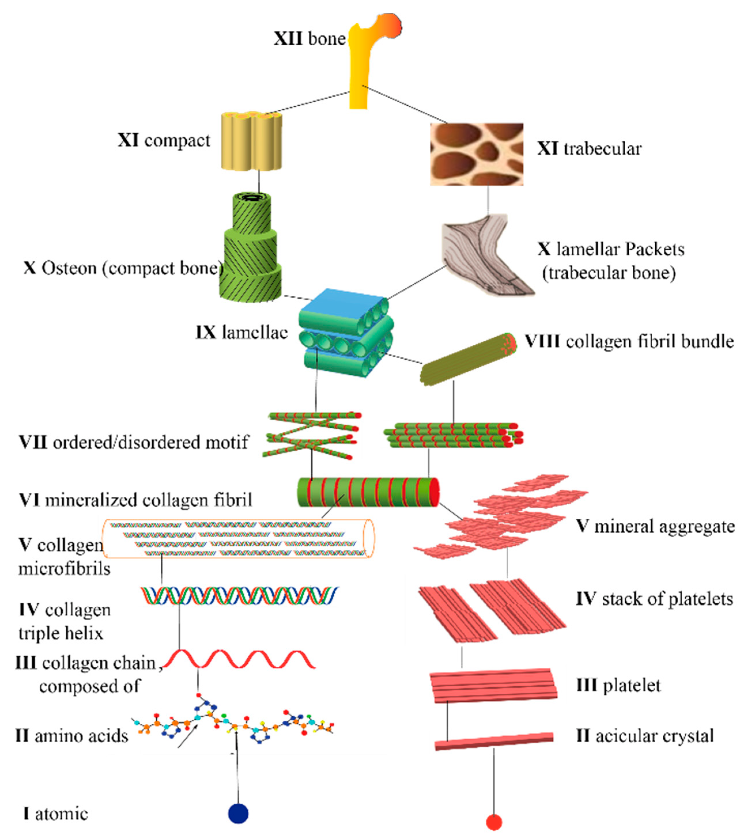
1.1. Composition and Structure of Natural Bone
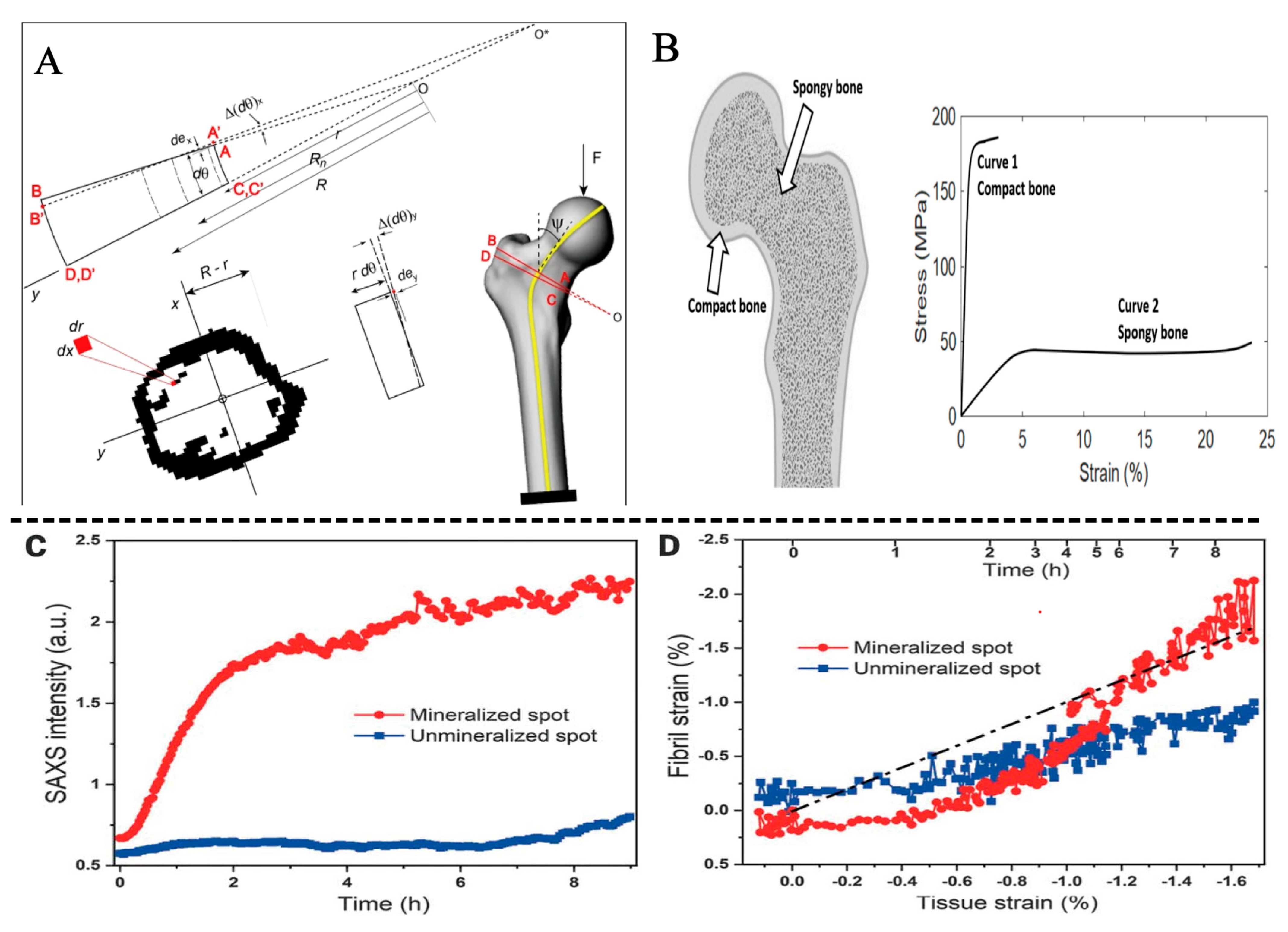
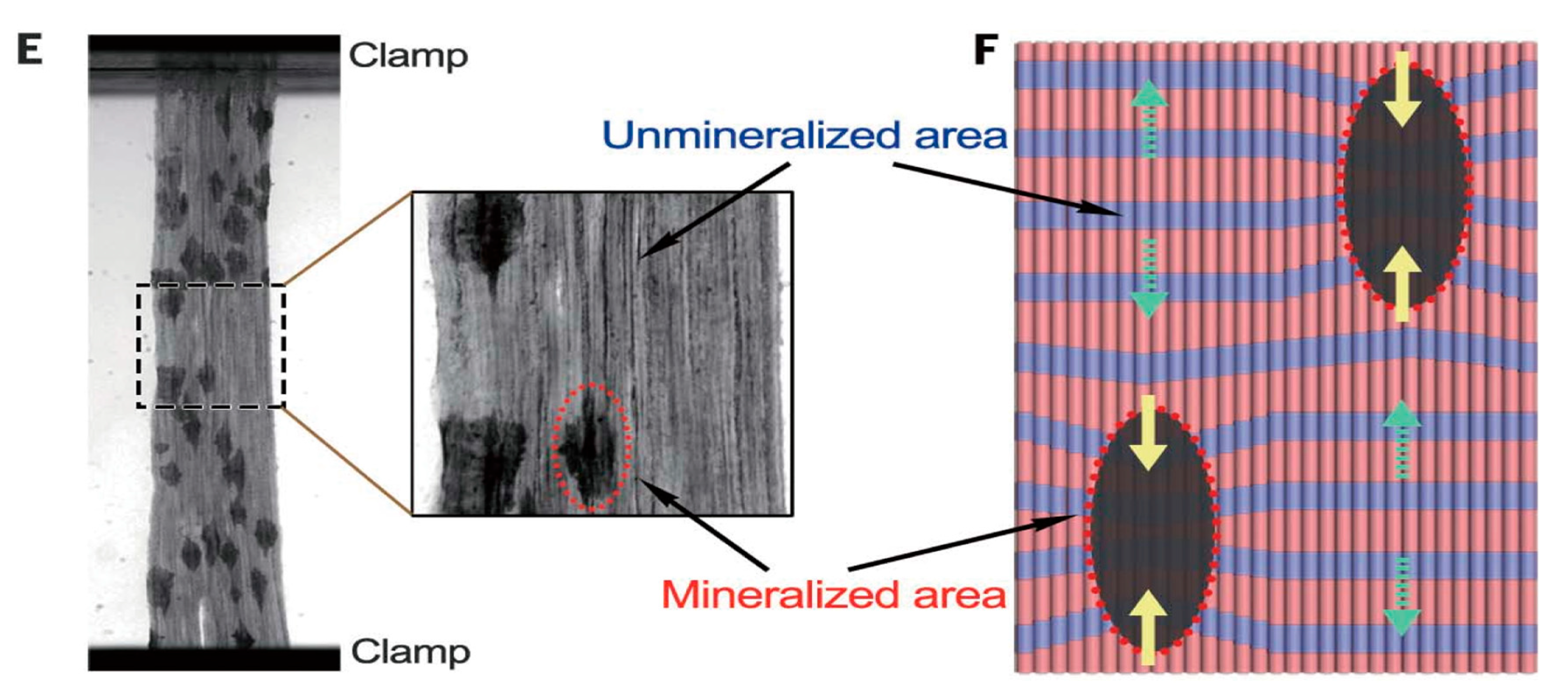
1.2. Mechanical Properties of Natural Bone


2. Biomechanical Properties of Biomimetic Bone Materials
2.1. Common Types of Bone Repair

| Repair Site | Vickers Hardness [19,20,21,22,23] | Material Properties | Application Features | Example | |
|---|---|---|---|---|---|
| Load bearing bone | Limb bones | 40.39–44.59 HV | Metals and Alloys | Weight-bearing, Correction, Immobilization | Nano-titanium and Ti-6Al-4V alloy [29] |
| Joints | 38.55 HV | ||||
| Spine | 25.47–32.80 HV | ||||
| Ribs | 37.35 HV | ||||
| Skull | 39.86 HV | ||||
| Non-weight-bearing bone | Maxillofacial | 43.54 HV | Bioceramics | Fill, Support, Protect | Calcium Phosphate, HA [33,34,35] |
| Orbital | 42.95 HV | ||||
| Dental | 278–285 HV | ||||
| Middle ear bone | 54.11 HV | ||||
| Cartilage | 0.317 HB | Polymer | Fill, patch, join, join | Collagen and PLA nanofibers [36,37] | |
| Maxillofacial | 43.54 HV | Composite material | Fill, repair | HA-Collagen [38,39,40,41] | |
| Dental | 278–285 HV |
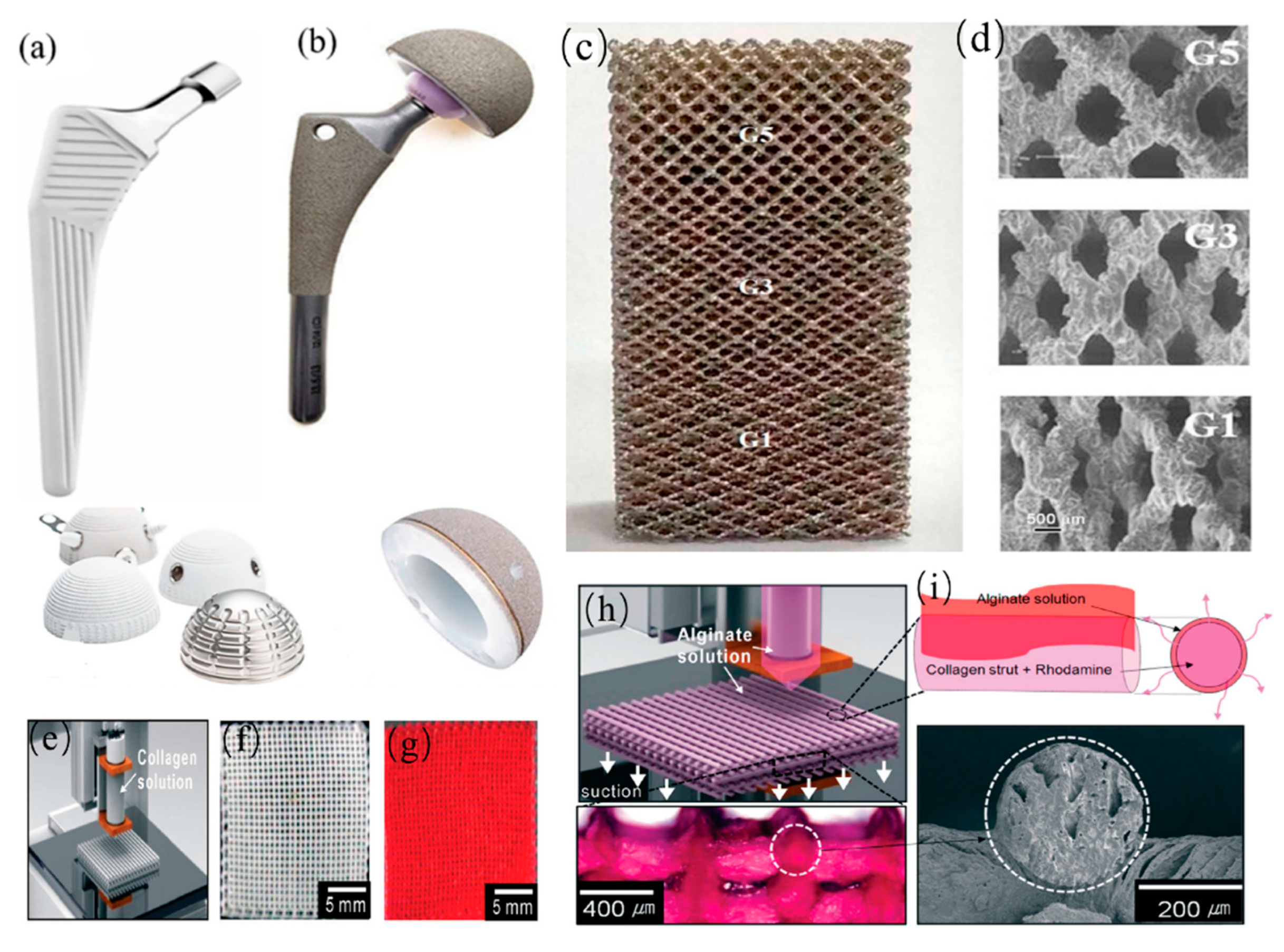
2.2. Load Bearing Implant
2.3. Non-Weight-Bearing Bone Implants
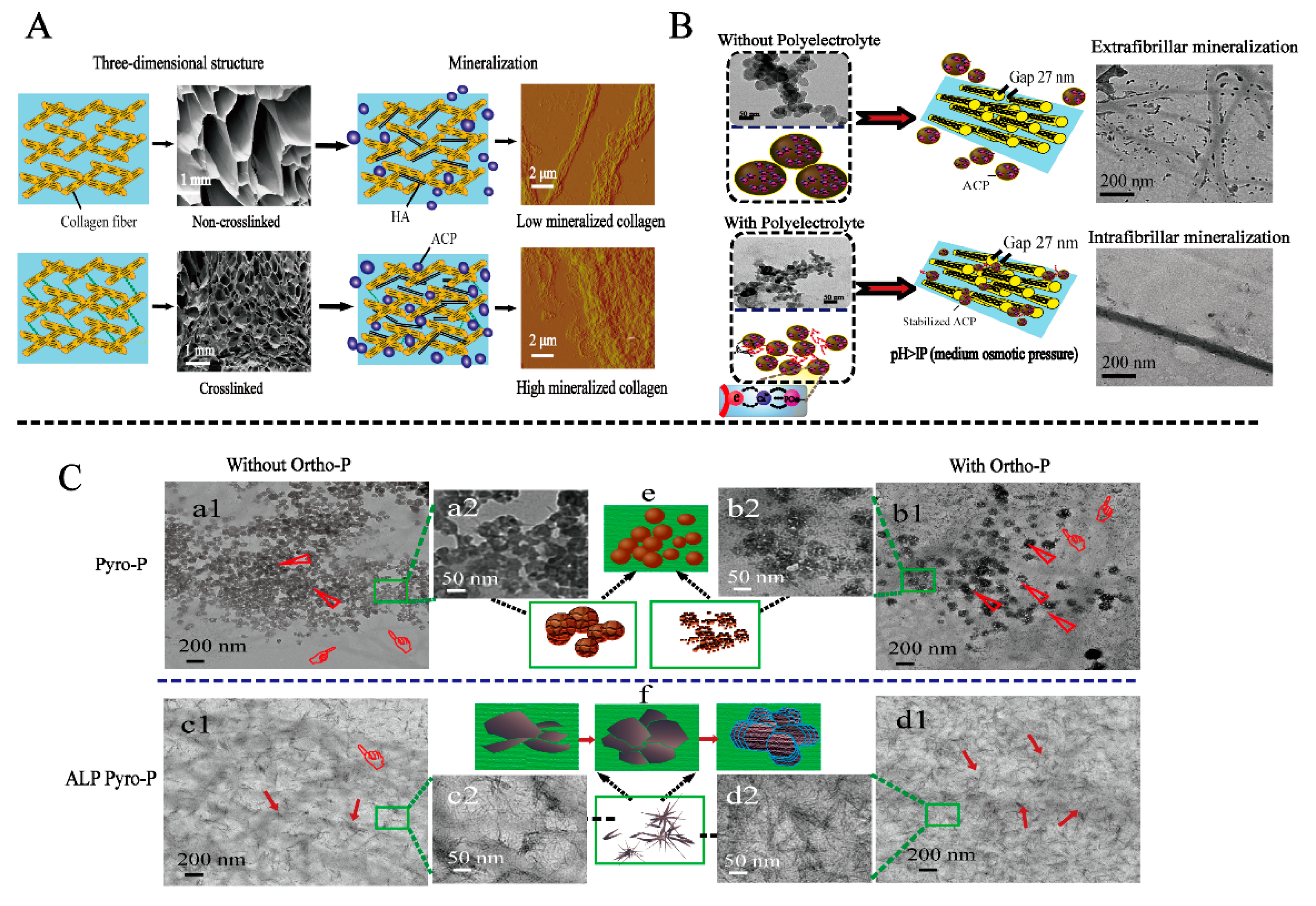
3. Study to Improve the Mechanical Strength of Mineralized Collagen
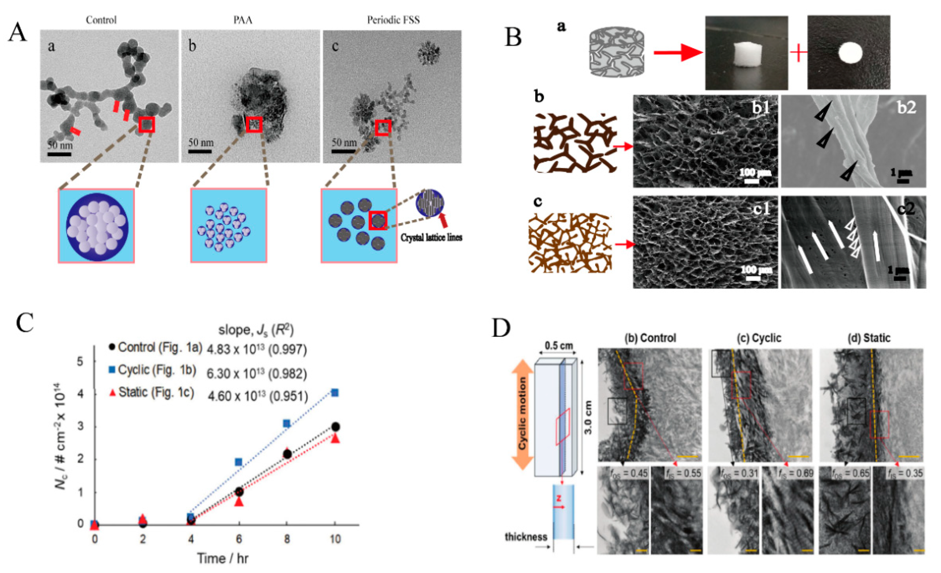
3.1. Force to Promote the Mineralization of Collagen
3.2. Collagen Fiber Arrangement Affects Mineralization
3.3. Other Methods to Promote the Mineralization of Collagen
4. Method for Detecting Mechanical Properties of Mineralized Collagen
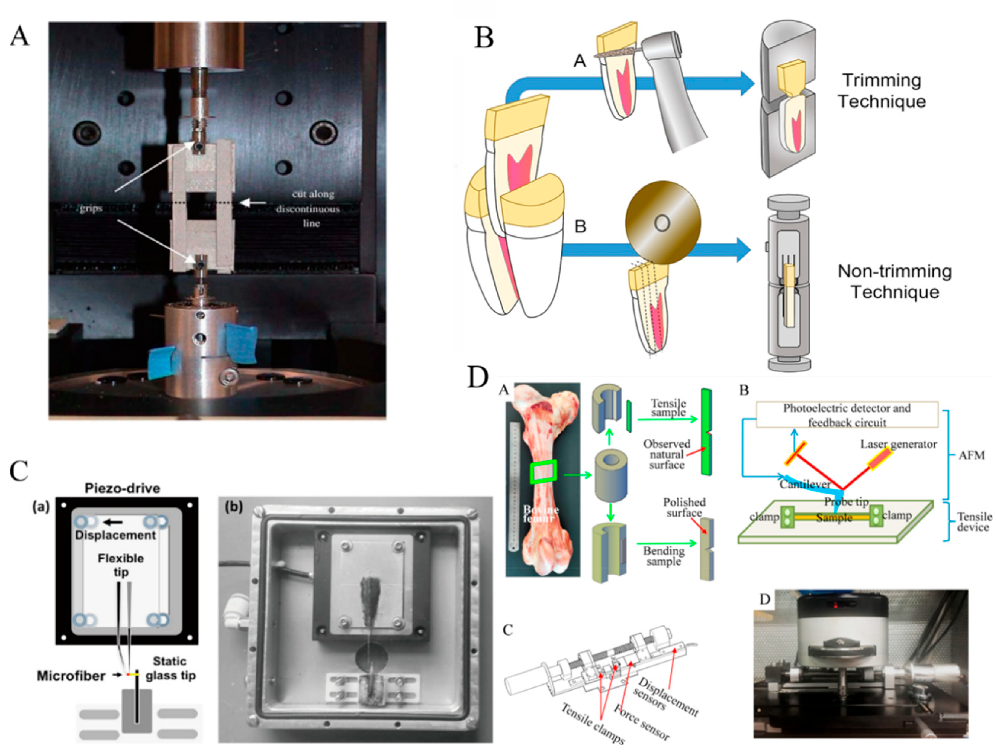
4.1. Macroscopic Mechanics Analysis Methods
4.2. Microscopic Mechanics Analysis Methods
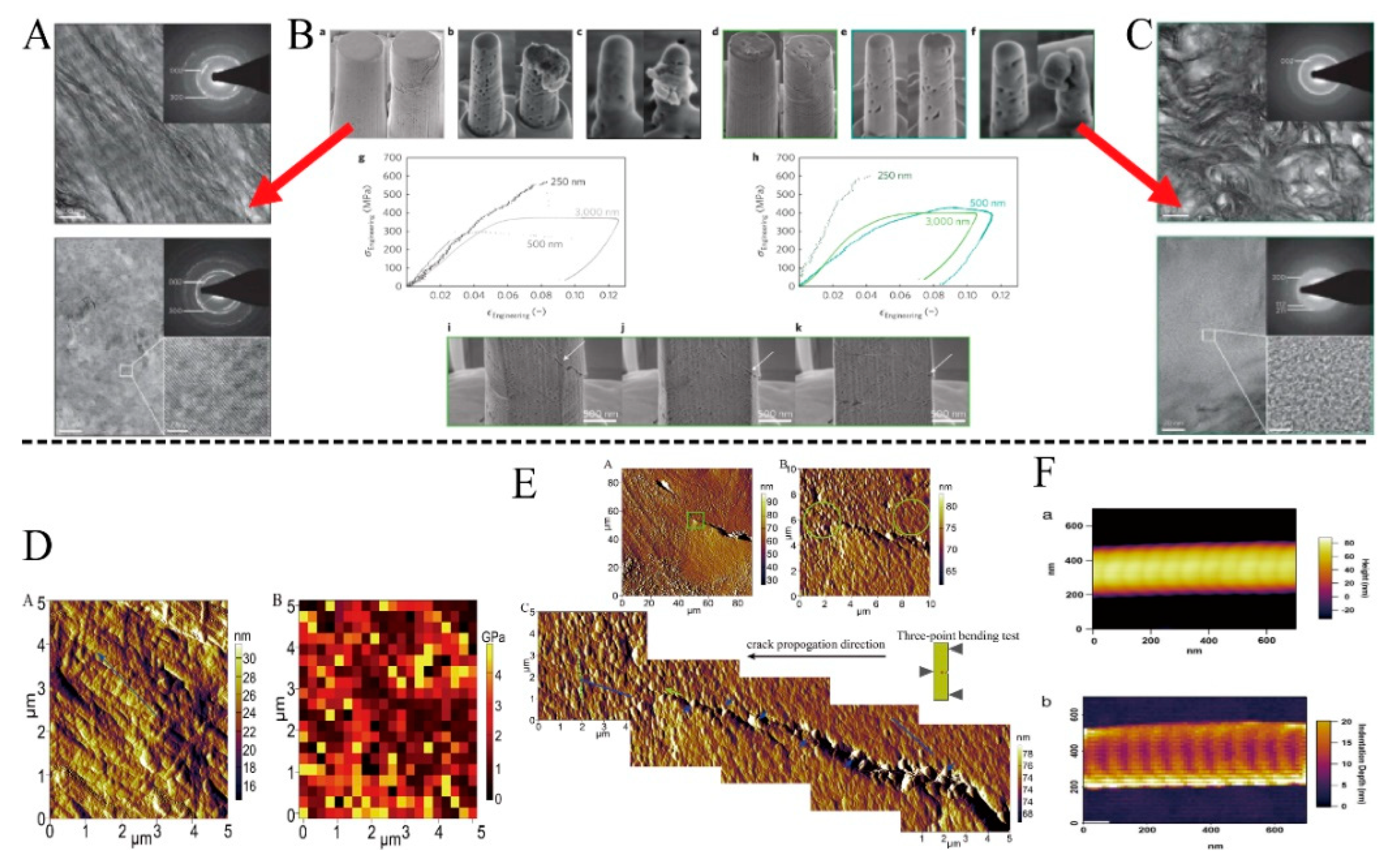
4.2.1. Micro Stretching
4.2.2. Nanoindentation and Scratch Experiments
4.2.3. AFM
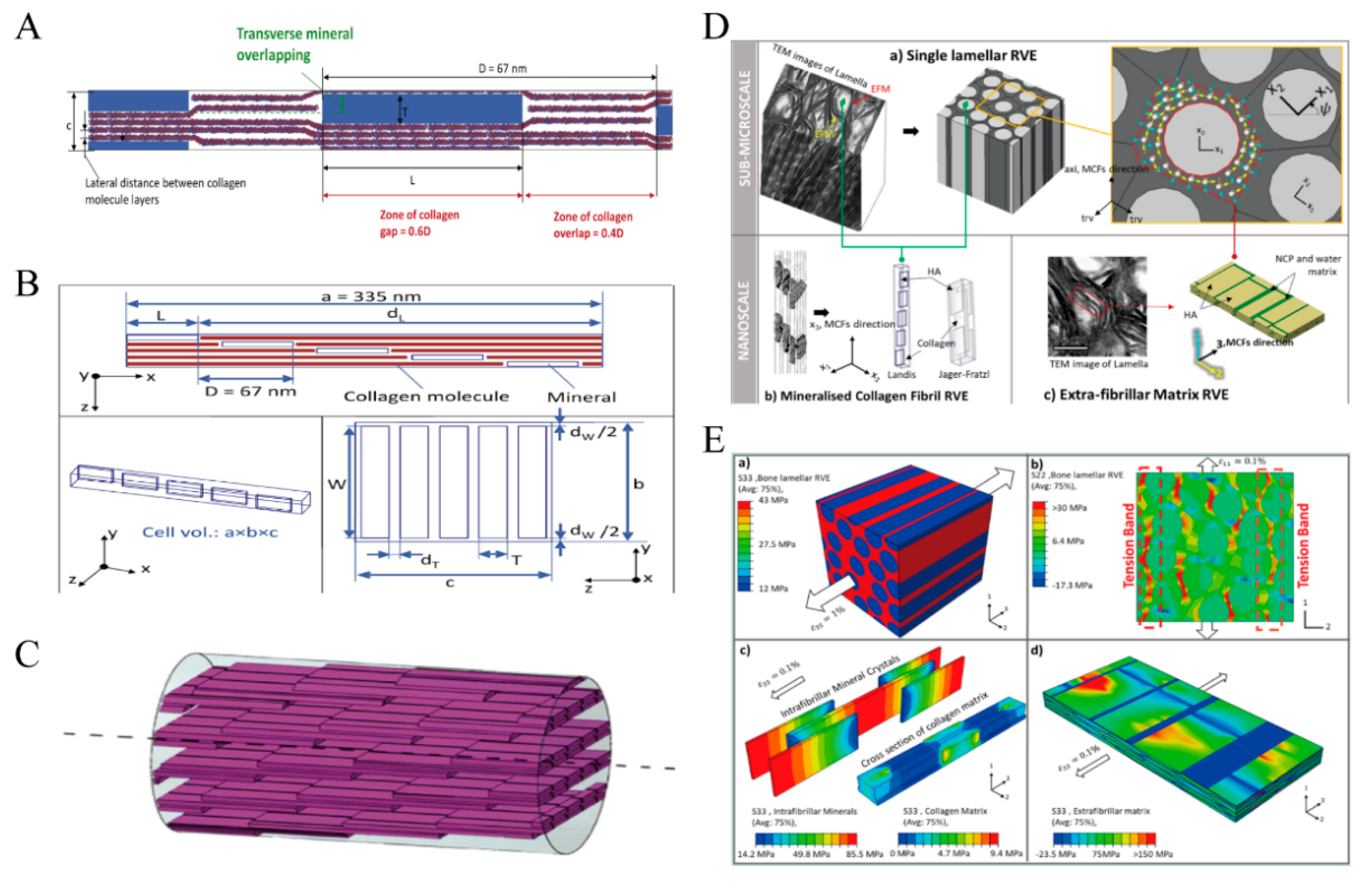
4.3. Simulation Analysis Method
| Reference | Subject | Method | Detecting Parameter |
|---|---|---|---|
| Tan [133] | polycaprolactone electrospun ultrafine fiber | fiber stretching and ex situ observation | tensile malleability |
| Sano [132] | dentin | fiber stretching and in situ observation | bond strength |
| Koester [138] | bone | In situ mechanical test with SEM | mechanical properties of the longitudinal and transverse orientations of the bone |
| Hengsberger [139] | cortical bone of cow | nanoindentation | elastic modulus |
| Isaksson [142] | cortical bone of rabbit | nanoindentation | elastic modulus and viscoelastic parameters |
| Stanishevsky [145] | HA nanoparticle-collagen composites electrospinning | nanoindentation | Young’s modulus and hardness |
| Grant [149] | collagen fibrils | AFM | elastic (static) and viscous (dynamic) responses |
| Qian [136] | Bovine Cortical Bone | AFM | crack propagation |
| Jäger [151] | Bone (submicron) | FEM staggered arrangement model | elastic modulus and fracture stress |
| Wang [152] | Bone (submicron) | FEM 2D shear lag model | an initial crack |
| Vercher [153] | lamellar bone | FEM a 3D finite element model | elastic properties |
| Alijani [154] | lamellar bone | FEM intra and extra-fibrillar mineralization model | elastic properties |
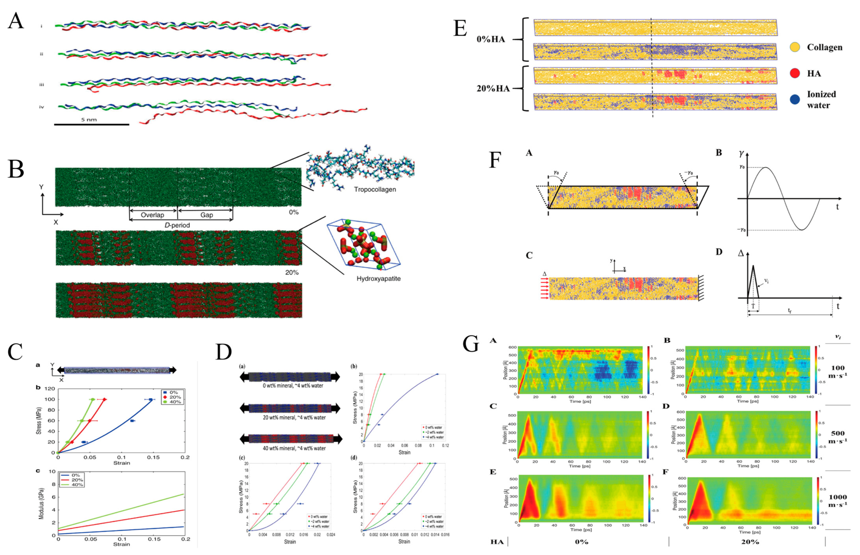
| Buehler [155] | collagen microfibril | MD simulation | Young’s modulus fracture stress (mineral) |
| Nair [4] | collagen microfibril | MD simulation | modulus of tension (mineral) |
| Nair [156] | collagen microfibril | MD simulation | modulus of compression (mineral) |
| Milazzo [158] | collagen microfibril | MD simulation | Viscoelasticity (mineral and water content) |
5. Conclusions
Author Contributions
Funding
Data Availability Statement
Conflicts of Interest
References
- Hamed, E.; Jasiuk, I. Multiscale damage and strength of lamellar bone modeled by cohesive finite elements. J. Mech. Behav. Biomed. Mater. 2013, 28, 94–110. [Google Scholar] [CrossRef] [PubMed]
- Wingender, B.; Bradley, P.; Saxena, N.; Ruberti, J.W.; Gower, L. Biomimetic organization of collagen matrices to template bone-like microstructures. Matrix Biol. 2016, 52–54, 384–396. [Google Scholar] [CrossRef] [Green Version]
- Kikuchi, M.; Ikoma, T.; Itoh, S.; Matsumoto, H.N.; Koyama, Y.; Takakuda, K.; Shinomiya, K.; Tanaka, J. Biomimetic synthesis of bone-like nanocomposites using the self-organization mechanism of hydroxyapatite and collagen. Compos. Sci. Technol. 2004, 64, 819–825. [Google Scholar] [CrossRef]
- Nair, A.K.; Gautieri, A.; Chang, S.W.; Buehler, M.J. Molecular mechanics of mineralized collagen fibrils in bone. Nat. Commun. 2013, 4, 1724–1729. [Google Scholar] [CrossRef] [Green Version]
- Noitup, P.; Garnjanagoonchorn, W.; Morrissey, M.T. Fish skin type I collagen: Characteristic comparison of albacore tuna (Thunnus alalunga) and silver-line grunt (Pomadasys kaakan). J. Aquat. Food Prod. Technol. 2005, 14, 17–28. [Google Scholar] [CrossRef]
- Rho, J.; Kuhn-Spearing, L.; Zioupos, P. Mechanical properties and the hierarchical structure of bone. Med. Eng. Phys. 1998, 20, 92–102. [Google Scholar] [CrossRef]
- Fang, W.J.; Ping, H.; Huang, Y.; Xie, H.; Wang, H.; Wang, W.M.; Fu, Z.Y. Growth of mineralized collagen films by oriented calcium fluoride nanocrystal assembly with enhanced cell proliferation. J. Mat. Chem. B 2021, 9, 6668–6677. [Google Scholar] [CrossRef] [PubMed]
- Cui, F.; Li, Y.; Ge, J. Self-assembly of mineralized collagen composites. Mat. Sci. Eng. R 2007, 57, 1–27. [Google Scholar] [CrossRef]
- Weiner, S.; Wagner, H.D. The material bone: Structure mechanical function relations. Annu. Rev. Mater. Sci. 1998, 28, 271–298. [Google Scholar] [CrossRef]
- Reznikov, N.; Shahar, R.; Weiner, S. Bone hierarchical structure in three dimensions. Acta Biomater. 2014, 10, 3815–3826. [Google Scholar] [CrossRef]
- Reznikov, N.; Bilton, M.; Lari, L.; Stevens, M.M.; Kroger, R. Fractal-like hierarchical organization of bone begins at the nanoscale. Science 2018, 360, 507. [Google Scholar] [CrossRef] [PubMed] [Green Version]
- Tertuliano, O.A.; Greer, J.R. The nanocomposite nature of bone drives its strength and damage resistance. Nat. Mater. 2016, 15, 1195–1202. [Google Scholar] [CrossRef] [Green Version]
- Skedros, J.G.; Dayton, M.R.; Sybrowsky, C.L.; Bloebaum, R.D.; Bachus, K.N. The influence of collagen fiber orientation and other histocompositional characteristics on the mechanical properties of equine cortical bone. J. Exp. Biol. 2006, 209, 3025–3042. [Google Scholar] [CrossRef] [Green Version]
- Oftadeh, R.; Perez-Viloria, M.; Villa-Camacho, J.C.; Vaziri, A.; Nazarian, A. Biomechanics and Mechanobiology of Trabecular Bone: A Review. J. Biomech. Eng.-Trans. ASME 2015, 137, 010802–01080215. [Google Scholar] [CrossRef] [Green Version]
- Launey, M.E.; Buehler, M.J.; Ritchie, R.O. On the Mechanistic Origins of Toughness in Bone. Annu. Rev. Mater. Rev. 2010, 40, 25–53. [Google Scholar] [CrossRef] [Green Version]
- Lu, S.Y.; Jiang, D.J.; Liu, S.H.; Liang, H.F.; Lu, J.R.; Xu, H.; Li, J.; Xiao, J.; Zhang, J.; Fei, Q.M. Effect of different structures fabricated by additive manufacturing on bone ingrowth. J. Biomater. Appl. 2022, 36, 1863–1872. [Google Scholar] [CrossRef] [PubMed]
- Oftadeh, R.; Karimi, Z.; Villa-Camacho, J.; Tanck, E.; Verdonschot, N.; Goebel, R.; Snyder, B.D.; Hashemi, H.N.; Vaziri, A.; Nazarian, A. Curved Beam Computed Tomography based Structural Rigidity Analysis of Bones with Simulated Lytic Defect: A Comparative Study with Finite Element Analysis. Sci. Rep. 2016, 6, 32397. [Google Scholar] [CrossRef] [PubMed] [Green Version]
- Lin, C.Y.; Kang, J.H. Mechanical Properties of Compact Bone Defined by the Stress-Strain Curve Measured Using Uniaxial Tensile Test: A Concise Review and Practical Guide. Materials 2021, 14, 4224. [Google Scholar] [CrossRef]
- Li, S.; Wang, J.Z.; Yin, B.; Hu, Z.S.; Zhang, X.J.; Wu, W.; Liu, G.B.; Liu, Y.K.; Fu, L.; Zhang, Y.Z. Atlas of Human Skeleton Hardness Obtained Using the Micro-indentation Technique. Orthop. Surg. 2021, 13, 1417–1422. [Google Scholar] [CrossRef]
- Duboeuf, F.; Burt-Pichat, B.; Farlay, D.; Suy, P.; Truy, E.; Boivin, G. Bone quality and biomechanical function: A lesson from human ossicles. Bone 2015, 73, 105–110. [Google Scholar] [CrossRef]
- Mieloch, A.A.; Richter, M.; Trzeciak, T.; Giersig, M.; Rybka, J.D. Osteoarthritis Severely Decreases the Elasticity and Hardness of Knee Joint Cartilage: A Nanoindentation Study. J. Clin. Med. 2019, 8, 1865. [Google Scholar] [CrossRef] [Green Version]
- Franke, O.; Durst, K.; Maier, V.; Göken, M.; Birkholz, T.; Schneider, H.; Hennig, F.; Gelse, K. Mechanical properties of hyaline and repair cartilage studied by nanoindentation. Acta Biomater. 2007, 3, 873–881. [Google Scholar] [CrossRef]
- Gnjato, S. Addition to the methodology of research into permanent teeth hardness. Arch. Biol. Sci. 2010, 62, 739–746. [Google Scholar] [CrossRef]
- Hart, N.H.; Nimphius, S.; Rantalainen, T.; Ireland, A.; Siafarikas, A.; Newtonet, R.U. Mechanical basis of bone strength: Influence of bone material, bone structure and muscle action. J. Musculoskelet. Neuronal Interact. 2017, 17, 114. [Google Scholar] [PubMed]
- Ping, H.; Wagermaier, W.; Horbelt, N.; Scoppola, E.; Li, C.; Werner, P.; Fu, Z.; Fratzl, P. Mineralization generates megapascal contractile stresses in collagen fibrils. Sci. (Am. Assoc. Adv. Sci.) 2022, 376, 188–192. [Google Scholar] [CrossRef] [PubMed]
- Levingstone, T.J.; Matsiko, A.; Dickson, G.R.; Brien, F.J.O.; Gleeson, J.P. A biomimetic multi-layered collagen-based scaffold for osteochondral repair. Acta Biomater. 2014, 10, 1996–2004. [Google Scholar] [CrossRef] [PubMed]
- Parkinson, I.H.; Fazzalari, N.L. Interrelationships between structural parameters of cancellous bone reveal accelerated structural change at low bone volume. J. Bone Miner. Res. 2003, 18, 2200–2205. [Google Scholar] [CrossRef]
- Currey, J.D. The design of mineralised hard tissues for their mechanical functions. J. Exp. Biol. 1999, 202, 3285–3294. [Google Scholar] [CrossRef]
- Michael, F.M.; Khalid, M.; Walvekar, R.; Ratnam, C.T.; Ramarad, S.; Siddiqui, H.; Hoque, M.E. Effect of nanofillers on the physico-mechanical properties of load bearing bone implants. Mat. Sci. Eng. 2016, 67, 792–806. [Google Scholar] [CrossRef]
- Stevens, M.M. Biomaterials for bone tissue engineering. Mater. Today 2008, 11, 18–25. [Google Scholar] [CrossRef]
- Paital, S.R.; Dahotre, N.B. Review of laser based biomimetic and bioactive Ca-P coatings. Mater. Sci. Technol. 2008, 24, 1144–1161. [Google Scholar] [CrossRef]
- Sozen, T.; Ozisik, L.; Calik Basaran, N. An overview and management of osteoporosis. Eur. J. Rheumatol. 2017, 4, 46–56. [Google Scholar] [CrossRef]
- Ma, H.; Feng, C.; Chang, J.; Wu, C. 3D-printed bioceramic scaffolds: From bone tissue engineering to tumor therapy. Acta Biomater. 2018, 79, 37–59. [Google Scholar] [CrossRef]
- Ben-Nissan, B. Natural bioceramics: From coral to bone and beyond. Curr. Opin. Solid. State Mat. Sci. 2003, 7, 283–288. [Google Scholar] [CrossRef]
- El-Ghannam, A. Bone reconstruction: From bioceramics to tissue engineering. Expert Rev. Med. Devices 2005, 2, 87–101. [Google Scholar] [CrossRef] [PubMed]
- Liu, X.; Ma, P.X. Polymeric scaffolds for bone tissue engineering. Ann. Biomed. Eng. 2004, 32, 477–486. [Google Scholar] [CrossRef] [PubMed] [Green Version]
- Bharadwaz, A.; Jayasuriya, A.C. Recent trends in the application of widely used natural and synthetic polymer nanocomposites in bone tissue regeneration. Mat. Sci. Eng. C-Mater. 2020, 110, 110698. [Google Scholar] [CrossRef] [PubMed]
- Deshpande, H.; Schindler, C.; Dean, D.; Clem, W.; Bellis, S.L.; Nyairo, E.; Mishra, M.; Thomas, V. Nanocomposite Scaffolds Based on Electrospun Pollycaprolactone/Modified CNF/Nanohydroxyapatite by Electrophoretic Deposition. J. Biomater. Tissue Eng. 2011, 1, 177–184. [Google Scholar] [CrossRef]
- Siddiqui, H.A.; Pickering, K.L.; Mucalo, M.R. A Review on the Use of Hydroxyapatite-Carbonaceous Structure Composites in Bone Replacement Materials for Strengthening Purposes. Materials 2018, 11, 1813. [Google Scholar] [CrossRef] [Green Version]
- Anandagoda, N.; Ezra, D.G.; Cheema, U.; Bailly, M.; Brown, R.A. Hyaluronan hydration generates three-dimensional meso-scale structure in engineered collagen tissues. J. R. Soc. Interface 2012, 9, 2680–2687. [Google Scholar] [CrossRef] [Green Version]
- Lee, S.S.; Hughes, P.; Ross, A.D.; Robinson, M.R. Biodegradable implants for sustained drug release in the eye. Pharm. Res. 2010, 27, 2043–2053. [Google Scholar] [CrossRef]
- Vamsi Krishna, B.; Xue, W.; Bose, S.; Bandyopadhyay, A. Engineered porous metals for implants. JOM 2008, 60, 45–48. [Google Scholar] [CrossRef]
- Weisgerber, D.W.; Erning, K.; Flanagan, C.L.; Hollister, S.J.; Harley, B.A.C. Evaluation of multi-scale mineralized collagen–polycaprolactone composites for bone tissue engineering. J. Mech. Behav. Biomed. Mater. 2016, 61, 318–327. [Google Scholar] [CrossRef] [Green Version]
- Matthews, J.A.; Wnek, G.E.; Simpson, D.G.; Bowlin, G.L. Electrospinning of Collagen Nanofibers. Biomacromolecules 2002, 3, 232–238. [Google Scholar] [CrossRef]
- Nazari, K.A.; Nouri, A.; Hilditch, T. Effects of milling time on powder packing characteristics and compressive mechanical properties of sintered Ti-10Nb-3Mo alloy. Mater. Lett. 2015, 140, 55–58. [Google Scholar] [CrossRef]
- Rony, L.; Lancigu, R.; Hubert, L. Intraosseous metal implants in orthopedics: A review. Morphologie 2018, 102, 231–242. [Google Scholar] [CrossRef] [PubMed]
- Elias, C.N.; Lima, J.; Valiev, R.; Meyers, M.A. Biomedical applications of titanium and its alloys. JOM 2008, 60, 46–49. [Google Scholar] [CrossRef]
- Khorasani, A.M.; Goldberg, M.; Doeven, E.H.; Littlefair, G. Titanium in Biomedical Applications-Properties and Fabrication: A Review. J. Biomater. Tissue Eng. 2015, 5, 593–619. [Google Scholar] [CrossRef]
- Glassman, A.H.; Bobyn, J.D.; Tanzer, M. New femoral designs—Do they influence stress shielding? Clin. Orthop. Rel. Res. 2006, 453, 64–74. [Google Scholar] [CrossRef]
- Zhang, M.; Gregory, T.; Hansen, U.; Cheng, C.K. Effect of stress-shielding-induced bone resorption on glenoid loosening in reverse total shoulder arthroplasty. J. Orthop. Res. 2020, 38, 1566–1574. [Google Scholar] [CrossRef]
- Asri, R.; Harun, W.; Samykano, M.; Lah, N.; Ghani, S.; Tarlochan, F.; Raza, M.R. Corrosion and surface modification on biocompatible metals: A review. Mater. Sci. Eng. C-Mater. Biol. Appl. 2017, 77, 1261–1274. [Google Scholar] [CrossRef] [PubMed] [Green Version]
- Zhang, S.; Li, C.; Hou, W.; Zhao, S.; Li, S. Longitudinal compression behavior of functionally graded Ti–6Al–4V meshes. J. Mater. Sci. Technol. 2016, 32, 1098–1104. [Google Scholar] [CrossRef]
- Lee, H.; Ahn, S.H.; Kim, G.H. Three-dimensional collagen/alginate hybrid scaffolds functionalized with a drug delivery system (DDS) for bone tissue regeneration. Chem. Mater. 2012, 24, 881–891. [Google Scholar] [CrossRef]
- Thompson, M.K.; Moroni, G.; Vaneker, T.; Fadel, G.; Campbell, R.I.; Gibson, I.; Bernard, A.; Schulz, J.; Graf, P.; Ahuja, B.; et al. Design for Additive Manufacturing: Trends, opportunities, considerations, and constraints. Cirp Ann.-Manuf. Technol. 2016, 65, 737–760. [Google Scholar] [CrossRef] [Green Version]
- Pattanayak, D.K.; Fukuda, A.; Matsushita, T.; Takemoto, M.; Fujibayashi, S.; Sasaki, K.; Nishida, N.; Nakamura, T.; Kokubo, T. Bioactive Ti metal analogous to human cancellous bone: Fabrication by selective laser melting and chemical treatments. Acta Biomater. 2011, 7, 1398–1406. [Google Scholar] [CrossRef]
- Liu, Y.; Rath, B.; Tingart, M.; Eschweiler, J. Role of implants surface modification in osseointegration: A systematic review. J. Biomed. Mater. Res. Part A 2020, 108, 470–484. [Google Scholar] [CrossRef] [Green Version]
- Polo-Corrales, L.; Latorre-Esteves, M.; Ramirez-Vick, J.E. Scaffold Design for Bone Regeneration. J. Nanosci. Nanotechnol. 2014, 14, 15–56. [Google Scholar] [CrossRef] [Green Version]
- Yang, H.T.; Qu, X.H.; Wang, M.Q.; Cheng, H.W.; Jia, B.; Nie, J.F.; Dai, K.R.; Zheng, Y.F. Zn-0.4Li alloy shows great potential for the fixation and healing of bone fractures at load-bearing sites. Chem. Eng. J. 2021, 417, 129317. [Google Scholar] [CrossRef]
- McBane, J.E.; Sharifpoor, S.; Cai, K.H.; Labow, R.S.; Santerre, J.P. Biodegradation and in vivo biocompatibility of a degradable, polar/hydrophobic/ionic polyurethane for tissue engineering applications. Biomaterials 2011, 32, 6034–6044. [Google Scholar] [CrossRef]
- Wisniewska, K.; Rybak, Z.; Watrobinski, M.; Struszczyk, M.H.; Filipiak, J.; Zywicka, B.; Szymonowicz, M. Bioresorbable polymeric materials—Current state of knowledge. Polimery 2021, 66, 3–10. [Google Scholar] [CrossRef]
- Ramesh, N.; Moratti, S.C.; Dias, G.J. Hydroxyapatite-polymer biocomposites for bone regeneration: A review of current trends. J. Biomed. Mater. Res. Part B 2018, 106, 2046–2057. [Google Scholar] [CrossRef] [PubMed]
- Park, S.; Gwon, Y.; Kim, W.; Kim, J. Rebirth of the Eggshell Membrane as a Bioactive Nanoscaffold for Tissue Engineering. ACS Biomater. Sci. Eng. 2021, 7, 2219–2224. [Google Scholar] [CrossRef]
- Dorozhkin, S.V. Calcium orthophosphate bioceramics. Ceram. Int. 2015, 41, 13913–13966. [Google Scholar] [CrossRef]
- Dorozhkin, S.V. Calcium Orthophosphates as Bioceramics: State of the Art. J. Funct. Biomater. 2010, 1, 22–107. [Google Scholar] [CrossRef] [PubMed] [Green Version]
- Jakus, A.E.; Rutz, A.L.; Jordan, S.W.; Kannan, A.; Mitchell, S.M.; Yun, C.; Koube, K.D.; Yoo, S.C.; Whiteley, H.E.; Richter, C.; et al. Hyperelastic “bone”: A highly versatile, growth factor-free, osteoregenerative, scalable, and surgically friendly biomaterial. Sci. Transl. Med. 2016, 8, 127r–358r. [Google Scholar] [CrossRef] [PubMed] [Green Version]
- Priyadarshini, B.; Rama, M.; Chetan; Vijayalakshmi, U. Bioactive coating as a surface modification technique for biocompatible metallic implants: A review. J. Asian Ceram. Soc. 2019, 7, 397–406. [Google Scholar] [CrossRef] [Green Version]
- Du, T.; Niu, X.; Cao, P.; Zhang, Y.; Liu, Y.; Yang, H.; Qiao, A. Multifarious roles of metal elements in bone mineralization. Appl. Mater. Today 2023, 32, 101810. [Google Scholar] [CrossRef]
- Devi, K.B.; Tripathy, B.; Kumta, P.N.; Nandi, S.K.; Roy, M. In vivo biocompatibility of zinc-doped magnesium silicate bio-ceramics. ACS Biomater. Sci. Eng. 2018, 4, 62126–62133. [Google Scholar] [CrossRef] [PubMed]
- Balakrishnan, S.; Padmanabhan, V.P.; Kulandaivelu, R.; Nellaiappan, T.S.N.; Sagadevan, S.; Paiman, S.; Mohammad, F.; Al-Lohedan, H.A.; Obulapuram, P.K.; Oh, W.C. Influence of iron doping towards the physicochemical and biological characteristics of hydroxyapatite. Ceram. Int. 2021, 47, 5061–5070. [Google Scholar] [CrossRef]
- Zhang, L.; Zhang, C.; Ji, Y.; Xu, E.; Liu, X.; Zhao, F.; Yuan, H.; Cui, J.; Cui, J. Effects of Z-value on physicochemical and biological properties of β-SiAlONs ceramics. Ceram. Int. 2021, 47, 34810–34819. [Google Scholar] [CrossRef]
- Qian, J.; Zhang, W.; Chen, Y.; Zeng, P.; Wang, J.; Zhou, C.; Zeng, H.; Sang, H.; Huang, N.; Zhang, H.; et al. Osteogenic and angiogenic bioactive collagen entrapped calcium/zinc phosphates coating on biodegradable Zn for orthopedic implant applications. Biomater. Adv. 2022, 136, 212792. [Google Scholar] [CrossRef] [PubMed]
- Pang, L.; Zhao, R.; Chen, J.; Ding, J.; Chen, X.; Chai, W.; Cui, X.; Li, X.; Wang, D.; Pan, H. Osteogenic and anti-tumor Cu and Mn-doped borosilicate nanoparticles for syncretic bone repair and chemodynamic therapy in bone tumor treatment. Bioact. Mater. 2022, 12, 1–15. [Google Scholar] [CrossRef]
- Wu, C.; Ramaswamy, Y.; Kwik, D.; Zreiqat, H. The effect of strontium incorporation into CaSiO3 ceramics on their physical and biological properties. Biomaterials 2007, 28, 3171–3181. [Google Scholar] [CrossRef] [PubMed]
- Borciani, G.; Ciapetti, G.; Vitale-Brovarone, C.; Baldini, N. Strontium functionalization of biomaterials for bone tissue engineering purposes: A biological point of view. Materials 2022, 15, 1724. [Google Scholar] [CrossRef] [PubMed]
- Peng, S.; Liu, X.S.; Huang, S.; Li, Z.; Pan, H.; Zhen, W.; Luk, K.D.K.; Guo, X.E.; Lu, W.W. The cross-talk between osteoclasts and osteoblasts in response to strontium treatment: Involvement of osteoprotegerin. Bone 2011, 49, 1290–1298. [Google Scholar] [CrossRef]
- Devi, K.B.; Tripathy, B.; Roy, A.; Lee, B.; Kumta, P.N.; Nandi, S.K.; Roy, M. In vitro biodegradation and in vivo biocompatibility of forsterite bio-ceramics: Effects of strontium substitution. ACS Biomater. Sci. Eng. 2018, 5, 530–543. [Google Scholar] [CrossRef]
- Ibrahimzade, L.; Kaygili, O.; Dundar, S.; Ates, T.; Dorozhkin, S.V.; Bulut, N.; Koytepe, S.; Ercan, F.; Gürses, C.; Hssain, A.H. Theoretical and experimental characterization of Pr/Ce co-doped hydroxyapatites. J. Mol. Struct. 2021, 1240, 30557. [Google Scholar] [CrossRef]
- Shekhawat, D.; Singh, A.; Banerjee, M.K.; Singh, T.; Patnaik, A. Bioceramic composites for orthopaedic applications: A comprehensive review of mechanical, biological, and microstructural properties. Ceram. Int. 2021, 47, 3013–3030. [Google Scholar] [CrossRef]
- Albalwi, H.A.; El-Naggar, M.E.; Abou Taleb, M.; Kalam, A.; Alghamdi, N.A.; Mostafa, M.S.; Salem, S.; Afifi, M. Medical applications of ternary nanocomposites based on hydroxyapatite/ytterbium oxide/graphene oxide: Potential bone tissue engineering and antibacterial properties. J. Mater. Res. Technol. 2022, 18, 4834–4845. [Google Scholar] [CrossRef]
- Jazayeri, H.E.; Rodriguez-Romero, M.; Razavi, M.; Tahriri, M.; Ganjawalla, K.; Rasoulianboroujeni, M.; Malekoshoaraie, M.H.; Khoshroo, K.; Tayebi, L. The cross-disciplinary emergence of 3D printed bioceramic scaffolds in orthopedic bioengineering. Ceram. Int. 2018, 44, 1–9. [Google Scholar]
- Conrad, B.; Yang, F. Hydroxyapatite-coated gelatin microribbon scaffolds induce rapid endogenous cranial bone regeneration in vivo. Biomater. Adv. 2022, 140, 213050. [Google Scholar] [CrossRef]
- Chen, Z.J.; Zhang, Y.; Zheng, L.; Zhang, H.; Shi, H.H.; Zhang, X.C.; Liu, B. Mineralized self-assembled silk fibroin/cellulose interpenetrating network aerogel for bone tissue engineering. Biomater. Adv. 2022, 134, 112549. [Google Scholar] [CrossRef]
- Lu, H.H.; El-Amin, S.F.; Scott, K.D.; Laurencin, C.T. Three-dimensional, bioactive, biodegradable, polymer–bioactive glass composite scaffolds with improved mechanical properties support collagen synthesis and mineralization of human osteoblast-like cells in vitro. J. Biomed. Mater. Res. A 2003, 64, 465–474. [Google Scholar] [CrossRef] [PubMed]
- Tsigkou, O.; Hench, L.L.; Boccaccini, A.R.; Polak, J.M. Enhanced differentiation and mineralization of human fetal osteoblasts on PDLLA containing Bioglass® composite films in the absence of osteogenic supplements. J. Biomed. Mater. Res. A 2007, 80, 837–851. [Google Scholar] [CrossRef] [PubMed]
- Yang, C.R.; Wang, Y.J.; Chen, X.F. Mineralization regulation and biological influence of bioactive glass-collagen-phosphatidylserine composite scaffolds. Sci. China Life. Sci. 2012, 55, 236–240. [Google Scholar] [CrossRef] [Green Version]
- Miri, A.K.; Muja, N.; Kamranpour, N.O.; Lepry, W.C.; Boccaccini, A.R.; Clarke, S.A.; Nazhat, S.N. Ectopic bone formation in rapidly fabricated acellular injectable dense collagen-Bioglass hybrid scaffolds via gel aspiration-ejection. Biomaterials 2016, 85, 128–141. [Google Scholar] [CrossRef] [PubMed] [Green Version]
- Vuornos, K.; Ojansivu, M.; Koivisto, J.T.; Häkkänen, H.; Belay, B.; Montonen, T.; Huhtala, H.; Kääriäinen, M.; Hupa, L.; Kellomäki, M.; et al. Bioactive glass ions induce efficient osteogenic differentiation of human adipose stem cells encapsulated in gellan gum and collagen type I hydrogels. Mat. Sci. Eng. C Mater. 2019, 99, 905–918. [Google Scholar] [CrossRef]
- Gurumurthy, B.; Pal, P.; Griggs, J.A.; Janorkar, A.V. Optimization of collagen-elastin-like polypeptide-bioglass scaffold composition for osteogenic differentiation of adipose-derived stem cells. Materialia 2020, 9, 100572. [Google Scholar] [CrossRef]
- Ferreira, S.A.; Young, G.; Jones, J.R.; Rankin, S. Bioglass/carbonate apatite/collagen composite scaffold dissolution products promote human osteoblast differentiation. Mat. Sci. Eng. C Mater. 2021, 118, 111393. [Google Scholar] [CrossRef]
- Brovold, M.; Almeida, J.I.; Pla-Palacin, I.; Sainz-Arnal, P.; Sanchez-Romero, N.; Rivas, J.J.; Almeida, H.; Dachary, P.R.; Serrano-Aullo, T.; Soker, S.; et al. Naturally-derived biomaterials for tissue engineering applications. Adv. Exp. Med. Biol. 2018, 1077, 421–449. [Google Scholar]
- Wang, S.; Zhao, Z.J.; Yang, Y.D.; Mikos, A.G.; Qiu, Z.Y.; Song, T.X.; Cui, F.Z.; Wang, X.M.; Zhang, C.Y. A high-strength mineralized collagen bone scaffold for large-sized cranial bone defect repair in sheep. Regen. Biomater. 2018, 5, 283–292. [Google Scholar] [CrossRef] [Green Version]
- Wang, S.; Yang, Y.D.; Koons, G.L.; Mikos, A.G.; Qiu, Z.Y.; Song, T.X.; Cui, F.Z.; Wang, X.M. Tuning pore features of mineralized collagen/PCL scaffolds for cranial bone regeneration in a rat model. Mat. Sci. Eng. C Mater. 2020, 106, 110186. [Google Scholar] [CrossRef]
- Ding, Q.F.; Cui, J.J.; Shen, H.Q.; He, C.L.; Wang, X.D.; Shen, S.; Lin, K.L. Advances of nanomaterial applications in oral and maxillofacial tissue regeneration and disease treatment. Wiley Interdiscip. Rev.-Nanomed. Nanobiotechnol. 2021, 13, e1669. [Google Scholar] [CrossRef]
- Boda, S.K.; Almoshari, Y.; Wang, H.; Wang, X.; Reinhardt, R.A.; Duan, B.; Wang, D.; Xie, J. Mineralized nanofiber segments coupled with calcium-binding BMP-2 peptides for alveolar bone regeneration. Acta Biomater. 2019, 85, 282–293. [Google Scholar] [CrossRef]
- Jain, G.; Blaauw, D.; Chang, S. A Comparative Study of Two Bone Graft Substitutes-InterOss((R)) Collagen and OCS-B Collagen((R)). J. Funct. Biomater. 2022, 13, 28. [Google Scholar] [CrossRef] [PubMed]
- Sionkowska, A.; Kozlowska, J. Properties and modification of porous 3-D collagen/hydroxyapatite composites. Int. J. Biol. Macromol. 2013, 52, 250–259. [Google Scholar] [CrossRef] [PubMed]
- Calabrese, G.; Giuffrida, R.; Fabbi, C.; Figallo, E.; Lo Furno, D.; Gulino, R.; Colarossi, C.; Fullone, F.; Giuffrida, R.; Parenti, R.; et al. Collagen-Hydroxyapatite Scaffolds Induce Human Adipose Derived Stem Cells Osteogenic Differentiation In Vitro. PLoS ONE 2016, 11, e0151181. [Google Scholar] [CrossRef] [Green Version]
- Cunniffe, G.M.; Dickson, G.R.; Partap, S.; Stanton, K.T.; O’Brien, F.J. Development and characterisation of a collagen nano-hydroxyapatite composite scaffold for bone tissue engineering. J. Mater. Sci.-Mater. Med. 2010, 21, 2293–2298. [Google Scholar] [CrossRef]
- Wang, L.; You, X.; Zhang, L.; Zhang, C.; Zou, W. Mechanical regulation of bone remodeling. Bone Res. 2022, 10, 16. [Google Scholar] [CrossRef] [PubMed]
- Almeida, M.; Han, L.; Martin-Millan, M.; O’Brien, C.A.; Manolagas, S.C. Oxidative Stress Antagonizes Wnt Signaling in Osteoblast Precursors by Diverting β-Catenin from T Cell Factor- to Forkhead Box O-mediated Transcription. J. Biol. Chem. 2007, 282, 27298–27305. [Google Scholar] [CrossRef] [Green Version]
- Aguirre, J.I.; Plotkin, L.I.; Stewart, S.A.; Weinstein, R.S.; Parfitt, A.M.; Manolagas, S.C.; Bellido, T. Osteocyte apoptosis is induced by weightlessness in mice and precedes osteoclast recruitment and bone loss. J. Bone Miner. Res. 2006, 21, 605–615. [Google Scholar] [CrossRef]
- Ziros, P.G.; Gil, A.P.; Georgakopoulos, T.; Habeos, I.; Kletsas, D.; Basdra, E.K.; Papavassiliou, A.G. The bone-specific transcriptional regulator Cbfa1 is a target of mechanical signals in osteoblastic cells. J. Biol. Chem. 2002, 277, 23934–23941. [Google Scholar] [CrossRef] [PubMed] [Green Version]
- Schaffler, M.B.; Cheung, W.Y.; Majeska, R.; Kennedy, O. Osteocytes: Master orchestrators of bone. Calcif. Tissue Int. 2014, 94, 5–24. [Google Scholar] [CrossRef] [PubMed] [Green Version]
- Bonewald, L.F. The amazing osteocyte. J. Bone Miner. Res. 2011, 26, 229–238. [Google Scholar] [CrossRef] [PubMed]
- Kurata, K.; Uemura, T.; Nemoto, A.; Tateishi, T.; Murakami, T.; Higaki, H.; Miura, H.; Iwamoto, Y. Mechanical strain effect on bone-resorbing activity and messenger RNA expressions of marker enzymes in isolated osteoclast culture. J. Bone Miner. Res. 2001, 16, 722–730. [Google Scholar] [CrossRef] [PubMed]
- Huiskes, R.; Ruimerman, R.; van Lenthe, G.H.; Janssen, J.D. Effects of mechanical forces on maintenance and adaptation of form in trabecular bone. Nature 2000, 405, 704–706. [Google Scholar] [CrossRef]
- Kivell, T.L. A review of trabecular bone functional adaptation: What have we learned from trabecular analyses in extant hominoids and what can we apply to fossils? J. Anat. 2016, 228, 569–594. [Google Scholar] [CrossRef] [PubMed] [Green Version]
- Peyroteo, M.; Belinha, J.; Vinga, S.; Dinis, L.; Natal Jorge, R. A Model for Bone Remodeling: Cellular Dynamics and Mechanical Loading. In Proceedings of the 2017 IEEE 5th Portuguese Meeting on Bioengineering, Coimbra, Portugal, 16–18 February 2017; pp. 1–4. [Google Scholar]
- Lee, P.; Lin, R.; Moon, J.; Lee, L.P. Microfluidic alignment of collagen fibers for in vitro cell culture. Biomed. Microdevices 2006, 8, 35–41. [Google Scholar] [CrossRef]
- Niu, X.; Fan, R.; Guo, X.; Du, T.; Yang, Z.; Feng, Q.; Fan, Y. Shear-mediated orientational mineralization of bone apatite on collagen fibrils. J. Mat. Chem. B 2017, 5, 9141–9147. [Google Scholar] [CrossRef]
- Du, T.; Niu, X.; Hou, S.; Xu, M.; Li, Z.; Li, P.; Fan, Y. Highly aligned hierarchical intrafibrillar mineralization of collagen induced by periodic fluid shear stress. J. Mat. Chem. B 2020, 8, 2562–2572. [Google Scholar] [CrossRef]
- Kim, D.; Lee, B.; Marshall, B.; Thomopoulos, S.; Jun, Y.S. Cyclic strain enhances the early stage mineral nucleation and the modulus of demineralized bone matrix. Biomater. Sci. 2021, 9, 5907–5916. [Google Scholar] [CrossRef] [PubMed]
- Daood, U.; Fawzy, A.S. Minimally invasive high-intensity focused ultrasound (HIFU) improves dentine remineralization with hydroxyapatite nanorods. Dent. Mater. 2020, 36, 456–467. [Google Scholar] [CrossRef]
- Du, T.M.; Niu, Y.M.; Liu, Y.J.; Yang, H.S.; Qiao, A.K.; Niu, X.F. Physical and Chemical Characterization of Biomineralized Collagen with Different Microstructures. J. Funct. Biomater. 2022, 13, 57. [Google Scholar] [CrossRef]
- Giraud-Guille, M.M. Twisted Plywood Architecture of Collagen Fibrils in Human Compact Bone Osteons. Calcif. Tissue Int. 1988, 42, 167–180. [Google Scholar] [CrossRef]
- Stockhausen, K.E.; Qwamizadeh, M.; Wölfel, E.M.; Hemmatian, H.; Fiedler, I.A.K.; Flenner, S.; Longo, E.; Amling, M.; Greving, I.; Ritchie, R.O.; et al. Collagen fiber orientation is coupled with specific nano-compositional patterns in dark and bright osteons modulating their biomechanical properties. ACS Nano 2021, 15, 455–467. [Google Scholar] [CrossRef]
- Wang, Y.; Ural, A. A finite element study evaluating the influence of mineralization distribution and content on the tensile mechanical response of mineralized collagen fibril networks. J. Biomed. Mater. Res. A 2019, 100, 103361. [Google Scholar] [CrossRef]
- Wang, Y.; Ural, A. Effect of modifications in mineralized collagen fibril and extra-fibrillar matrix material properties on submi-croscale mechanical behavior of cortical bone. J. Mech. Behav. Biomed. Mater. 2018, 82, 18–26. [Google Scholar] [CrossRef] [PubMed]
- Siegmund, T.; Allen, M.R.; Burr, D.B. Failure of mineralized collagen fibrils: Modeling the role of collagen cross-linking. J. biomech. 2008, 41, 1427–1435. [Google Scholar] [CrossRef]
- Dhand, C.; Ong, S.T.; Dwivedi, N.; Diaz, S.M.; Venugopal, J.R.; Navaneethan, B.; Fazil, M.H.; Liu, S.; Seitz, V.; Wintermantel, E.; et al. Bio-inspired in situ crosslinking and mineralization of electrospun collagen scaffolds for bone tissue engineering. Biomaterials 2016, 104, 323–338. [Google Scholar] [CrossRef]
- Du, T.M.; Niu, X.F.; Li, Z.W.; Li, P.; Feng, Q.L.; Fan, Y.B. Crosslinking induces high mineralization of apatite minerals on collagen fibers. Int. J. Biol. Macromol. 2018, 113, 450–457. [Google Scholar] [CrossRef] [PubMed]
- Kikuchi, M.; Matsumoto, H.N.; Yamada, T.; Koyama, Y.; Takakuda, K.; Tanaka, J. Glutaraldehyde cross-linked hydroxyapatite/collagen self-organized nanocomposites. Biomaterials 2004, 25, 63–69. [Google Scholar] [CrossRef] [PubMed]
- Du, T.; Niu, X.; Hou, S.; Li, Z.; Li, P.; Fan, Y. Apatite minerals derived from collagen phosphorylation modification induce the hierarchical intrafibrillar mineralization of collagen fibers. J. Biomed. Mater. Res. Part A 2019, 107, 2403–2413. [Google Scholar] [CrossRef] [PubMed]
- Nassif, N.; Martineau, F.; Syzgantseva, O.; Gobeaux, F.; Willinger, M.; Coradin, T.; Cassaignon, S.; Azaïs, T.; Giraud-Guille, M.M. In Vivo Inspired Conditions to Synthesize Biomimetic Hydroxyapatite. Chem. Mat. 2010, 22, 3653–3663. [Google Scholar] [CrossRef]
- Niederberger, M.; Colfen, H. Oriented attachment and mesocrystals: Non-classical crystallization mechanisms based on nanoparticle assembly. Phys. Chem. Chem. Phys. 2006, 8, 3271–3287. [Google Scholar] [CrossRef] [PubMed]
- Liu, Y.; Luo, D.; Wang, T. Hierarchical structures of bone and bioinspired bone tissue engineering. Small 2016, 12, 4611–4632. [Google Scholar] [CrossRef]
- Lima, D.B.; de Souza, M.; de Lima, G.G.; Souto, E.; Oliveira, H.; Fook, M.; de Sa, M. Injectable bone substitute based on chitosan with polyethylene glycol polymeric solution and biphasic calcium phosphate microspheres. Carbohydr. Polym. 2020, 245, 116575. [Google Scholar] [CrossRef] [PubMed]
- Du, T.; Niu, Y.; Jia, Z.; Liu, Y.; Qiao, A.; Yang, H.; Niu, X. Orthophosphate and alkaline phosphatase induced the formation of apatite with different multilayered structures and mineralization balance. Nanoscale 2022, 14, 1814–1825. [Google Scholar] [CrossRef]
- Thula, T.T.; Rodriguez, D.E.; Lee, M.H.; Pendi, L.; Podschun, J.; Gower, L.B. In vitro mineralization of dense collagen substrates: A biomimetic approach toward the development of bone-graft materials. Acta Biomater. 2011, 7, 3158–3169. [Google Scholar] [CrossRef] [Green Version]
- Thula, T.T.; Svedlund, F.; Rodriguez, D.E.; Podschun, J.; Pendi, L.; Gower, L.B. Mimicking the Nanostructure of Bone: Comparison of Polymeric Process-Directing Agents. Polymers 2011, 3, 10–35. [Google Scholar] [CrossRef] [Green Version]
- Höhling, H.J.; Barckhaus, R.H.; Krefting, E.-R.; Althoff, J.; Quint, P. Collagen mineralization: Aspects of the structural relationship between collagen and the apatitic crystallites. Ultrastruct. Skelet. Tissues 1990, 7, 41–62. [Google Scholar]
- Sano, H.; Chowdhury, A.F.M.A.; Saikaew, P.; Matsumoto, M.; Hoshika, S.; Yamauti, M. The microtensile bond strength test: Its historical background and application to bond testing. Jpn. Dent. Sci. Rev. 2020, 56, 24–31. [Google Scholar] [CrossRef]
- Tan, E.P.; Ng, S.Y.; Lim, C.T. Tensile testing of a single ultrafine polymeric fiber. Biomaterials 2005, 26, 1453–1456. [Google Scholar] [CrossRef]
- Xiang, D.H.; Zhang, Z.M.; Wu, B.F.; Feng, H.R.; Shi, Z.L.; Zhao, B. Effect of Ultrasonic Vibration Tensile on the Mechanical Properties of High-Volume Fraction SiCp/Al Composite. Int. J. Precis. Eng. Manuf. 2020, 21, 2051–2066. [Google Scholar] [CrossRef]
- Chatzipanagis, K.; Baumann, C.G.; Sandri, M.; Sprio, S.; Tampieri, A.; Kröger, R. In situ mechanical and molecular investigations of collagen/apatite biomimetic composites combining Raman spectroscopy and stress-strain analysis. Acta Biomater. 2016, 46, 278–285. [Google Scholar] [CrossRef] [PubMed] [Green Version]
- Qian, T.; Chen, X.; Hang, F. Investigation of nanoscale failure behaviour of cortical bone under stress by AFM. J. Mech. Behav. Biomed. Mater. 2020, 112, 103989. [Google Scholar] [CrossRef]
- Ritchie, R.O.; Nalla, R.K.; Kruzic, J.J.; Ager, J.W.; Balooch, G.; Kinney, J.H. Fracture and ageing in bone: Toughness and structural characterization. Strain 2006, 42, 225–232. [Google Scholar] [CrossRef]
- Koester, K.J.; Ager, J.W.; Ritchie, R.O. The true toughness of human cortical bone measured with realistically short cracks. Nat. Mater. 2008, 7, 672–677. [Google Scholar] [CrossRef]
- Hengsberger, S.; Enstroem, J.; Peyrin, F.; Zysset, P. How is the indentation modulus of bone tissue related to its macroscopic elastic response? A validation study. J. Biomech. 2003, 36, 1503–1509. [Google Scholar] [CrossRef]
- Hoffler, C.E.; Guo, X.E.; Zysset, P.K.; Goldstein, S.A. An application of nanoindentation technique to measure bone tissue lamellae properties. J. Biomech. Eng.-Trans. ASME 2005, 127, 1046–1053. [Google Scholar] [CrossRef]
- Hengsberger, S.; Kulik, A.; Zysset, P. Nanoindentation discriminates the elastic properties of individual human bone lamellae under dry and physiological conditions. Bone 2002, 30, 178–184. [Google Scholar] [CrossRef] [PubMed]
- Isaksson, H.; Malkiewicz, M.; Nowak, R.; Helminen, H.J.; Jurvelin, J.S. Rabbit cortical bone tissue increases its elastic stiffness but becomes less viscoelastic with age. Bone 2010, 47, 1030–1038. [Google Scholar] [CrossRef] [PubMed]
- Wu, Z.H.; Baker, T.A.; Ovaert, T.C.; Niebur, G.L. The effect of holding time on nanoindentation measurements of creep in bone. J. Biomech. 2011, 44, 1066–1072. [Google Scholar] [CrossRef] [PubMed] [Green Version]
- Ibrahim, A.; Magliulo, N.; Groben, J.; Padilla, A.; Akbik, F.; Abdel, H.Z. Hardness, an Important Indicator of Bone Quality, and the Role of Collagen in Bone Hardness. J. Funct. Biomater. 2020, 11, 85. [Google Scholar] [CrossRef]
- Stanishevsky, A.; Chowdhury, S.; Chinoda, P.; Thomas, V. Hydroxyapatite nanoparticle loaded collagen fiber composites: Microarchitecture and nanoindentation study. J. Biomed. Mater. Res. Part A 2008, 86A, 873–882. [Google Scholar] [CrossRef] [PubMed]
- Rodriguez-Florez, N.; Oyen, M.L.; Shefelbine, S.J. Insight into differences in nanoindentation properties of bone. J. Mech. Behav. Biomed. Mater. 2013, 18, 90–99. [Google Scholar] [CrossRef]
- Johnson, T.B.; Siderits, B.; Nye, S.; Jeong, Y.H.; Han, S.H.; Rhyu, I.C.; Han, J.S.; Deguchi, T.; Beck, F.M.; Kim, D.G. Effect of guided bone regeneration on bone quality surrounding dental implants. J. Biomech. 2018, 80, 166–170. [Google Scholar] [CrossRef]
- Zhao, X.; Hu, T.; Li, H.; Chen, M.; Cao, S.; Zhang, L.; Hou, X. Electrochemically assisted co-deposition of calcium phosphate/collagen coatings on carbon/carbon composites. Appl. Surf. Sci. 2011, 257, 3612–3619. [Google Scholar] [CrossRef]
- Grant, C.A.; Phillips, M.A.; Thomson, N.H. Dynamic mechanical analysis of collagen fibrils at the nanoscale. J. Mech. Behav. Biomed. Mater. 2012, 5, 165–170. [Google Scholar] [CrossRef] [PubMed]
- Yang, L.; Fitié, C.F.C.; Van Der Werf, K.O. Mechanical properties of single electrospun collagen type I fibers. Biomaterials 2008, 29, 955–962. [Google Scholar] [CrossRef] [PubMed]
- Jäger, I.; Fratzl, P. Mineralized Collagen Fibrils: A Mechanical Model with a Staggered Arrangement of Mineral Particles. Biophys. J. 2000, 79, 1737–1746. [Google Scholar] [CrossRef] [Green Version]
- Wang, X.; Qian, C. Prediction of microdamage formation using a mineral-collagen composite model of bone. J. Biomech. 2006, 39, 595–602. [Google Scholar] [CrossRef] [PubMed] [Green Version]
- Vercher-Martínez, A.; Giner, E.; Arango, C.; Javier Fuenmayor, F. Influence of the mineral staggering on the elastic properties of the mineralized collagen fibril in lamellar bone. J. Mech. Behav. Biomed. Mater. 2015, 42, 243–256. [Google Scholar] [CrossRef] [PubMed] [Green Version]
- Alijani, H.; Vaughan, T.J. A multiscale finite element investigation on the role of intra- and extra-fibrillar mineralisation on the elastic properties of bone tissue. J. Mech. Behav. Biomed. Mater. 2022, 129, 105139. [Google Scholar] [CrossRef] [PubMed]
- Buehler, M.J. Molecular nanomechanics of nascent bone: Fibrillar toughening by mineralization. Nanotechnology 2007, 18, 295102. [Google Scholar] [CrossRef]
- Nair, A.K.; Gautieri, A.; Buehler, M.J. Role of Intrafibrillar Collagen Mineralization in Defining the Compressive Properties of Nascent Bone. Biomacromolecules 2014, 15, 2494–2500. [Google Scholar] [CrossRef]
- Fielder, M.; Nair, A.K. Effects of hydration and mineralization on the deformation mechanisms of collagen fibrils in bone at the nanoscale. Biomech. Model. Mechanobiol. 2019, 18, 57–68. [Google Scholar] [CrossRef]
- Milazzo, M.; David, A.; Jung, G.S.; Danti, S.; Buehler, M.J. Molecular origin of viscoelasticity in mineralized collagen fibrils. Biomater. Sci. 2021, 9, 3390–3400. [Google Scholar] [CrossRef]
- Zitnay, J.L.; Jung, G.S.; Lin, A.H.; Qin, Z.; Li, Y.; Yu, S.M.; Buehler, M.J.; Weiss, J.A. Accumulation of collagen molecular unfolding is the mechanism of cyclic fatigue damage and failure in collagenous tissues. Sci. Adv. 2020, 6, a2795. [Google Scholar] [CrossRef] [PubMed]


Disclaimer/Publisher’s Note: The statements, opinions and data contained in all publications are solely those of the individual author(s) and contributor(s) and not of MDPI and/or the editor(s). MDPI and/or the editor(s) disclaim responsibility for any injury to people or property resulting from any ideas, methods, instructions or products referred to in the content. |
© 2023 by the authors. Licensee MDPI, Basel, Switzerland. This article is an open access article distributed under the terms and conditions of the Creative Commons Attribution (CC BY) license (https://creativecommons.org/licenses/by/4.0/).
Share and Cite
Niu, Y.; Du, T.; Liu, Y. Biomechanical Characteristics and Analysis Approaches of Bone and Bone Substitute Materials. J. Funct. Biomater. 2023, 14, 212. https://doi.org/10.3390/jfb14040212
Niu Y, Du T, Liu Y. Biomechanical Characteristics and Analysis Approaches of Bone and Bone Substitute Materials. Journal of Functional Biomaterials. 2023; 14(4):212. https://doi.org/10.3390/jfb14040212
Chicago/Turabian StyleNiu, Yumiao, Tianming Du, and Youjun Liu. 2023. "Biomechanical Characteristics and Analysis Approaches of Bone and Bone Substitute Materials" Journal of Functional Biomaterials 14, no. 4: 212. https://doi.org/10.3390/jfb14040212