
Full-Mouth Rehabilitation of a Patient with Sjogren’s Syndrome with Maxillary Titanium-Zirconia and Mandibular Monolithic Zirconia Implant Prostheses Fabricated with CAD/CAM Technology: A Clinical Report
 ,
,  ,
,  , and
, and 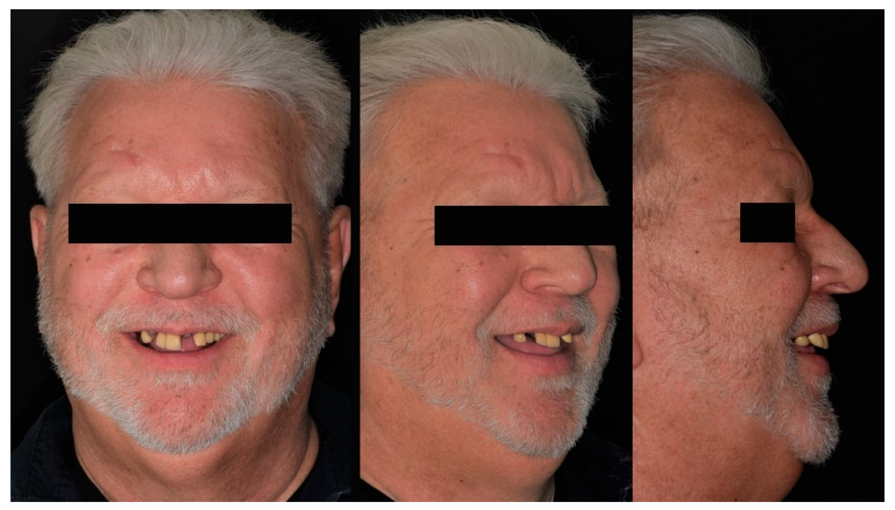
Abstract
:1. Introduction
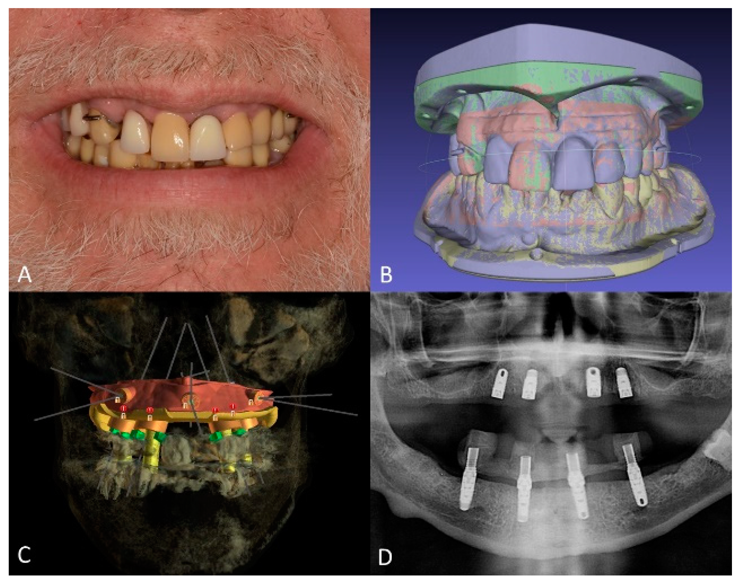
2. Materials and Methods
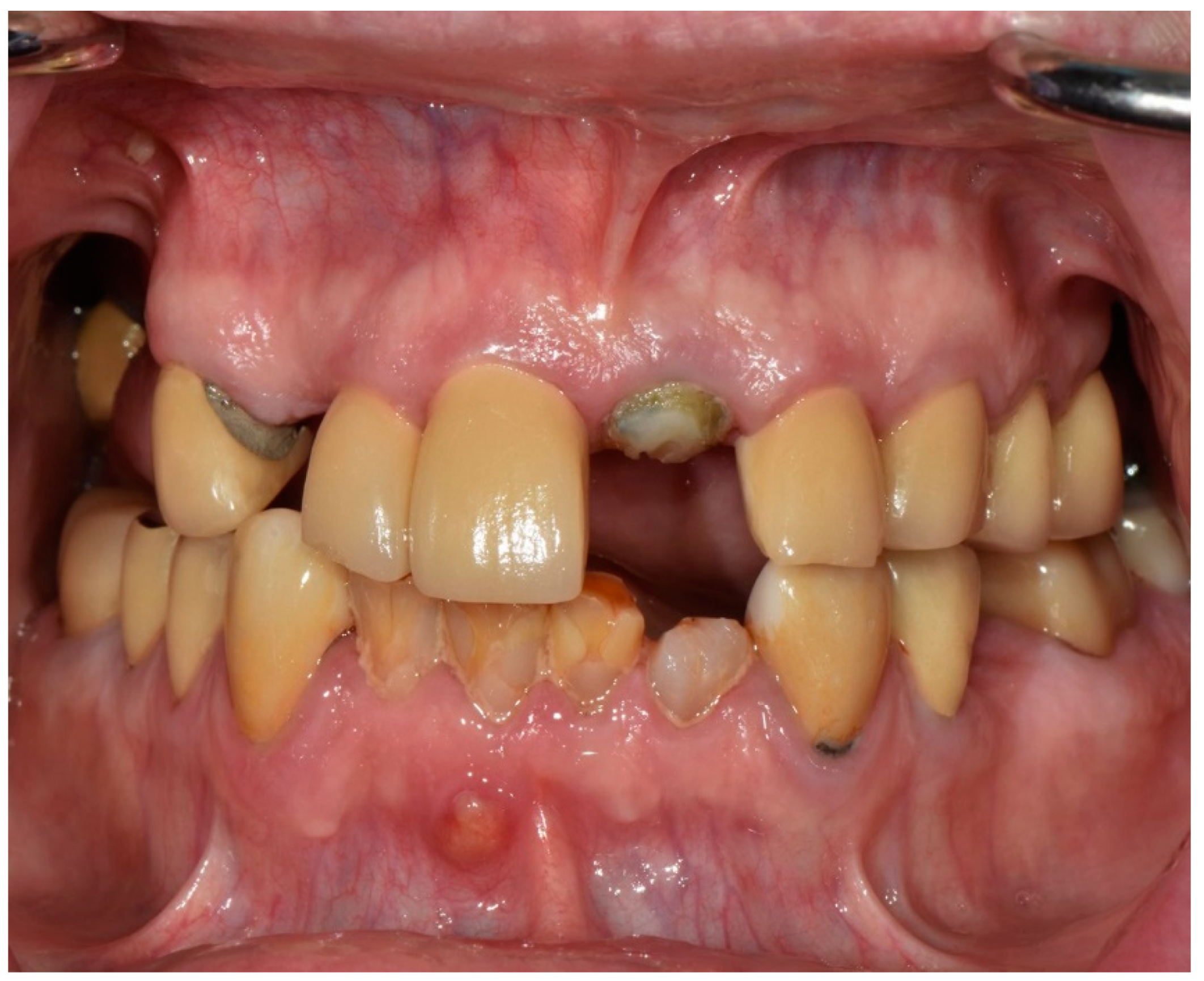
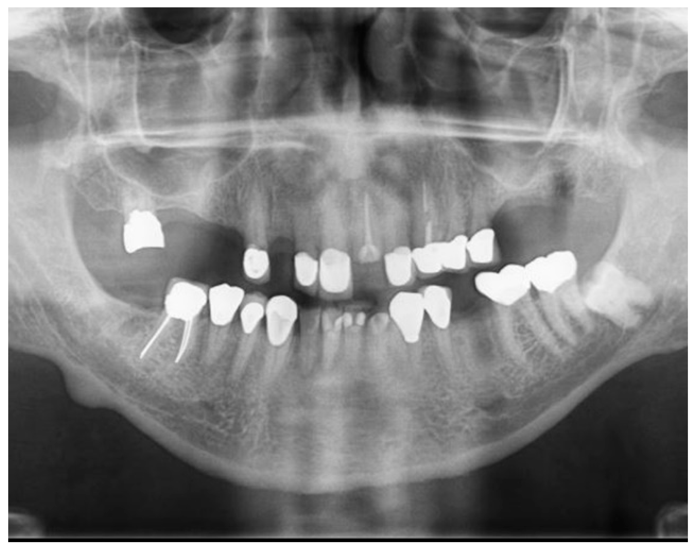
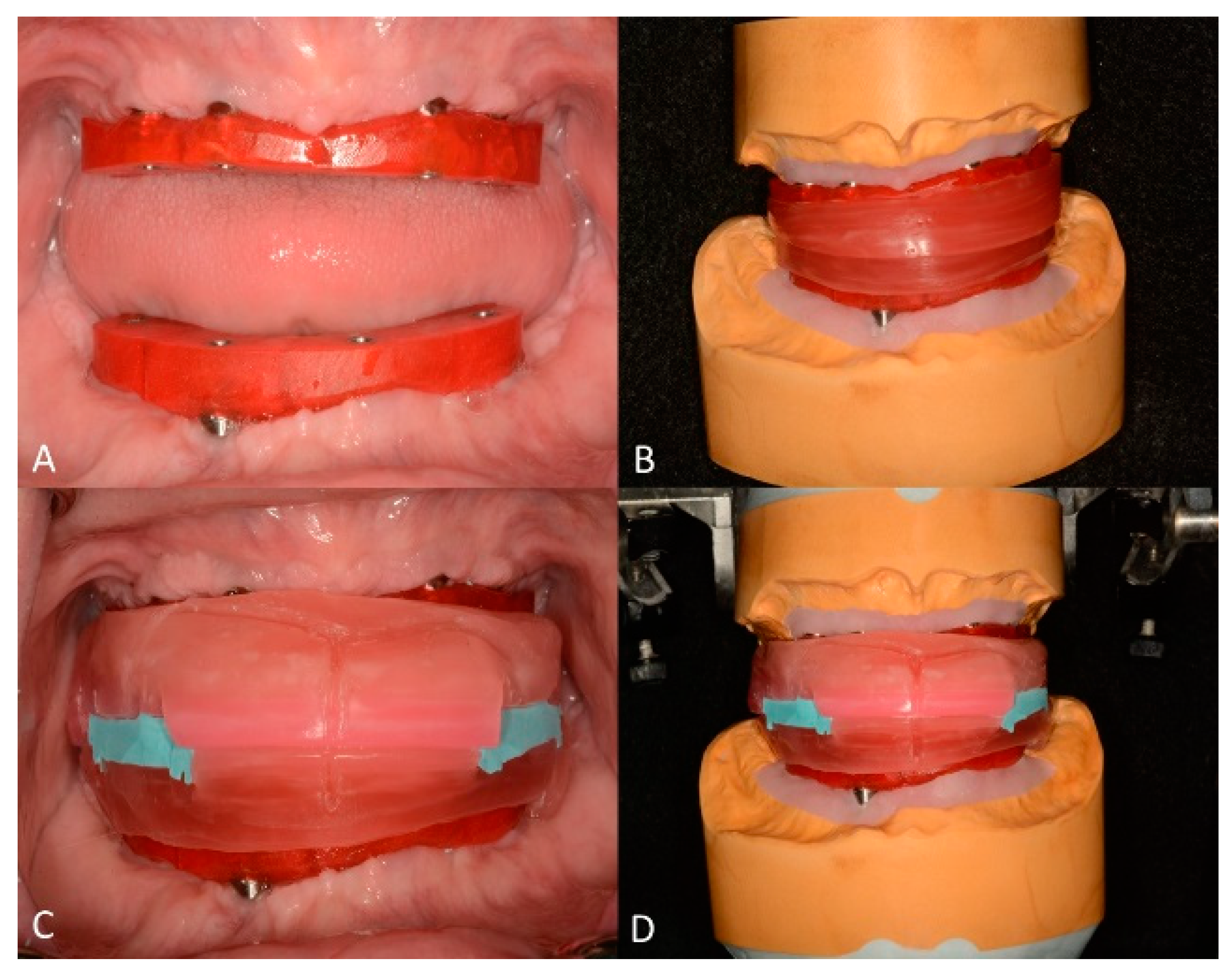
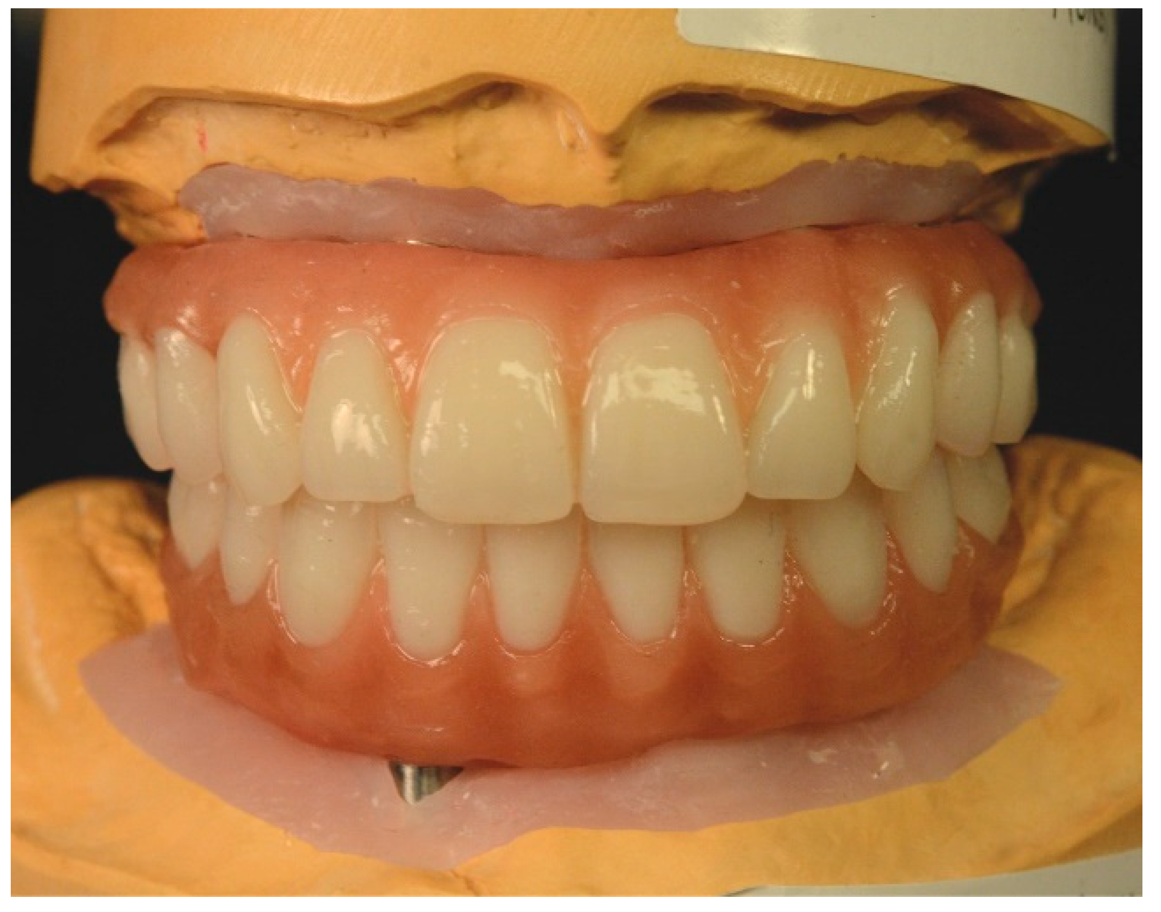
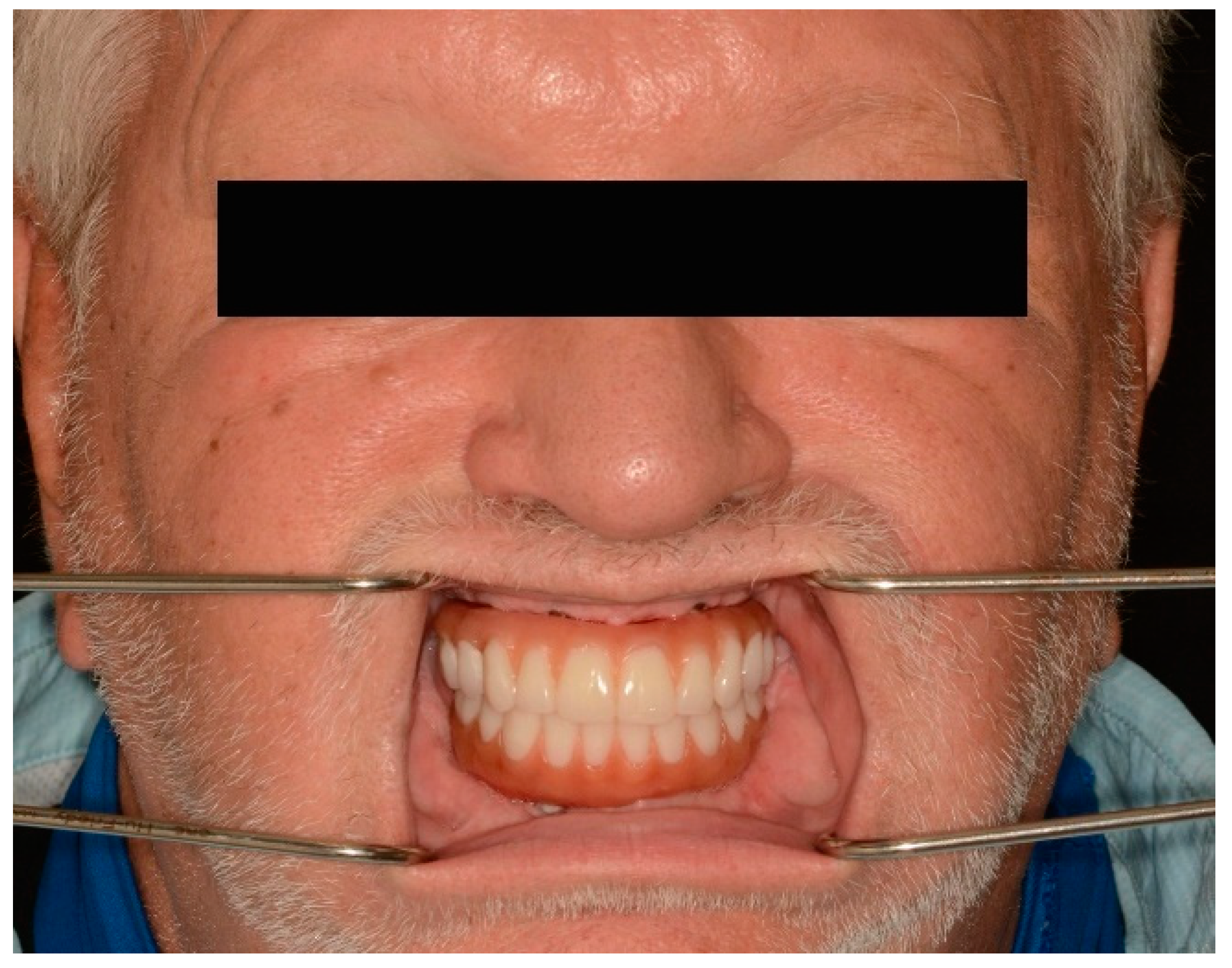
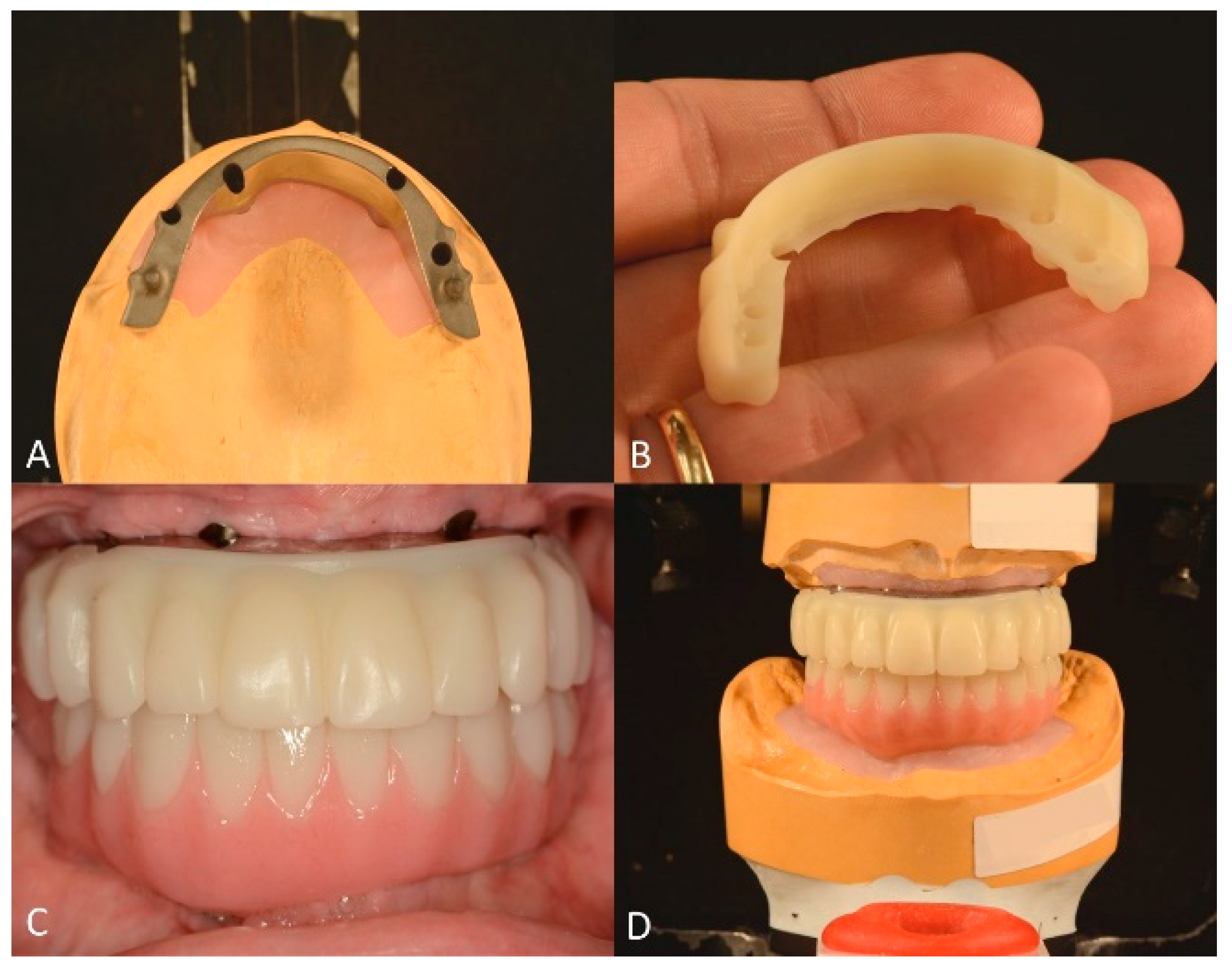
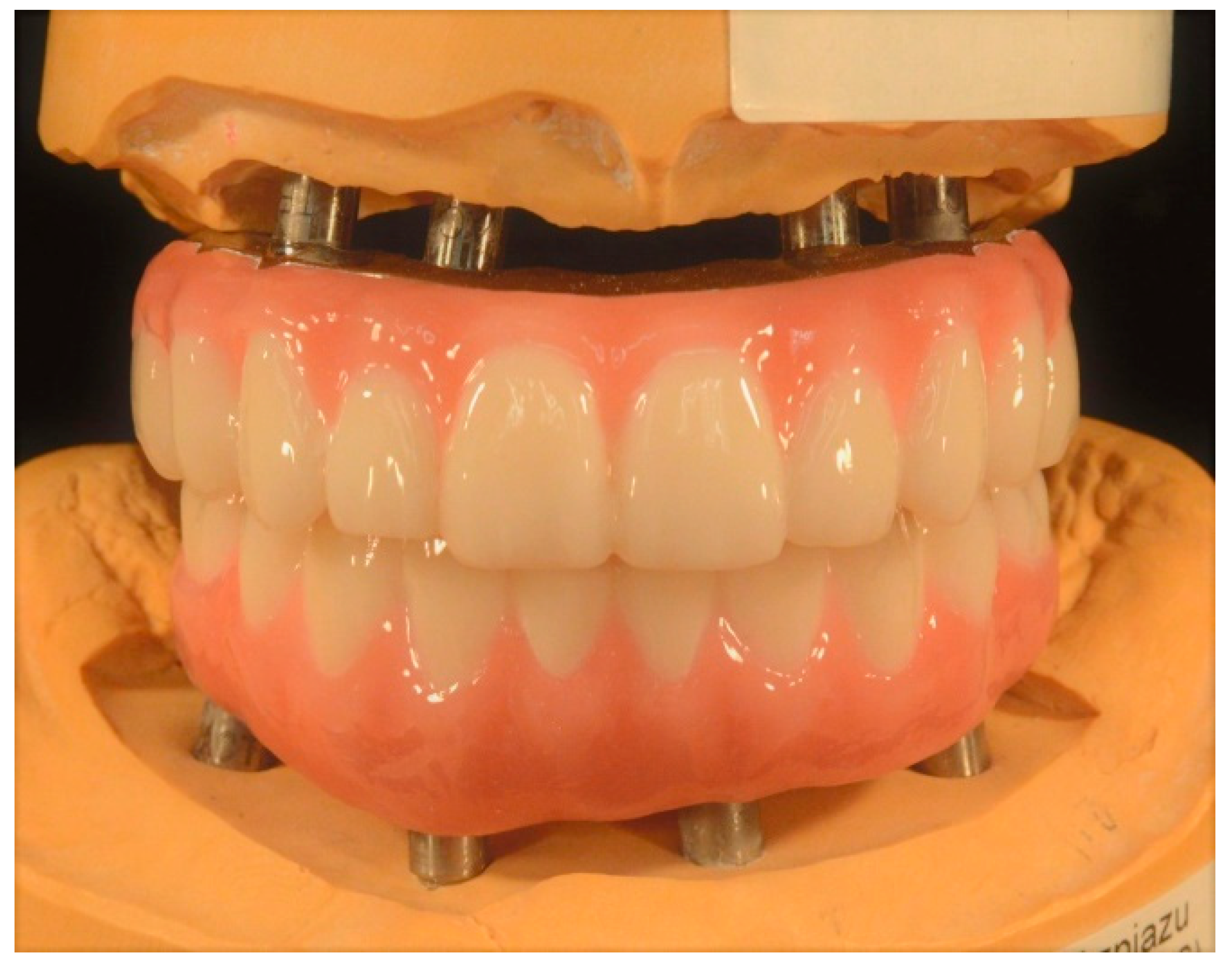
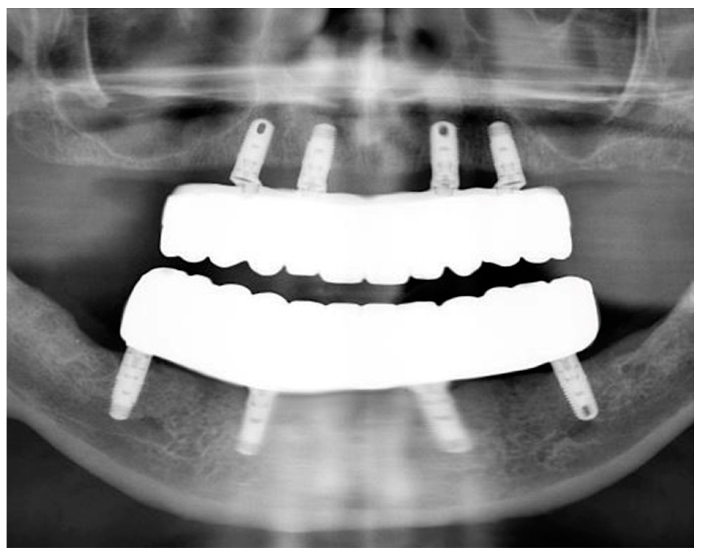
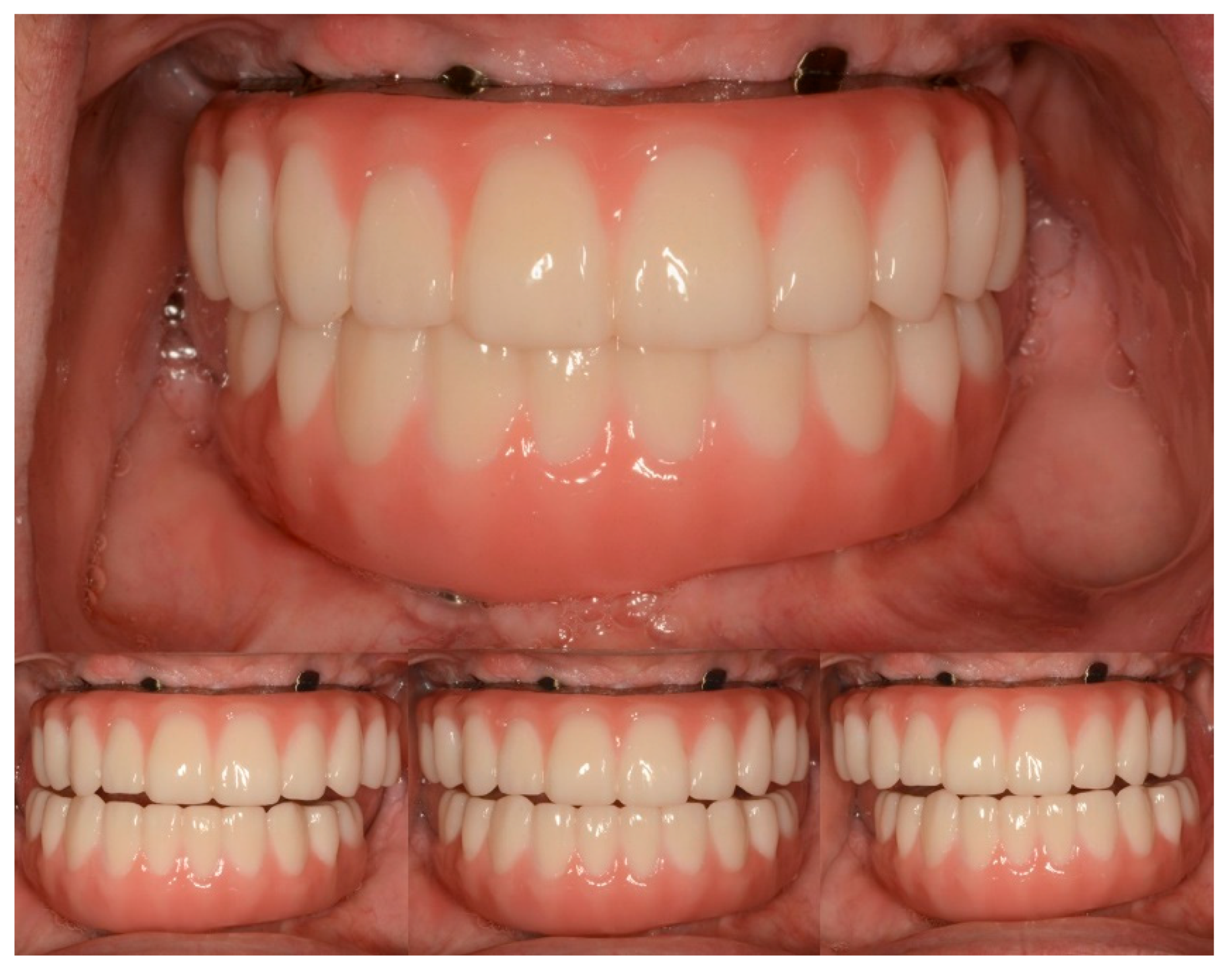
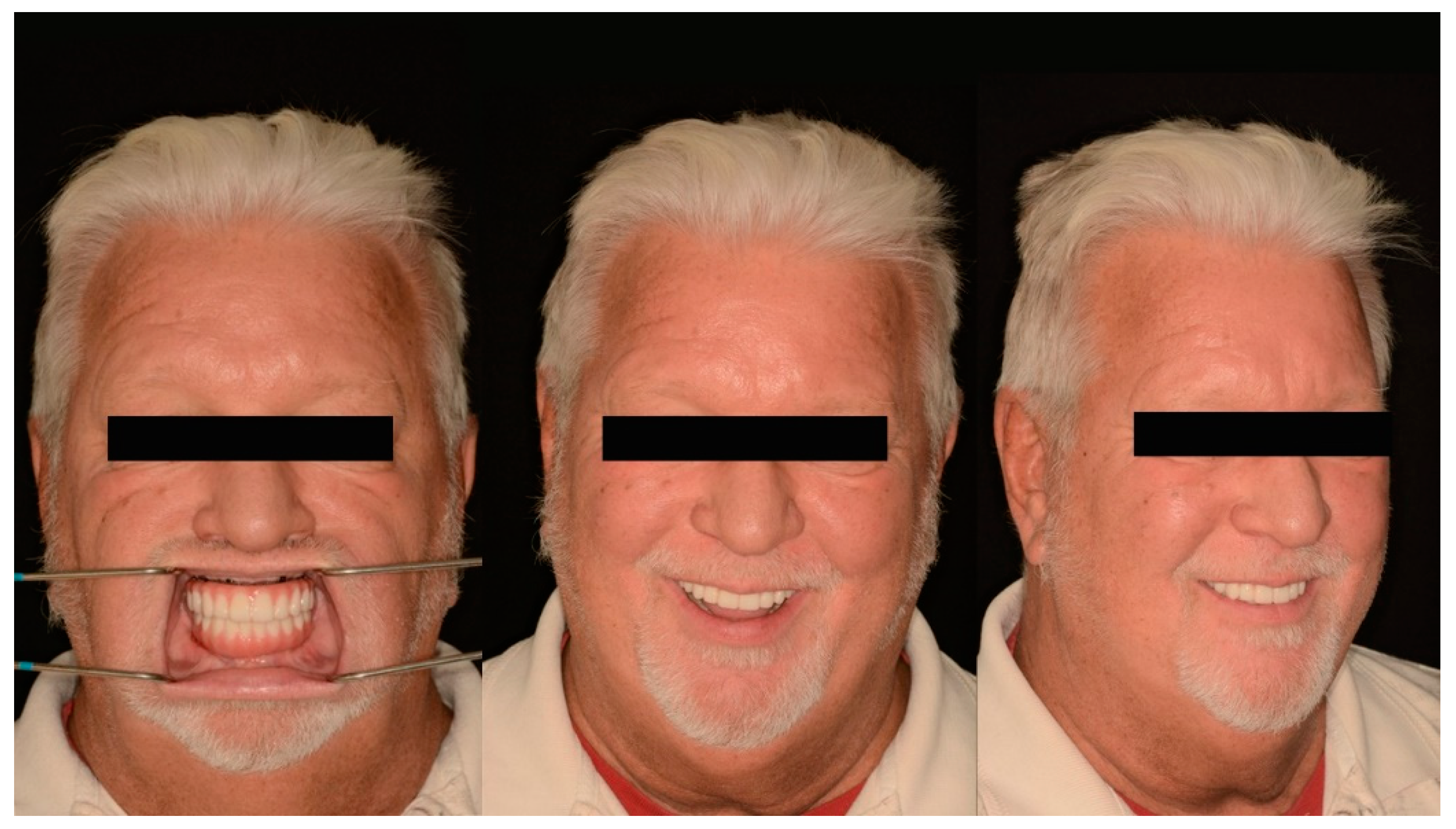
3. Results
4. Discussion
5. Conclusions
Author Contributions
Funding
Institutional Review Board Statement
Informed Consent Statement
Data Availability Statement
Acknowledgments
Conflicts of Interest
References
- Slutzkey, G.S.; Cohen, O.; Chaushu, L.; Rahmanov, A.; Mijiritsky, E.; Beitlitum, I.; Kolerman, R. Immediate Maxillary Full-Arch Rehabilitation of Periodontal Patients with Terminal Dentition Using Tilted Implants and Bone Augmentation: A 5-Year Retrospective Cohort Study. J. Clin. Med. 2022, 11, 2902. [Google Scholar] [CrossRef] [PubMed]
- Mathews, S.; Kurien, B.; Scofield, R. Oral Manifestations of Sjögren’s Syndrome. J. Dent. Res. 2008, 87, 308–318. [Google Scholar] [CrossRef]
- Thompson, M.J. Masticatory Efficiency as Related to Cusp form in Denture Prosthesis. J. Am. Dent. Assoc. Dent. Cosm. 1937, 24, 207–219. [Google Scholar] [CrossRef]
- Iegami, C.M.; Barbosa, W.F.; Furuyama, R.J.; Lima, J.R.B.; De Campos, T.T.; Minagi, S.; Tamaki, R. Masticatory efficiency in complete denture wearers with reduced dental arches—A randomised cross-over study. J. Oral Rehabil. 2014, 41, 619–623. [Google Scholar] [CrossRef]
- Albrektson, T.; Wenneberg, A. The Impact of Oral Implants—Past and Future, 1966–2042. J. Can. Dent. Assoc. 2005, 71, 327. [Google Scholar]
- French, D.; Larjava, H.; Ofec, R. Retrospective cohort study of 4591 Straumann implants in private practice setting, with up to 10-year follow-up. Part 1: Multivariate survival analysis. Clin. Oral Implant. Res. 2015, 26, 1345–1354. [Google Scholar] [CrossRef]
- French, D.; Ofec, R.; Levin, L. Long term clinical performance of 10 871 dental implants with up to 22 years of follow-up: A cohort study in 4247 patients. Clin. Implant. Dent. Relat. Res. 2021, 23, 289–297. [Google Scholar] [CrossRef] [PubMed]
- Jensen, O.T.; Adams, M.W.; Cottam, J.R.; Parel, S.M.; Phillips, W.R. The all on 4 shelf: Mandible. J. Oral Maxillofac. Surg. 2011, 69, 175–181. [Google Scholar] [CrossRef] [PubMed]
- Jensen, O.T.; Adams, M.W.; Cottam, J.R.; Parel, S.M.; Phillips, W.R. The All-on-4 shelf: Maxilla. J. Oral Maxillofac. Surg. 2010, 68, 2520–2527. [Google Scholar] [CrossRef]
- Maló, P.; Rangert, B.; Nobre, M. “All-on-Four” immediate-function concept with Brånemark System implants for completely eden-tulous mandibles: A retrospective clinical study. Clin. Implant. Dent. Relat. Res. 2003, 5 (Suppl. 1), 2–9. [Google Scholar] [CrossRef]
- van Assche, N.; Vercruyssen, M.; Coucke, W.; Teughels, W.; Jacobs, R.; Quirynen, M. Accuracy of computer-aided implant placement. Clin. Oral Implants Res. 2012, 23 (Suppl. 6), 112–123. [Google Scholar] [CrossRef] [PubMed]
- Papaspyridakos, P.; Bedrossian, A.; De Souza, A.; Bokhary, A.; Gonzaga, L.; Chochlidakis, K. Digital Workflow in Implant Treatment Planning for Terminal Dentition Patients. J. Prosthodont. 2022, 31, 543–548. [Google Scholar] [CrossRef]
- Van Noort, R. The future of dental devices is digital. Dent. Mater. 2012, 28, 3–12. [Google Scholar] [CrossRef]
- Revilla-León, M.; Özcan, M. Additive Manufacturing Technologies Used for Processing Polymers: Current Status and Potential Application in Prosthetic Dentistry. J. Prosthodont. 2019, 28, 146–158. [Google Scholar] [CrossRef]
- Ashurko, I.; Trofimov, A.; Tarasenko, S.; Mekhtieva, S. Full-Mouth Screw-Retained Implant-Supported Rehabilitation with Multiunit Abutments Using Virtual Guided Surgery and Digital Prosthetics Protocol. Case Rep. Dent. 2020, 2020, 3585169. [Google Scholar] [CrossRef] [PubMed]
- Massad, J.; Wicks, R.; Ahuja, S.; Cagna, D.R. A Prosthesis Retention System for Full-Arch, Fixed, Implant-Supported Prosthesis. J. Prosthodont. 2019, 28, e912–e916. [Google Scholar] [CrossRef] [PubMed]
- Chaar, M.S.; Att, W.; Strub, J.R. Prosthetic outcome of cement-retained implant-supported fixed dental restorations: A systematic review. J. Oral Rehabil. 2011, 38, 697–711. [Google Scholar] [CrossRef]
- Chee, W.; Jivraj, S. Screw versus cemented implant supported restorations. Br. Dent. J. 2006, 201, 501–507. [Google Scholar] [CrossRef] [PubMed]
- Gaddale, R.; Mishra, S.K.; Chowdhary, R. Complications of screw- and cement-retained implant-supported full-arch restorations: A systematic review and meta-analysis. Int. J. Oral Implantol. 2020, 13, 11–40. [Google Scholar]
- Misch, C.E. Dental Implant Prosthetics. In Principles of Fixed Implant Prosthodontics: Cement-Retained Restorations, 2nd ed.; Elsevier: St. Louis, MO, USA, 2015; pp. 650–699. [Google Scholar]
- Michael, K.; Winfried, W. Retrospective analysis of loosening of cement-retained vs screw-retained fixed implant-supported reconstructions. Quintessence Int. 2015, 46, 583–589. [Google Scholar] [CrossRef]
- Guichet, D.L.; Caputo, A.A.; Choi, H.; Sorensen, J.A. Passivity of fit and marginal opening in screw-or cement-retained implant fixed partial denture designs. Int. J. Oral Maxillofac. Implant. 2000, 15, 239–246. [Google Scholar]
- Torrado, E.; Ercoli, C.; Al Mardini, M.; Graser, G.N.; Tallents, R.H.; Cordaro, L. A comparison of the porcelain fracture resistance of screw-retained and cementretained implant supported metal ceramic crowns. J. Prosthet. Dent. 2004, 91, 532–537. [Google Scholar] [CrossRef] [PubMed]
- Korsch, M.; Obst, U.; Walther, W. Cement-associated peri-implantitis: A retrospective clinical observational study of fixed implant-supported restorations using a methacrylate cement. Clin. Oral Implant Res. 2014, 25, 797–802. [Google Scholar] [CrossRef] [PubMed]
- D’Souza, K.M.; Aras, M.A. Types of Implant Surgical Guides in Dentistry: A Review. J. Oral Implant. 2012, 38, 643–652. [Google Scholar] [CrossRef] [PubMed]
- Anadioti, E.; Musharbash, L.; Blatz, M.B.; Papavasiliou, G.; Kamposiora, P. 3D printed complete removable dental prostheses: A narrative review. BMC Oral Health 2020, 20, 343. [Google Scholar] [CrossRef]
- Anusavice, K.J.; Shen, C.; Rawls, H.R. Philips Science of Dental Materials, 12th ed.; Elsevier: Amsterdam, The Netherlands, 2012. [Google Scholar]
- Papynov, E.; Shichalin, O.; Apanasevich, V.; Portnyagin, A.; Yu, M.V.; Yu, B.I.; Merkulov, E.; Kaidalova, T.; Modin, E.; Afonin, I.; et al. Sol-gel (template) synthesis of osteoplastic CaSiO3/HAp powder biocomposite: “In vitro” and “in vivo” biocompatibility assessment. Powder Technol. 2020, 367, 762–773. [Google Scholar] [CrossRef]
- Papynov, E.; Shichalin, O.; Skurikhina, Y.; Turkutyukov, V.; Medkov, M.; Grishchenko, D.; Portnyagin, A.; Merkulov, E.; Apanasevich, V.; Geltser, B.; et al. ZrO2-phosphates porous ceramic obtained via SPS-RS “in situ” technique: Bacteria test assessment. Ceram. Int. 2019, 45, 13838–13846. [Google Scholar] [CrossRef]
- Papynov, E.K.; Shichalin, O.O.; Apanasevich, V.I.; Afonin, I.S.; Evdokimov, I.O.; Mayorov, V.Y.; Portnyagin, A.S.; Agafonova, I.G.; Skurikhina, Y.E.; Medkov, M.A. Synthetic CaSiO3 sol-gel powder and SPS ceramic derivatives: “In vivo” toxicity assessment. Prog. Nat. Sci. 2019, 29, 569–575. [Google Scholar] [CrossRef]
- Bidra, A.S.; Tischler, M.; Patch, C. Survival of 2039 complete arch fixed implant-supported zirconia prostheses: A retrospective study. J. Prosthet. Dent. 2018, 119, 220–224. [Google Scholar] [CrossRef]
- Tischler, M.; Patch, C.; Bidra, A.S. Rehabilitation of edentulous jaws with zirconia complete-arch fixed implant-supported prostheses: An up to 4-year retrospective clinical study. J. Prosthet. Dent. 2018, 120, 204–209. [Google Scholar] [CrossRef]
- Bidra, A.S. Three-Dimensional Esthetic Analysis in Treatment Planning for Implant-Supported Fixed Prosthesis in the Edentulous Maxilla: Review of the Esthetics Literature. J. Esthet. Restor. Dent. 2011, 23, 219–236. [Google Scholar] [CrossRef] [PubMed]
- Bidra, A.S. Technique for systematic bone reduction for fixed implant-supported prosthesis in the edentulous maxilla. J. Prosthet. Dent. 2015, 113, 520–523. [Google Scholar] [CrossRef] [PubMed]
- Azpiazu-Flores, F.X.; Mata-Mata, S.J. Overlay occlusion rim technique to facilitate the recording of maxillomandibular relationships. J. Prosthet. Dent. 2021, 126, 715–717. [Google Scholar] [CrossRef]
- Balshi, T.J.; Mingledorff, E.B.; Olbrys, B.H.; Cantor, S.J. Restorative occlusion utilizing a custom incisal guide table. J. Prosthet. Dent. 1976, 36, 468–471. [Google Scholar] [CrossRef]
- Venezia, P.; Torsello, F.; Cavalcanti, R.; D’Amato, S. Retrospective analysis of 26 complete-arch implant-supported monolithic zir-conia prostheses with feldspathic porcelain veneering limited to the facial surface. J. Prosthet. Dent. 2015, 114, 506–512. [Google Scholar] [CrossRef] [PubMed]
- Adell, R.; Lekholm, U.; Rockler, B.; Brånemark, P.-I. Review Article. Economica 2003, 70, 691–697. [Google Scholar]
- Azpiazu-Flores, F.X.; Lee, D.J.; Mata-Mata, S.J.; Zheng, F. Rehabilitation of a patient with mandibular flexure using contemporary glass-infiltrated high performance CAD-CAM polymers: A clinical report with 1-year follow-up. J. Prosthet. Dent. 2023. [Google Scholar] [CrossRef]
- Azpiazu-Flores, F.X.; Knobloch, L.A.; Larsen, P.E. Interdisciplinary Management of a Patient with Dentinogenesis Imperfecta Type II Using a Combination of CAD-CAM and Analog Techniques: A Clinical Report. J. Prosthodont. 2022, 31, 647–654. [Google Scholar] [CrossRef]
- Almeida, D.; Vianna, K.; Arriaga, P.; Moraschini, V. Dental implants in Sjögren’s syndrome patients: A systematic review. PLoS ONE 2018, 12, e0189507. [Google Scholar] [CrossRef]
- Korfage, A.; Raghoebar, G.M.; Arends, S.; Meiners, P.M.; Visser, A.; Kroese, F.G.; Bootsma, H.; Vissink, A. Dental Implants in Patients with Sjögren’s Syndrome. Clin. Implant. Dent. Relat. Res. 2015, 18, 937–945. [Google Scholar] [CrossRef]
- Spinato, S.; Soardi, C.M.; Zane, A.M. A Mandibular Implant-Supported Fixed Complete Dental Prosthesis in a Patient with Sjogren Syndrome: Case Report. Implant. Dent. 2010, 19, 178–183. [Google Scholar] [CrossRef] [PubMed]
- Binon, P.P. Thirteen-year follow-up of a mandibular implant-supported fixed complete denture in a patient with Sjogren’s syndrome: A clinical report. J. Prosthet. Dent. 2005, 94, 409–413. [Google Scholar] [CrossRef] [PubMed]
- Albrecht, K.; Callhoff, J.; Westhoff, G.; Dietrich, T.; Dörner, T.; Zink, A. The Prevalence of Dental Implants and Related Factors in Patients with Sjögren Syndrome: Results from a Cohort Study. J. Rheumatol. 2016, 43, 1380–1385. [Google Scholar] [CrossRef] [PubMed]


Disclaimer/Publisher’s Note: The statements, opinions and data contained in all publications are solely those of the individual author(s) and contributor(s) and not of MDPI and/or the editor(s). MDPI and/or the editor(s) disclaim responsibility for any injury to people or property resulting from any ideas, methods, instructions or products referred to in the content. |
© 2023 by the authors. Licensee MDPI, Basel, Switzerland. This article is an open access article distributed under the terms and conditions of the Creative Commons Attribution (CC BY) license (https://creativecommons.org/licenses/by/4.0/).
Share and Cite
Azpiazu-Flores, F.X.; Lee, D.J.; Jurado, C.A.; Afrashtehfar, K.I.; Alhotan, A.; Tsujimoto, A. Full-Mouth Rehabilitation of a Patient with Sjogren’s Syndrome with Maxillary Titanium-Zirconia and Mandibular Monolithic Zirconia Implant Prostheses Fabricated with CAD/CAM Technology: A Clinical Report. J. Funct. Biomater. 2023, 14, 174. https://doi.org/10.3390/jfb14040174
Azpiazu-Flores FX, Lee DJ, Jurado CA, Afrashtehfar KI, Alhotan A, Tsujimoto A. Full-Mouth Rehabilitation of a Patient with Sjogren’s Syndrome with Maxillary Titanium-Zirconia and Mandibular Monolithic Zirconia Implant Prostheses Fabricated with CAD/CAM Technology: A Clinical Report. Journal of Functional Biomaterials. 2023; 14(4):174. https://doi.org/10.3390/jfb14040174
Chicago/Turabian StyleAzpiazu-Flores, Francisco X., Damian J. Lee, Carlos A. Jurado, Kelvin I. Afrashtehfar, Abdulaziz Alhotan, and Akimasa Tsujimoto. 2023. "Full-Mouth Rehabilitation of a Patient with Sjogren’s Syndrome with Maxillary Titanium-Zirconia and Mandibular Monolithic Zirconia Implant Prostheses Fabricated with CAD/CAM Technology: A Clinical Report" Journal of Functional Biomaterials 14, no. 4: 174. https://doi.org/10.3390/jfb14040174