
Evaluation of Antidiabetic Effect of Luteolin in STZ Induced Diabetic Rats: Molecular Docking, Molecular Dynamics, In Vitro and In Vivo Studies

 , , , ,
  ,
,  and
and
Abstract
:1. Introduction
2. Results
2.1. Effect of LUT on α-glucosidase
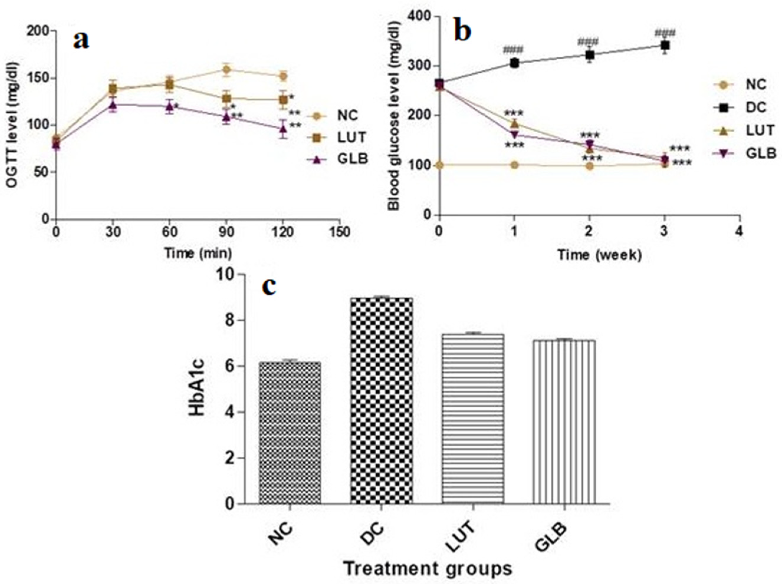
2.2. Oral Glucose Tolerance Test (OGTT)
2.3. Effect of LUT Treatment on HbA1c Level in Diabetic Rats
2.4. Body Weight
2.5. Effect of LUT on Serum Lipid Profiles in Diabetic Rats
2.6. Effect of LUT on Liver Function Tests in Diabetic Rats
2.7. Effect of LUT on Kidney Function Tests in Diabetic Rats
2.8. Effect of LUT on Antioxidant Status
2.9. Effect of LUT on TNF-α and IL-6 Expression Levels in Serum
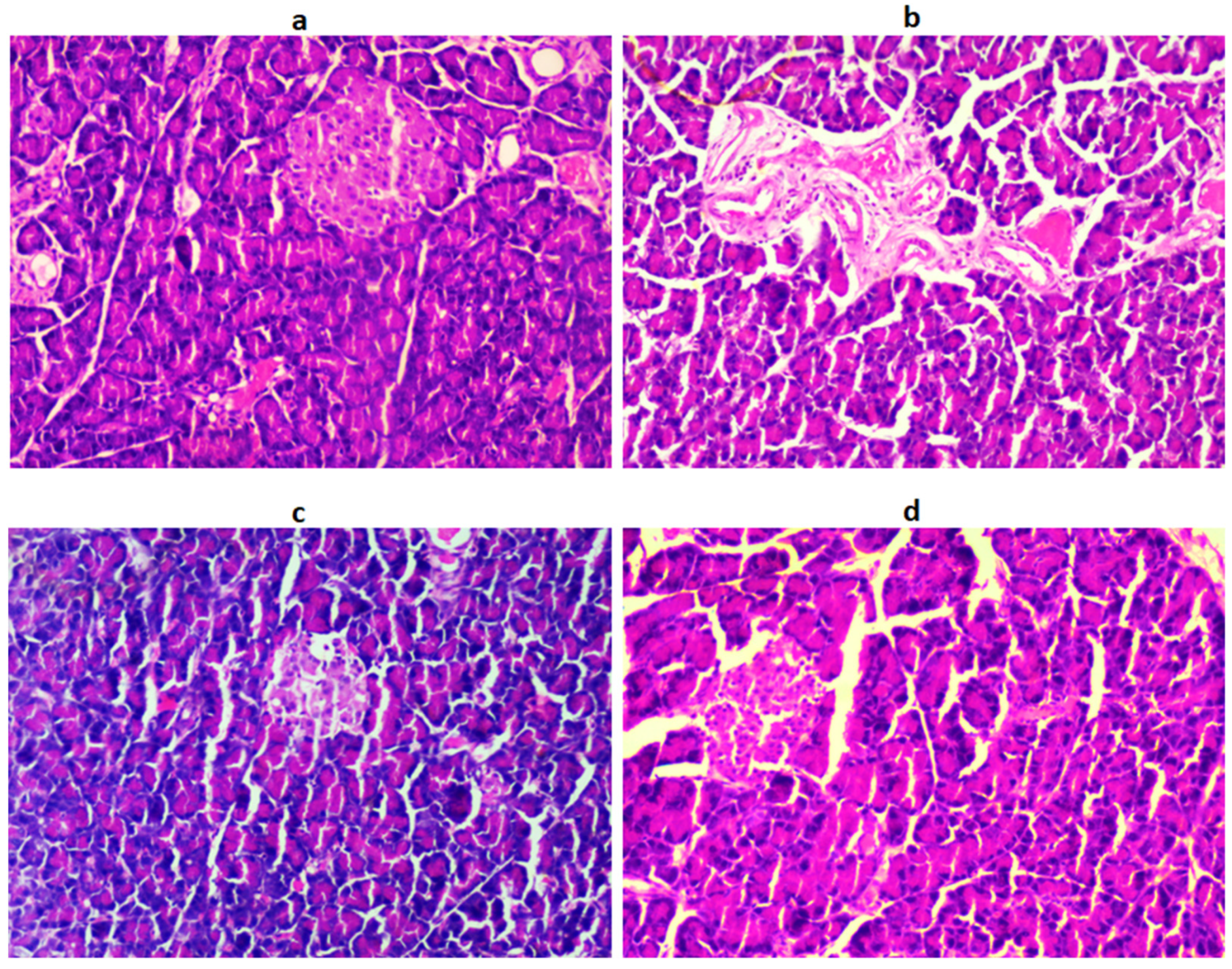
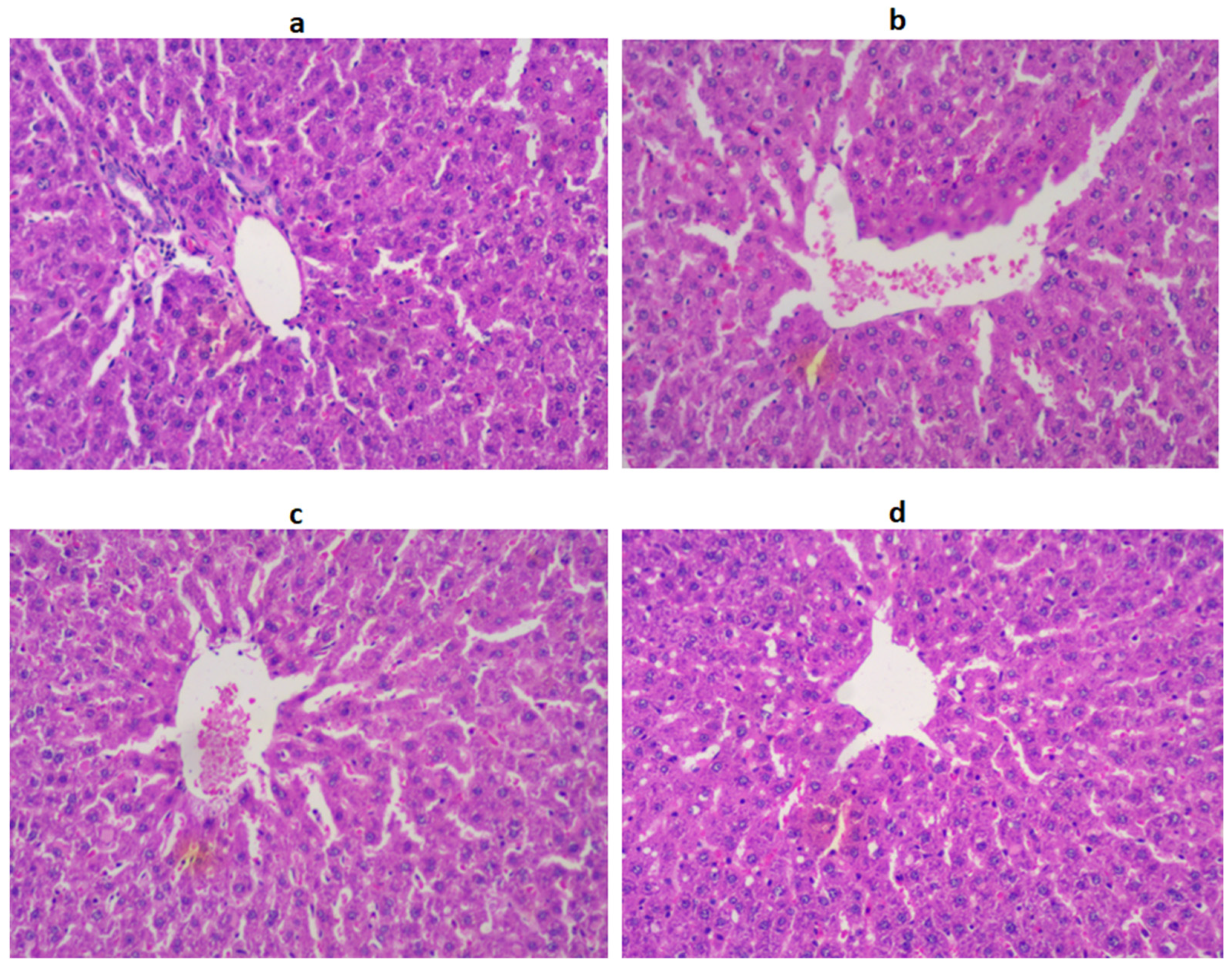
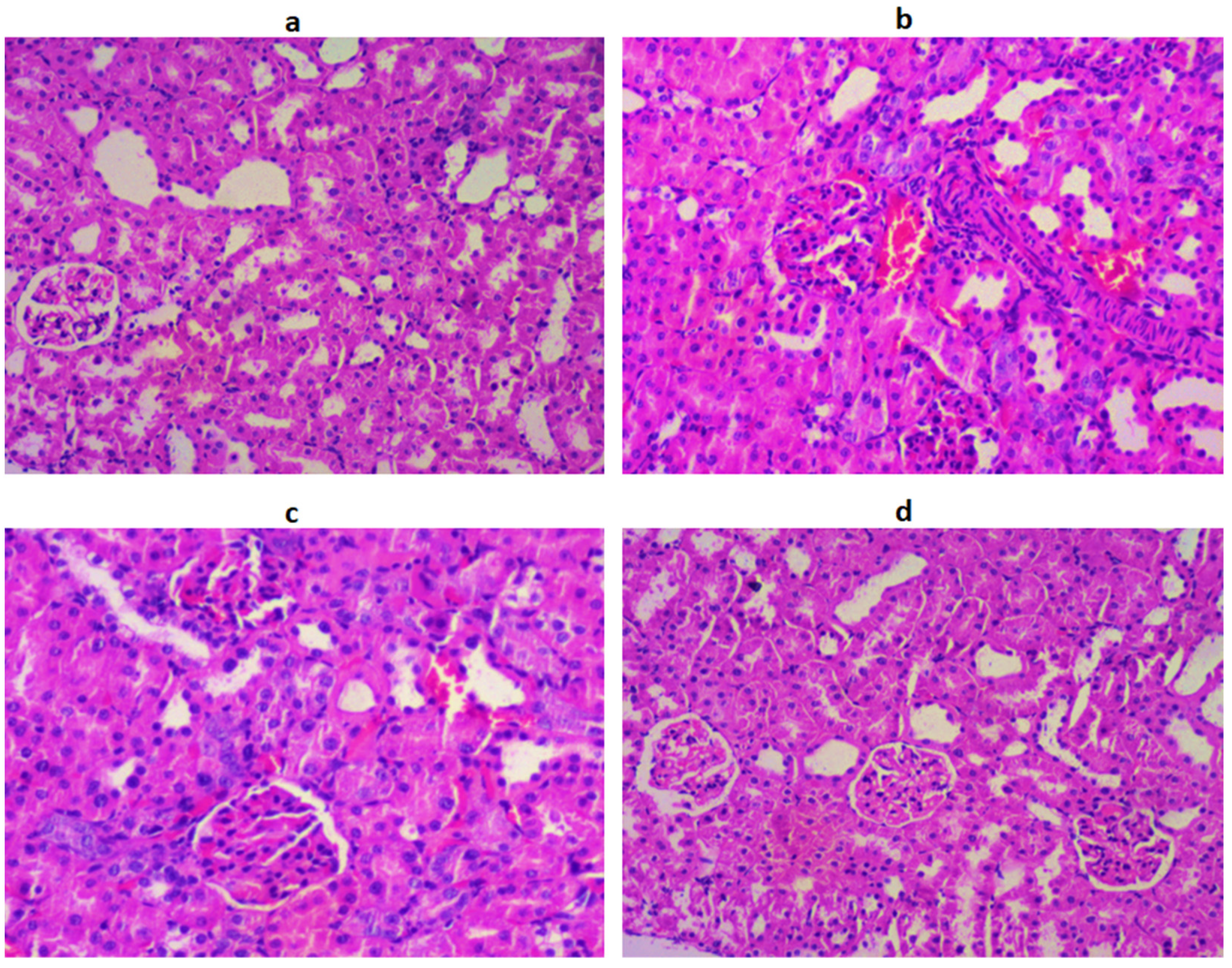
2.10. Histopathology of the Pancreas, Liver, and Kidneys
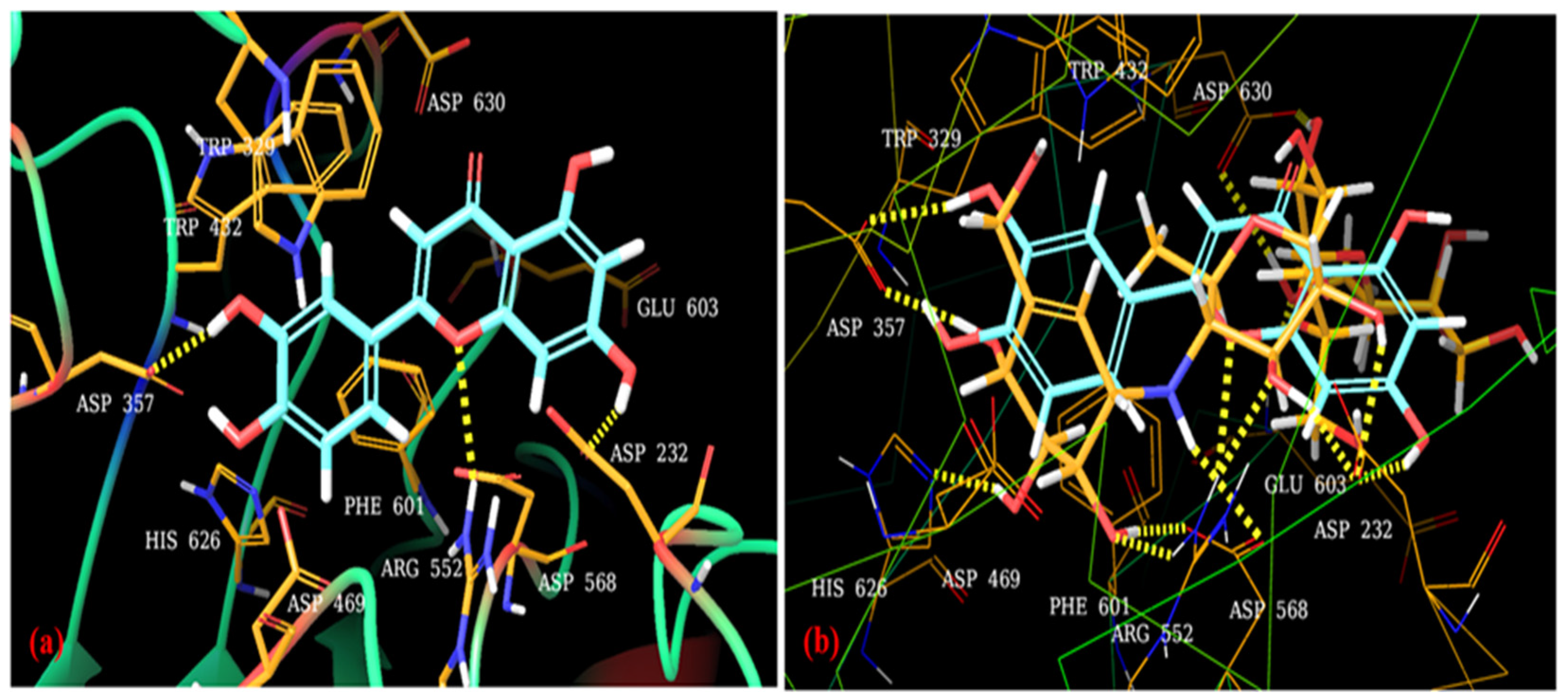
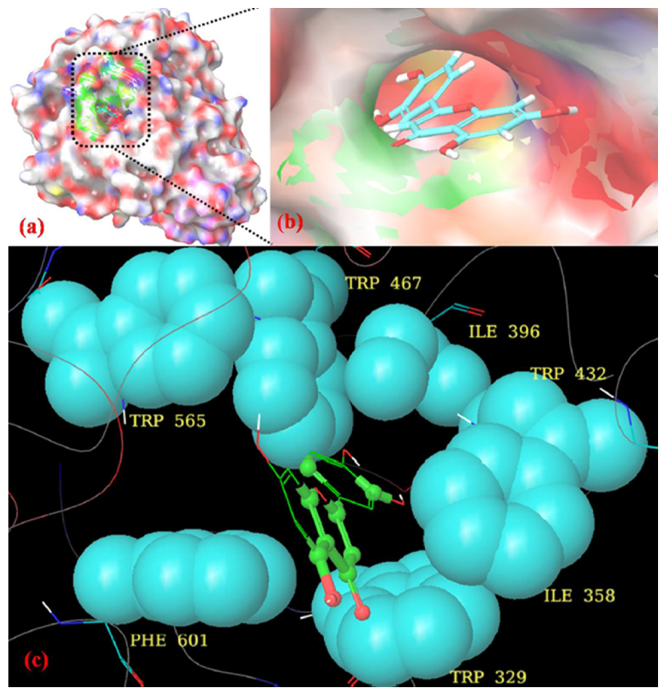
2.11. Molecular Docking
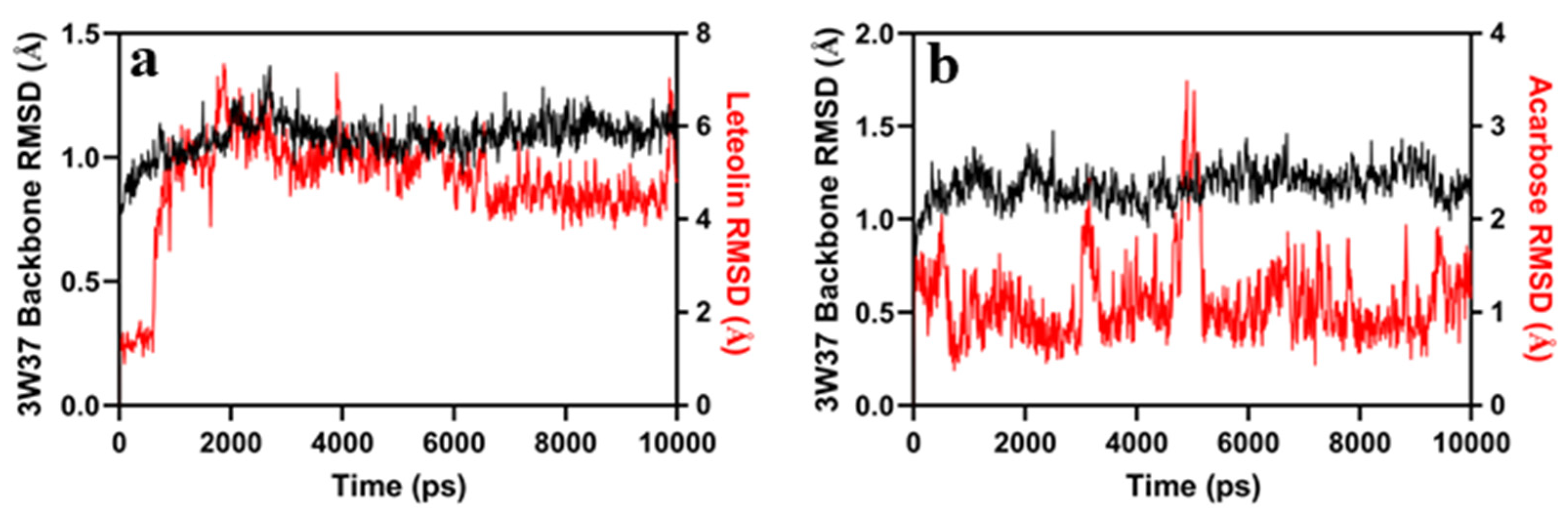
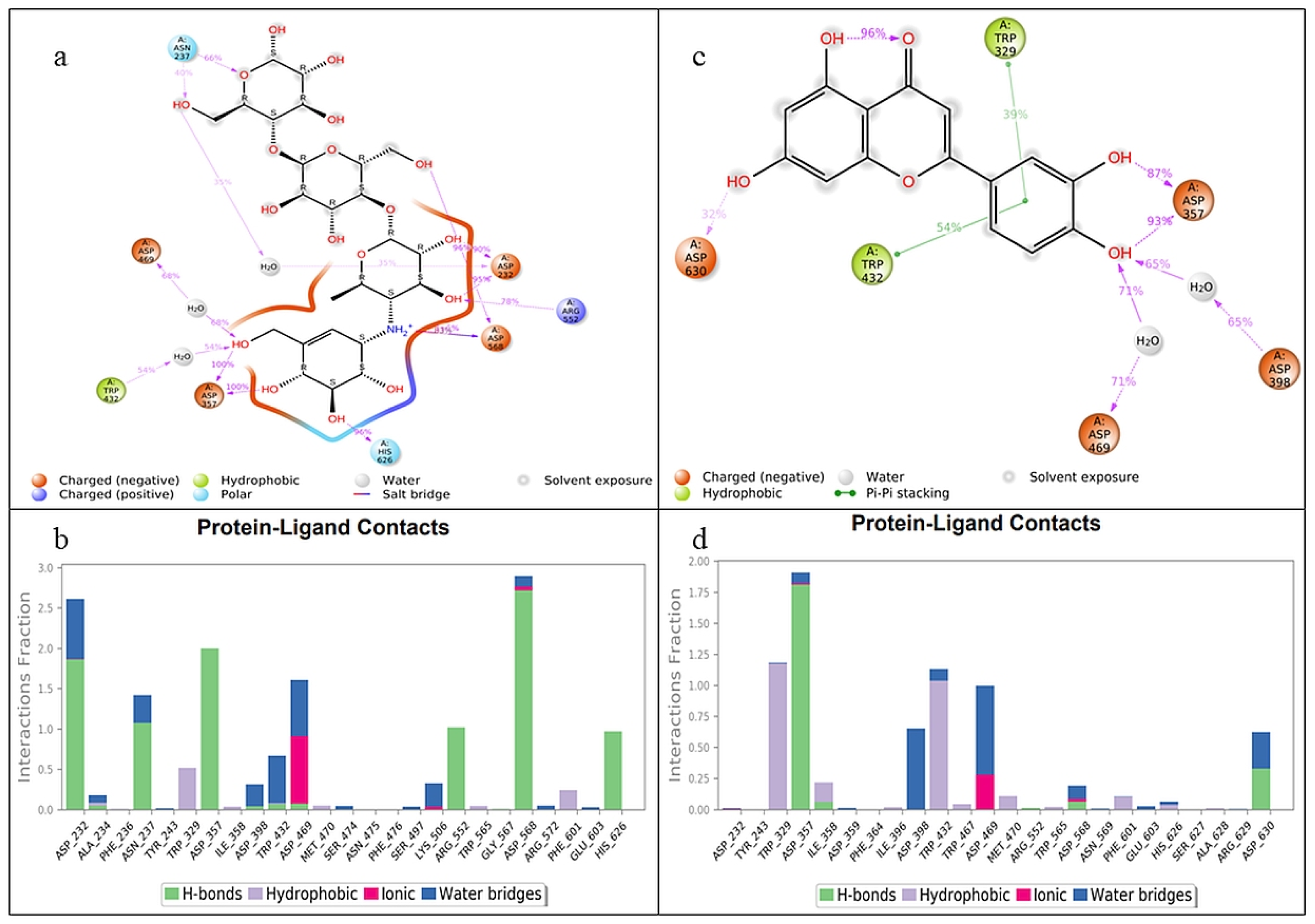
2.12. Molecular Dynamics Simulation
3. Discussion
4. Materials and Methods
4.1. In Vitro α-glucosidase Activity
4.2. Animals
4.2.1. Induction of Type 2 Diabetes and Grouping of Animals
4.2.2. Oral Glucose Tolerance Test (OGTT)
4.2.3. Blood Glucose Level
4.2.4. Glycated Hemoglobin Level
4.3. Body Weight
4.4. Biochemical Analysis
4.5. Detection of TNF-α and IL-6 Levels in Serum
4.6. In Vivo Antioxidant Activity
4.7. Histopathological Studies
4.8. Molecular Docking
4.9. Molecular Dynamics Simulation
4.10. Statistical Analysis
5. Conclusions
Author Contributions
Funding
Institutional Review Board Statement
Informed Consent Statement
Data Availability Statement
Acknowledgments
Conflicts of Interest
Abbreviations
| LUT | Luteolin |
| STZ | Streptozotocin |
| b.w. | Body weight |
| OGTT | Oral glucose tolerance test |
| DM | Diabetes mellitus |
| MD | Molecular dynamics |
| GLB | Glibenclamide |
| HbA1c | Glycated hemoglobin A1c |
| NC | Normal control |
| DC | Diabetic control |
| TG | Triglycerides |
| TC | Total cholesterol |
| HDL | High-density lipoproteins |
| LDL | Low-density lipoproteins |
| ALT | Alanine transaminase |
| AST | Aspartate transaminase |
| ALP | Alkaline phosphatase |
| SOD | Superoxide dismutase |
| CAT | Catalase enzyme |
| GPx | Glutathione peroxidase |
| TNF-α | Tumor necrosis factor α |
| IL-6 | Interleukin 6 |
| RMSD | Root-mean-square deviation |
| PS | Picoseconds |
| SD | Standard deviation |
| H&E | Hematoxylin and eosin |
| IAEC | Institutional Animal Ethics Committee |
| CPCSEA | Committee for the Purpose of Control and Supervision of Experiments on Animals |
References
- Sun, H.; Saeedi, P.; Karuranga, S.; Pinkepank, M.; Ogurtsova, K.; Duncan, B.B.; Stein, C.; Basit, A.; Chan, J.C.N.; Mbanya, J.C.; et al. IDF Diabetes Atlas: Global, regional and country-level diabetes prevalence estimates for 2021 and projections for 2045. Diabetes Res. Clin. Pract. 2022, 183, 109119. [Google Scholar] [CrossRef]
- Kahksha; Alam, O.; Naaz, S.; Sharma, V.; Manaithiya, A.; Khan, J.; Alam, A. Recent developments made in the assessment of the antidiabetic potential of gymnema species—From 2016 to 2020. J. Ethnopharmacol. 2022, 286, 114908. [Google Scholar] [CrossRef] [PubMed]
- Ibrahim, M.; Parveen, B.; Zahiruddin, S.; Gautam, G.; Parveen, R.; Khan, M.A.; Gupta, A.; Ahmad, S. Analysis of polyphenols in Aegle marmelos leaf and ameliorative efficacy against diabetic mice through restoration of antioxidant and anti-inflammatory status. J. Food Biochem. 2022, 46, e13852. [Google Scholar] [CrossRef] [PubMed]
- Hameed, I.; Masoodi, S.R.; Mir, S.A.; Nabi, M.; Ghazanfar, K.; Ganai, B.A. Type 2 diabetes mellitus: From a metabolic disorder to an inflammatory condition. World J. Diabetes 2015, 6, 598. [Google Scholar] [CrossRef] [PubMed]
- Dirir, A.M.; Daou, M.; Yousef, A.F.; Yousef, L.F. A Review of Alpha-Glucosidase Inhibitors from Plants as Potential Candidates for the Treatment of Type-2 Diabetes. Phytochem. Rev. 2022, 21, 1049–1079. [Google Scholar] [CrossRef]
- Rajasekaran, P.; Ande, C.; Vankar, Y.D. Synthesis of (5,6 & 6,6)-oxa-oxa annulated sugars as glycosidase inhibitors from 2-formyl galactal using iodocyclization as a key step. Arkivoc 2022, 2022, 5–23. [Google Scholar] [CrossRef]
- Chaudhury, A.; Duvoor, C.; Reddy Dendi, V.S.; Kraleti, S.; Chada, A.; Ravilla, R.; Marco, A.; Shekhawat, N.S.; Montales, M.T.; Kuriakose, K.; et al. Clinical Review of Antidiabetic Drugs: Implications for Type 2 Diabetes Mellitus Management. Front. Endocrinol. 2017, 8, 6. [Google Scholar] [CrossRef] [PubMed] [Green Version]
- Ekor, M. The growing use of herbal medicines: Issues relating to adverse reactions and challenges in monitoring safety. Front. Pharmacol. 2014, 4, 177. [Google Scholar] [CrossRef] [Green Version]
- Khan, S.; Ibrahim, M. A systematic review on hepatoprotective potential of grape and polyphenolic compounds: Molecular mechanism and future prospective. Nat. Resour. Hum. Health 2022, 3, 196–213. [Google Scholar] [CrossRef]
- Muruganathan, N.; Dhanapal, A.R.; Baskar, V.; Muthuramalingam, P.; Selvaraj, D.; Aara, H.; Abdullah, M.Z.S.; Sivanesan, I. Recent Updates on Source, Biosynthesis, and Therapeutic Potential of Natural Flavonoid Luteolin: A Review. Metabolites 2022, 12, 1145. [Google Scholar] [CrossRef]
- Lin, Y.; Shi, R.; Wang, X.; Shen, H.-M. Luteolin, a Flavonoid with Potential for Cancer Prevention and Therapy. Curr. Cancer Drug Targets 2008, 8, 634–646. [Google Scholar] [CrossRef]
- Caporali, S.; De Stefano, A.; Calabrese, C.; Giovannelli, A.; Pieri, M.; Savini, I.; Tesauro, M.; Bernardini, S.; Minieri, M.; Terrinoni, A. Anti-Inflammatory and Active Biological Properties of the Plant-Derived Bioactive Compounds Luteolin and Luteolin 7-Glucoside. Nutrients 2022, 14, 1155. [Google Scholar] [CrossRef]
- Aghajani, J.; Farnia, P.; Farnia, P.; Ghanavi, J.; Velayati, A.A. Molecular Dynamic Simulations and Molecular Docking as a Potential Way for Designed New Inhibitor Drug without Resistance. Tanaffos 2022, 21, 1–14. [Google Scholar] [PubMed]
- Hawash, M.; Jaradat, N.; Abualhasan, M.; Qaoud, M.T.; Joudeh, Y.; Jaber, Z.; Sawalmeh, M.; Zarour, A.; Mousa, A.; Arar, M. Molecular docking studies and biological evaluation of isoxazole-carboxamide derivatives as COX inhibitors and antimicrobial agents. 3 Biotech 2022, 12, 342. [Google Scholar] [CrossRef] [PubMed]
- Gong, L.; Feng, D.; Wang, T.; Ren, Y.; Liu, Y.; Wang, J. Inhibitors of α-amylase and α-glucosidase: Potential linkage for whole cereal foods on prevention of hyperglycemia. Food Sci. Nutr. 2020, 8, 6320–6337. [Google Scholar] [CrossRef] [PubMed]
- Sangeetha, R. Luteolin in the Management of Type 2 Diabetes Mellitus. Curr. Res. Nutr. Food Sci. J. 2019, 7, 393–398. [Google Scholar] [CrossRef] [Green Version]
- Maiti, R.; Das, U.K.; Ghosh, D. Attenuation of hyperglycemia and hyperlipidemia in streptozotocin-induced diabetic rats by aqueous extract of seed of Tamarindus indica. Biol. Pharm. Bull. 2005, 28, 1172–1176. [Google Scholar] [CrossRef] [PubMed] [Green Version]
- Giacco, F.; Brownlee, M. Oxidative Stress and Diabetic Complications. Circ. Res. 2010, 107, 1058–1070. [Google Scholar] [CrossRef] [Green Version]
- Magalhaes, D.A.; Kume, D.E.; Correia, W.T.; Queiroz, F.S.; Allebrandt Neto, T.S.; Santos, E.W.; dos Kawashita, M.P.; França, N.H.; De, S.A. High-Fat diet and streptozotocin in the induction of type 2 diabetes mellitus: A new proposal. An. Acad. Bras. Cienc. 2019, 91, e20180314. [Google Scholar] [CrossRef] [Green Version]
- Elangovan, A.; Subramanian, A.; Durairaj, S.; Ramachandran, J.; Lakshmanan, D.K.; Ravichandran, G.; Nambirajan, G.; Thilagar, S. Antidiabetic and hypolipidemic efficacy of skin and seed extracts of Momordica cymbalaria on alloxan induced diabetic model in rats. J. Ethnopharmacol. 2019, 241, 111989. [Google Scholar] [CrossRef]
- Kooti, W.; Farokhipour, M.; Asadzadeh, Z.; Ashtary-Larky, D.; Asadi-Samani, M. The role of medicinal plants in the treatment of diabetes: A systematic review. Electron. Physician 2016, 8, 1832–1842. [Google Scholar] [CrossRef] [PubMed] [Green Version]
- Sheng, Y.; Zheng, S.; Ma, T.; Zhang, C.; Ou, X.; He, X.; Xu, W.; Huang, K. Mulberry leaf alleviates streptozotocin-induced diabetic rats by attenuating NEFA signaling and modulating intestinal microflora. Sci. Rep. 2017, 7, 12041. [Google Scholar] [CrossRef] [PubMed] [Green Version]
- Alqahtani, N.; Khan, W.A.G.; Alhumaidi, M.H.; Ahmed, Y.A.A.R. Use of glycated hemoglobin in the diagnosis of diabetes mellitus and pre-diabetes and role of fasting plasma glucose, oral glucose tolerance test. Int. J. Prev. Med. 2013, 4, 1025–1029. [Google Scholar]
- Ramkumar, K.M.; Vanitha, P.; Uma, C.; Suganya, N.; Bhakkiyalakshmi, E.; Sujatha, J. Antidiabetic activity of alcoholic stem extract of Gymnema montanum in streptozotocin-induced diabetic rats. Food Chem. Toxicol. 2011, 49, 3390–3394. [Google Scholar] [CrossRef]
- Nouri, Z.; Hajialyani, M.; Izadi, Z.; Bahramsoltani, R.; Farzaei, M.H.; Abdollahi, M. Nanophytomedicines for the Prevention of Metabolic Syndrome: A Pharmacological and Biopharmaceutical Review. Front. Bioeng. Biotechnol. 2020, 8, 425. [Google Scholar] [CrossRef] [PubMed]
- Gbadamosi, I.T.; Adeyi, A.O.; Oyekanmi, O.O.; Somade, O.T. Launaea taraxacifolia leaf partitions ameliorate alloxan-induced pathophysiological complications via antioxidant mechanisms in diabetic rats. Metab. Open 2020, 6, 100029. [Google Scholar] [CrossRef] [PubMed]
- Jialal, I.; Singh, G. Management of diabetic dyslipidemia: An update. World J. Diabetes 2019, 10, 280–290. [Google Scholar] [CrossRef]
- Sobczak, A.I.S.; Blindauer, C.A.; JStewart, A. Changes in Plasma Free Fatty Acids Associated with Type-2 Diabetes. Nutrients 2019, 11, 2022. [Google Scholar] [CrossRef] [Green Version]
- Zang, Y.; Igarashi, K.; Li, Y. Anti-diabetic effects of luteolin and luteolin-7-O-glucoside on KK- A y mice. Biosci. Biotechnol. Biochem. 2016, 80, 1580–1586. [Google Scholar] [CrossRef] [Green Version]
- Ben Salem, M.; Ben Abdallah Kolsi, R.; Dhouibi, R.; Ksouda, K.; Charfi, S.; Yaich, M.; Hammami, S.; Sahnoun, Z.; Zeghal, K.M.; Jamoussi, K.; et al. Protective effects of Cynara scolymus leaves extract on metabolic disorders and oxidative stress in alloxan-diabetic rats. BMC Complement. Altern. Med. 2017, 17, 328. [Google Scholar] [CrossRef] [Green Version]
- Lu, Z.; Zhong, Y.; Liu, W.; Xiang, L.; Deng, Y. The Efficacy and Mechanism of Chinese Herbal Medicine on Diabetic Kidney Disease. J. Diabetes Res. 2019, 2019, 2697672. [Google Scholar] [CrossRef]
- Junejo, J.A.; Rudrapal, M.; Nainwal, L.M.; Zaman, K. Antidiabetic activity of hydro-alcoholic stem bark extract of Callicarpa arborea Roxb. with antioxidant potential in diabetic rats. Biomed. Pharmacother. 2017, 95, 84–94. [Google Scholar] [CrossRef] [PubMed]
- Donath, M.Y.; Böni-Schnetzler, M.; Ellingsgaard, H.; Halban, P.A.; Ehses, J.A. Cytokine production by islets in health and diabetes: Cellular origin, regulation and function. Trends Endocrinol. Metab. 2010, 21, 261–267. [Google Scholar] [CrossRef]
- Abdelli, I.; Benariba, N.; Adjdir, S.; Fekhikher, Z.; Daoud, I.; Terki, M.; Benramdane, H.; Ghalem, S. In Silico evaluation of phenolic compounds as inhibitors of A-amylase and A-glucosidase. J. Biomol. Struct. Dyn. 2021, 39, 816–822. [Google Scholar] [CrossRef] [PubMed]
- Villa-Rodriguez, J.A.; Kerimi, A.; Abranko, L.; Tumova, S.; Ford, L.; Blackburn, R.S.; Rayner, C.; Williamson, G. Acute metabolic actions of the major polyphenols in chamomile: An in vitro mechanistic study on their potential to attenuate postprandial hyperglycaemia. Sci. Rep. 2018, 8, 5471. [Google Scholar] [CrossRef] [PubMed] [Green Version]
- Gaurav; Zahiruddin, S.; Parveen, B.; Ibrahim, M.; Sharma, I.; Sharma, S.; Sharma, A.K.; Parveen, R.; Ahmad, S. TLC-MS bioautography-based identification of free-radical scavenging, α-amylase, and α-glucosidase inhibitor compounds of antidiabetic tablet BGR-34. ACS Omega 2020, 5, 29688–29697. [Google Scholar] [CrossRef]
- Khan, J.; Sheoran, S.; Khan, W.; Panda, B.P. Metabolic differentiation and quantification of gymnemic acid in Gymnema sylvestre (Retz.) R.Br. ex Sm. leaf extract and its fermented products. Phytochem. Anal. 2020, 31, 488–500. [Google Scholar] [CrossRef]
- Chenafa, H.; Mesli, F.; Daoud, I.; Achiri, R.; Ghalem, S.; Neghra, A. In silico design of enzyme α-amylase and α-glucosidase inhibitors using molecular docking, molecular dynamic, conceptual DFT investigation and pharmacophore modelling. J. Biomol. Struct. Dyn. 2022, 40, 6308–6329. [Google Scholar] [CrossRef]
- Ahmed, S.; Ali, M.; Ruma, R.; Mahmud, S.; Paul, G.; Saleh, M.; Alshahrani, M.; Obaidullah, A.; Biswas, S.; Rahman, M.; et al. Molecular Docking and Dynamics Simulation of Natural Compounds from Betel Leaves (Piper betle L.) for Investigating the Potential Inhibition of Alpha-Amylase and Alpha-Glucosidase of Type 2 Diabetes. Molecules 2022, 27, 4526. [Google Scholar] [CrossRef]


| Tests Parameters | Treatment Groups | |||||
|---|---|---|---|---|---|---|
| Body weight | Treatment Day | NC | DC | LUT | GLB | |
| Day 0 | 161.87 ± 4.86 | 164.37 ± 5.24 ns | 160.68 ± 3.73 ns | 161.13 ± 5.94 ns | ||
| Day 7 | 171.85 ± 3.27 | 154.78 ± 4.97 ɸ | 164.18 ± 4.21 € | 166.46 ± 3.55 £ | ||
| Day 14 | 184.40 ± 3.44 | 141.52 ± 5.85 ɸ | 180.19 ± 5.71 £ | 176.87 ± 5.12 £ | ||
| Day 21 | 200.80 ± 3.96 | 132.00 ± 5.96 ɸ | 194.07 ± 4.75 £ | 202. 09 ± 5.02 £ | ||
| Lipid profile | TC (mg/dL) | Day 0 | 78.78 ± 2.44 | 183.38 ± 4.42 ɸ | 95.79 ± 3.86 £ | 89.38 ± 3.00 £ |
| TG (mg/dL) | Day 7 | 111.75 ± 2.36 | 164.10 ± 2.43 ɸ | 117.13 ± 4.31 € | 107.05 ± 4.40 £ | |
| HDL (mg/dL) | Day 14 | 30.14 ± 0.98 | 22.68 ± 1.11 ɸ | 27.34 ± 0.57 € | 27.18 ± 1.21 £ | |
| LDL (mg/dL) | Day 21 | 30.40 ± 1.51 | 103.42 ± 2.90 ɸ | 46.95 ± 2.55 £ | 36.51 ± 2.51 £ | |
| Biochemical Parameters | Treatment Groups | ||||
|---|---|---|---|---|---|
| Liver function tests | NC | DC | LUT | GLB | |
| AST (U/L) | 51.72 ± 1.01 | 98.24 ± 2.78 ɸ | 68.08 ± 2.83 £ | 59.71 ± 2.25 £ | |
| ALT (U/L) | 68.14 ± 2.06 | 111.06 ± 4.95 ɸ | 95.23 ± 4.50 € | 70.75 ± 3.87 £ | |
| ALP (U/L) | 117.63 ± 3.44 | 213.30 ± 5.63 ɸ | 147.41 ± 3.51 £ | 142.59 ± 3.50 £ | |
| Kidney function tests | Serum urea (mg/dL) | 38.70 ± 1.36 | 68.54 ± 2.43 ɸ | 46.40 ± 1.48 £ | 41.20 ± 1.16 £ |
| Serum creatinine (mg/dL) | 0.91 ± 0.06 | 1.34 ± 0.06 ɸ | 1.01 ± 0.07 € | 0.92 ± 0.07 £ | |
| Uric acid (mg/dL) | 8.41 ± 0.23 | 19.05 ± 0.96 ɸ | 10.66 ± 0.56 £ | 8.56 ± 0.89 £ | |
| Antioxidant status | SOD (U/mg protein) | 9.10 ± 0.09 | 5.82 ± 0.11 ɸ | 7.22 ± 0.09 £ | 8.83 ± 0.06 £ |
| CAT (U/mg protein) | 66.37 ± 2.33 | 47.07 ± 2.61 ɸ | 57.24 ± 2.65 € | 60.80 ± 1.72 £ | |
| GSH (U/mg protein) | 12.73 ± 0.87 | 6.27 ± 0.80 ɸ | 9.27 ± 0.93 € | 9.63 ± 0.92 € | |
| Antioxidant status | TNF-α | 52.28 ± 3.47 | 179.08 ± 7.54 ɸ | 82.95 ± 3.17 £ | 87.77 ± 3.18 £ |
| IL-6 | 40.42 ± 3.29 | 108.75 ± 5.14 ɸ | 68.19 ± 2.91 £ | 71.31 ± 3.01 £ | |
| Protein (PDB) | Docking Score | Glide Energy (Kcal/mol) | Hydrogen Bond Interaction | |||
|---|---|---|---|---|---|---|
| 3W37 | −7.701 | −54.698 | Atom of Ligand | Atom of Amino acids | Amino acids | Dist. (Å) |
| ASP 232 | H | O | 1.76 | |||
| ASP 552 | O | H | 3.34 | |||
| ASP 357 | H | O | 1.89 | |||
Disclaimer/Publisher’s Note: The statements, opinions and data contained in all publications are solely those of the individual author(s) and contributor(s) and not of MDPI and/or the editor(s). MDPI and/or the editor(s) disclaim responsibility for any injury to people or property resulting from any ideas, methods, instructions or products referred to in the content. |
© 2023 by the authors. Licensee MDPI, Basel, Switzerland. This article is an open access article distributed under the terms and conditions of the Creative Commons Attribution (CC BY) license (https://creativecommons.org/licenses/by/4.0/).
Share and Cite
Kahksha; Alam, O.; Al-Keridis, L.A.; Khan, J.; Naaz, S.; Alam, A.; Ashraf, S.A.; Alshammari, N.; Adnan, M.; Beg, M.A. Evaluation of Antidiabetic Effect of Luteolin in STZ Induced Diabetic Rats: Molecular Docking, Molecular Dynamics, In Vitro and In Vivo Studies. J. Funct. Biomater. 2023, 14, 126. https://doi.org/10.3390/jfb14030126
Kahksha, Alam O, Al-Keridis LA, Khan J, Naaz S, Alam A, Ashraf SA, Alshammari N, Adnan M, Beg MA. Evaluation of Antidiabetic Effect of Luteolin in STZ Induced Diabetic Rats: Molecular Docking, Molecular Dynamics, In Vitro and In Vivo Studies. Journal of Functional Biomaterials. 2023; 14(3):126. https://doi.org/10.3390/jfb14030126
Chicago/Turabian StyleKahksha, Ozair Alam, Lamya Ahmed Al-Keridis, Jalaluddin Khan, Sameena Naaz, Afshar Alam, Syed Amir Ashraf, Nawaf Alshammari, Mohd Adnan, and Md Amjad Beg. 2023. "Evaluation of Antidiabetic Effect of Luteolin in STZ Induced Diabetic Rats: Molecular Docking, Molecular Dynamics, In Vitro and In Vivo Studies" Journal of Functional Biomaterials 14, no. 3: 126. https://doi.org/10.3390/jfb14030126