
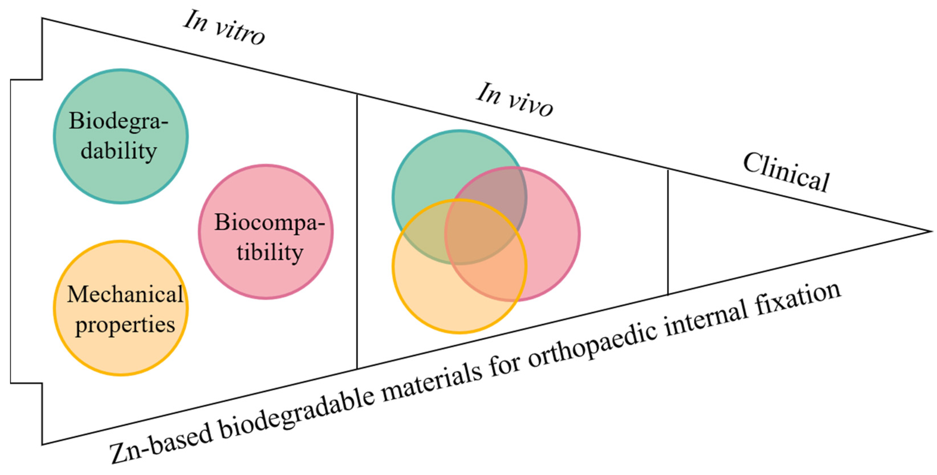
Zinc-Based Biodegradable Materials for Orthopaedic Internal Fixation


Abstract
:1. Introduction

| Classification | Materials | Biodegradability | Mechanical Properties | Biocompatibility | Applications or Potential Applications | Ref. |
|---|---|---|---|---|---|---|
| Non-biodegradable metallicmaterials | 316L SS | Non-biodegradable | High elastic modulus, low wear and corrosion resistance, high tensile strength | High biocompatibility | Acetabular cup, bone screws, bone plates, pins, etc. | [11] |
| Co–Cr alloys | Non-biodegradable | High elastic modulus, high wear and corrosion resistance | Low biocompatibility | Bone screws, bone plates, femoral stems, total hip replacements, etc. | [12] | |
| Ti alloys | Non-biodegradable | Poor fatigue strength, light weight | High biocompatibility | Dental implants, bone screws, bone plates, etc. | [11,13] | |
| Biodegradable metallic materials | Mg-based alloys | Biodegradable, high degradation rate | Poor mechanical properties, elastic modulus are close to cortical bone | High biocompatibility, H2 evolution | Bone screws, bone plates (non-load bearing parts), etc. | [2,9] |
| Fe-based alloys | Biodegradable, low degradation rate | High elastic modulus, poor mechanical properties | Low biocompatibility | Bone screws, bone plates, etc. | [9] | |
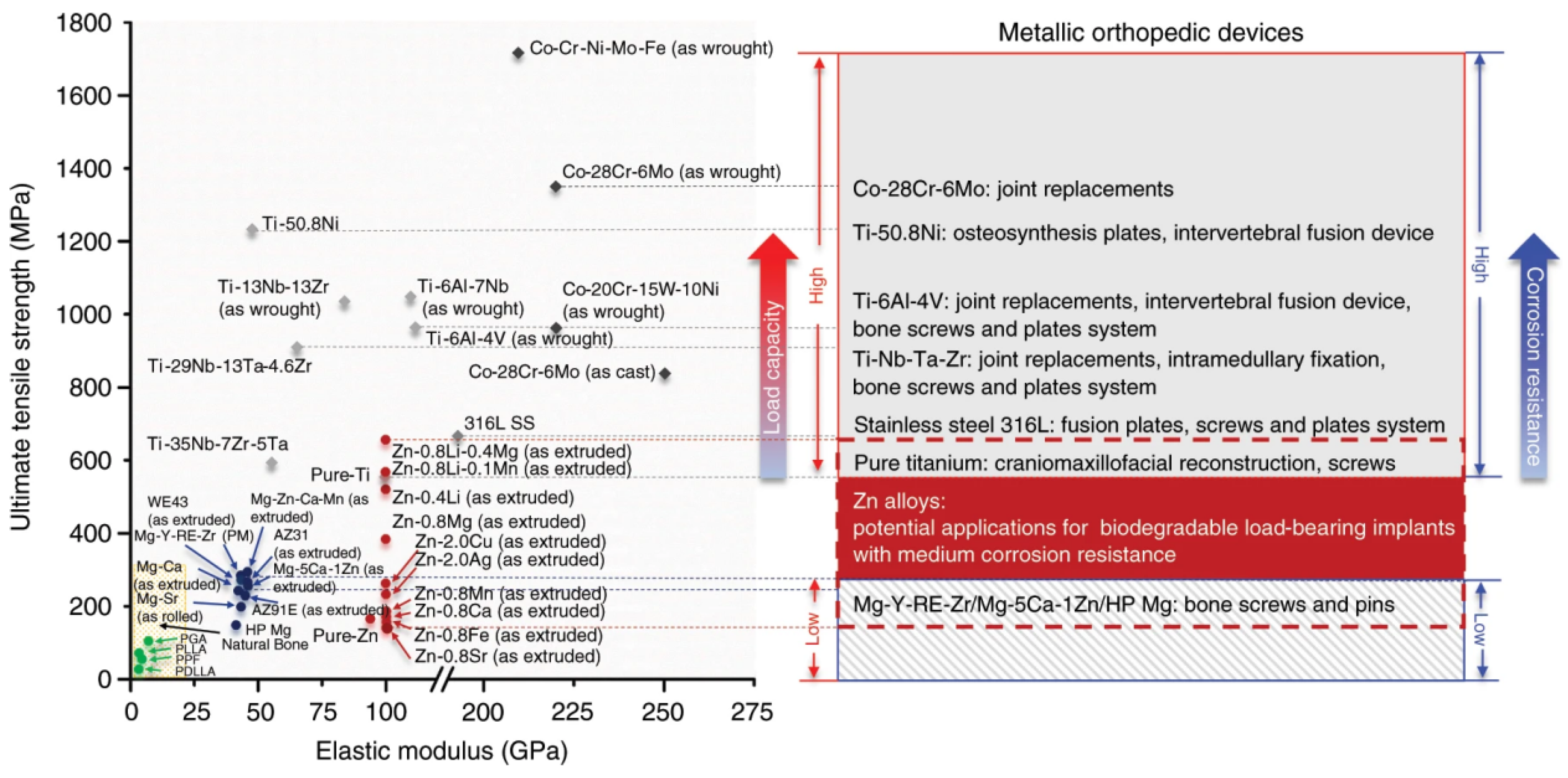
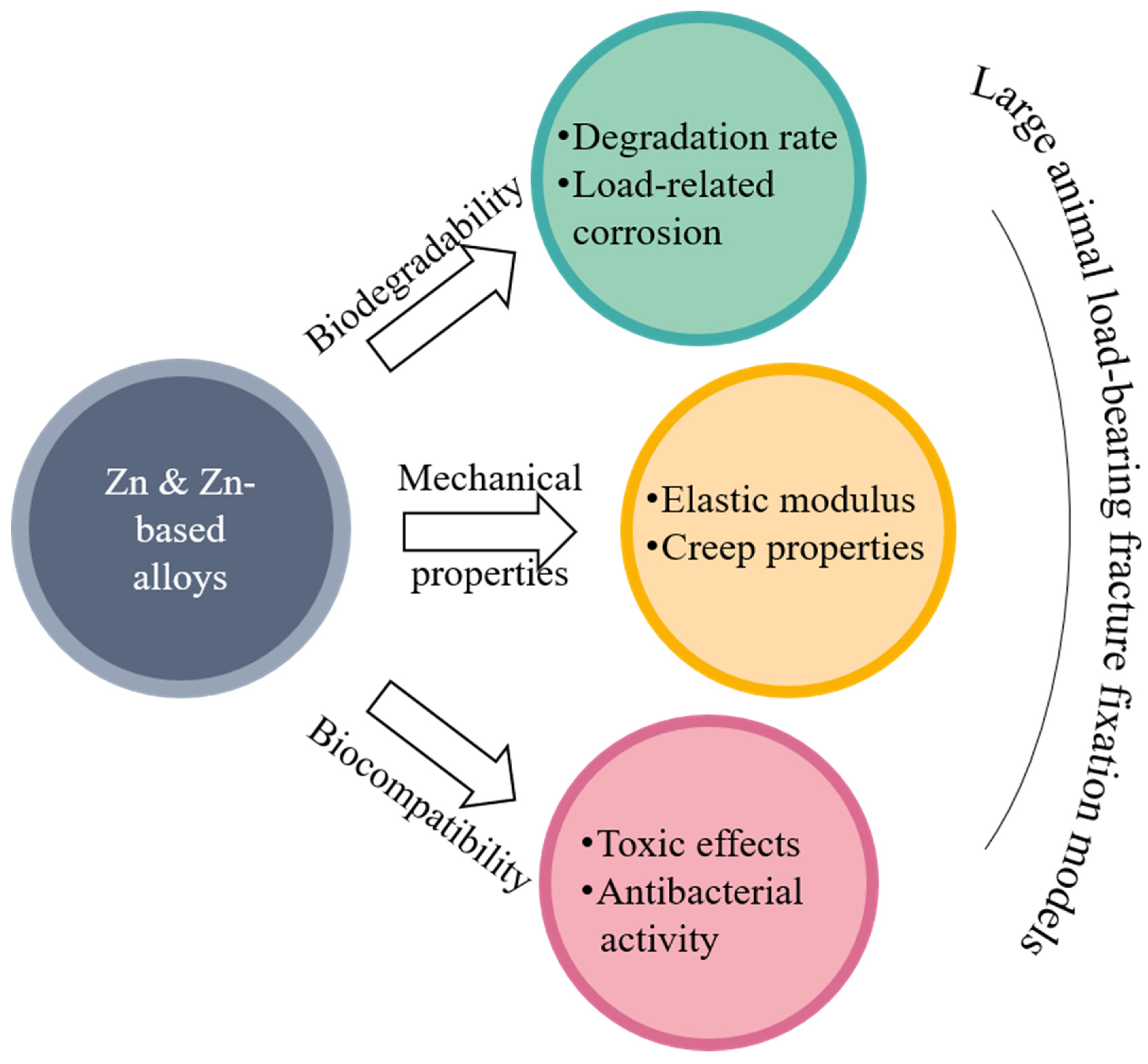
| Zn-based alloys | Biodegradable, moderate corrosion rate | High elastic modulus, high mechanical properties, low creep resistance | Cytotoxicity, no gas production, high biocompatibility | Bone screws, bone plates (load-bearing parts (potential applications)), etc. | [3,9,10] |
2. Biodegradability of Zn-Based Biodegradable Materials
2.1. Biodegradability of Pure Zn
2.2. Biodegradability of Zn-Based Alloys
2.3. Biodegradability of the Zn-Based Composites
| Composition (wt%) | Mechanical Properties | Corrosion Test | Cytocompatibility | Ref. | ||||
|---|---|---|---|---|---|---|---|---|
| σYS (MPa) | σUTS (MPa) | ε (%) | Corrosion Medium | Corrosion Rate (mm y−1) | Cell Type | Key Findings | ||
| Zn-0.8Mg | 203 | 301 | 13 | MEM | 0.071 | U-2OS, L-929 | Zn is less biocompatible than magnesium and the maximum safe concentrations of Zn2+ for the U-2OS and L929 cells are 120 μM and 80 μM. | [50] |
| Zn-1.0Ca | 206 | 252 | 12.7 | HBSS | 0.09 | MG63 | Adding the alloying elements Ca into Zn can significantly increase the viability of MG63 and can promote the MG63 cell proliferation compared with the pure Zn and negative control groups. | [51] |
| Zn-1.1Sr | 220 | 250 | 22 | SBF | 0.4 | HOBs, hMSCs | The proliferation ability of the two kinds of cells did not decrease in the zinc alloy leaching solution. When the concentration of the leaching solution was low, the growth of the two kinds of cells was promoted. | [32] |
| Zn-0.4Li | 387 | 520 | 5.0 | SBF | 0.019 | MC3T3-E1 | Zn-0.4Li alloy extract can significantly promote the proliferation of MC3T3-E1 cells. | [24] |
| Zn-5.0Ge | 175 | 237 | 22 | HBSS | 0.051 | MC3T3-E1 | The diluted extracts at a concentration 12.5% of both the as-cast Zn-5Ge alloy and pure Zn showed grade 0 cytotoxicity; the diluted extracts at the concentrations of 50% and 25% of Zn-5Ge alloy showed a significantly higher cell viability than those of pure Zn. | [52] |
| Zn-6.0Ag | - | 290 | - | SBF | 0.114 | - | - | [44] |
| Zn-0.8Fe | 127 | 163 | 28.1 | SBF | 0.022 | MC3T3-E1 | MC3T3-E1 cells had unhealthy morphology and low cell viability. | [24] |
| Zn-4Cu | 327 | 393 | 44.6 | HBSS | 0.13 | L-929, TAG, SAOS-2 | Zn-4Cu alloy had no obvious cytotoxic effect on L929, TAG and Saos-2 cells. | [53] |
| Zn-0.8Mn | 98.4 | 104.7 | 1.0 | - | - | L-929 | Zn-0.8Mn alloy showed 29% to 44% cell viability in 100% extract, indicating moderate cytotoxicity. | [40] |
| Zn-2Al | 142 | 192 | 12 | SBF | 0.13 | MG63 | Cell viability decreased to 67.5 ± 5.3% in 100% extract cultured for one day, indicating that high concentrations of ions have a negative effect on cell growth. With the extension of culture time, the number of cells increased significantly. | [42] |
| Zn-0.0.5Zr | 104 | 157 | 22 | - | - | - | - | [54] |
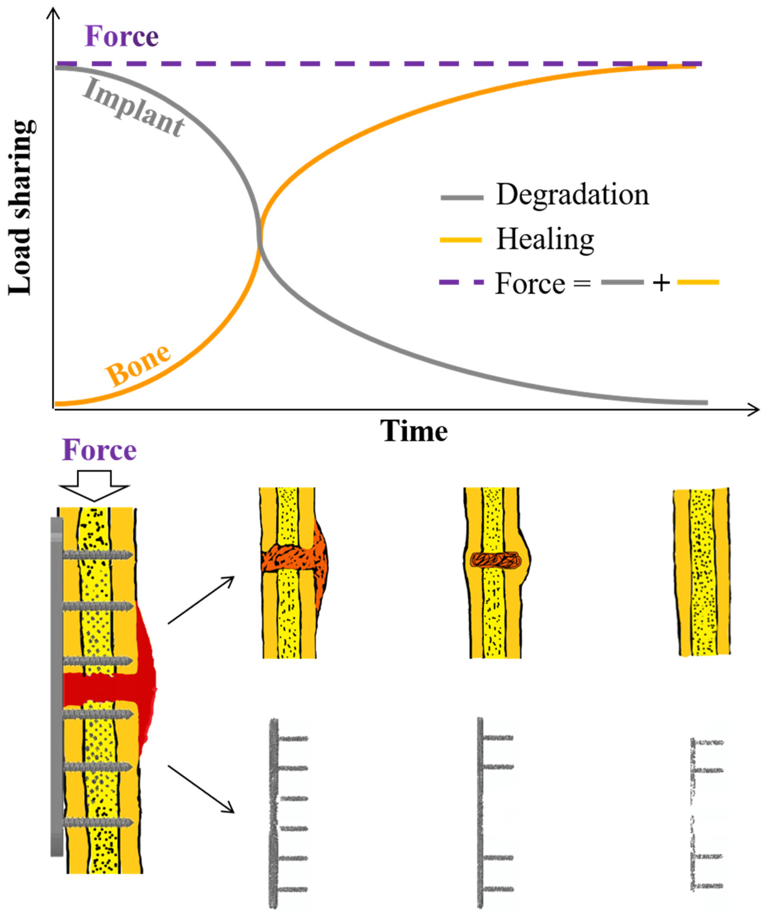
2.4. Biodegradability of Zn-Based Biomaterials under Mechanical Loading
3. Mechanical Properties of Zn-Based Biodegradable Materials
3.1. Mechanical Properties of Pure Zn
3.2. Mechanical Properties of Zn-Based Alloys
3.3. Mechanical Properties of Zn-Based Composites
| Composition (wt%) and Manufacturing Process | Mechanical Properties | Corrosion Test | Cytocompatibility | Ref. | ||||
|---|---|---|---|---|---|---|---|---|
| σYS (MPa) | σUTS (MPa) | ε (%) | Corrosion Medium | Corrosion Rate (mm/y) | Cell Type | Key Findings | ||
| Zn-1.5Mg-0.5Zr HE | 350 | 425 | 12 | - | - | L-929 | Overall, the L-929 cells exhibit polygonal or spindle shape, and well spread and proliferated in the extracts of pure Zn and Zn alloys. | [39] |
| Zn-1.0Ca-1Sr Cast | 86 | 140 | 1.2 | SBF | - | MG63 | Adding the alloying elements Mg, Ca and Sr into Zn can significantly increase the viability of MG63 and can promote the MG63 cell proliferation compared with the pure Zn and negative control groups. | [77] |
| Zn-1.0Ca-1Sr HE | 212 | 260 | 6.7 | SBF | 0.11 | |||
| Zn-1.0Ca-1Sr HR | 144 | 203 | 8.8 | SBF | - | |||
| Zn-0.8Li-0.4Mg HE | 438 | 646 | 3.68 | - | - | - | - | [24] |
| Zn-3Ge-0.5Mg Cast | 66.9 | 88.3 | 1.4 | HBSS | 0.062 | MG63 | The extract with a concentration of 100% had obvious cytotoxicity to MG63 cells. When the concentration of the extract was diluted to 12.5% or lower, the survival rate of MG-63 cells was all above 90%. | [78] |
| Zn-3Ge-0.5Mg HR | 253 | 208 | 9.2 | HBSS | 0.075 | |||
| Zn-4Ag-0.6Mn HE | - | 302 | 35 | HBSS | 0.012 | - | - | [79] |
| Zn-1Fe-1Mg Cast | 146 | 157 | 2.3 | SBF | 0.027 | - | - | [80] |
| Zn-0.8Mn-0.4 Cast | 112 | 120 | 0.3 | - | - | - | - | [68] |
| Zn-0.8Mn-0.4 HE | 253 | 343 | 8 | - | - | |||
| Zn-0.8Mn-0.4 HR | 245 | 323 | 12 | - | - | |||
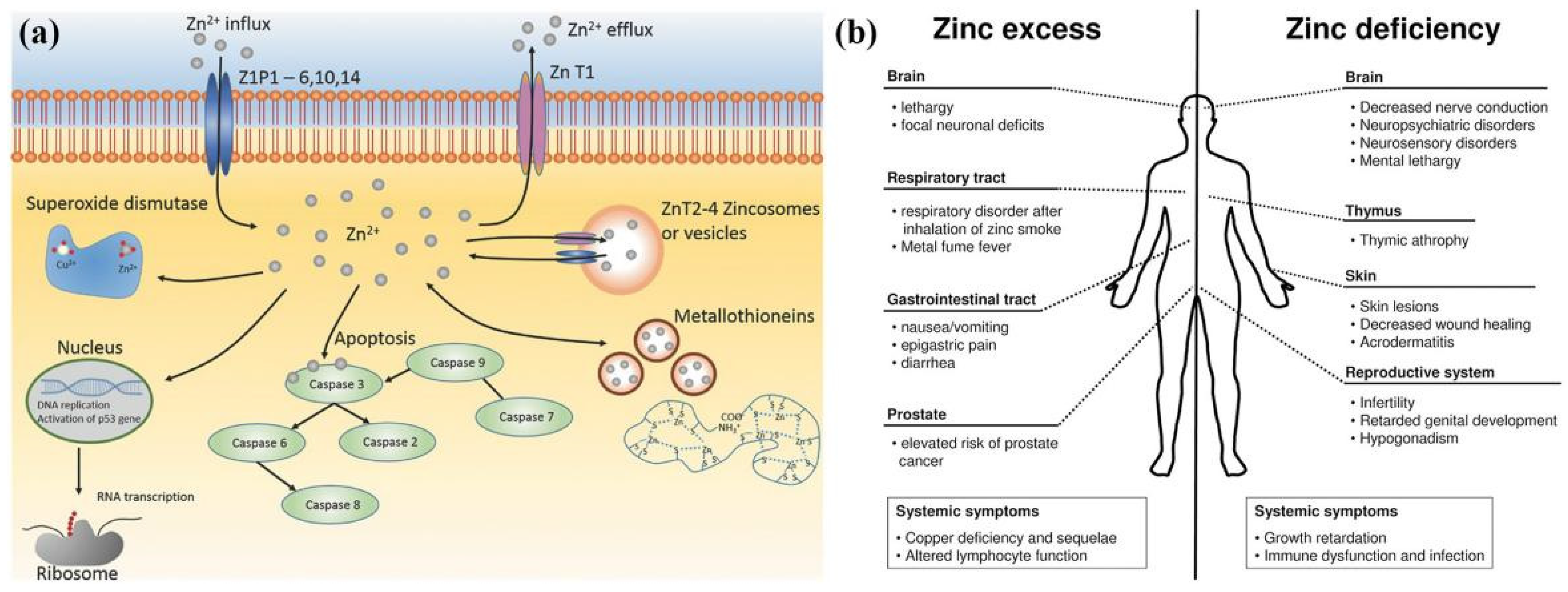
4. Biocompatibility of Zn-Based Biodegradable Materials
4.1. Biocompatibility of Pure Zn
4.2. Biocompatibility of Zn-Based Alloys
4.3. Biocompatibility of Zn-Based Composites
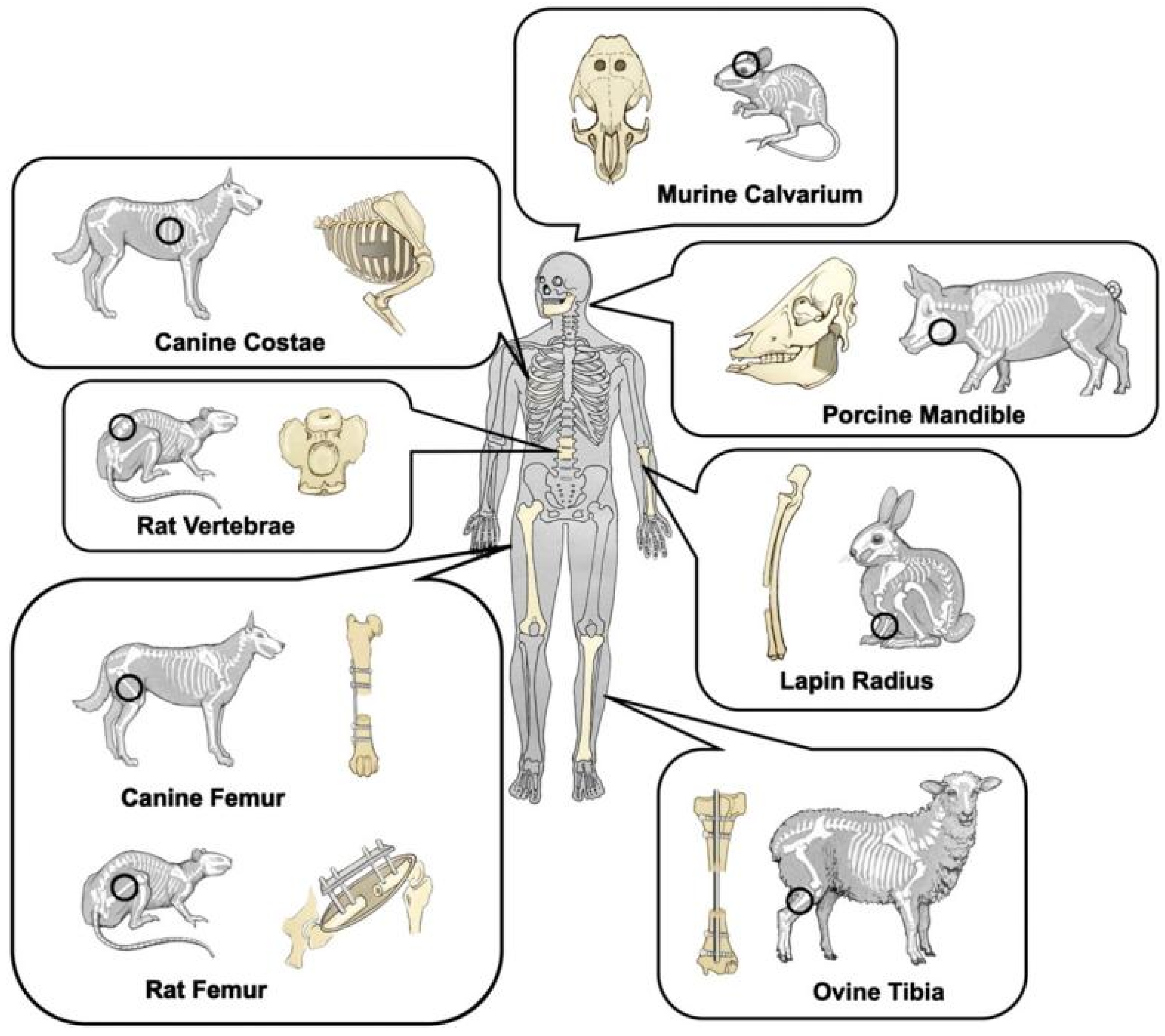
5. In Vivo Evaluation of Zn-Based Biodegradable Materials with Animal Models
| Zn-Based Metals | Designed Implants | Control | Surgeries | Animal Species | Major Findings | Ref. |
|---|---|---|---|---|---|---|
| Zn-Mn | Scaffold | Pure Ti | Insertion into femoral condyle | Rats | The new bone tissues at the bone defect sites gradually increased with time in both groups, and numerous new bone tissues were observed around the Zn-0.8Mn alloy scaffold | [70] |
| Zn-1Mg, Zn-1Ca, Zn-1Sr | Intramedullary nails | NA | Insertion into femoral marrow medullary cavity | Mice | There was no inflammation observed around the implantation site and no mouse died after operation. The new bone thickness of Zn-1Mg, Zn-1Ca and Zn-1Sr pin groups are significantly larger than the sham control group. | [51] |
| Zn-HA | Pin | Pure Zn | Insertion into femoral condyle | Rats | There was new bone formation around the Zn-HA composite, and the bone mass increased over time. With prolonged implantation time, the Zn-HA composite was more effective than pure Zn in promoting new bone formation. | [55] |
| Zn-0.05Mg | Pin | Pure Zn | Insertion into femoral condyle | Rabbits | No inflammatory cells were found at the fracture site, and new bone tissue formation was confirmed at the bone/implant interface, proving that the Zn-0.05Mg alloy promoted the formation of new bone tissue. | [46] |
| Zn-(0.001% Mg 2.5%, 0.01% Fe 2.5%) | Screw and plate | PLLA, Ti-based alloys | Mandible fracture | Beagles | The new bone formation in the Zn alloy group and the titanium alloy group was significantly higher than that in the PLLA group. In addition, the new bone formation in the Zn-based alloys group was slightly higher than that in the Ti-based alloys group. The degradation of Zn implants in vivo would not increase the concentration of Zn2+. | [97] |
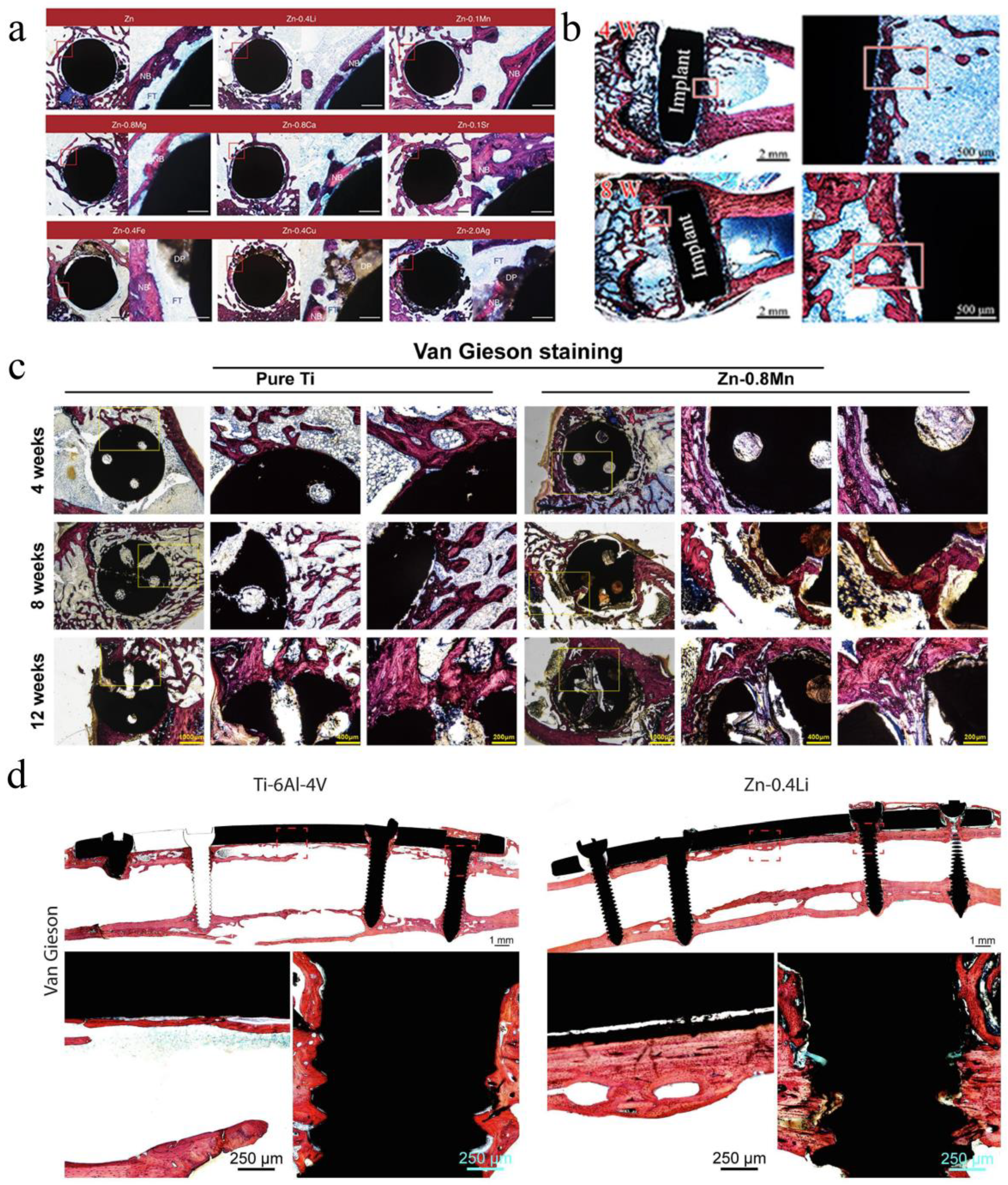
| Zn-X (Fe, Cu, Ag, Mg, Ca, Sr, Mn, Li) | Intramedullary nails | Pure Zn | Insertion into femoral marrow medullary cavity | Rats | Pure Zn, Zn-0.4Fe, Zn-0.4Cu and Zn-2.0Ag alloy implants showed localized degradation patterns with local accumulation of products. In contrast, the degradation of Zn-0.8Mg, Zn-0.8Ca, Zn-0.1Sr, Zn-0.4Li and Zn-0.1Mn was more uniform on the macroscopic scale. | [24] |
| Zn-0.8Sr | Scaffold | Pure Ti | Insertion into femoral condyle | Rats | Zn-based alloys promote bone regeneration by promoting the proliferation and differentiation of MC3T3-E1 cells, upregulating the expression of osteogenesis-related genes and proteins, and stimulating angiogenesis. | [36] |
| Zn-0.8Li-0.1Ca | Scaffold | Pure Ti | Insertion into radial defect | Rabbits | The Zn-0.8Li-0.1Ca alloy has a similar level of biocompatibility to pure titanium, but it promotes regeneration significantly faster than pure Ti. | [33] |
| Zn-0.4Li | Screw and plate | Ti-6Al-4V | Femoral shaft fracture | Rabbits | Plates and screws made of Zn-0.4Li alloy showed comparable performance to Ti-6Al-4V in fracture fixation, and the fractured bone healed completely six months after surgery. | [25] |
| Zn-1Mg-nvol%β-TCP (n = 0, 1) | Columnar samples | Zn-1Mg | Specimens in lateral thighs. | Rats | Zn-1Mg alloy and Zn-1Mg-β-TCP composites had no significant tissue inflammation and showed good biocompatibility. | [56] |
6. Summary and Future Directions
Author Contributions
Funding
Data Availability Statement
Conflicts of Interest
References
- Shuai, C.; Li, S.; Peng, S.; Feng, P.; Lai, Y.; Gao, C. Biodegradable metallic bone implants. Mater. Chem. Front. 2019, 3, 544–562. [Google Scholar] [CrossRef]
- Fu, R.; Feng, Y.; Liu, Y.; Willie, B.M.; Yang, H. The combined effects of dynamization time and degree on bone healing. J. Orthop. Res. 2022, 40, 634–643. [Google Scholar] [CrossRef] [PubMed]
- Fu, R.; Feng, Y.; Bertrand, D.; Du, T.; Liu, Y.; Willie, B.M.; Yang, H. Enhancing the efficiency of distraction osteogenesis through rate-varying distraction: A computational study. Int. J. Mol. Sci. 2021, 22, 11734. [Google Scholar] [CrossRef] [PubMed]
- Tucker, A. Management of common upper limb fractures in adults. Surgery 2022, 40, 184–191. [Google Scholar] [CrossRef]
- Wu, A.M.; Bisignano, C.; James, S.L.; Abady, G.G.; Abedi, A.; Abu-Gharbieh, E.; Alhassan, R.K.; Alipour, V.; Arabloo, J.; Asaad, M.; et al. Global, regional, and national burden of bone fractures in 204 countries and territories, 1990–2019: A systematic analysis from the Global Burden of Disease Study 2019. Lancet Healthy Longev. 2021, 2, e580–e592. [Google Scholar] [CrossRef]
- Fu, R.; Feng, Y.; Liu, Y.; Yang, H. Mechanical regulation of bone regeneration during distraction osteogenesis. Med. Nov. Technol. Devices 2021, 11, 100077. [Google Scholar] [CrossRef]
- Fu, R.; Bertrand, D.; Wang, J.; Kavaseri, K.; Feng, Y.; Du, T.; Liu, Y.; Willie, B.M.; Yang, H. In vivo and in silico monitoring bone regeneration during distraction osteogenesis of the mouse femur. Comput. Methods Programs Biomed. 2022, 216, 106679. [Google Scholar] [CrossRef]
- Stiffler, K.S. Internal fracture fixation. Clin. Tech. Small Anim. Pract. 2004, 19, 105–113. [Google Scholar] [CrossRef]
- Taljanovic, M.S.; Jones, M.D.; Ruth, J.T.; Benjamin, J.B.; Sheppard, J.E.; Hunter, T.B. Fracture Fixation. Radiographics 2003, 23, 1569–1590. [Google Scholar] [CrossRef]
- Manam, N.S.; Harun, W.S.W.; Shri, D.N.A.; Ghani, S.A.C.; Kurniawan, T.; Ismail, M.H.; Ibrahim, M.H.I. Study of corrosion in biocompatible metals for implants: A review. J. Alloys Compd. 2017, 701, 698–715. [Google Scholar] [CrossRef] [Green Version]
- Hasiak, M.; Sobieszczanska, B.; Laszcz, A.; Bialy, M.; Checmanowski, J.; Zatonski, T.; Bozemska, E.; Wawrzynska, M. Production, mechanical properties and biomedical characterization of ZrTi-based bulk metallic glasses in comparison with 316L stainless steel and Ti6Al4V alloy. Materials 2021, 15, 252. [Google Scholar] [CrossRef] [PubMed]
- Iatecola, A.; Longhitano, G.A.; Antunes, L.H.M.; Jardini, A.L.; Miguel, E.C.; Beres, M.; Lambert, C.S.; Andrade, T.N.; Buchaim, R.L.; Buchaim, D.V.; et al. Osseointegration improvement of Co-Cr-Mo alloy produced by additive manufacturing. Pharmaceutics 2021, 13, 724. [Google Scholar] [CrossRef] [PubMed]
- Okazaki, Y.; Katsuda, S.I. Biological safety evaluation and surface modification of biocompatible Ti-15Zr-4Nb alloy. Materials 2021, 14, 731. [Google Scholar] [CrossRef] [PubMed]
- Kabir, H.; Munir, K.; Wen, C.; Li, Y. Recent research and progress of biodegradable zinc alloys and composites for biomedical applications: Biomechanical and biocorrosion perspectives. Bioact. Mater. 2021, 6, 836–879. [Google Scholar] [CrossRef]
- Wang, J.L.; Xu, J.K.; Hopkins, C.; Chow, D.H.; Qin, L. Biodegradable magnesium-based implants in orthopedics-A general review and perspectives. Adv. Sci. 2020, 7, 1902443. [Google Scholar] [CrossRef]
- Witte, F.; Hort, N.; Vogt, C.; Cohen, S.; Kainer, K.U.; Willumeit, R.; Feyerabend, F. Degradable biomaterials based on magnesium corrosion. Curr. Opin. Solid State Mater. Sci. 2008, 12, 63–72. [Google Scholar] [CrossRef]
- Jang, H.Y.; Shin, J.Y.; Oh, S.H.; Byun, J.H.; Lee, J.H. PCL/HA hybrid microspheres for effective osteogenic differentiation and bone regeneration. ACS Biomater. Sci. Eng. 2020, 6, 5172–5180. [Google Scholar] [CrossRef]
- Yang, Y.; Zhao, Y.; Tang, G.; Li, H.; Yuan, X.; Fan, Y. In vitro degradation of porous poly(l-lactide-co-glycolide)/β-tricalcium phosphate (PLGA/β-TCP) scaffolds under dynamic and static conditions. Polym. Degrad. Stab. 2008, 93, 1838–1845. [Google Scholar] [CrossRef]
- Zhou, H.; Lawrence, J.G.; Bhaduri, S.B. Fabrication aspects of PLA-CaP/PLGA-CaP composites for orthopedic applications: A review. Acta Biomater. 2012, 8, 1999–2016. [Google Scholar] [CrossRef]
- Zhang, Y.; Xu, J.; Ruan, Y.C.; Yu, M.K.; O’Laughlin, M.; Wise, H.; Chen, D.; Tian, L.; Shi, D.; Wang, J.; et al. Implant-derived magnesium induces local neuronal production of CGRP to improve bone-fracture healing in rats. Nat. Med. 2016, 22, 1160–1169. [Google Scholar] [CrossRef] [Green Version]
- Lee, J.W.; Han, H.S.; Han, K.J.; Park, J.; Jeon, H.; Ok, M.R.; Seok, H.K.; Ahn, J.P.; Lee, K.E.; Lee, D.H.; et al. Long-term clinical study and multiscale analysis of in vivo biodegradation mechanism of Mg alloy. Proc. Natl. Acad. Sci. USA 2016, 113, 716–721. [Google Scholar] [CrossRef] [PubMed]
- Zheng, Y.F.; Gu, X.N.; Witte, F. Biodegradable metals. Mater. Sci. Eng. R Rep. 2014, 77, 1–34. [Google Scholar] [CrossRef]
- Kumar, K.; Gill, R.S.; Batra, U. Challenges and opportunities for biodegradable magnesium alloy implants. Mater. Technol. 2017, 33, 153–172. [Google Scholar] [CrossRef]
- Yang, H.; Jia, B.; Zhang, Z.; Qu, X.; Li, G.; Lin, W.; Zhu, D.; Dai, K.; Zheng, Y. Alloying design of biodegradable zinc as promising bone implants for load-bearing applications. Nat. Commun. 2020, 11, 401. [Google Scholar] [CrossRef] [PubMed]
- Yang, H.; Qu, X.; Wang, M.; Cheng, H.; Jia, B.; Nie, J.; Dai, K.; Zheng, Y. Zn-0.4Li alloy shows great potential for the fixation and healing of bone fractures at load-bearing sites. Chem. Eng. J. 2021, 417, 129317. [Google Scholar] [CrossRef]
- Li, G.; Yang, H.; Zheng, Y.; Chen, X.H.; Yang, J.A.; Zhu, D.; Ruan, L.; Takashima, K. Challenges in the use of zinc and its alloys as biodegradable metals: Perspective from biomechanical compatibility. Acta Biomater. 2019, 97, 23–45. [Google Scholar] [CrossRef] [PubMed]
- Li, H.F.; Shi, Z.Z.; Wang, L.N. Opportunities and challenges of biodegradable Zn-based alloys. J. Mater. Sci. Technol. 2020, 46, 136–138. [Google Scholar] [CrossRef]
- Torne, K.; Larsson, M.; Norlin, A.; Weissenrieder, J. Degradation of Zinc in saline solutions, plasma, and whole blood. J. Biomed. Mater. Res. B Appl. Biomater. 2016, 104, 1141–1151. [Google Scholar] [CrossRef]
- Andreini, C.; Banci, L.; Bertini, I.; Rosato, A. Counting the Zinc-proteins encoded in the human genome. J. Proteome Res. 2006, 5, 196–201. [Google Scholar] [CrossRef]
- Weiss, A.; Murdoch, C.C.; Edmonds, K.A.; Jordan, M.R.; Monteith, A.J.; Perera, Y.R.; Rodriguez Nassif, A.M.; Petoletti, A.M.; Beavers, W.N.; Munneke, M.J.; et al. Zn-regulated GTPase metalloprotein activator 1 modulates vertebrate zinc homeostasis. Cell 2022, 185, 2148–2163. [Google Scholar] [CrossRef]
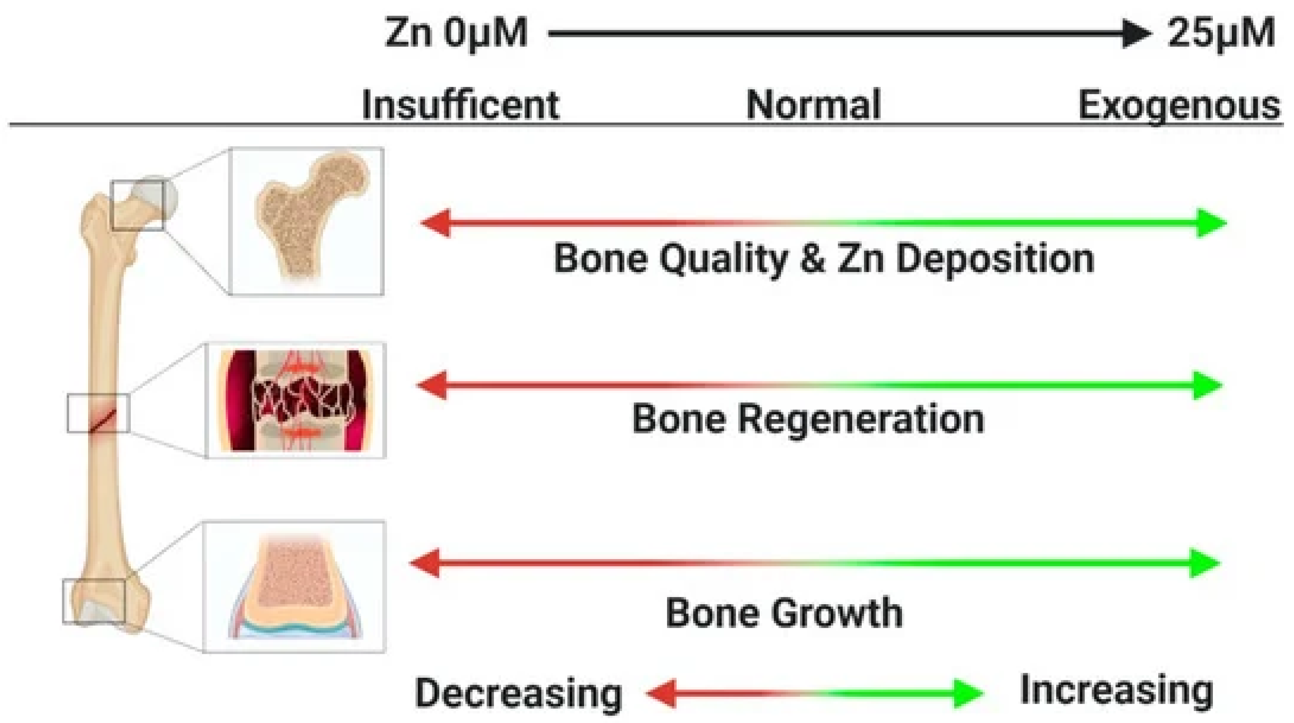
- Qiao, W.; Pan, D.; Zheng, Y.; Wu, S.; Liu, X.; Chen, Z.; Wan, M.; Feng, S.; Cheung, K.M.C.; Yeung, K.W.K.; et al. Divalent metal cations stimulate skeleton interoception for new bone formation in mouse injury models. Nat. Commun. 2022, 13, 535. [Google Scholar] [CrossRef]
- Zhu, D.; Cockerill, I.; Su, Y.; Zhang, Z.; Fu, J.; Lee, K.W.; Ma, J.; Okpokwasili, C.; Tang, L.; Zheng, Y.; et al. Mechanical strength, biodegradation, and in vitro and in vivo biocompatibility of Zn biomaterials. ACS Appl. Mater. Interfaces 2019, 11, 6809–6819. [Google Scholar] [CrossRef] [PubMed]
- Zhang, Z.; Jia, B.; Yang, H.; Han, Y.; Wu, Q.; Dai, K.; Zheng, Y. Biodegradable ZnLiCa ternary alloys for critical-sized bone defect regeneration at load-bearing sites: In vitro and in vivo studies. Bioact. Mater. 2021, 6, 3999–4013. [Google Scholar] [CrossRef]
- Bowen, P.K.; Seitz, J.M.; Guillory, R.J.; Braykovich, J.P.; Zhao, S.; Goldman, J.; Drelich, J.W. Evaluation of wrought Zn-Al alloys (1; 3; and 5 wt % Al) through mechanical and in vivo testing for stent applications. J. Biomed. Mater. Res. B Appl. Biomater. 2018, 106, 245–258. [Google Scholar] [CrossRef] [PubMed]
- Dambatta, M.S.; Izman, S.; Kurniawan, D.; Hermawan, H. Processing of Zn-3Mg alloy by equal channel angular pressing for biodegradable metal implants. J. King Saud. Univ. Sci. 2017, 29, 455–461. [Google Scholar] [CrossRef]
- Jia, B.; Yang, H.; Zhang, Z.; Qu, X.; Jia, X.; Wu, Q.; Han, Y.; Zheng, Y.; Dai, K. Biodegradable Zn-Sr alloy for bone regeneration in rat femoral condyle defect model: In vitro and in vivo studies. Bioact. Mater. 2021, 6, 1588–1604. [Google Scholar] [CrossRef]
- Kafri, A.; Ovadia, S.; Goldman, J.; Drelich, J.; Aghion, E. The suitability of Zn–1.3%Fe alloy as a biodegradable implant material. Metals 2018, 8, 153. [Google Scholar] [CrossRef]
- Krezel, A.; Maret, W. The biological inorganic chemistry of zinc ions. Arch. Biochem. Biophys. 2016, 611, 3–19. [Google Scholar] [CrossRef] [PubMed]
- Ren, T.; Gao, X.; Xu, C.; Yang, L.; Guo, P.; Liu, H.; Chen, Y.; Sun, W.; Song, Z. Evaluation of as-extruded ternary Zn–Mg–Zr alloys for biomedical implantation material: In vitro and in vivo behavior. Mater. Corros. 2019, 70, 1056–1070. [Google Scholar] [CrossRef]
- Shi, Z.Z.; Yu, J.; Liu, X.F.; Zhang, H.J.; Zhang, D.W.; Yin, Y.X.; Wang, L.N. Effects of Ag, Cu or Ca addition on microstructure and comprehensive properties of biodegradable Zn-0.8Mn alloy. Mater. Sci. Eng. C Mater. Biol. Appl. 2019, 99, 969–978. [Google Scholar] [CrossRef]
- Shi, Z.; Yu, J.; Liu, X. Microalloyed Zn-Mn alloys: From extremely brittle to extraordinarily ductile at room temperature. Mater. Des. 2018, 144, 343–352. [Google Scholar] [CrossRef]
- Shuai, C.; Cheng, Y.; Yang, Y.; Peng, S.; Yang, W.; Qi, F. Laser additive manufacturing of Zn-2Al part for bone repair: Formability; microstructure and properties. J. Alloys Compd. 2019, 798, 606–615. [Google Scholar] [CrossRef]
- Shuai, C.; Xue, L.; Gao, C.; Peng, S.; Zhao, Z. Rod-like eutectic structure in biodegradable Zn-Al-Sn alloy exhibiting enhanced mechanical strength. ACS Biomater. Sci. Eng. 2020, 6, 3821–3831. [Google Scholar] [CrossRef]
- Shuai, C.; Xue, L.; Gao, C.; Yang, Y.; Peng, S.; Zhang, Y. Selective laser melting of Zn–Ag alloys for bone repair: Microstructure; mechanical properties and degradation behavior. Virtual Phys. Prototyp. 2018, 13, 146–154. [Google Scholar] [CrossRef]
- Shi, Z.; Yu, J.; Liu, X.; Wang, L. Fabrication and characterization of novel biodegradable Zn-Mn-Cu alloys. J. Mater. Sci. Technol. 2018, 34, 1008–1015. [Google Scholar] [CrossRef]
- Xiao, C.; Wang, L.; Ren, Y.; Sun, S.; Zhang, E.; Yan, C.; Liu, Q.; Sun, X.; Shou, F.; Duan, J.; et al. Indirectly extruded biodegradable Zn-0.05wt%Mg alloy with improved strength and ductility: In vitro and in vivo studies. J. Mater. Sci. Technol. 2018, 34, 1618–1627. [Google Scholar] [CrossRef]
- Yuan, W.; Xia, D.; Wu, S.; Zheng, Y.; Guan, Z.; Rau, J.V. A review on current research status of the surface modification of Zn-based biodegradable metals. Bioact. Mater. 2022, 7, 192–216. [Google Scholar] [CrossRef] [PubMed]
- Chen, K.; Lu, Y.; Tang, H.; Gao, Y.; Zhao, F.; Gu, X.; Fan, Y. Effect of strain on degradation behaviors of WE43, Fe and Zn wires. Acta Biomater. 2020, 113, 627–645. [Google Scholar] [CrossRef]
- Yang, J.; Yim, C.D.; You, B.S. Effects of solute Zn on corrosion film of Mg–Sn–Zn alloy formed in NaCl solution. J. Electrochem. Soc. 2016, 163, C839–C844. [Google Scholar] [CrossRef]
- Kubasek, J.; Vojtech, D.; Jablonska, E.; Pospisilova, I.; Lipov, J.; Ruml, T. Structure, mechanical characteristics and in vitro degradation; cytotoxicity; genotoxicity and mutagenicity of novel biodegradable Zn-Mg alloys. Mater. Sci. Eng. C Mater. Biol. Appl. 2016, 58, 24–35. [Google Scholar] [CrossRef]
- Li, H.F.; Xie, X.H.; Zheng, Y.F.; Cong, Y.; Zhou, F.Y.; Qiu, K.J.; Wang, X.; Chen, S.H.; Huang, L.; Tian, L.; et al. Development of biodegradable Zn-1X binary alloys with nutrient alloying elements Mg, Ca and Sr. Sci. Rep. 2015, 5, 10719. [Google Scholar] [CrossRef] [PubMed]
- Tong, X.; Zhang, D.; Zhang, X.; Su, Y.; Shi, Z.; Wang, K.; Lin, J.; Li, Y.; Lin, J.; Wen, C. Microstructure; mechanical properties; biocompatibility; and in vitro corrosion and degradation behavior of a new Zn-5Ge alloy for biodegradable implant materials. Acta Biomater. 2018, 82, 197–204. [Google Scholar] [CrossRef] [PubMed]
- Li, P.; Zhang, W.; Dai, J.; Xepapadeas, A.B.; Schweizer, E.; Alexander, D.; Scheideler, L.; Zhou, C.; Zhang, H.; Wan, G.; et al. Investigation of zinccopper alloys as potential materials for craniomaxillofacial osteosynthesis implants. Mater. Sci. Eng. C Mater. Biol. Appl. 2019, 103, 109826. [Google Scholar] [CrossRef]
- Wątroba, M.; Bednarczyk, W.; Kawałko, J.; Bała, P. Effect of zirconium microaddition on the microstructure and mechanical properties of Zn-Zr alloys. Mater. Charact. 2018, 142, 187–194. [Google Scholar] [CrossRef]
- Yang, H.; Qu, X.; Lin, W.; Wang, C.; Zhu, D.; Dai, K.; Zheng, Y. In vitro and in vivo studies on zinc-hydroxyapatite composites as novel biodegradable metal matrix composite for orthopedic applications. Acta Biomater. 2018, 71, 200–214. [Google Scholar] [CrossRef]
- Pan, C.; Sun, X.; Xu, G.; Su, Y.; Liu, D. The effects of beta-TCP on mechanical properties; corrosion behavior and biocompatibility of beta-TCP/Zn-Mg composites. Mater. Sci. Eng. C Mater. Biol. Appl. 2020, 108, 110397. [Google Scholar] [CrossRef]
- Li, X.; Chu, C.; Chu, P.K. Effects of external stress on biodegradable orthopedic materials: A review. Bioact. Mater. 2016, 1, 77–84. [Google Scholar] [CrossRef]
- Li, N.; Zheng, Y. Novel magnesium alloys developed for biomedical application: A review. J. Mater. Sci. Technol. 2013, 29, 489–502. [Google Scholar] [CrossRef]
- Kirkland, N.T.; Birbilis, N.; Staiger, M.P. Assessing the corrosion of biodegradable magnesium implants: A critical review of current methodologies and their limitations. Acta Biomater. 2012, 8, 925–936. [Google Scholar] [CrossRef]
- Li, G.N.; Zhu, S.M.; Nie, J.F.; Zheng, Y.; Sun, Z. Investigating the stress corrosion cracking of a biodegradable Zn-0.8 wt%Li alloy in simulated body fluid. Bioact. Mater. 2021, 6, 1468–1478. [Google Scholar] [CrossRef]
- Li, P.; Feng, X.; Jia, X.; Fan, Y. Influences of tensile load on in vitro degradation of an electrospun poly(L-lactide-co-glycolide) scaffold. Acta Biomater. 2010, 6, 2991–2996. [Google Scholar] [CrossRef] [PubMed]
- Guo, M.; Chu, Z.; Yao, J.; Feng, W.; Wang, Y.; Wang, L.; Fan, Y. The effects of tensile stress on degradation of biodegradable PLGA membranes: A quantitative study. Polym. Degrad. Stab. 2016, 124, 95–100. [Google Scholar] [CrossRef]
- Gao, Y.; Wang, L.; Li, L.; Gu, X.; Zhang, K.; Xia, J.; Fan, Y. Effect of stress on corrosion of high-purity magnesium in vitro and in vivo. Acta Biomater. 2019, 83, 477–486. [Google Scholar] [CrossRef] [PubMed]
- Gao, Y.; Wang, L.; Gu, X.; Chu, Z.; Guo, M.; Fan, Y. A quantitative study on magnesium alloy stent biodegradation. J. Biomech. 2018, 74, 98–105. [Google Scholar] [CrossRef]
- Vasudevan, A.K.; Sadananda, K. Classification of environmentally assisted fatigue crack growth behavior. Int. J. Fatigue 2009, 31, 1696–1708. [Google Scholar] [CrossRef]
- Jafari, S.; Singh Raman, R.K.; Davies, C.H.J. Corrosion fatigue of a magnesium alloy in modified simulated body fluid. Eng. Fract. Mech. 2015, 137, 2–11. [Google Scholar] [CrossRef]
- Zhao, J.; Gao, L.L.; Gao, H.; Yuan, X.; Chen, X. Biodegradable behaviour and fatigue life of ZEK100 magnesium alloy in simulated physiological environment. Fatigue Fract. Eng. Mater. Struct. 2015, 38, 904–913. [Google Scholar] [CrossRef]
- Shi, Z.; Li, H.; Xu, J.; Gao, X.; Liu, X. Microstructure evolution of a high-strength low-alloy Zn–Mn–Ca alloy through casting; hot extrusion and warm caliber rolling. Mater. Sci. Eng. A 2020, 771, 138626. [Google Scholar] [CrossRef]
- Guan, Z.; Linsley, C.S.; Pan, S.; Yao, G.; Wu, B.M.; Levi, D.S.; Li, X. Zn-Mg-WC nanocomposites for bioresorbable cardiovascular stents: Microstructure, mechanical properties, fatigue, shelf life, and corrosion. ACS Biomater. Sci. Eng. 2022, 8, 328–339. [Google Scholar] [CrossRef]
- Jia, B.; Yang, H.; Han, Y.; Zhang, Z.; Qu, X.; Zhuang, Y.; Wu, Q.; Zheng, Y.; Dai, K. In vitro and in vivo studies of Zn-Mn biodegradable metals designed for orthopedic applications. Acta Biomater. 2020, 108, 358–372. [Google Scholar] [CrossRef]
- Katarivas Levy, G.; Leon, A.; Kafri, A.; Ventura, Y.; Drelich, J.W.; Goldman, J.; Vago, R.; Aghion, E. Evaluation of biodegradable Zn-1%Mg and Zn-1%Mg-0.5%Ca alloys for biomedical applications. J. Mater. Sci. Mater. Med. 2017, 28, 174. [Google Scholar] [CrossRef] [PubMed]
- Liu, C.; Li, Y.; Ge, Q.; Liu, Z.; Qiao, A.; Mu, Y. Mechanical characteristics and in vitro degradation of biodegradable Zn-Al alloy. Mater. Lett. 2021, 300, 130181. [Google Scholar] [CrossRef]
- Tang, Z.; Niu, J.; Huang, H.; Zhang, H.; Pei, J.; Ou, J.; Yuan, G. Potential biodegradable Zn-Cu binary alloys developed for cardiovascular implant applications. J. Mech. Behav. Biomed. Mater. 2017, 72, 182–191. [Google Scholar] [CrossRef]
- Lin, J.; Tong, X.; Shi, Z.; Zhang, D.; Zhang, L.; Wang, K.; Wei, A.; Jin, L.; Lin, J.; Li, Y.; et al. A biodegradable Zn-1Cu-0.1Ti alloy with antibacterial properties for orthopedic applications. Acta Biomater. 2020, 106, 410–427. [Google Scholar] [PubMed]
- Zhu, S.; Wu, C.; Li, G.; Zheng, Y.; Nie, J. Creep properties of biodegradable Zn-0.1Li alloy at human body temperature: Implications for its durability as stents. Mater. Res. Lett. 2019, 7, 347–353. [Google Scholar] [CrossRef]
- Yuan, W.; Xia, D.; Zheng, Y.; Liu, X.; Wu, S.; Li, B.; Han, Y.; Jia, Z.; Zhu, D.; Ruan, L.; et al. Controllable biodegradation and enhanced osseointegration of ZrO2-nanofilm coated Zn-Li alloy: In vitro and in vivo studies. Acta Biomater. 2020, 105, 290–303. [Google Scholar] [CrossRef]
- Li, H.; Yang, H.; Zheng, Y.; Zhou, F.; Qiu, K.; Wang, X. Design and characterizations of novel biodegradable ternary Zn-based alloys with IIA nutrient alloying elements Mg, Ca and Sr. Mater. Des. 2015, 83, 95–102. [Google Scholar] [CrossRef]
- Lin, J.; Tong, X.; Sun, Q.; Luan, Y.; Zhang, D.; Shi, Z.; Wang, K.; Lin, J.; Li, Y.; Dargusch, M.; et al. Biodegradable ternary Zn-3Ge-0.5X (X = Cu; Mg; and Fe) alloys for orthopedic applications. Acta Biomater. 2020, 115, 432–446. [Google Scholar] [CrossRef]
- Mostaed, E.; Sikora-Jasinska, M.; Ardakani, M.S.; Mostaed, A.; Reaney, I.M.; Goldman, J.; Drelich, J.W. Towards revealing key factors in mechanical instability of bioabsorbable Zn-based alloys for intended vascular stenting. Acta Biomater. 2020, 105, 319–335. [Google Scholar] [CrossRef]
- Xue, P.; Ma, M.; Li, Y.; Li, X.; Yuan, J.; Shi, G.; Wang, K.; Zhang, K. Microstructure, mechanical properties, and in vitro corrosion behavior of biodegradable Zn-1Fe-xMg alloy. Materials 2020, 13, 4835. [Google Scholar] [CrossRef]
- Bowen, P.K.; Shearier, E.R.; Zhao, S.; Guillory II, R.J.; Zhao, F.; Goldman, J.; Drelich, J.W. Biodegradable metals for cardiovascular stents: From clinical concerns to recent Zn-alloys. Adv. Healthc. Mater. 2016, 5, 1121–1140. [Google Scholar] [CrossRef]
- Plum, L.M.; Rink, L.; Haase, H. The essential toxin: Impact of zinc on human health. Int. J. Environ. Res. Publ. Health. 2010, 7, 1342–1365. [Google Scholar] [CrossRef] [PubMed]
- O’Connor, J.P.; Kanjilal, D.; Teitelbaum, M.; Lin, S.S.; Cottrell, J.A. Zinc as a therapeutic agent in bone regeneration. Materials 2020, 13, 2211. [Google Scholar] [CrossRef] [PubMed]
- Tapiero, H.; Tew, K.D. Trace elements in human physiology and pathology: Zinc and metallothioneins. Biomed. Pharmacother. 2003, 57, 399–411. [Google Scholar] [CrossRef]
- Glutsch, V.; Hamm, H.; Goebeler, M. Zinc and skin: An update. J. Dtsch. Dermatol. Ges. 2019, 17, 589–596. [Google Scholar] [CrossRef]
- Jimenez, M.; Abradelo, C.; San Roman, J.; Rojo, L. Bibliographic review on the state of the art of strontium and zinc based regenerative therapies. Recent developments and clinical applications. J. Mater. Chem. B 2019, 7, 1974–1985. [Google Scholar] [CrossRef] [PubMed]
- Hernandez-Escobar, D.; Champagne, S.; Yilmazer, H.; Dikici, B.; Boehlert, C.J.; Hermawan, H. Current status and perspectives of zinc-based absorbable alloys for biomedical applications. Acta Biomater. 2019, 97, 1–22. [Google Scholar] [CrossRef]
- Venezuela, J.; Dargusch, M.S. The influence of alloying and fabrication techniques on the mechanical properties, biodegradability and biocompatibility of zinc: A comprehensive review. Acta Biomater. 2019, 87, 1–40. [Google Scholar] [CrossRef] [PubMed]
- Bakhsheshi-Rad, H.R.; Hamzah, E.; Low, H.T.; Kasiri-Asgarani, M.; Farahany, S.; Akbari, E.; Cho, M.H. Fabrication of biodegradable Zn-Al-Mg alloy: Mechanical properties, corrosion behavior, cytotoxicity and antibacterial activities. Mater. Sci. Eng. C Mater. Biol. Appl. 2017, 73, 215–219. [Google Scholar] [CrossRef] [PubMed]
- Sukhodub, L.B. Antimicrobial activity of Ag+, Cu2+, Zn2+, Mg2+ ions doped chitosan nanoparticles. Ann. Mechnikov’s Inst. 2015, 1, 39–43. [Google Scholar]
- Serrano-Aroca, A.; Cano-Vicent, A.; Sabater, I.S.R.; El-Tanani, M.; Aljabali, A.; Tambuwala, M.M.; Mishra, Y.K. Scaffolds in the microbial resistant era: Fabrication, materials, properties and tissue engineering applications. Mater Today Bio 2022, 16, 100412. [Google Scholar] [CrossRef]
- Riduan, S.N.; Zhang, Y. Recent Advances of Zinc-based Antimicrobial Materials. Chem. Asian J. 2021, 16, 2588–2595. [Google Scholar] [CrossRef]
- de Lima, C.O.; de Oliveira, A.L.M.; Chantelle, L.; Silva Filho, E.C.; Jaber, M.; Fonseca, M.G. Zn-doped mesoporous hydroxyapatites and their antimicrobial properties. Colloids Surf. 2021, 198, 111471. [Google Scholar] [CrossRef] [PubMed]
- Tong, X.; Shi, Z.; Xu, L.; Lin, J.; Zhang, D.; Wang, K.; Li, Y.; Wen, C. Degradation behavior, cytotoxicity, hemolysis, and antibacterial properties of electro-deposited Zn-Cu metal foams as potential biodegradable bone implants. Acta Biomater. 2020, 102, 481–492. [Google Scholar] [CrossRef] [PubMed]
- Ren, L.; Yang, K. Antibacterial design for metal implants. In Metallic Foam Bone; Woodhead Publishing: Sawston, UK, 2017; pp. 203–216. [Google Scholar]
- Cui, Z.; Zhang, Y.; Cheng, Y.; Gong, D.; Wang, W. Microstructure; mechanical, corrosion properties and cytotoxicity of betacalcium polyphosphate reinforced ZK61 magnesium alloy composite by spark plasma sintering. Mater. Sci. Eng. C Mater. Biol. Appl. 2019, 99, 1035–1047. [Google Scholar] [CrossRef] [PubMed]
- Wang, X.; Shao, X.; Dai, T.; Xu, F.; Zhou, J.G.; Qu, G.; Tian, L.; Liu, B.; Liu, Y. In vivo study of the efficacy, biosafety, and degradation of a zinc alloy osteosynthesis system. Acta Biomater. 2019, 92, 351–361. [Google Scholar] [CrossRef]
- Taguchi, T.; Lopez, M.J. An overview of de novo bone generation in animal models. J. Orthop. Res. 2021, 39, 7–21. [Google Scholar] [CrossRef]


Publisher’s Note: MDPI stays neutral with regard to jurisdictional claims in published maps and institutional affiliations. |
© 2022 by the authors. Licensee MDPI, Basel, Switzerland. This article is an open access article distributed under the terms and conditions of the Creative Commons Attribution (CC BY) license (https://creativecommons.org/licenses/by/4.0/).
Share and Cite
Liu, Y.; Du, T.; Qiao, A.; Mu, Y.; Yang, H. Zinc-Based Biodegradable Materials for Orthopaedic Internal Fixation. J. Funct. Biomater. 2022, 13, 164. https://doi.org/10.3390/jfb13040164
Liu Y, Du T, Qiao A, Mu Y, Yang H. Zinc-Based Biodegradable Materials for Orthopaedic Internal Fixation. Journal of Functional Biomaterials. 2022; 13(4):164. https://doi.org/10.3390/jfb13040164
Chicago/Turabian StyleLiu, Yang, Tianming Du, Aike Qiao, Yongliang Mu, and Haisheng Yang. 2022. "Zinc-Based Biodegradable Materials for Orthopaedic Internal Fixation" Journal of Functional Biomaterials 13, no. 4: 164. https://doi.org/10.3390/jfb13040164