Article

Bioremediation of Multiple Heavy Metals Mediated by Antarctic Marine Isolated Dietzia psychralcaliphila JI1D

by
Janardan Ausuri
1,†,
Filippo Dell’Anno
1,†,
Giovanni Andrea Vitale
1,
Fortunato Palma Esposito
1,
Valerio Funari
1,2,
Gianluigi Franci
3,
Massimiliano Galdiero
4,
Gerardo Della Sala
1,
Pietro Tedesco
1,
Daniela Coppola
1,* and
Donatella de Pascale
1,*
1
Department of Ecosustainable Marine Biotechnology, Stazione Zoologica Anton Dohrn, Via Ammiraglio Ferdinando Acton 55, 80133 Naples, Italy
2
Institute of Marine Science (ISMAR), National Research Council (CNR), via P. Gobetti 101, 40129 Bologna, Italy
3
Department of Medicine, Surgery and Dentistry “Scuola Medica Salernitana”, University of Salerno, 84081 Baronissi, Italy
4
Department of Experimental Medicine, University of Campania “Luigi Vanvitelli”, 80138 Naples, Italy
*
Authors to whom correspondence should be addressed.
These authors contributed equally to this work.
J. Mar. Sci. Eng. 2022, 10(11), 1669; https://doi.org/10.3390/jmse10111669
Submission received: 11 October 2022 / Revised: 28 October 2022 / Accepted: 1 November 2022 / Published: 5 November 2022
(This article belongs to the Special Issue Feature Papers in Marine Biology)

Abstract

:
Extreme environments host numerous microorganisms perfectly adapted to survive in such harsh conditions. In recent years, many bacteria isolated from these inhospitable environments have shown interesting biotechnological applications, including the bioremediation of polluted sites by hydrocarbons and heavy metals. In this work, we present Dietzia psychralcaliphila JI1D, a psychrophilic bacterium, isolated from Deception Island, Antarctica, which is able to resist high concentrations (up to 1000 ppm) of heavy metals and to favor their removal from polluted water systems. In detail, D. psychralcaliphila JI1D can actively promote the sequestration of arsenic, copper, and zinc from the medium up to a maximum of 31.6%, 49.4%, and 38.9%, respectively. Moreover, genome analysis allowed for the identification of heavy metal tolerance genes, thus shedding light on the mechanisms underlying the detoxification ability of the bacterium. Other than the demonstrated ability of D. psychralcaliphila JI1D to degrade polycyclic aromatic hydrocarbons, this study indicates the possibility of using this bacterium in the bioremediation of contaminated matrices, for example, those containing inorganic pollutants.

1. Introduction

Global industrialization coupled with human activities have led to growing heavy metal contamination of soils and water. The primary source of heavy metal contamination is linked to the improper release of industrial effluents and run-off water from water treatment facilities [1]. Heavy metals comprise of large group of inorganic elements, which are physiologically and biologically significant in trace levels [2]. Their presence beyond the permissible levels pose a serious threat to human beings and the ecosystem. Notably, zinc is one of the major metals discharged from industrial activity, particularly during the process of electroplating, galvanization, smelting, and in the manufacture of batteries [3]. In its metallic form, zinc does not represent a threat to biological systems, but in the presence of or in contact with other chemicals, such as acid and/or organic solvents, it is highly reactive and causes damage to biological systems [4]. Copper is an essential micronutrient for living organisms as it is responsible for antioxidant reactions, and it plays a preponderant role in the electron transport chain of cellular systems. However, elevated copper concentrations, mainly resulting from industrial outlets like paint factories, landfills, and combustion of fossil fuels, have the potential to cause severe damage to the kidneys, liver, and cause cancer in human beings [5].
Another notable element is arsenic, a metalloid that is widespread in many environmental habitats due primarily to coal combustion, pesticide formulations, and metal refining activities [6,7]. The effects of arsenic on human health include neurological disorders, nephrotoxic conditions, and interference in the repair mechanisms of genetic elements [8]. In general, heavy metal toxicity leads to fatal enzymatic reactions, deactivation of the ion regulation mechanisms, and degeneration of the genetic material in the living organisms [9].
Unlike other organic contaminants, heavy metals are persistent pollutants that cannot be degraded or converted into smaller nontoxic products. This leads to the biomagnification phenomenon of these heavy metals in the human food chain, which can lead to ingestion by living organisms beyond prescribed levels [10]. Conventional physical and chemical methods to remove heavy metals include treatments with organic and inorganic acids and chelating agents, soil vapor extraction, and excavation, all of which have several drawbacks, such as the high cost and the generation of toxic sludge, which, in turn, requires further removal activities [11].
In this scenario, remediation using microorganisms could be an alternative method. The cleanup of contaminated sites using microorganisms is economical and eco-friendly. Moreover, their ability to produce promising secondary metabolites represents a further feature of biotechnological relevance [12].
Generally, bioremediation is applied with two approaches, namely, bioaugmentation and biostimulation. The first involves the application of autochthonous or allochthonous (wild-type or genetically modified) microbes to the polluted sites in order to speed up the remediation process [13]. In the second, rate-limiting nutrients are added to improve the degradation efficiency of inhabitant microbes [14].
In this context, extreme marine environments are a source of novel microbes that can be used for the bioaugmentation process of remediating polluted systems.
The ability of these microorganisms to tolerate high concentrations of pollutants can be attributed to their ability to survive in extreme environments, which, over time, may have selected genetic features capable of conferring resistance to different biotic and abiotic stressors [15]. Nonetheless, as reported by Lo Giudice and collaborators [16], extreme environments such as Antarctica are not exempt from anthropogenic contaminants (e.g., hydrocarbons, polychlorinated biphenyls, antibiotics, and heavy metals) which, combined with the need to survive in harsh conditions, helps to shape a genetic makeup capable of responding to most different challenges. Therefore, Antarctic marine bacteria could represent a promising tool for bioremediation.
To date, bacteria such as Methanofollis tationis, Acidithiobacillus ferrooxidans, Leptospirillum ferriphilum, Shewanella sp. Asc-3, Streptomyces sp. HKF-8, Exiguobacterium sp. SH31, Streptomyc atacamensis, Deinococcus peraridilitoris, and Pseudomonas putida ATH-43, all of which have promising biotechnological applications, have been isolated from harsh environments, including geysers, mining sites, halophilic/alkaliphilic deserts, and Antarctica [17]. Generally, Antarctic marine bacteria with ability in the remediation of hydrocarbons and heavy metals belong to the genera Arthrobacter, Psychrobacter, Pseudoalteromonas, Rhodococcus, and Rhodanobacter [17,18,19,20].
In this work, we report the ability to degrade a selection of metals by Dietzia psychralcaliphila JI1D, a psychrophile bacterium isolated from marine sediments from Deception Island, Antarctica, which has previously demonstrated a phenanthrene degradation ability [21]. In detail, Deception Island, located in the South Shetland Islands, is a volcanic island. It is one of the most active volcanoes in the Antarctic Peninsula region and is characterized by collapsed morphologies in which an irregular sequence of low- and high-energy eruptions of compositionally different magmas (basalt-andesite) shaped the present-day caldera. As such, the high concentrations of arsenic found in the soil is very likely due to the volcanic eruptions [22]. It is known for significant geothermal activity; moreover, anthropic activities are a factor around Deception Island. [23]. Therefore, geogenic and anthropogenic source-to-sink transport processes can affect metal distribution in the sampling station and the surrounding environments. In this study, arsenic, copper, and zinc, which are among the most common and dangerous heavy metals to be found in wastewater [24,25,26,27], were selected and their interaction with D. psychralcaliphila JI1D was explored. Initially, we investigated the Maximum Tolerable Concentration (MTC) of the bacterium with those metals. Subsequently we analyzed the effective ability of such bacterium to sequester metals using the Inductively Coupled Plasma—Mass Spectrometry (ICP-MS) technique. The functional annotation of the genome was carried out to identify the putative catabolic genes involved in heavy metal tolerance. Overall, this study suggests the possibility of employing this bacterium for the bioremediation of sites co-polluted by organic and inorganic toxic compounds.

2. Materials and Methods

2.1. Media and Chemicals

The TYP growth medium (per liter) contained Bacto-tryptone 16 g, yeast extract 16 g, NaCl 10 g. For the solid media, 18 g of bacteriological agar was added for 1 L of liquid medium. The heavy metal salts of arsenic, copper, and zinc (NaAsO2, CuCl3, ZnCl2) were dissolved in deionized water, and sterilized using a filter membrane with a pore size of 0.22 µm.
Solutions at different concentrations (from 10 ppm to 10,000 ppm) of heavy metals were prepared. All the nutrients were provided by Condalab (Madrid, Spain) and the soluble salts of arsenic, copper, and zinc by Sigma-Aldrich (Darmstadt, Germany).

2.2. Dietzia Psychralcaliphila JI1D Culture Condition

The bacterium D. psychralcaliphila JI1D was previously isolated from marine sediments obtained from Deception Island (62°58′34.6″ S 60°40′31.7″ W), Antarctica [21]. The bacterium D. psychralcaliphila JI1D was plated on agar TYP medium and incubated at 28 °C overnight. A single colony was selected and pre-inoculated into 5 mL of liquid TYP bacteria (1 mL) at the exponential growth stage and were subsequently added to 100 mL of liquid media in a shake flask. The growth was monitored at regular intervals by measuring the optical density at 600 nm (OD600) using a Cary 3500 UV-Vis Spectrophotometer (Agilent, Austria, Vienna).

2.3. Determination of Maximum Tolerance Concentration (MTC)

D. psychralcaliphila JI1D was screened for heavy metal tolerance with salts of arsenic, copper, and zinc (NaAsO2, CuCl3, ZnCl2). Four different concentrations: 10, 100, 1000, and 10,000 ppm, of the mentioned heavy metal solutions were inoculated with 0.2 OD of culture and incubated at 25 °C. The growth was observed for 48 h using a spectrophotometer measuring OD600. The heavy metals supplemented with TYP media without the bacteria served as a control. Each experiment was conducted in triplicates. The highest metal concentration at which the bacterium showed any growth was selected as the concentration for further analysis.

2.4. Experimental Setup for Growth with Heavy Metals

The experimental setup for growth in the presence of heavy metals consisted of three flasks, one for each heavy metal (arsenic, copper, and zinc). Moreover, three flasks with no inoculum but with heavy metals (TYP plus heavy metal) and one flask with the bacterium without heavy metal (TYP plus cells) were used as control. The experiment was carried out in triplicates. In detail, 350 mL of TYP media containing 0.2 OD of the bacterial inoculum was mixed with a 1000 ppm concentration of heavy metals (arsenic, copper, or zinc). For this purpose, TPP tissue culture flasks (500 mL) fitted with a hydrophobic nonwetting PTFE membrane with a pore size of 0.22 μm were used. The flasks were incubated at 25 °C for 25 days in the dark. The OD600 of each bacterial culture was measured to monitor bacterial growth using a Cary 3500 UV-Vis Spectrophotometer (Agilent, Austria, Vienna). To track the changes in acidity or alkalinity of the inoculum, the pH was analyzed.

2.5. Heavy Metal Analysis by ICP-MS

Accurate determination of selected analytes was performed by ICP-MS to assess the bioaccumulation in D. psychralcaliphila JI1D. For this purpose, 50 mL of samples were withdrawn at 0, 10, 18, 25 days, aseptically. Following the method described in [28], each inoculum was centrifuged at 10,000 rpm for 30 min at 4 °C. Both pellet and supernatant were subjected to analysis. The quantification of heavy metals through ICP-MS was conducted by Ambiente spa (Via Frassina, 21, 54,033 Carrara MS, IT), following the analytical method EPA6010 [29]. The heavy metal concentration in the pellet was used to calculate metal yields from the chemical composition at the start of incubations. The control conditions were used to estimate the amount of metals actively accumulated by bacterial biomass since partial metal precipitation occurred following the addition of the medium.

2.6. Genomic Analysis for Heavy Metal Resistance

Whole genome sequencing for this bacterium was performed as mentioned in [21]. The assembled genome was annotated using PROKKA annotation v1.14.6 as described by [30] and by rapid annotation using the Subsystem Technology (RAST) server [31]. The pathway analysis, ortholog assignment, and mapping of genes were carried using KEGG automatic annotation service (KAAS) [32]. The genome of this bacterium was analyzed for the presence of heavy metal resistance operons using Blast KOALA (https://www.kegg.jp/blastkoala/ (accessed on 2 April 2021)). The arrangement of the genes in the genome was visualized using the SnapGene software (from Insightful Science; available at www.snapgene.com (accessed on 2 April 2021)).

3. Results and Discussion

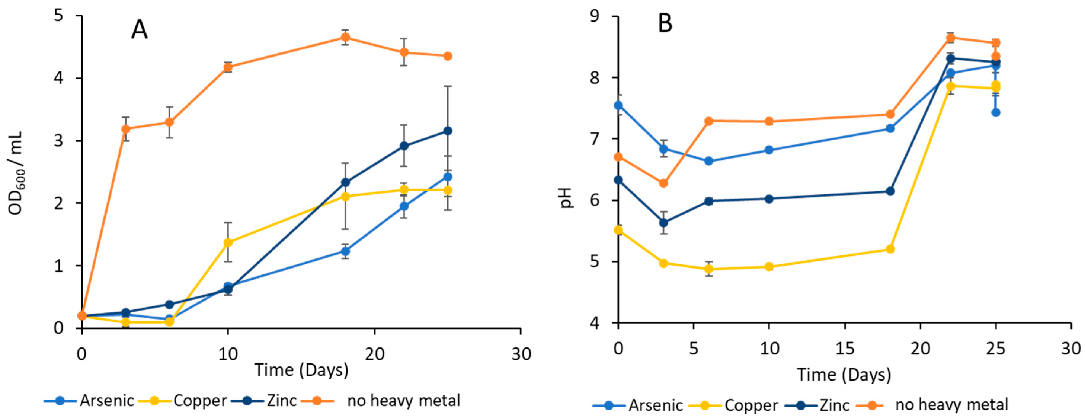
3.1. Measurement of MTC with D. psychralcaliphila JI1D

To select the highest limit of heavy metal tolerance, four different concentrations (from 10 ppm to 10,000 ppm) of arsenic, copper, and zinc were used to evaluate the MTC for each metal. The bacterium showed growth up to 1000 ppm. No growth was observed for all heavy metals on 10,000 ppm. Based on the MTC experiment, the 1000 ppm concentration was chosen for further experiments.

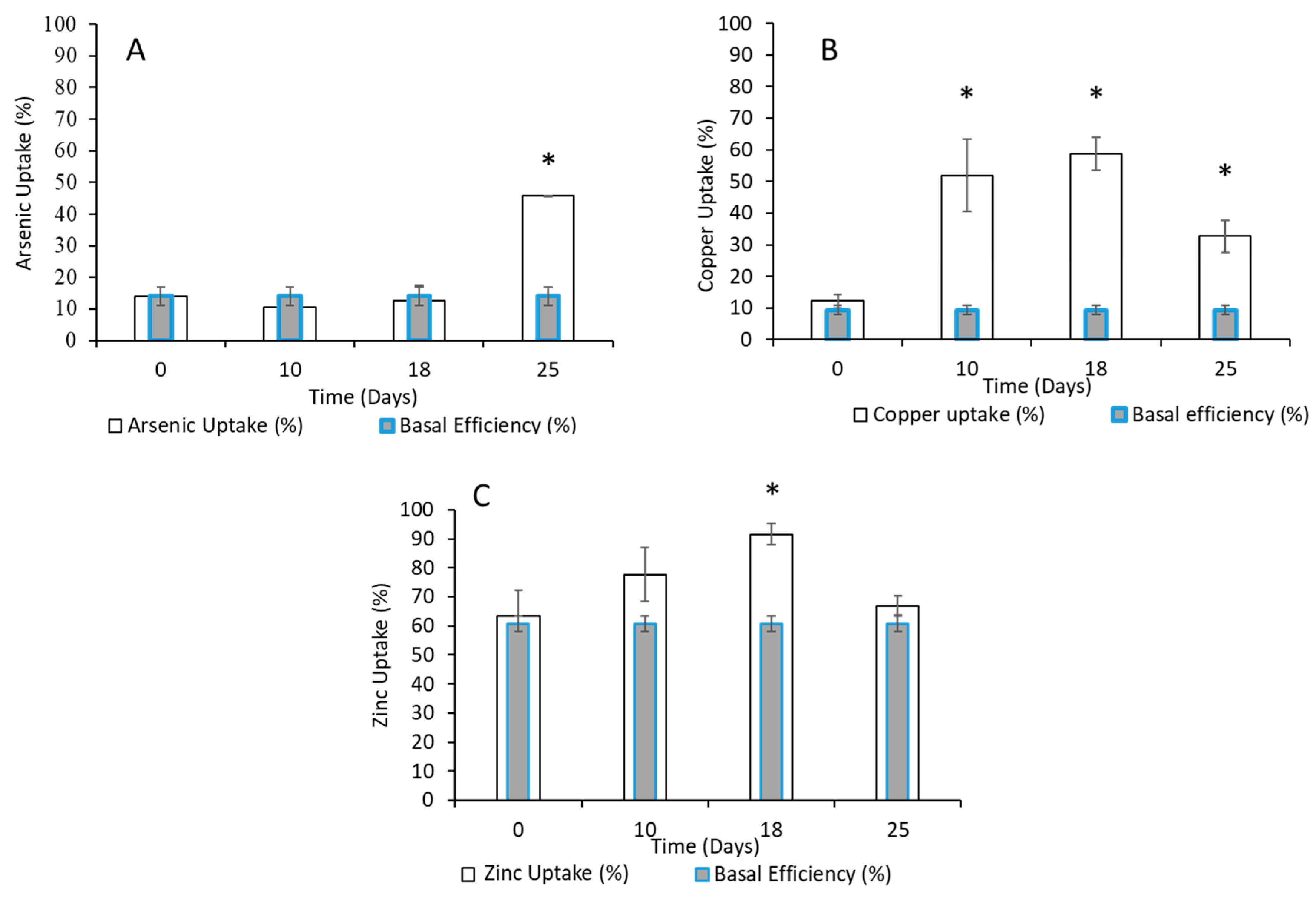
3.2. Quantification of Heavy Metals by ICP-MS

To investigate the bioaccumulation capacity of selected heavy metals by the bacterium, ICP-MS was used.
The growth pattern of D. psychralcaliphila JI1D reported in Figure 1A shows its ability to propagate in the presence of 1000 ppm of arsenic. Figure 2A indicates that there is no arsenic internalization after 10 and 18 days. Arsenic uptake (45.7%) was only detected on day 25, and its bioaccumulation capacity was found to be 31.6%.
The variation in the arsenic uptake process in the initial phase seems to be well related to the growth profile, as it shows an extended lag phase. The time it took for the bacterium to become adapted to such a high concentration of arsenic probably led to lag state [33]. In fact, many resources are generally spent to repair the damage caused by the toxic effect of arsenic, as previously reported [34,35,36]. Interestingly, the pH (Figure 1B) varied from 7.5 to 6.8 during the first 10 days, whereas it changed from 7.2 to 8.2 from day 18 to 25, indicating the increasing alkalinity of the growth medium.
The copper uptake was 51.9% and 58.7% on days 10 and 18, respectively, while the basal efficiency was 9.3% (Figure 2B). These values of copper uptake were higher when compared with day 25, which stood at 32.6%. Moreover, the copper bioaccumulation capacity for days 10 and 18 was 42.6% and 49.4%, respectively, while at day 25 only 23.3% of copper was actively precipitated by the bacteria biomass (Figure 2A). The decrease in the efficiency of bioaccumulation could be due to the depletion of essential nutrients throughout the timeline of the experiments with copper, or to the presence of damaged cells, which release intracellular material into the culture medium. A common rate-limiting step in heavy metal uptake mechanisms involves the bacteria compromising the copper uptake ratio for survival under stress conditions, while the growth of the bacteria remains unaffected [37]. Posacka and collaborators hypothesized that marine heterotrophic bacteria have a highly controlled Cu-homeostasis system, resulting from contrasting early evolutionary metal conditions [38]. Interestingly, in the presence of copper, the growth curve showed a surge of growth after day 10 until day 25, reaching OD600 2.20 (Figure 1A). Moreover, there was a variation in pH values (Figure 1B). The pH differed from 5.5 on day 0 to 7.9 on day 25, showing an alkaline condition similar to that of the arsenic treatment.
For zinc, there was a 63.6% metal uptake rate on day zero, suggesting that the zinc salt added in the form of ZnCl2 precipitated in a higher concentration with the contents of the media (Figure 2C). The overall precipitated metal steadily increased during the first 18 days, reaching 77.6% and 91.6% on days 10 and 18, respectively. On day 18, with higher metal uptake, the bioaccumulation capacity reached the maximum value of 38.9%. On day 25, there was a reduction in both the metal uptake rate and bioaccumulation capacity of 66.9% and 6.2%, respectively. Interestingly, zinc was the element that exhibited the highest basal metal removal among the three heavy metals investigated (Figure 2). It is reasonable to assume that zinc in the form of zinc chloride salt is highly reactive and forms zinc oxy chlorides, which, in turn, form white precipitates in the media [8]. This assumption is supported by the high basal efficiency (60.7%). Finally, in presence of zinc, the bacterium showed exponential growth between days 10 and 18 (Figure 1B), where we observed the highest heavy metal uptake. Interestingly, the uptake of zinc is thus directly linked to the D. psychralcaliphila JI1D growth curve.
Overall, the ability to tolerate the metals herein described is consistent with the heavy metal resistance patterns of bacteria isolated from King George Island, which exhibited the ability to tolerate arsenite (18 mM), arsenate (1000 mM), and copper (8 mM) [39].
In any case, although the mechanisms capable of leading to the removal of metals from the medium require further investigations, our results show high bacterial efficiency in metal removal, especially considering the high starting concentrations. Indeed, Gholami and Etemadifar [40] isolated the Dietzia sp. MG4 strain, which showed growth over heavy metals, such as mercury, cadmium, and nickel at concentrations between 3 and 0.3 mM. Moreover, moderate heavy metal resistance was reported for D. psychralcaliphila BGN5 against lead and copper, and very low resistance against zinc (<1 mM) [41]. Similarly, the heavy metal resistance of Dietzia sp. 29ETS and Dietzia sp. 30ETS was also reported against lead and copper at a 200 ppm concentration [42]. Apart from single culture resistance studies, D. maris in combination with Lysinibacillus sp. showed cadmium resistance with the production of hydroxamate siderophores, making them an efficient bioinoculant for rhizoremediation purposes [43].
The quantification of the different heavy metal uptake rates with D. psychralcaliphila JI1D by chemical analysis showed that the accumulation of metals reached its maximum during the exponential phase of bacterial growth, which correlates with previous reports [33].
It is important to note that the pH affects the activity of enzymes in microorganisms, which, in turn, affects the rate of microbial metabolism of some metals [44]. Moreover, the pH influences the surface charge of the microorganism, which dictates the adsorption rate of heavy metals in the cell membrane [45]. Previous studies [46,47] showed that the heavy metal removal rate of microorganisms increases with an increase in the pH. According to Babu et al. [48], heavy metal removal efficiency from a culture medium is due to the ability of strains to increase cell density, which leads to the saturation of metal adsorption sites on the cell surface and to different influx and efflux system strategies to maintain cellular homeostasis [49].

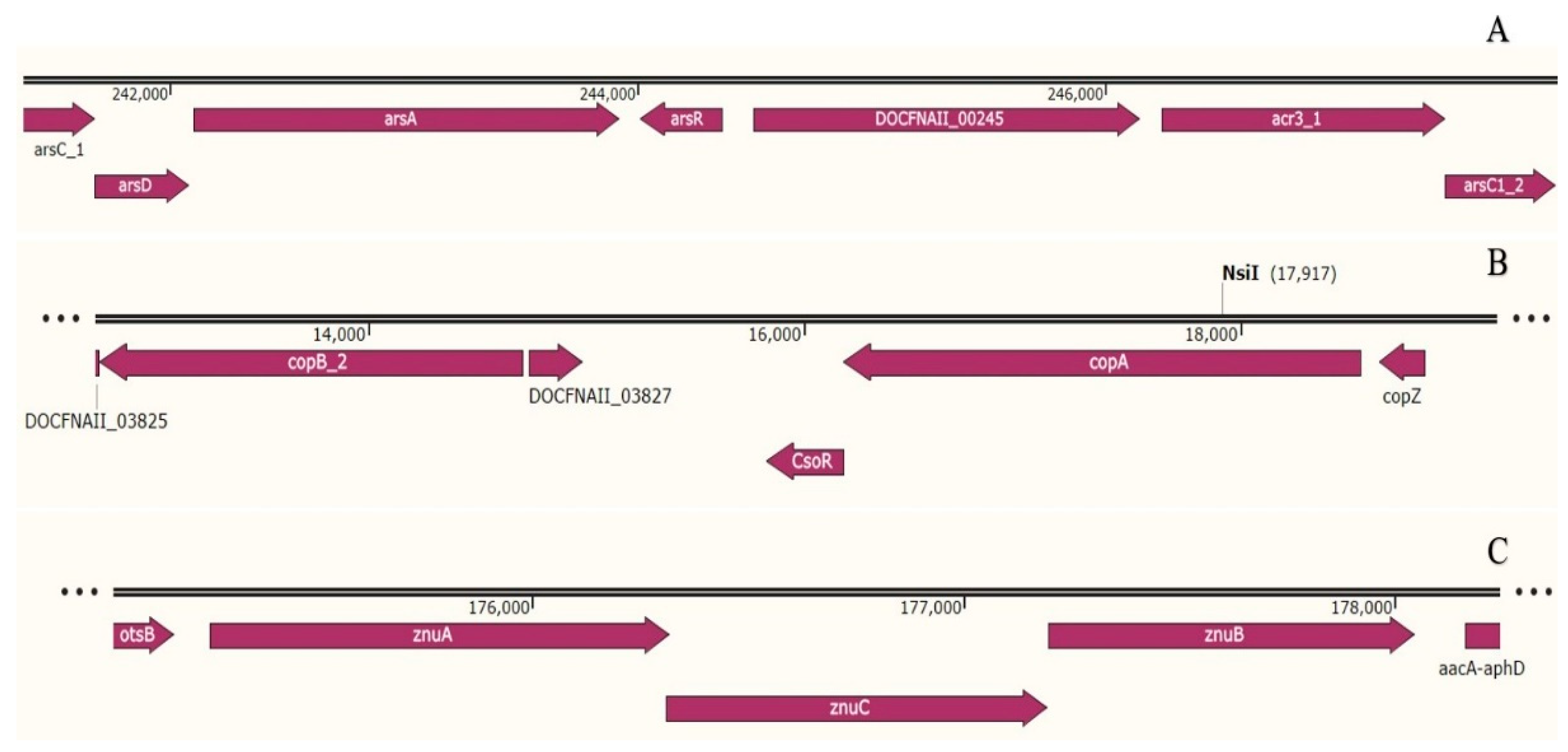
3.3. Genomic Insights of Heavy Metal Tolerance and Stress Response

Due to the promising results reported in Section 3.2, the D. psychralcaliphila JI1D genome was investigated in an attempt to identify the specific genes putatively responsible for arsenic, copper, and zinc transformation and the stress response.
In general, the genome annotation of D. psychralcaliphila JI1D highlighted the presence of numerous genes involved in resistance to arsenic, copper, and zinc. These data support and confirm what Centurion and colleagues [23] reported in a previous work. Indeed, the authors, following the annotation of metagenomes from the sediment sampled in Deception Island, showed that most of the genes involved in metal resistance were directed towards arsenic, copper, and zinc, among the main metals present in the area.
The results are discussed below and summarized in the Table 1.

3.3.1. Arsenic Resistance Determinants

Bacteria thrive in an arsenic environment by resisting via several mechanisms, including active extrusion, extracellular precipitation, compartmentalization, and sequestration in order to reduce the mobility and bioavailability of arsenic [50]. The most commonly described system for arsenic detoxification in bacteria is the arsenic resistance module system (ARS), which is represented by the presence of arsRDABC components [7]. The ARS, which is also present in JI1D, consists of genetic components that are involved in detoxification of arsenic metalloid [51]. The catabolic gene DOCFNAII_00244 containing arsR acts as a DNA binding arsenate/arsenite responsive transcriptional repressor, which regulates its own expression and confers arsenical resistance (COG0640) [52]. Seven others transcriptional repressor genes belonging to the arsR family have been identified in this genome. The next gene in the arsRDABC system is arsD, which was found to be encoded in DOCFNAII_00242, an arsenical resistance operon trans-acting repressor (PF06953.10). This is a metallochaperone, which enhances the rate of arsenite extrusion by interacting with arsA [53].The presence of DOCFNAII_00243 containing the arsA arsenite tail-anchored protein represents the transporting ATP-ase, which acts as an efficient transmembrane arsenical efflux pump (TIGR04291) [7]. The presence of COG0798 corresponds to the catabolic gene DOCFNAII_00246, which contains the arsB arsenite transporter. This is supposed to act as a transporter of arsenite generated by the action of arsC. More importantly, DOCFNAII_00247 encodes for arsenate-mycothiol transferase, which belongs to the low molecular weight phosphotyrosine protein phosphatase family. This gene is believed to play a major role in the reduction of arsenate As(V) to arsenite As(III), which represents a first step in detoxifying arsenic. It is interesting to note that it possesses the conserved domain cd16345, which contains the reductase enzyme that couples with reducing equivalent thioredoxin (Trx)/thioredoxin reductase (TrxR) (PRK13530) [54]. The entry of As(III) is usually mediated by the glycerol uptake facilitator protein glpF, which was found in DOCFNAII_00656 [55]. The previous studies of Procopio et al. demonstrated the presence of arsenic resistance genes in D. cinnamea P4 [56]. In co-culture studies, Ye and collaborators reported arsenic reduction following combined treatment with Brevundimonas spp., Massilia spp., and Planococcus spp. isolated from arsenic aquifers in Jianghan Plain, China [57]. Moreover, arsenic resistance genes in Dietzia spp. were identified, suggesting that the presence of the ARS module is prevalent among the genus Dietzia [58].

3.3.2. Copper Resistant Determinants

Copper is an essential trace element that is required for several biochemical processes, such as oxidative respiration and electron transport chain reactions; it also serves as a structural component or catalytic co-factor for a wide range of enzymes in bacteria. However, high levels of copper are toxic for the physiological functioning of the bacterial system. Bacteria overcome these challenges via various mechanisms, such as active efflux systems, cellular sequestration, and reduction reactions by multicopper oxidase enzymes [59]. When the genome of D. psychralcaliphila JI1D was investigated, 15 genes related to copper resistance were found. The presence of two copies of the copper sensing transcriptional repressor csoR DOCFNAII_03828 and DOCFNAII_00088, which functionally belong to COG1937, is supposed to be involved in resisting cytoplasmic copper toxicity [20,28,52,60]. The presence of (a) two copies of cadA-like heavy metal translocating P-type ATP-ase copA in DOCFNAII-03829 and DOCFNAII_00090 and (b) three copies of the copB P-type Cu2+ transporter in DOCFNAII_03960, DOCFNAII_02731, and DOCFNAII_03826, both belonging to the PIB subclass, infers their significance in maintaining copper homeostasis across biological membranes [61]. Apart from copA and copB, two copies of copC catabolic genes in DOCFNAII_03084 and DOCFNAII_01147 suggest copper resistance in the periplasmic membrane [62]. The presence of DOCFNAII_00341, which contains periplasmic copper chaperone copZ and is supposed to aid in the transfer of Cu(I) to cytoplasm belonging to the PCu(A)C family, was observed in a similar orientation in the downstream region [51]. The presence of two copies of multicopper oxidase mmco catabolic genes in DOCFNAII_03834 and DOCFNAII_03956 with COG2132 implies its role in oxidizing Cu(I) to Cu(II) in copper resistance [63]. It is interesting to note the conserved domains of the first, second, and third cupredoxin domain of multicopper oxidases with trinuclear copper binding sites [54]. Similar copper resistance genes were reported for the draft genome of the D. cinnamea strain P4 [56]. The co-occurrence of antibiotic resistance genes (ARGs) and copper resistance genes was reported in a community of bacterium constituting Dietzia spp. [64]. Apart from copper resistance genes, antioxidative-natured SOD1 superoxide dismutase belonging to the Cu-Zn family was found at DOCFNAII_00093, suggesting the elimination of superoxide anions, thus providing a defense against oxygen toxicity [65].

3.3.3. Zinc Resistant Determinants

Zinc is a micronutrient that is essential for various metabolic reactions in the form of a coenzyme, cofactor, or in a stabilizing protein structure in bacteria. Excessive concentrations of zinc in a bacterial cell leads to interference in the electron transport chain and the generation of reactive oxygen species [66]. Thus, the intracellular zinc concentration must be strictly balanced, which is controlled by transporter genes that act based on specific metalloregulators. The catabolic gene DOCFNAII_03463 encodes for zinc-transporting ATPase Znt A, which acts as the zinc efflux system. This catabolic gene contains the conserved domains cd02079 and cd07546, which play an important role in generating and maintaining electrochemical gradient across cellular membranes [54]. The presence of a Zn2+ uptake system—the ABC zinc/manganese transport system (substrate-binding protein) (DOCFNAII_02177), ABC zinc/manganese transport system ATP-binding protein (DOCFNAII_02178), and ABC zinc/manganese transport system permease protein (DOCFNAII_02179)—indicates the utilization of zinc ions inside and outside the cell membrane [67]. The Zn2+ uptake system is known as the znuABC cluster, which is upregulated by the catabolic gene DOCFNAII_02964 zur, belonging to the metalloregulatory protein family. The presence of COG2072 from to cation diffusion facilitator family of cobalt/zinc/cadmium efflux system components (czcD-like), which are present in DOCFNAII_01529 and DOCFNAII_03837, suggests a heavy metal resistance mechanism across cellular membrane [68]. The genetic arrangements of the catabolic genes involved in heavy metal resistance are shown in Figure 3. The above-listed catabolic genes of metal resistance are important as they are presumably involved in converting toxic metal ions into their less toxic forms by reducing the oxidation state and increasing the solubility of ions within the cell.

4. Conclusions

In this study, the bioaccumulation capacity of D. psychralcaliphila JI1D for the removal of three different metals was investigated. This strain displayed a metabolic versatility with high heavy metal resistance, showing a tolerance up to 1000 ppm. The bioaccumulation was found to be in order of copper > arsenic > zinc, thus lowering the bioavailability in the medium. The genome analysis also provided indications of the presence of specific resistance genes that are potentially involved in the removal process. In accordance with previous studies that confirmed its application in xenobiotic degradation, i.e., hydrocarbon processing, this study demonstrates that this bacterium is also capable of remediating polluted environments from inorganic components such as potentially toxic elements.

Author Contributions

Conceptualization, D.d.P., J.A., F.D., D.C. and M.G.; investigation and formal analysis, J.A., F.D., F.P.E., G.A.V., V.F., G.D.S., P.T., G.F. and D.C.; data curation, J.A. and F.D.; writing—original draft preparation, J.A., F.D. and D.C.; writing and editing, J.A., F.D., F.P.E., G.A.V., V.F., G.F., M.G., P.T., G.D.S., D.C. and D.d.P.; supervision, D.C. and D.d.P.; funding acquisition, D.d.P. All authors have read and agreed to the published version of the manuscript.

Funding

This research received no external funding.

Institutional Review Board Statement

Not applicable.

Informed Consent Statement

Not applicable.

Data Availability Statement

Not applicable.

Acknowledgments

The authors thank the University of Campania Luigi Vanvitelli for partial support.

Conflicts of Interest

The authors declare no conflict of interest.

References

  1. Ayangbenro, A.S.; Babalola, O.O. A New Strategy for Heavy Metal Polluted Environments: A Review of Microbial Biosorbents. Int. J. Environ. Res. Public Health 2017, 14, 94. [Google Scholar] [CrossRef] [PubMed]
  2. Photolo, M.; Sitole, L.; Mavumengwana, V.; Tlou, M. Genomic and Physiological Investigation of Heavy Metal Resistance from Plant Endophytic Methylobacterium radiotolerans MAMP 4754, Isolated from Combretum erythrophyllum. Int. J. Environ. Res. Public Health 2021, 18, 997. [Google Scholar] [CrossRef] [PubMed]
  3. Plum, L.M.; Rink, L.; Haase, H. The Essential Toxin: Impact of Zinc on Human Health. Int. J. Environ. Res. Public Health 2010, 7, 1342–1365. [Google Scholar] [CrossRef] [PubMed] [Green Version]
  4. Tarekegn, M.M.; Salilih, F.Z.; Ishetu, A.I. Microbes Used as a Tool for Bioremediation of Heavy Metal from the Environment. Cogent Food Agric. 2020, 6, 1783174. [Google Scholar] [CrossRef]
  5. Batey, T.; Berryman, C.; Line, C. The Disposal of Copper-Enriched Pig-Manure Slurry on Grassland. Grass Forage Sci. 2006, 27, 139–144. [Google Scholar] [CrossRef]
  6. Chung, J.-Y.; Yu, S.-D.; Hong, Y.-S. Environmental Source of Arsenic Exposure. J. Prev. Med. Public Health 2014, 47, 253–257. [Google Scholar] [CrossRef] [Green Version]
  7. Soto, J.; Charles, T.; Lynch, M.; Larama, G.; Herrera, H.; Arriagada, C. Genome Sequence of Brevundimonas sp., an Arsenic Resistant Soil Bacterium. Diversity 2021, 13, 344. [Google Scholar] [CrossRef]
  8. Kapahi, M.; Sachdeva, S. Bioremediation Options for Heavy Metal Pollution. J. Health Pollut. 2019, 9, 191203. [Google Scholar] [CrossRef] [PubMed] [Green Version]
  9. Abioye, O.P. Biological Remediation of Hydrocarbon and Heavy Metals Contaminated Soil. In Soil Contamination; Pascucci, S., Ed.; IntechOpen: Rijeka, Croatia, 2011. [Google Scholar]
  10. Gambino, E.; Chandrasekhar, K.; Nastro, R.A. SMFC as a tool for the removal of hydrocarbons and metals in the marine environment: A concise research update. Environ. Sci. Pollut. Res. 2021, 28, 30436–30451. [Google Scholar] [CrossRef]
  11. Arora, V.; Khosla, B. Conventional and Contemporary Techniques for Removal of Heavy Metals from Soil. In Biodegradation Technology of Organic and Inorganic Pollutants; Mendes, K.F., de Sousa, R.N., Mielke, K.C., Eds.; IntechOpen: Rijeka, Croatia, 2021. [Google Scholar]
  12. Mosa, K.A.; Saadoun, I.; Kumar, K.; Helmy, M.; Dhankher, O.P. Potential Biotechnological Strategies for the Cleanup of Heavy Metals and Metalloids. Front. Plant Sci. 2016, 7, 303. [Google Scholar] [CrossRef]
  13. Perelo, L.W. Review: In situ and bioremediation of organic pollutants in aquatic sediments. J. Hazard. Mater. 2010, 177, 81–89. [Google Scholar] [CrossRef] [PubMed] [Green Version]
  14. Kuppusamy, S.; Thavamani, P.; Venkateswarlu, K.; Lee, Y.B.; Naidu, R.; Megharaj, M. Remediation Approaches for Polycyclic Aromatic Hydrocarbons (PAHs) Contaminated Soils: Technological Constraints, Emerging Trends and Future Directions. Chemosphere 2017, 168, 944–968. [Google Scholar] [CrossRef] [PubMed]
  15. Orellana, R.; Macaya, C.; Bravo, G.; Dorochesi, F.; Cumsille, A.; Valencia, R.; Rojas, C.; Seeger, M. Living at the Frontiers of Life: Extremophiles in Chile and Their Potential for Bioremediation. Front. Microbiol. 2018, 9, 2309. [Google Scholar] [CrossRef] [PubMed]
  16. Giudice, A.L.; Caruso, G.; Rizzo, C.; Papale, M.; Azzaro, M. Bacterial communities versus anthropogenic disturbances in the Antarctic coastal marine environment. Environ. Sustain. 2019, 2, 297–310. [Google Scholar] [CrossRef]
  17. Alvarez, L.M.; Bolhuis, H.; Mau, G.K.; Kok-Gan, C.; Sing, C.C.; Mac Cormack, W.; Ruberto, L. Identification of key bacterial players during successful full-scale soil field bioremediation in Antarctica. Int. Biodeterior. Biodegradation 2022, 168, 105354. [Google Scholar] [CrossRef]
  18. Darham, S.; Syed-Muhaimin, S.N.; Subramaniam, K.; Zulkharnain, A.; Shaharuddin, N.A.; Khalil, K.A.; Ahmad, S.A. Optimisation of Various Physicochemical Variables Affecting Molybdenum Bioremediation Using Antarctic Bacterium, Arthrobacter sp. Strain AQ5-05. Water 2021, 13, 2367. [Google Scholar] [CrossRef]
  19. Michaud, L.; Di Marco, G.; Bruni, V.; Giudice, A.L. Biodegradative potential and characterization of psychrotolerant polychlorinated biphenyl-degrading marine bacteria isolated from a coastal station in the Terra Nova Bay (Ross Sea, Antarctica). Mar. Pollut. Bull. 2007, 54, 1754–1761. [Google Scholar] [CrossRef] [PubMed]
  20. Acevedo-Barrios, R.; Rubiano-Labrador, C.; Navarro-Narvaez, D.; Escobar-Galarza, J.; González, D.; Mira, S.; Moreno, D.; Contreras, A.; Miranda-Castro, W. Perchlorate-reducing bacteria from Antarctic marine sediments. Environ. Monit. Assess. 2022, 194, 654. [Google Scholar] [CrossRef] [PubMed]
  21. Ausuri, J.; Vitale, G.; Coppola, D.; Esposito, F.P.; Buonocore, C.; de Pascale, D. Assessment of the Degradation Potential and Genomic Insights towards Phenanthrene by Dietzia psychralcaliphila JI1D. Microorganisms 2021, 9, 1327. [Google Scholar] [CrossRef] [PubMed]
  22. Centurion, V.; Silva, J.; Duarte, A.; Rosa, L.; Oliveira, V. Comparing resistome profiles from anthropogenically impacted and non-impacted areas of two South Shetland Islands—Maritime Antarctica. Environ. Pollut. 2022, 304, 119219. [Google Scholar] [CrossRef] [PubMed]
  23. Centurion, V.; Delforno, T.; Lacerda-Júnior, G.; Duarte, A.; Silva, L.; Bellini, G.; Rosa, L.; Oliveira, V. Unveiling resistome profiles in the sediments of an Antarctic volcanic island. Environ. Pollut. 2019, 255, 113240. [Google Scholar] [CrossRef] [PubMed]
  24. Balali-Mood, M.; Naseri, K.; Tahergorabi, Z.; Khazdair, M.R.; Sadeghi, M. Toxic Mechanisms of Five Heavy Metals: Mercury, Lead, Chromium, Cadmium, and Arsenic. Front. Pharmacol. 2021, 12, 643972. [Google Scholar] [CrossRef]
  25. Jaishankar, M.; Tseten, T.; Anbalagan, N.; Mathew, B.B.; Beeregowda, K.N. Toxicity, mechanism and health effects of some heavy metals. Interdiscip. Toxicol. 2014, 7, 60–72. [Google Scholar] [CrossRef] [Green Version]
  26. Tchounwou, P.B.; Yedjou, C.G.; Patlolla, A.K.; Sutton, D.J. Molecular, Clinical and Environmental Toxicicology Volume 3: Environmental Toxicology. Mol. Clin. Environ. Toxicol. 2012, 101, 133–164. [Google Scholar] [CrossRef]
  27. Lambert, M.; Leven, B.A.; Green, R.M. New Methods of Cleaning Up Heavy Metal in Soils and Water Innovative Solutions to an Environmental Problem. Environ. Sci. Technol. Briefs Citiz. 2000, 7, 1–3. [Google Scholar]
  28. Kuznetsova, O.V.; Khlebtsov, N.G.; Jarosz, M.; Timerbaev, A.R. Metal-Specific Response of High-Resolution ICP-MS for Proteins Binding to Gold Nanoparticles in Human Serum. Anal. Chem. 2021, 93, 14918–14922. [Google Scholar] [CrossRef] [PubMed]
  29. EPA. U.S. Method 6020B Inductively Coupled Plasma-Mass Spectrometry; EPA: Washington, DC, USA, 2014; pp. 1–33. [Google Scholar]
  30. Seemann, T. Prokka: Rapid Prokaryotic Genome Annotation. Bioinformatics 2014, 30, 2068–2069. [Google Scholar] [CrossRef] [PubMed] [Green Version]
  31. Aziz, R.K.; Bartels, D.; Best, A.A.; De Jongh, M.; Disz, T.; Edwards, R.A.; Formsma, K.; Gerdes, S.; Glass, E.M.; Kubal, M.; et al. The RAST server: Rapid annotations using subsystems technology. BMC Genom. 2008, 9, 75. [Google Scholar] [CrossRef] [Green Version]
  32. Moriya, Y.; Itoh, M.; Okuda, S.; Yoshizawa, A.C.; Kanehisa, M. KAAS: An automatic genome annotation and pathway reconstruction server. Nucleic Acids Res. 2007, 35 (Suppl. 2), W182–W185. [Google Scholar] [CrossRef] [PubMed] [Green Version]
  33. Pandey, N.; Bhatt, R. Arsenic Resistance and Accumulation by Two Bacteria Isolated from a Natural Arsenic Contaminated Site. J. Basic Microbiol. 2015, 55, 1275–1286. [Google Scholar] [CrossRef] [PubMed]
  34. Gikas, P.; Sengör, S.S.; Ginn, T.; Moberly, J.; Peyton, B. The Effects of Heavy Metals and Temperature on Microbial Growth and Lag. Glob. Nest J. 2009, 11, 325–332. [Google Scholar] [CrossRef]
  35. Abbas, S.Z.; Riaz, M.; Ramzan, N.; Zahid, M.T.; Shakoori, F.R.; Rafatullah, M. Isolation and Characterization of Arsenic Resistant Bacteria from Wastewater. Braz. J. Microbiol. 2014, 45, 1309–1315. [Google Scholar] [CrossRef] [Green Version]
  36. Prosun, T.A.; Younus, A.; Monira, Y.; Brun, M.S.; Badiuzzaman, K.; Harun-Or, R. Identification and Evaluation of Arsenic Tolerant Bacteria for Arsenic Mitigation in Contaminated Soil. J. Bacteriol. Mycol. 2020, 7, 1156. [Google Scholar]
  37. Porcheron, G.; Garénaux, A.; Proulx, J.; Sabri, M.; Dozois, C.M. Iron, Copper, Zinc, and Manganese Transport and Regulation in Pathogenic Enterobacteria: Correlations between Strains, Site of Infection and the Relative Importance of the Different Metal Transport Systems for Virulence. Front. Cell. Infect. Microbiol. 2013, 3, 90. [Google Scholar] [CrossRef] [Green Version]
  38. Posacka, A.M.; Semeniuk, D.M.; Maldonado, M.T. Effects of Copper Availability on the Physiology of Marine Heterotrophic Bacteria. Front. Mar. Sci. 2019, 5, 523. [Google Scholar] [CrossRef]
  39. Romaniuk, K.; Ciok, A.; Decewicz, P.; Uhrynowski, W.; Budzik, K.; Nieckarz, M.; Pawlowska, J.; Zdanowski, M.K.; Bartosik, D.; Dziewit, L. Insight into heavy metal resistome of soil psychrotolerant bacteria originating from King George Island (Antarctica). Polar Biol. 2018, 41, 1319–1333. [Google Scholar] [CrossRef] [Green Version]
  40. Gholami, M.; Etemadifar, Z. Isolation and characterization of a novel strain of genus Dietzia capable of multiple-extreme resistance. Microbiology 2015, 84, 389–397. [Google Scholar] [CrossRef]
  41. Máthé, I.; Benedek, T.; Táncsics, A.; Palatinszky, M.; Lányi, S.; Márialigeti, K. Diversity, activity, antibiotic and heavy metal resistance of bacteria from petroleum hydrocarbon contaminated soils located in Harghita County (Romania). Int. Biodeterior. Biodegrad. 2012, 73, 41–49. [Google Scholar] [CrossRef]
  42. Freitas, D.; Lima-Bittencourt, C.; Reis, M.; Costa, P.; Assis, P.; Chartone-Souza, E.; Nascimento, A. Molecular characterization of early colonizer bacteria from wastes in a steel plant. Lett. Appl. Microbiol. 2008, 47, 241–249. [Google Scholar] [CrossRef]
  43. Gusain, P.; Paliwal, R.; Singh, V. Rhizoremediation of cadmium-contaminated soil associated with hydroxamate siderophores isolated from Cd-resistant plant growth–promoting Dietzia maris and Lysinibacillus strains. Int. J. Phytoremediation 2017, 19, 290–299. [Google Scholar] [CrossRef]
  44. Morton-Bermea, O.; Álvarez, E.H.; Gaso, I.; Segovia, N. Heavy Metal Concentrations in Surface Soils from Mexico City. Bull. Environ. Contam. Toxicol. 2002, 68, 0383–0388. [Google Scholar] [CrossRef] [PubMed]
  45. Galiulin, R.V.; Galiulina, R.A. Removing heavy metals from soil with plants. Her. Russ. Acad. Sci. 2008, 78, 141–143. [Google Scholar] [CrossRef]
  46. Rodríguez-Tirado, V.; Green-Ruiz, C.; Gómez-Gil, B. Cu and Pb biosorption on Bacillus thioparans strain U3 in aqueous solution: Kinetic and equilibrium studies. Chem. Eng. J. 2012, 181–182, 352–359. [Google Scholar] [CrossRef]
  47. Wierzba, S. Biosorption of lead(II), zinc(II) and nickel(II) from industrial wastewater by Stenotrophomonas maltophilia and Bacillus subtilis. Pol. J. Chem. Technol. 2015, 17, 79–87. [Google Scholar] [CrossRef] [Green Version]
  48. Babu, A.G.; Shea, P.J.; Sudhakar, D.; Jung, I.-B.; Oh, B.-T. Potential use of Pseudomonas koreensis AGB-1 in association with Miscanthus sinensis to remediate heavy metal(loid)-contaminated mining site soil. J. Environ. Manag. 2015, 151, 160–166. [Google Scholar] [CrossRef]
  49. Diep, P.; Mahadevan, R.; Yakunin, A.F. Heavy Metal Removal by Bioaccumulation Using Genetically Engineered Microorganisms. Front. Bioeng. Biotechnol. 2018, 6, 157. [Google Scholar] [CrossRef] [Green Version]
  50. Slyemi, D.; Bonnefoy, V. How prokaryotes deal with arsenic†. Environ. Microbiol. Rep. 2012, 4, 571–586. [Google Scholar] [CrossRef]
  51. Kotoky, R.; Pandey, P. The genomic attributes of Cd-resistant, hydrocarbonoclastic Bacillus subtilis SR1 for rhizodegradation of benzo(a)pyrene under co-contaminated conditions. Genomics 2020, 113, 613–623. [Google Scholar] [CrossRef]
  52. Rawle, R.; Saley, T.C.; Kang, Y.-S.; Wang, Q.; Walk, S.; Bothner, B.; McDermott, T.R. Introducing the ArsR-Regulated Arsenic Stimulon. Front. Microbiol. 2021, 12, 630562. [Google Scholar] [CrossRef]
  53. Lin, Y.-F.; Walmsley, A.R.; Rosen, B.P. An arsenic metallochaperone for an arsenic detoxification pump. Proc. Natl. Acad. Sci. USA 2006, 103, 15617–15622. [Google Scholar] [CrossRef] [Green Version]
  54. Lu, S.; Wang, J.; Chitsaz, F.; Derbyshire, M.K.; Geer, R.C.; Gonzales, N.R.; Gwadz, M.; Hurwitz, D.; Marchler, G.H.; Song, J.S.; et al. CDD/SPARCLE: The conserved domain database in 2020. Nucleic Acids Res. 2020, 48, D265–D268. [Google Scholar] [CrossRef] [PubMed] [Green Version]
  55. Nanda, M.; Kumar, V.; Sharma, D. Multimetal tolerance mechanisms in bacteria: The resistance strategies acquired by bacteria that can be exploited to ‘clean-up’ heavy metal contaminants from water. Aquat. Toxicol. 2019, 212, 1–10. [Google Scholar] [CrossRef] [PubMed]
  56. Procópio, L.; Alvarez, V.M.; Jurelevicius, D.A.; Hansen, L.; Sørensen, S.J.; Cardoso, J.S.; Pádula, M.; Leitão, Á.C.; Seldin, L.; Van Elsas, J.D. Insight from the Draft Genome of Dietzia Cinnamea P4 Reveals Mechanisms of Survival in Complex Tropical Soil Habitats and Biotechnology Potential. Antonie Van Leeuwenhoek Int. J. Gen. Mol. Microbiol. 2012, 101, 289–302. [Google Scholar] [CrossRef] [Green Version]
  57. Ye, H.; Yang, Z.; Wu, X.; Wang, J.; Du, D.; Cai, J.; Lv, K.; Chen, H.; Mei, J.; Chen, M.; et al. Sediment biomarker, bacterial community characterization of high arsenic aquifers in Jianghan Plain, China. Sci. Rep. 2017, 7, srep42037. [Google Scholar] [CrossRef]
  58. Sanyal, S.K.; Mou, T.J.; Chakrabarty, R.P.; Hoque, S.; Hossain, M.A.; Sultana, M. Diversity of arsenite oxidase gene and arsenotrophic bacteria in arsenic affected Bangladesh soils. AMB Express 2016, 6, 21. [Google Scholar] [CrossRef] [PubMed] [Green Version]
  59. Thummeepak, R.; Pooalai, R.; Harrison, C.; Gannon, L.; Thanwisai, A.; Chantratita, N.; Millard, A.D.; Sitthisak, S. Essential Gene Clusters Involved in Copper Tolerance Identified in Acinetobacter baumannii Clinical and Environmental Isolates. Pathogens 2020, 9, 60. [Google Scholar] [CrossRef] [Green Version]
  60. Chang, F.M.J.; Coyne, H.J.; Cubillas, C.; Vinuesa, P.; Fang, X.; Ma, Z.; Ma, D.; Helmann, J.D.; García-De Los Santos, A.; Wang, Y.X.; et al. Cu(I)-mediated Allosteric Switching in a Copper-sensing Operon Repressor (CsoR). J. Biol. Chem. 2014, 289, 19204–19217. [Google Scholar] [CrossRef] [Green Version]
  61. Völlmecke, C.; Drees, S.L.; Reimann, J.; Albers, S.-V.; Lübben, M. The ATPases CopA and CopB both contribute to copper resistance of the thermoacidophilic archaeon Sulfolobus solfataricus. Microbiology 2012, 158, 1622–1633. [Google Scholar] [CrossRef]
  62. Zhang, L.; Koay, M.; Maher, M.J.; Xiao, Z.; Wedd, A.G. Intermolecular Transfer of Copper Ions from the CopC Protein of Pseudomonas syringae. Crystal Structures of Fully Loaded CuICuII Forms. J. Am. Chem. Soc. 2006, 128, 5834–5850. [Google Scholar] [CrossRef]
  63. Li, L.-G.; Cai, L.; Zhang, X.-X.; Zhang, T. Potentially novel copper resistance genes in copper-enriched activated sludge revealed by metagenomic analysis. Appl. Microbiol. Biotechnol. 2014, 98, 10255–10266. [Google Scholar] [CrossRef]
  64. Xie, J.; Gu, J.; Wang, X.; Hu, T.; Sun, W.; Lei, L.; Zhang, R.; Guo, H. Insights into the beneficial effects of woody peat for reducing abundances of antibiotic resistance genes during composting. Bioresour. Technol. 2021, 342, 125903. [Google Scholar] [CrossRef]
  65. Kobayashi, Y.; Nojima, Y.; Sakamoto, T.; Iwabuchi, K.; Nakazato, T.; Bono, H.; Toyoda, A.; Fujiyama, A.; Kanost, M.; Tabunoki, H. Comparative analysis of seven types of superoxide dismutases for their ability to respond to oxidative stress in Bombyx mori. Sci. Rep. 2019, 9, 2170. [Google Scholar] [CrossRef] [PubMed]
  66. Park, S.; Ely, R.L. Genome-Wide Transcriptional Responses of Nitrosomonas Europaea to Zinc. Arch. Microbiol. 2008, 189, 541–548. [Google Scholar] [CrossRef] [PubMed]
  67. Patzer, S.I.; Hantke, K. The Zinc-responsive Regulator Zur and Its Control of theznu Gene Cluster Encoding the ZnuABC Zinc Uptake System in Escherichia coli. J. Biol. Chem. 2000, 275, 24321–24332. [Google Scholar] [CrossRef] [Green Version]
  68. Anton, A.; Große, C.; Reißmann, J.; Pribyl, T.; Nies, D.H. CzcD Is a Heavy Metal Ion Transporter Involved in Regulation of Heavy Metal Resistance in Ralstonia sp. Strain CH34. J. Bacteriol. 1999, 181, 6876–6881. [Google Scholar] [CrossRef] [PubMed]
Figure 1. Growth profile (A) and pH variation (B) of D. psychralcaliphila JI1D, in the absence (orange) or the presence of arsenic (light blue), copper (yellow), and zinc (blue).
Jmse 10 01669 g001
Figure 2. Uptake of arsenic (A), copper (B), and zinc (C) by D. psychralcaliphila JI1D biomass. The white bars represent the yield (%) of metal uptake during the treatment with D. psychralcaliphila JI1D. The grey bars indicate the yield (%) in the controls in the absence of bacterial cells (medium plus metal), due to metal precipitation. Data are presented as mean ± standard deviation (SD); n = 3. Student’s t-test was used to compare means between groups. The * indicates a statistically significant (p-value < 0.05) difference between metal uptake and the basal efficiency.
Jmse 10 01669 g002
Figure 3. The genetic arrangement of genes with respect to arsenic (A), copper (B), and zinc (C) resistance and transporting systems in D. psychralcaliphila JI1D.
Jmse 10 01669 g003
Table 1. D. psychralcaliphila JI1D genes putatively responsible for arsenic, copper, and zinc transformation and the stress response. Accession number refers to the UniProt database.
Gene IDGene NameDescriptionAccession Number
DOCFNAII_00244arsRarsenate/arsenite-responsive transcriptional repressorP37309
DOCFNAII_00242arsDarsenical resistance operon trans-acting repressorO52028
DOCFNAII_00243arsAtransmembrane arsenical efflux pumpP52145
DOCFNAII_00246arsBarsenite transporterP0AB93
DOCFNAII_00247arsCarsenate reductaseP0A006
DOCFNAII_00656glpFglycerol uptake facilitator proteinQ51389
DOCFNAII_03828 DOCFNAII_00088csoRcopper sensing transcriptional repressorP9WP49
DOCFNAII-03829 DOCFNAII_00090copACadA-like heavy metal translocating P-type ATP-aseO32220
DOCFNAII_03960 DOCFNAII_02731
DOCFNAII_03826
copBP-type Cu2+ transporterQ59385
DOCFNAII_03084
DOCFNAII_01147
copCcopper resistance protein CP12376
DOCFNAII_00341copZperiplasmic copper chaperoneO32221
DOCFNAII_03834 DOCFNAII_03956mmcomulticopper oxidaseI6WZK7
DOCFNAII_00093sod1superoxide dismutase belonging to Cu-Zn familyP53649
DOCFNAII_03463znt Azinc efflux systemQ8NEW0
DOCFNAII_02177znuAABC zinc/manganese transport system (substrate-binding protein)O34966
DOCFNAII_02178znuBABC zinc/manganese transport system ATP-binding proteinP39832
DOCFNAII_02179znuCABC zinc/manganese transport system permease proteinP0A9X1
DOCFNAII_02964zurmetalloregulatory protein familyP54479
DOCFNAII_01529 DOCFNAII_03837czcDcobalt/zinc/cadmium efflux system componentsP13512
Publisher’s Note: MDPI stays neutral with regard to jurisdictional claims in published maps and institutional affiliations.

