Article

Characterisation of Pectinolytic Bacillus pumilus and Paenibacillus amyloliticus Strains, New Pathogens of Potato in Tunisia

by
Anissa Yahyaoui
1,
Maroua Oueslati
1,
Agata Motyka-Pomagruk
2,3,
Natalia Kaczynska
2,3,
Wojciech Sledz
2,3,
Belhassen Tarhouni
4,
Mohamed Rabeh Hajlaoui
5,
Ewa Lojkowska
2,3,* and
Najla Sadfi-Zouaoui
1,*
1
Laboratory of Mycology, Pathologies and Biomarkers (LR16ES05), Department of Biology, Faculty of Sciences of Tunis, University of Tunis El Manar, Tunis 2092, Tunisia
2
Laboratory of Plant Protection and Biotechnology, Intercollegiate Faculty of Biotechnology UG & MUG, University Gdansk, 80-307 Gdansk, Poland
3
Research and Development Laboratory, Intercollegiate Faculty of Biotechnology UG & MUG, University Gdansk, 80-824 Gdansk, Poland
4
Technical Center of Potato and Artichoke, Route Jedaida, Saïda 2031, Tunisia
5
Laboratory of Biotechnology Applied to Agriculture, INRA Tunisia, University of Carthage, Ariana 2094, Tunisia
*
Authors to whom correspondence should be addressed.
Agriculture 2023, 13(6), 1275; https://doi.org/10.3390/agriculture13061275
Submission received: 16 April 2023 / Revised: 12 June 2023 / Accepted: 15 June 2023 / Published: 20 June 2023
(This article belongs to the Special Issue Molecular Diagnosis and Control of Plant Diseases)

Abstract

:
Soft rot disease in potato is a major problem in fields and warehouses all over the world. Although it is known that bacteria from the genera Pectobacterium and Dickeya are the main causative agents of soft rot diseases, recent studies indicate the involvement of pectinolytic Bacillus and Paenibacillus in this disease. In the present research, samples of potato with soft rot symptoms were collected from eight governorates of Tunisia. Two hundred and seventy bacterial isolates were acquired from tubers. Twenty of the isolated strains indicted pectinolytic activity by forming deep cavities on crystal violet pectate medium. All pectinolytic isolates were able to macerate potato tuber tissue. Phenotypic characterisation showed that these isolates were Gram-positive bacilli, exhibiting pectinolytic, cellulolytic, proteolytic and amylolytic activity. The majority of the isolates indicated swimming and swarming motility. The application of API test, MALDI-TOF MS and 16S rDNA sequencing allowed for the assignment of nineteen of the tested isolates to the species Bacillus pumilus and one to the species Paenibacillus amyloliticus. To the best of our knowledge, this is the first report of soft rot in potato caused by pectinolytic B. pumilus in Tunisia.

1. Introduction

Solanum tuberosum L. has been widely cultivated in Tunisia as an important vegetable crop. Potato is grown year-round as a seasonal, late-season, early-season and extra-early-season crop. In 2011, this crop covered about 23,200 hectares and the potato production rate in Tunisia reached about 367,000 tonnes [1]. Faced with the multiplicity of growing seasons and different climates in the cultivation areas, potato production is threatened by several serious diseases and pests in the field or during storage. The biotic factors responsible for soft rot diseases in this country are most commonly diagnosed visually, which leads to confusion between rots caused by bacteria in contrast to Pythium ultimum, Fusarium spp. and Phytophthora spp.
The most widely known agents responsible for potato blackleg and soft rot diseases are pectinolytic bacteria from the genera Dickeya and Pectobacterium [2,3,4]. Both genera belong to the recently established Pectobacteriaceae family [5], and have been classified among the top ten bacterial pathogens responsible for severe economic damage in multiple crops [6]. The disease symptoms caused by pectinolytic bacteria during vegetation are brown to black discolouration of the stem base starting from below ground level, which is usually accompanied by wet rot. These disorders may aggravate and progress to the upper parts of the stem. In particular, brown discolouration may be recognised inside the stems with sagging pith [7].
Wet rot of the basal part of stem results in wilting, curling and yellowing of the leaves, and subsequently, leads to dieback. In the case of tuber tissue, a moist, grainy soft rot, white to cream in colour and that tends to blacken at the periphery, is observed. In addition, secondary invaders including bacteria and fungi may colonise the macerated tissue, culminating in the development of a characteristic odour [8].
Recent studies performed in Africa, Asia and Europe have shown that apart from Dickeya and Pectobacterium spp., bacteria from the genus Bacillus, especially pectinolytic Bacillus pumilus, are capable of causing soft rot in different crops and trees [9,10,11,12]. In more detail, B. pumilus infections led to the development of soft rot in stored potato tubers in Mali [10], bacterial blotch in peach in Egypt [13], wetwood disorder in young Scots pine trees of Ukraine and Belarus [12], ginger rhizome rot in China [11], leaf blight in mango trees in Egypt [9], bacterial fruit rot in muskmelon in China [14] and Ficus lacor in Pakistan [15], Persian oak decline in Iran [16], trunk bulges in rubber trees in Malaysia [17] and interveinal purple-brown leaf spots in common bean in Spain [18]. Additionally, the screening of Belarusian bacterial collections also revealed the occurrence of B. pumilus-triggered infections in Cucumis sativus, Solanum lycopersicum, Linum usitatissimum, Solanum tuberosum and Pinus sylvestris [19]. So far, little attention has been paid to the virulence factors responsible for the pathogenic potential of B. pumilus strains [9,10,12,13,14,17]. To the best of our knowledge, only Evdokimova at al. [18] have reported the activity of plant cell wall-degrading enzymes, i.e., pectinases, cellulases, proteases and lipases, among the investigated plant pathogenic B. pumilus isolates.
Considering that the previous research on major bacterial and fungal diseases in potato fields in Tunisia was conducted in the growing seasons of 1993–1994 [20], the objective of this work was to identify, via the MALDI-TOF MS- and 16S rDNA-based approaches, the currently dominating causative agents of soft rot in S. tuberosum in this region.

2. Materials and Methods

2.1. Potato Sampling and Isolation of Bacterial Strains and Their Growth Conditions

During the three growing seasons of 2018, 2019 and 2020, soft rot-affected potato tubers from the cultivars ‘Spunta’, ‘Daiffla’ and ‘Donia’ were collected in eight governorates of Tunisia, i.e., Nabeul and Bizerte situated in the north–east and the extreme north of the country, respectively; El Kef, Jendouba and Manouba in the north–west; Kairouan and Sidi Bouzid in the centre; and Gafsa in the south–west. All samples were collected from 12 arbitrarily selected potato fields from the listed governorates. The total number of collected samples was 1001 (126 in 2018, 525 in 2019 and 350 in 2020). During collection, the sampled soft rotten potato tubers were put into sterile plastic bags, properly labelled and transported to the Laboratory of Mycology, Pathologies and Biomarkers at the Faculty of Sciences of Tunis.
The collected tubers were disinfected with 0.5% sodium hypochlorite and subsequently washed with sterile distilled water. From each sample, 0.5 g of the edges of healthy and rotten tubers was cut out, homogenised in 8.75 g/l K2HPO4 (BIOMATIQ, Medchal, India) and 6.75 g/l KH2PO4 (BIOMATIQ, India) phosphate buffer at pH 7.5 and vortexed for 4 min. Each homogenate was diluted to 10−4, and 100 µL of each dilution was spread on King’s B medium (Biolife, Monza, Italy) supplemented with 200 mg/l of cycloheximide (SIGMA, Lezennes, France). After 2 days of incubation at 28 °C, the non-fluorescent colonies were subcultured on King B plates to obtain axenic bacterial colonies. A total of 270 isolates were collected and kept for a long storage period in 40% glycerol at −80 °C and at 4 °C for short-term use as described before [21].

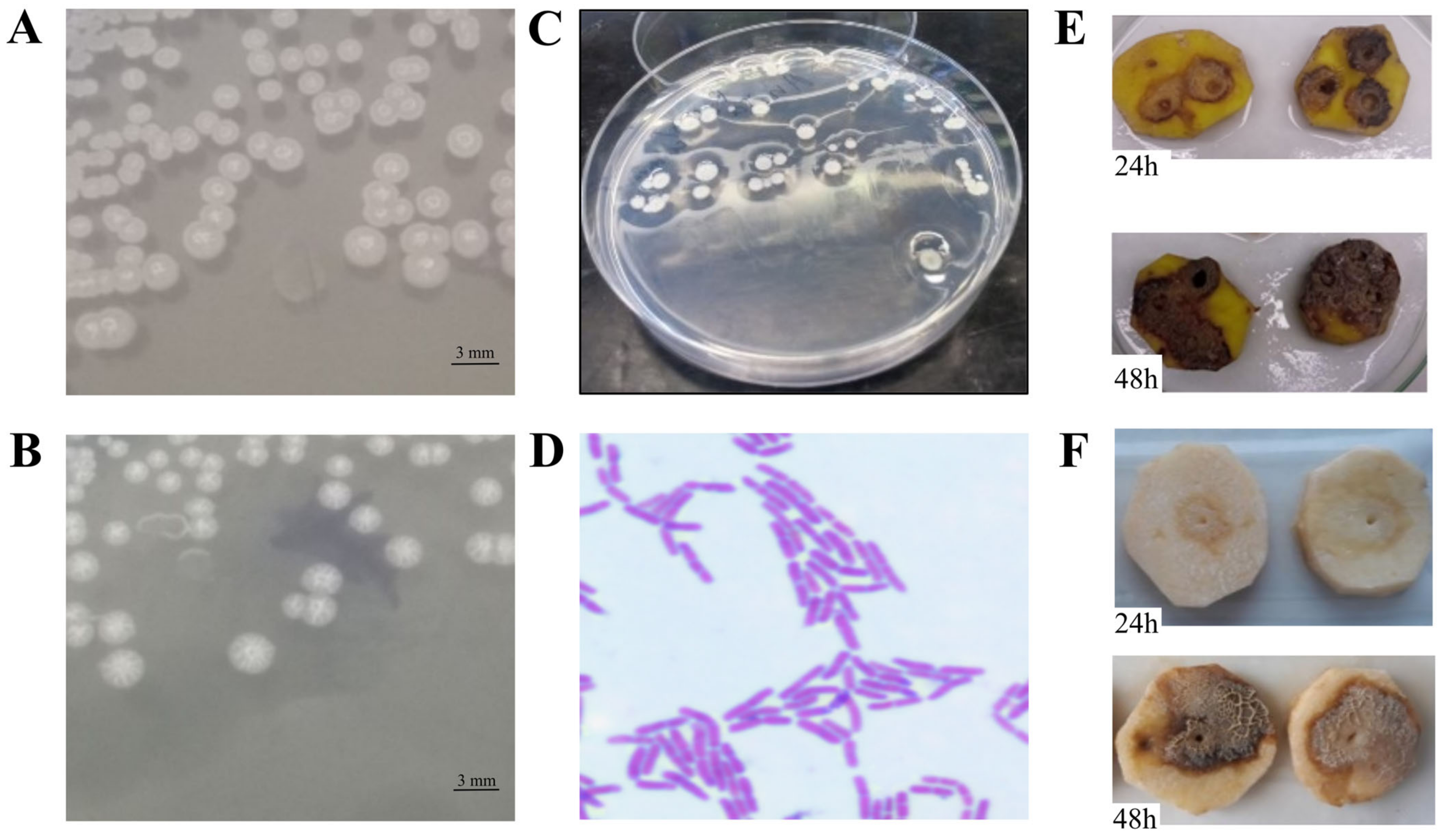
2.2. Isolation of Pectinolytic Bacteria

The collected bacterial strains were plated on a semi-selective crystal violet pectate (CVP) medium [7] and incubated at 28 °C for 48 h. Pectinolytic bacteria were detected by identifying the formation of cavities around colonies resulting from the degradation of polygalacturonic acid.

2.3. MALDI-TOF MS Analysis

Matrix Assisted Laser Desorption/Ionisation Mass Spectrometry with Time-of-Flight (MALDI-TOF MS) analysis was performed on a MALDI Biotyper® device (Bruker, Billerica, MA, USA) in the Laboratoria Medyczne Bruss (Gdansk, Poland) as a commercial service according to the procedure recommended by the manufacturer. The following classification criteria were applied: a score ≥ 2.0 allowed for identification of the species and a score between 1.7 and 1.9 supported genus identification, while a score < 1.70 was considered unreliable for identification purposes. All MALDI-TOF MS spectra for the studied strains were recorded, and two representative spectra for the isolates were identified.

2.4. Sequencing of 16S rDNA

The genomic DNA of the obtained pectinolytic isolates was extracted using a GF-1 Bacterial DNA Extraction Kit (Vivantis Technologies, Heeßen, Germany) according to the manufacturer’s instructions. The DNA purity was assessed using a NanoDrop spectrophotometer (Thermo Fisher Scientific, Waltham, MA, USA). DNA was stored at −20 °C for long-term preservation or at 4 °C for immediate use.
The 16S rRNA encoding region was PCR-amplified using the universal primer pair forward S1 (5′-AGAGTTTGATCMTGGCTCAG-3′) and reverse S2 (5′-GGMTACCTTGTTACGAYTTC-3′) [22]. The PCR reaction was performed in a total volume of 50 µL containing 50 ng of the template DNA, 0.1 U Taq polymerase, 10× Taq-buffer, 25 mM MgCl2, 25 mM dNTPs and 2 mM of each primer. The following thermal conditions were used: 95 °C for 5 min, followed by 35 cycles at 94 °C for 1 min, 55 °C for 45 s and 72 °C for 90 s, with a final extension at 72 °C for 2 min [22]. PCR was performed using a thermocycler (Biometra TRIO, Analytic Jena, Jena, Germany).
After electrophoretic separation, PCR products were visualised under UV light (Vilber Lourmat TFX-35.MC UV-Lamp, Vilber, Marne-la-Vallée, France) and the size of the amplicons was determined via comparison with a 1 kb DNA ladder (Promega, Madison, WI, USA) before they were sequenced. The obtained nucleotide sequences were compared with the sequences deposited in the GenBank database (National Center for Biotechnology Information; https://www.ncbi.nlm.nih.gov/, accessed on 5 July 2022) with the use of BLAST algorithm. The herein acquired 16S rDNA sequences of B. pumilus and Paenibacillus sp. strains were submitted to the GenBank database and deposited under the following accession numbers: ON898611, ON899811, ON899941, ON899942, ON900081-ON900086, ON921248-ON921252 and ON922531-ON922535.

2.5. Phenotypic Characterisation of the Isolated Strains

Out of 270 collected isolates, 20 showing pectinolytic activity were selected for further phenotypic studies involving: Gram staining, the examination of oxidase and catalase production, levan production from sucrose, bacterial abilities to macerate potato tuber tissue, the production of plant cell wall-degrading enzymes (PCWDEs), motility, and biochemical tests included in the commercial test kit API20E (bioMérieux, Craponne, France). Two reference strains of established causative agents of soft rot, D. solani IFB0099 and P. parmentieri IFB5308, originating from the bacterial collection of Laboratory of Plant Protection and Biotechnology IFB UG & MUG (Gdansk, Poland), were used for comparative purposes [23,24].

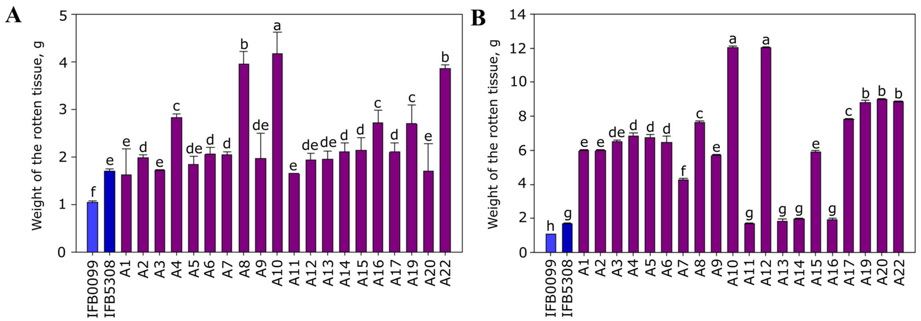
2.5.1. Bacterial Ability to Macerate Potato Tuber Tissue

Potato tubers were sterilised as mentioned above. Then, the tubers were aseptically cut into 10 mm slices. Subsequently, three holes (5 mm) per slice were made using a sterile cork borer. Bacterial suspensions of each isolate in M63 medium were adjusted to 0.5 McF, and 50 µL of the suspension was added to each hole in the potato slice. The tuber slices were placed in glass Petri dishes on absorbent paper, which had been previously moistened with sterile water. The dishes were incubated in plastic boxes at 28 °C for 48 h with 85–90% relative humidity. The holes designated as negative controls were filled with 50 µL of sterile 0.85% NaCl instead of the bacterial suspension. In terms of positive controls of efficient potato macerators, Dickeya solani IFB0099 and Pectobacterium parmentieri IFB5308 strains were applied. After 48 h of incubation, the rotten potato tissue was removed and weighed as performed before [25,26]. The experiment was repeated three times, each of which involved two technical replicates.

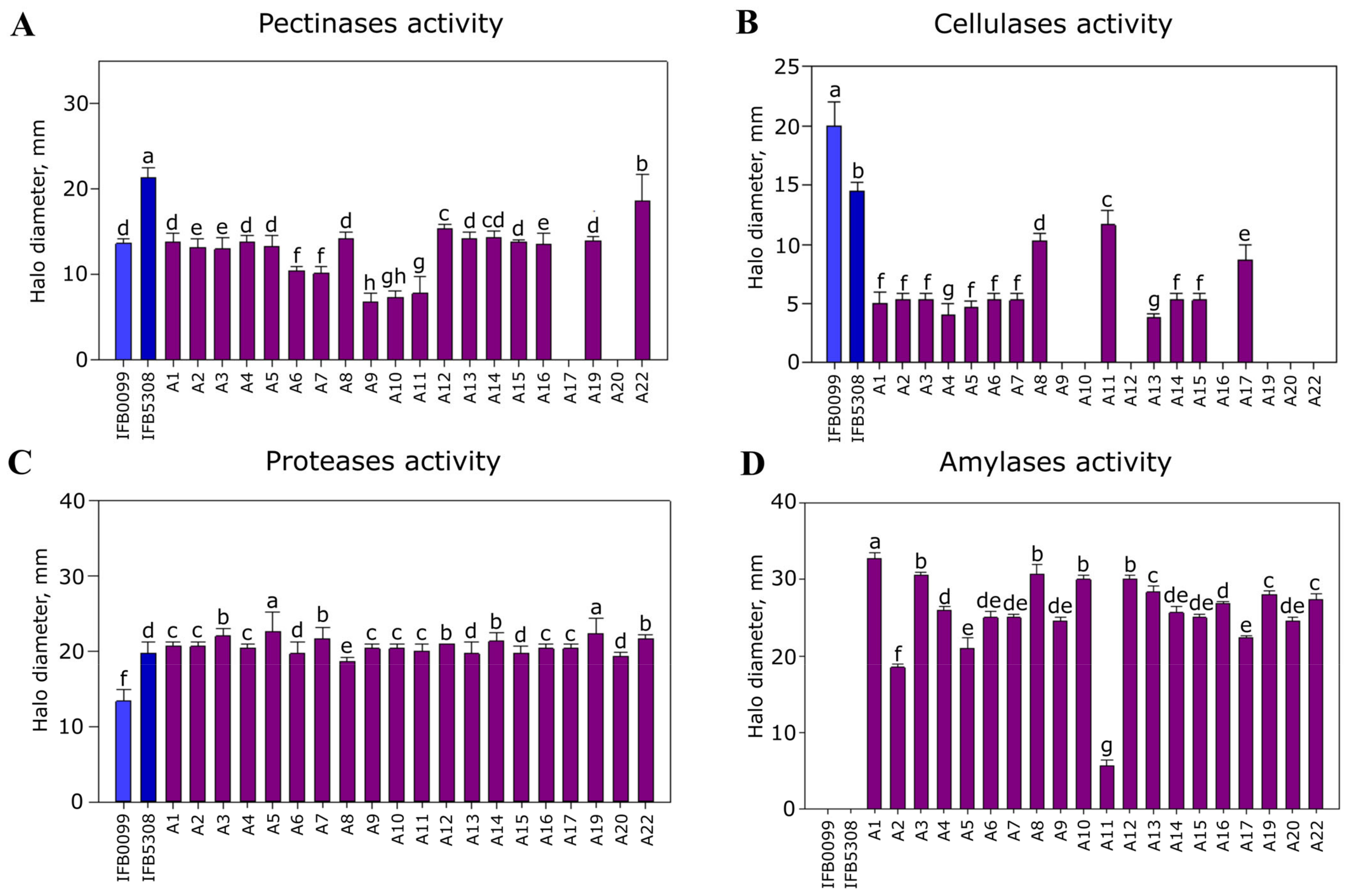
2.5.2. Production of Extracellular Enzymes and Bacterial Motility

The production of major PCWDEs (pectinases, cellulases, proteases and amylases) and cell motility of the selected isolates were assessed via semi-quantitative plate assays. D. solani IFB0099 and P. parmentieri IFB5308 were used as reference strains. For each assay, a single 2 µL drop of bacterial suspension, previously adjusted to 0.5 McF in 0.9% NaCl, was spotted on the below-listed solid media plates. All tests were performed three times with four technical replicates.
The activity of pectinases was tested on M63 agar medium supplemented with 0.25% polygalacturonate (PGA). After incubation for 48 h at 28 °C, the plates were flooded with 10% copper acetate, which forms a blue precipitate with PGA. The diameter of the pale white halo around the colony, corresponding to the PGA degradation capacity, was measured as mentioned in Reverchon et al. (1986) [27].
The activity of cellulases was evaluated on M63 agar medium supplemented with 1% carboxymethylcellulose (CMC). After incubation for 48 h at 28 °C, the plates were stained with 1% Congo red solution for 5 min and washed two times with 4 M NaCl. The diameter of the clear zone around the colony, corresponding to the exhibited cellulase activity, was measured [28].
Protease activity was tested on nutrient agar medium supplemented with 1% skim milk. After incubation for 48 h at 28 °C, the diameter of the clear zone around the colony was measured [29].
Amylase activity was evaluated on starch-containing medium. After incubation for 48 h at 28 °C, the amylase production was detected by adding an iodine solution. If the bacteria were able to degrade starch, clear zones around the bacterial colonies appeared [30]. The diameter of the clear zone around the colony was measured.
To evaluate swimming motility, bacterial strains were spotted on Tryptone Soy Agar (TSA) medium solidified with 0.3% agar. After 24 h incubation at 28 °C, the diameters of the bacterial colonies were measured [31].
To evaluate swarming motility, bacterial suspensions were spotted on TSA medium solidified with 0.6% agar. After 24 h of incubation at 28 °C, the diameters of the bacterial colonies were measured [31].

2.5.3. Biochemical Profiling

The oxidase and catalase assays were performed using bacterial colonies collected from TSA agar plates. For the oxidase test, filter paper soaked in a freshly prepared 1% Kovac’s oxidase reagent was used [32]. For the catalase test, 3% H2O2 was added and examined for the formation of gas bubbles [32]. Levan production from sucrose was determined on nutrient agar containing 5% sucrose. A glucosidase activity test was performed according to Carrim et al. [30]. β-galactosidase, acetoin, gelatinase, arginine dihydrolase, lysine decarboxylase, ornithine decarboxylase, H2S, urease, indole, acetoin, use of citrate and tartrate, fermentation of glucose, mannitol, inositol, sorbitol, rhamnose, saccharose, melibiose, amygdalin and arabinose assays were performed using an API 20E test kit (bioMérieux, France) following the manufacturer’s instructions.

2.6. Statistical Analysis

Statistical analysis involving analysis of variance (ANOVA), in addition to Pearson’s test for the evaluation of the significance of the differences between parameters, was conducted using SAS Institute, Cary, NC USA 2008. The data were depicted as means ± standard deviations following Duncan’s multiple-range tests at p < 0.05. For each measurement, at least three replicates were performed.

3. Results

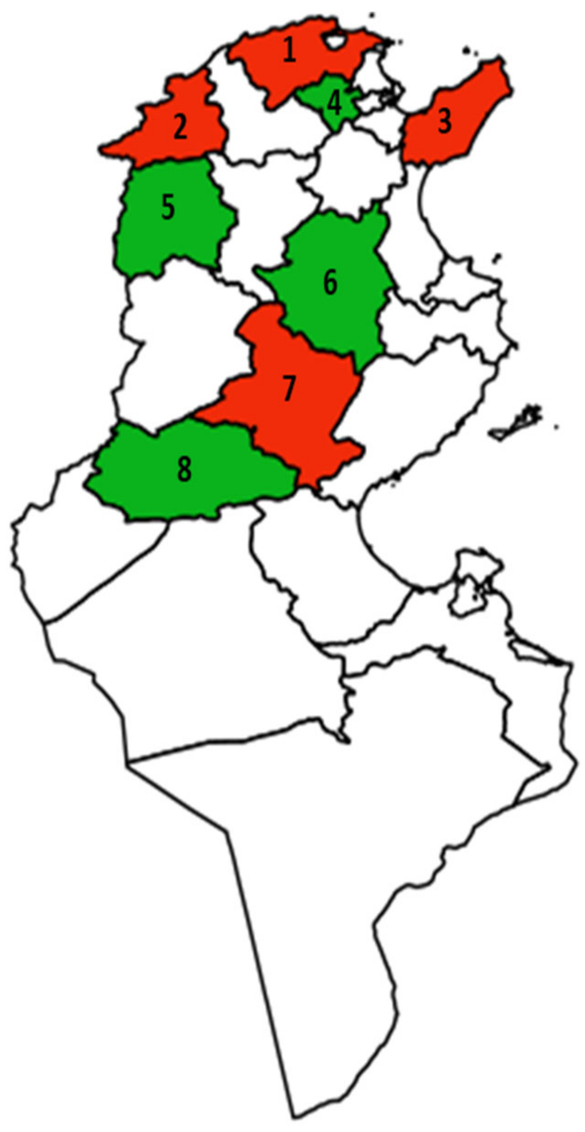
3.1. Occurrence of Pectinolytic Bacteria in Potato Fields in the Territory of Tunisia

In a three-year survey (2018, 2019 and 2020), potato tubers with soft rot symptoms were collected from eight different governorates of Tunisia that are well-known for their high potato production rates (Figure 1).
From a total of 1001 tuber samples obtained for study, 270 bacterial isolates were collected showing different morphological traits. In more detail, 26 strains were isolated in 2018, 160 in 2019 and 84 in 2020 (Supplementary Table S1). All bacterial isolates were screened for pectinolytic activity on a CVP medium. Twenty out of 270 isolates were able to form characteristic deep pits in this selective-differential medium (Figure 2C) and were chosen for further studies. These pectinolytic strains were isolated from soft-rotten potato tubers (cultivars ‘Spunta’ and ‘Daifla’) sampled in four Tunisian governorates (Bizerte, Jendouba, Nabeul and Sidi Bouzid) in 2019 and 2020 (Table 1). Most of the selected pectinolytic isolates were obtained from tubers of the potato cultivar ‘Spunta’ collected in the Bizerte territory, i.e., 25% and 30% in 2019 and 2020, respectively.

3.2. Identification of the Collected Pectinolytic Isolates

All pectinolytic strains included in this study were sub-cultured on TSA plates. Morphologically, the isolates showed some differences in the shape, edge, colour and size of their colonies on TSA medium. Nine isolates (A1–A6, A10, A19 and A22) formed round, transparent colonies of 1–3 mm in diameter with a white centre (Figure 2A), ten isolates (A7–A9, A11, A13–A17, A20) formed irregular colonies of 1–3 mm in diameter (Figure 2B), and one isolate (A12) exhibited a round, beige-coloured colony of 1–2 mm in diameter.
All pectinolytic isolates were rod-shaped and Gram-positive (Figure 2D), catalase-positive and oxidase-negative, and were able to grow at pH 7.5 both in the liquid and solid media at temperatures ranging from 14 °C to 37 °C, with an optimum at 28 °C.
Identification based on MALDI-TOF MS indicated that 19 pectinolytic strains (A1–A10, A13–A17, A19 and A22) belonged to the genus Bacillus spp. and one (A12) to the genus Paenibacillus spp. (Table 2). For all 20 isolates studied, the MALDI BioTyper scores exceeded 1.7, which supports bacterial identification at the genus level. Based on MALDI BioTyper identifications reaching scores ≥2.0, eight isolates (A1, A2, A6, A9, A13, A14, A18 and A20) were assigned to the species Bacillus pumilus, and one isolate (A12) to the species Paenibacillus amyloliticus (Table 2).
According to the subsequently conducted alignment of the approx. 1100 bp high-quality fragments of the sequenced 16S rDNA of 20 analysed strains, the Bacillus sp. isolates previously not assigned to the species level share 100% homology in this 16S rDNA fragment with the strains undoubtedly assigned to Bacillus pumilus species based on the acquired MALDI-TOF MS spectra. Afterwards, the fragments of 16S rDNA sequenced for the herein collected strains were compared using a BLAST algorithm, with the reference sequences available in the GenBank database, which confirmed the former taxonomic assignments and allowed for recognition of the closest sequences to the ones tested. To conclude, the sequencing of 16S rDNA proved that nineteen pectinolytic strains (A1–A11, A13–A17, A19–A20 and A22) belong to the Bacillus pumilus species, while one strain (A12) belongs to the genus Paenibacillus (similarity: 99%) (Table 2).

3.3. Phenotypic Characterisation of the Collected Pectinolytic Isolates

3.3.1. Bacterial Ability to Macerate Potato Tuber Tissue

The maceration ability of the pectinolytic strains originating from different regions of Tunisia was tested on tuber slices of two potato cultivars: cv. ‘Lilly’, acquired in Poland (Figure 3A), and cv. ‘Spunta’, the most widely grown variety in Tunisia (Figure 3B).
The rotten tissue was collected and weighed 48 h after inoculation to establish the maceration capacity of the tested strains. All 20 pectinolytic strains showed the ability to macerate potato tissue, but differences in the potency between certain strains were noted. In more detail, the highest bacterial ability to macerate the potato tuber tissue of cv. ‘Lilly’ was recorded for isolates A4, A8, A10 and A22 (Figure 3A), and in the case of cv. ‘Spunta’, the most prominent maceration potencies were shown by isolates A10 and A12 (Figure 3B).
It needs to be acknowledged that potato tubers of cv. ‘Spunta’ indicated a higher level of susceptibility to bacterial infection, as the recorded weights of the macerated tuber tissue of cv. ‘Spunta’ were approximately four times higher than those of cv. ‘Lilly’ and ranged from 2 to 12 g (Figure 3A,B).
Inoculation of the tuber slices with bacterial suspensions of D. solani strain IFB0099 and P. parmentieri strain IFB5308, used for comparison purposes, revealed that some pectinolytic strains collected in Tunisia have a higher ability to macerate potato tissue than the reference strains (Figure 3A,B). The presented data indicated that pectinolytic bacteria isolated from the rotten potato tuber in Tunisia were capable of macerating plant tissue under laboratory conditions.

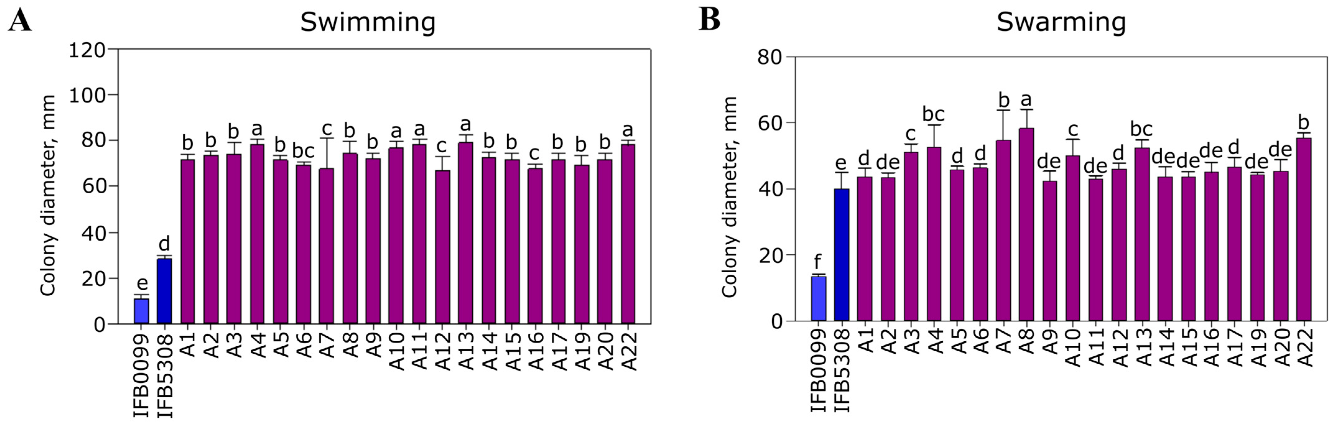
3.3.2. Major Pathogenicity-Associated Features

The analysis of pectinase activity on the M63 PGA plates revealed that the majority of the tested strains exhibited comparable pectinolytic activity to the reference strain D. solani IFB0099 (Figure 4A). Isolate A22 indicated the highest activity among the studied strains, similar to that of P. parmentieri strain IFB5308. Several isolates (A6–A7 and A9–A11) revealed significantly lower pectinolytic activity than the included reference strains (Figure 4A).
All tested pectinolytic isolates showed significantly lower cellulolytic activity than the reference strains D. solani IFB0099 and P. parmentieri IFB5308 (Figure 4B). Some of the characterised pectinolytic strains, i.e., A9, A10, A16, A19, A20, A22 and A12, did not exhibit any cellulolytic activity (Figure 4B).
All tested pectinolytic isolates originating from different regions of Tunisia revealed protease activity comparable to P. parmentieri IFB5308, reaching the highest value of 22 mm. Isolates A1–A5, A7, A9–A12, A14–A17 and A19 showed the highest protease activity (20–22 mm), and strains A6, A8, A13 and A19 exhibited the lowest protease activity (18–19 mm).
All investigated isolates showed significantly higher protease activity than the reference strain D. solani IFB0099, for which this trait approximated a value of 13 mm (Figure 4C).
The majority of the characterised pectinolytic Bacillus and Paenibacillus spp. strains yielded amylolytic activity (Figure 4D). Significantly lower amylase activity was shown by the A11 strain. In addition, it was confirmed that D. solani IFB0099 exhibited no amylase activity (Figure 4D).
All strains of pectinolytic Bacillus and Paenibacillus spp. isolated in Tunisia revealed similar swimming motility, which, at the same time, was higher than that of the reference strains, reflected by the colonies’ diameters on TSA supplemented with 0.3% agar, which ranged from 78 to 67 mm (Figure 5A).
In terms of swarming motility examined on TSA supplemented with 0.6% agar, all the investigated strains showed potency comparable to the reference P. parmentieri IFB5803 strain as the noted colonies’ diameters were within 43–58 mm. The performed analysis revealed that strains A7, A8 and A22 indicated the highest swarming motility among the investigated isolates (Figure 5B).

3.3.3. Biochemical Profiling

All tested pectinolytic isolates produced β-galactosidase and gelatinase in addition to fermented saccharose and amygdalin. Moreover, each of the studied bacterial strains lacked lysine decarboxylase and urease activity and was incapable of H2S, indole or levan production, in addition to showing an inability to utilize tartrate.
Based on the API 20E assay, it was found that the pectinolytic isolates were capable of β-galactosidase, saccharose and amygdalin fermentation and utilising D-mannitol. The examined isolates were also capable of producing gelatinase. Some isolates (A7–A10, A12–A17, A20 and A22) managed to degrade L-arabinose, L-rhamnose and melibiose. In addition, several strains (A1–A6, A11, A12 and A19) were shown to be unable to utilise sorbitol (Table 3).
Interestingly, the sole isolate of Paenibacillus spp. differed from all the analysed Bacillus spp. in terms of lack of capacity to ferment mannitol and glucose. It is also important to acknowledge that strain-dependent variation was identified among the herein investigated B. pumilus and Paenibacillus spp. strains in relation to the production of arginine dihydrolase, ornithine decarboxylase and acetoin, in addition to the utilisation of citrate, inositol, sorbitol, rhamnose, melibiose and arabinose.

4. Discussion

Over the past 10 years, an increasing incidence of soft rot or blackleg caused by Erwinia spp. has been reported. This disease is responsible for extensive damage in the field and in potato storage in Tunisia. Moreover, Erwinia spp. has been isolated from 20% of rotten imported seed tubers [20]. It has also been noted that latent infections with Erwinia spp. not only occurs in 90% of imported seed potatoes, but also does not decrease with successive progenies, thus posing a real threat to the cultivation and storage of potatoes [33]. Contrarily, previous research on major bacterial and fungal diseases in potato fields in Tunisia conducted in the growing seasons of 1993–1994 pointed to predominance of dry rot, with 87–90% of the isolates classified as Fusarium solani, followed by minor contributions of F. sambucinum in addition to either Verticillium dahliae and Phoma exigua or Sclerotina minor, depending on the year of study [20]. From the 1993 collections, only five isolates of E. caratovora were acquired among the 141 samples plated on CVP medium [20]. No members of the current family Pectobacteriaceae were identified during the survey performed in 1994 [20]. The current study, conducted in the 2018–2020 growing seasons, revealed that just 20 bacterial strains showed pectinolytic properties on CVP medium, and no soft rot Pectobacteriaceae were detected among the 270 isolates obtained from the diseased potatoes picked up in distinct Tunisian regions; this agrees with the data of Priou and Mahjoub collected over 20 years ago. Similarly to the previous suggestions of Priou and Mahjoub [20], the climatic conditions present in Tunisia do not seem to favour development of disease symptoms by Pectobacterium and Dickeya spp., in contrast to the common occurrence of these bacteria in temperate climates [26,34,35]. Thus, as shown in the current study, there was an ecological niche accessible to a different plant pathogen, reported herein for the first time as a causative agent of bacterial soft rot in potato in Tunisia.
Of the 20 pectinolytic isolates originating from the diseased potatoes collected in the territory of Tunisia, 19 were assigned as Bacillus pumilus, while 1 was assigned to the genus Paenibacillus. Interestingly, strains classified as Bacillus pumilus (former member of the Bacillus subtilis group) indicate various features, such as plant growth promotion [36,37], the potential to cause disease symptoms in plants [9,10,11,12], and the ability to cause human diseases [38,39], to spoil food [40], or to sustain themselves in demanding ecosystems [41,42]. B. pumilus strains are particularly resistant to environmental stressors, such as low nutrient availability, UV irradiation, desiccation, high salinity or various oxidisers [41], which may be the reason for their higher resilience and better fitness in the Tunisian climate of Bacillus pumilus in contrast to Dickeya and Pectobacterium spp. Additionally, the genus Paenibacillus encloses ubiquitous strains of over 200 species [43] of notable relevance to plants, humans, animals or the environment, which have been isolated from highly diverse habitats, e.g., polar regions, tropics, aquatic ecosystems or even deserts. These bacteria draw interest due to their plant growth-promoting properties, potential uses in bioremediation and effective production of enzymes, such as lipases, pectinases, amylases, cellulases or hemicellulases, which exhibit higher activity, stability and cost-effectiveness in contrast to the currently utilised alternatives [43]. However, the production of proteases, lipases and phospholipases, in addition to the ability of Paenibacillus spp. to multiply at low temperatures, contributes to the food spoilage process.
In this research, we have proven that all the investigated Bacillus and Paenibacillus spp. strains are able to cause disease symptoms on potato slices and that the vast majority of these isolates exhibit the activity of plant cell wall-degrading enzymes, i.e., pectinases, cellulases, proteases and amylases, which significantly contribute to the decay of plant tissue [44]. The manifestation of disease symptoms triggered by Bacillus pumilus in potato, referred to as water-soaked or yellowish-brown rot, agrees with the former observations of Bathily et al. [10] and further supports the previously reported [9,10,11,12] pathogenic potential of this species.
Contrarily, the pathogenic potential of Paenibacillus spp. is not commonly raised as, to the best of our knowledge, there are only two reports on the isolation of Paenibacillus spp. strains from diseased plants, i.e., wheat and barley, showing leaf deformation, wilting, spotting, strokes and stripes [45], in addition to symptomatic Solanum lycopersicum, Alium cepa, Pelargonium sp., Lilium sp., Begonia obliqua, Anthurium sp., Dianthus caryophyllus and Hypericum perforatum, which were sent to the Plant Disease Clinic in Poland in 2013–2019 for examination [46]. Considering that in this study, the initially observed disease symptoms in potato were reproduced under laboratory conditions post artificial inoculation of two potato cultivars (‘Lilly’ and ‘Spunta’), the pathogenicity of Paenibacillus spp. strains should be taken into consideration and further investigated. It is especially imperative to do so, as the recorded potato maceration capacities of the vast majority of the studied Bacillus and Paenibacillus spp. strains turned out to exceed the potency of the included reference Dickeya and Pectobacterium spp. strains, which are recognised as the main causative agents of soft rot and blackleg diseases [44,47].
Subsequently, we studied the activity of pivotal virulence factors associated with the development of soft rot symptoms among the 20 isolated Bacillus and Paenibacillus spp strains. So far, little information has been published on the activity of most significant plant cell wall-degrading enzymes among plant pathogenic strains of Bacillus pumilus [9,10,12,13,14,17]. Evdokimova et al. [19] are the sole reporters of lipase and protease action among all the investigated plant pathogenic B. pumilus strains. Notably, some intraspecies variation was noted in cellulase and pectinase activity among phytopathogenic B. pumilus by Evdokimova et al. [19], though the capacity to produce these enzymes dominated among the included isolates. Likewise, the common production of pectinases, cellulases, proteases and amylases was exhibited by Bacillus and Paenibacillus spp. strains for industrial use [43,48,49,50,51,52]. In terms of plant pathogens, Od23 Bacillus pumilus characterised by Bathily et al. [10], the GR8 isolate obtained by Peng et al. [11] and all the strains examined by Kovaleva et al. [12] were reported to be incapable of hydrolysing starch, in contrast to all the herein investigated strains showing amylase activity. Putatively, these discrepancies result from the application of diverse methodologies, as the action of amylases of Bacillus and Paenibacillus spp. found confirmation in research focused on industrial processes or the characterisation of this group of enzymes in the listed bacterial taxa [43,50,52]. It is also worth noting that we identified two Bacillus pumilus strains lacking the action of pectinases, and six B. pumilus and one Paenibacillus spp. deprived of cellulase activity. In spite of the absence of these virulence factors, all the herein investigated Bacillus and Paenibacillus spp. strains efficiently macerated potato tissue. Additionally, all isolates exhibited high swimming and swarming capacities, which confirmed former reports on the motility of plant pathogenic Bacillus pumilus strains [9,10,12,13,14,17]. Curiously, the intensities of the swimming and swarming of Bacillus and Paenibacillus spp. strains exceeded the ability to move of the two analysed Dickeya and Pectobacterium strains.
Biochemical characterisation of the phytopathogenic Bacillus and Paenibacillus spp. isolated in Tunisia was performed in order to reveal intra-species diversity. Similarly to the current research, each of the five Bacillus pumilus strains studied by Galal et al. [9] fermented glucose and mannitol, though they were incapable of gelatin liquefaction, contrarily to our work and the studies of Peng et al. [11,13,19]. Moreover, the inability of B. pumilus isolates to produce indole in the herein presented study agreed with that of Bathily et al. [10]. However, the Od23 strain investigated by the latter group gave inconsistent results in terms of H2S production, and was incapable of producing acetoin and fermenting arabinose, in opposition to the outcomes collected for some of the herein analysed isolates. Notably, the lack of oxidase activity for all the herein investigated strains is confirmed in several reports [13,14,17,19]. It is likewise worth mentioning that the abilities of phytopathogenic Bacillus pumilus to utilise mannitol, glucose and saccharose agreed with the study of Saleh et al. [13]. Furthermore, the raised intraspecies variability among B. pumilus strains regarding biochemical features is consistent with former research, which revealed such phenomena, for instance, in terms of glycerol and D-cellobiose utilisation [11], trehalose metabolisation [13] and arabinose degradation [9,10], in addition to H2S production [9].
To summarise, we identified pectinolytic plant pathogenic B. pumilus and Paenibacillus sp. as being responsible for potato diseases in the fields of Tunisia. As the studied strains macerated potato tissue, in addition to exhibiting activity of major virulence factors, their pathogenic potential seems well founded. We want to underline the high significance and novelty of the current research as there are few previous reports that describe the phytopathogenic properties of B. pumilus strains in diverse plant hosts throughout the world, and even less data concerning the plant disease-causing potential of Paenibacillus sp. Most of all, the pathogenic properties of B. pumilus and Paenibacillus sp. should be taken into consideration in view of the attempts to utilise the members of these taxa as biocontrol and/or plant growth-promoting agents. Previously, the beneficial effects of B. pumilus inoculations against fungal pathogens were associated with the production of peptide antibiotics, biosurfactants, chitinases, other fungus cell wall-degrading enzymes and volatiles, in addition to diverse stimulants of plant defence systems [36]. Moving to the plant growth-promoting Paenibacillus spp., these isolates may secrete phytohormones, antimicrobials or insecticides, induce plant systemic resistance or solubilise inaccessible forms of phosphorous or iron, and some strains even exhibit nitrogen-fixation properties [53]. Based on the data shown herein and the previous reports of Peng et al. [11], Galal et al. [9], Bathily et al. [10] and Kovaleva et al. [12], we suggest devoting the highest attention to both preventing fungal-derived diseases and stimulating plant growth with bacterial strains with disease-causing potential. This matter should be thoroughly investigated in the future.

5. Conclusions

The occurrence of new, virulent pectinolytic phytopathogenic bacteria from the genera Bacillus and Paenibacillus, in particular, B. pumilus and P. amyloliticus, is a serious problem for the producers of seed potatoes, as well as for the farmers in Tunisia. To the best of our knowledge, this is the first report on the occurrence of Bacillus and Paenibacillus spp. capable of causing soft rot in potato in Tunisia. It is important to conduct a complementary study on the interactions between pathogenic bacteria associated with soft rot during the different seasons and cultivar susceptibility in order to develop a potential strategy for controlling this disease in the field and in postharvest storage.

Supplementary Materials

The following supporting information can be downloaded at: https://www.mdpi.com/article/10.3390/agriculture13061275/s1, Table S1: Characteristics of the Tunisian orchards surveyed for bacterial soft rot and blackleg diseases in 2018, 2019 and 2020.

Author Contributions

Conceptualisation, A.Y., M.O., A.M.-P., W.S., E.L. and N.S.-Z.; methodology, A.Y., A.M.-P., W.S., N.K., E.L. and B.T.; sampling, A.Y. and M.R.H.; conducting experiments, A.Y.; supervision, E.L. and N.S.-Z.; project administration, N.S.-Z.; writing—original draft, A.Y., M.O., A.M.-P., N.K., E.L. and N.S.-Z.; writing—review and editing, N.S.-Z. and E.L. All authors have read and agreed to the published version of the manuscript.

Funding

This research was partly funded by the Ministry of Higher Education and Scientific Research of Tunisia (n°LR16ES05), the project “PROMETEO—Un village transfrontalier pour protéger les cultures arboricoles méditerranéennes en partageant les connaissances” Ref. n°_C-5_2.1-36, Strategic project ENI Italy-Tunisia 2014–2020 and by the Ministry of Education and Science of Poland (531-N107-D801-21).

Institutional Review Board Statement

Not applicable.

Data Availability Statement

The collected data is contained within the article or Supplementary Materials. Raw data presented in this study are available on reasonable request from the corresponding authors. The herein acquired 16S rDNA sequences of B. pumilus and Paenibacillus sp. strains are publicly available in the GenBank database under the following accession numbers: ON898611, ON899811, ON899941, ON899942, ON900081-ON900086, ON921248-ON921252 and ON922531-ON922535.

Acknowledgments

The authors want to thank Afef Zeddini from the Mycology, Pathologies and Biomarkers Laboratory of the Faculty of Sciences of Tunis (Tunisia) for her technical support on this project.

Conflicts of Interest

The authors declare no conflict of interest.

References

  1. Chemak, F.; Allagui, L.; Ali, Y. Analyse des performances techniques des producteurs de la pomme de terre en Tunisie. Une approche non paramétrique. New Medit. 2014, 13, 72–80. [Google Scholar]
  2. Perombelon, M.C.M.; Kelman, A. Ecology of the soft rot Erwinias. Ann. Rev. Phytopathol. 1980, 18, 361–387. [Google Scholar] [CrossRef]
  3. Toth, I.K.; Bell, K.S.; Holeva, M.C.; Birch, P.R.J. Soft rot erwiniae: From genes to genomes. Mol. Plant Pathol. 2003, 4, 17–30. [Google Scholar] [CrossRef] [PubMed]
  4. Toth, I.K.; van der Wolf, J.M.; Saddler, G.; Lojkowska, E.; Helias, V.; Pirhonen, M.; Tsror, L.; Elphinstone, J.G. Dickeya species: An emerging problem for potato production in Europe. Plant. Pathol 2011, 60, 385–399. [Google Scholar] [CrossRef]
  5. Adeolu, M.; Alnajar, S.; Naushad, S.; Gupta, R.S. Genome-based phylogeny and taxonomy of the ‘Enterobacteriales’: Proposal for Enterobacterales ord. nov. divided into the families Enterobacteriaceae, Erwiniaceae fam. nov., Pectobacteriaceae fam. nov., Yersiniaceae fam. nov., Hafniaceae fam. nov., Morganellaceae fam. nov., and Budviciaceae fam. nov. Int. J. Syst. Evol. Microbiol. 2016, 66, 5575–5599. [Google Scholar] [CrossRef]
  6. Mansfield, J.; Genin, S.; Magori, S.; Citovsky, V.; Sriariyanum, M.; Ronald, P.; Dow, M.; Verdier, V.; Beer, S.V.; Machado, M.A.; et al. Top 10 plant pathogenic bacteria in molecular plant pathology. Mol. Plant Pathol. 2012, 13, 614–629. [Google Scholar] [CrossRef] [Green Version]
  7. Hélias, V.; Hamon, P.; Huchet, E.; van der Wolf, J.M.; Andrivon, D. Two new effective semiselective crystal violet pectate media for isolation of Pectobacterium and Dickeya. Plant Pathol. 2012, 61, 339–345. [Google Scholar] [CrossRef]
  8. De Boer, S.H.; Li, X.; Ward, L.J. Pectobacterium spp. associated with bacterial stem rot syndrome of potato in Canada. Phytopathology 2012, 102, 937–947. [Google Scholar] [CrossRef] [Green Version]
  9. Galal, A.A.; El-Bana, A.A.; Janse, J. Bacillus pumilus, a new pathogen on mango plants. Egypt. J. Phytopathol. 2006, 34, 17–29. [Google Scholar]
  10. Bathily, H.; Babana, A.H.; Samaké, F. Bacillus pumilus, a new pathogen on potato tubers in storage in Mali. Afr. J. Microbiol. Res. 2010, 4, 2067–2071. [Google Scholar]
  11. Peng, Q.; Yuan, Y.; Gao, M. Bacillus pumilus, a novel ginger rhizome rot pathogen in China. Plant Dis. 2013, 97, 1308–1315. [Google Scholar] [CrossRef] [Green Version]
  12. Kovaleva, V.A.; Shalovylo, Y.I.; Gorovik, Y.N.; Lagonenko, A.L.; Evtushenkov, A.N.; Gout, R.T. Bacillus pumilus—A new phytopathogen of Scots pine. J. Forest Sci. 2015, 61, 131–137. [Google Scholar] [CrossRef] [Green Version]
  13. Saleh, O.I.; Huang, P.-Y.; Huang, J.-S. Bacillus pumilus, the cause of bacterial blotch of immature balady peach in Egypt. J. Phytopathol. 1997, 145, 447–453. [Google Scholar] [CrossRef]
  14. Song, J.H.; Wu, Z.R.; Zhang, L.X.; Tan, G.J.; Wang, S.; Wang, J.J. First report of Bacillus pumilus causing fruit rot on muskmelon (Cucumis melo) in China. Plant Dis. 2018, 102, 439. [Google Scholar] [CrossRef]
  15. Hakim, M.; Liaquat, F.; Gul, S.; Chaudhary, H.J.; Munis, M.F.H. Presence of Bacillus pumilus causing fruit rot of Ficus lacor in Pakistan. J. Plant Pathol. 2015, 97, 543. [Google Scholar]
  16. Ahmadi, E.; Kowsari, M.; Azadfar, D.; Jouzani, G.S. Bacillus pumilus and Stenotrophomonas maltophilia as two potentially causative agents involved in Persian oak decline in Zagros forests (Iran). Forest Pathol. 2019, 49, e12541. [Google Scholar] [CrossRef]
  17. Mazlan, S.; Zulperi, D.; Wahab, A.; Jaafar, N.M.; Sulaiman, Z.; Rajandas, H. First report of Bacillus pumilus causing trunk bulges of rubber tree (Hevea brasiliensis) in Malaysia. Plant Dis. 2019, 103, 1016. [Google Scholar] [CrossRef]
  18. Font, M.I.; Bassimba, D.D.M.; Cebrián, M.C.; Molina, L.M.; Jordá, C. First report of Bacillus pumilus on Phaseolus vulgaris in Spain. Plant Pathol. 2010, 59, 2. [Google Scholar] [CrossRef]
  19. Evdokimova, O.V.; Miamin, V.E.; Valentovich, L.N. Biochemical and molecular genetic characteristics of Bacillus pumilus bacteria, isolated on the territory of Belarus. J. Belarusian State Univ. Biol. 2018, 1, 39–47. [Google Scholar]
  20. Priou, S.; El-Mahjoub, M. Bacterial and fungal diseases in the major potato-growing areas of Tunisia. EPPO Bull. 1999, 29, 167–171. [Google Scholar] [CrossRef]
  21. Yahiaoui-Zaidi, R.; Ladjouzi, R.; Benallaoua, S. Pathogenic variability within biochemical groups of Pectobacterium carotovorum isolated in Algeria from seed potato tubers. Int. J. Biotech. Mol. Biol. Res. 2010, 1, 1–9. [Google Scholar]
  22. El Arbia, A.; Rochexa, A.; Chataignéa, G.; Bécheta, M.; Lecouturiera, D.; Arnaulda, S.; Gharsallah, N.; Jacques, P. The Tunisian oasis ecosystem is a source of antagonistic Bacillus spp. producing diverse antifungal lipopeptides. Res. Microbiol. 2015, 167, 46–57. [Google Scholar] [CrossRef] [PubMed]
  23. Potrykus, M.; Golanowska, M.; Hugouvieux-Cotte-Pattat, N.; Lojkowska, E. Regulators involved in Dickeya solani virulence, genetic conservation, and functional variability. Mol. Plant Microbe Interact. 2014, 27, 700–711. [Google Scholar] [CrossRef] [PubMed] [Green Version]
  24. Zoledowska, S.; Motyka-Pomagruk, A.; Sledz, W.; Mengoni, A.; Lojkowska, E. High genomic variability in the plant pathogenic bacterium Pectobacterium parmentieri deciphered from de novo assembled complete genomes. BMC Genom. 2018, 19, 751. [Google Scholar] [CrossRef] [Green Version]
  25. Golanowska, M.; Kielar, J.; Lojkowska, E. The effect of temperature on the phenotypic features and the maceration ability of Dickeya solani strains isolated in Finland, Israel and Poland. Eur. J. Plant Pathol. 2017, 147, 803–817. [Google Scholar] [CrossRef] [Green Version]
  26. Zoledowska, S.; Motyka, A.; Zukowska, D.; Sledz, W.; Lojkowska, E. Population structure and biodiversity of Pectobacterium parmentieri isolated from potato fields in temperate climate. Plant Dis. 2018, 102, 154–164. [Google Scholar] [CrossRef] [Green Version]
  27. Reverchon, S.; Van Gijsegem, F.; Rouve, M.; Kotoujansky, A.; Robert-Baudouy, J. Organization of a pectate lyase gene family in Erwinia chrysanthemi. Gene 1986, 49, 215–224. [Google Scholar] [CrossRef]
  28. Moran, F.; Nasuno, S.; Starr, M.P. Extracellular and Intracellular Polygalacturonic Acid h-arts-Eliminases of Erwinia carotovora. Arch. Biochem. Biophys. 1968, 123, 298–306. [Google Scholar] [CrossRef]
  29. Ji, J.; Hugouvieux-Cotte-Pattat, N.; Robert-Baudouy, J. Use of Mu-lac insertions to study the secretion of pectate lyases by Erwinia chrysanthemi. Microbiology 1987, 133, 793–802. [Google Scholar] [CrossRef] [Green Version]
  30. Carrim, A.J.I.; Barbosa, E.C.; Vieira, J.D.G. Enzymatic activity of endophytic bacterial isolates of Jacaranda decurrents cham.(Carobinha-do-campo). Braz. Archiv. Biol. Technol. 2006, 49, 353–359. [Google Scholar] [CrossRef]
  31. Harshey, R.M. Bacterial motility on a surface: Many ways to a common goal. Ann. Rev. Microbiol. 2003, 57, 249–273. [Google Scholar] [CrossRef]
  32. Borisov, V.B.; Forte, E.; Davletshin, A.; Mastronicola, D.; Sarti, P.; Giuffrè, A. Cytochrome bd oxidase from Escherichia coli displays high catalase activity: An additional defence against oxidative stres. FEBS Lett. 2013, 587, 2214–2218. [Google Scholar] [CrossRef]
  33. Romdhani, M.S.; El-Mahjoub, M. Latent infection of potato tubers with Erwinia spp. in Tunisia. Meded. Van Fac. Landbouwwet. Rijksuniv. Gent 1990, 55, 1119–1123. [Google Scholar]
  34. Motyka-Pomagruk, A.; Zoledowska, S.; Sledz, W.; Lojkowska, E. The occurrence of bacteria from different species of Pectobacteriaceae on seed potato plantations in Poland. Eur. J. Plant Pathol. 2021, 159, 309–325. [Google Scholar] [CrossRef]
  35. Potrykus, M.; Golanowska, M.; Sledz, W.; Zoledowska, S.; Motyka, A.; Kolodziejska, A.; Butrymowicz, J.; Lojkowska, E. Biodiversity of Dickeya spp. isolated from potato plants and water sources in temperate climate. Plant Dis. 2016, 100, 408–417. [Google Scholar] [CrossRef] [Green Version]
  36. Leifert, C.; Li, H.; Chidburee, S.; Hampson, S.; Workman, S.; Sigee, D.; Epton, H.A.; Harbour, A. Antibiotic production and biocontrol activity by Bacillus subtilis CL27 and Bacillus pumilus CL45. J. Appl. Bacteriol. 1995, 78, 97–108. [Google Scholar] [CrossRef]
  37. Gutiérrez-Mañero, F.J.; Ramos-Solano, F.; Probanza, A.; Mehouachi, J.; Tadeo, F.R.; Talon, M. The plant-growth-promoting rhizobacteria Bacillus pumilus and Bacillus licheniformis produce high amounts of physiologically active gibberellins. Physiologia Plantarum. 2001, 111, 206–211. [Google Scholar] [CrossRef]
  38. Kimouli, M.; Vrioni, M.; Papadopoulou, M.; Koumaki, V.; Petropoulou, D.; Gounaris, A.; Friedrich, A.W.; Tsakris, A. Two cases of severe sepsis caused by Bacillus pumilus in neonatal infants. J. Med. Microbiol. 2012, 61, 596–599. [Google Scholar] [CrossRef] [Green Version]
  39. Tena, D.; Martínez-Torres, J.A.; Pérez-Pomata, M.T.; Sáez-Nieto, J.A.; Rubio, V.; Bisquert, J. Cutaneous infection due to Bacillus pumilus: Report of 3 cases. Clin. Infect. Dis. 2007, 44, e40–e42. [Google Scholar] [CrossRef]
  40. From, C.; Hormazabal, V.; Granum, P.E. Food poisoning associated with pumilacidin-producing Bacillus pumilus in rice. Inter. J. Food Microbiol. 2007, 115, 319–324. [Google Scholar] [CrossRef]
  41. Vockler, C.J.; Greenfield, P.; Tran-Dinh, N.; Midgley, D.J. Draft genome sequence of Bacillus pumilus Fairview, an isolate recovered from a microbial methanogenic enrichment of coal seam gas formation water from Queensland, Australia. Genome Announc. 2014, 2, e00279-14. [Google Scholar] [CrossRef] [PubMed] [Green Version]
  42. Link, L.; Sawyer, J.; Venkateswaran, K.; Nicholson, W. Extreme spore UV resistance of Bacillus pumilus isolates obtained from an ultraclean spacecraft assembly facility. Microb. Ecol. 2004, 47, 159–163. [Google Scholar] [CrossRef] [PubMed]
  43. Grady, E.N.; MacDonald, J.; Liu, L.; Richman, A.; Yuan, Z.-C. Current knowledge and perspectives of Paenibacillus: A review. Microb. Cell Fact. 2016, 15, 203. [Google Scholar] [CrossRef] [PubMed] [Green Version]
  44. Van der Wolf, J.M.; Acuña, I.; de Boer, S.H.; Brurberg, M.B.; Cahill, G.; Charkowski, A.O.; Coutinho, T.; Davey, T.; Dees, M.W.; Degefu, Y.; et al. Diseases caused by Pectobacterium and Dickeya species around the world. In Plant Diseases Caused by Dickeya and Pectobacterium Species; van Gijsegem, F., van der Wolf, J.M., Toth, I.K., Eds.; Springer Nature: Cham, Switzerland, 2021; pp. 215–262. [Google Scholar]
  45. Slovareva, O.Y. Detection and identification of wheat and barley phytopathogens in Russia. Microbiol. Ind. Res. J. 2020, 7, 13–23. [Google Scholar]
  46. Zenelt, W.; Krawczyk, K.; Borodynko-Filas, N. Biodiversity and scope of endophytic and phytopathogenic bacterial species identified in plant samples investigated in the Plant Disease Clinic laboratory. J. Plant Prot. Res. 2021, 61, 63–82. [Google Scholar]
  47. Motyka, A.; Zoledowska, S.; Sledz, W.; Lojkowska, E. Molecular methods as tools to control plant diseases caused by Dickeya and Pectobacterium spp: A minireview. New Biotech. 2017, 39, 181–189. [Google Scholar] [CrossRef]
  48. Huang, Q.; Yong, P.; Xin, L.; Haifeng, W.; Yizheng, Z. Purification and characterization of an extracellular alkaline serine protease with dehairing function from Bacillus pumilus. Curr. Microbiol. 2003, 46, 169–173. [Google Scholar] [CrossRef]
  49. Basu, S.; Manabendra, N.S.; Dhrubajyoti, C.; Krishanu, C. Large-scale degumming of ramie fibre using a newly isolated Bacillus pumilus DKS1 with high pectate lyase activity. J. Ind. Microbiol. Biotech. 2009, 36, 239–245. [Google Scholar] [CrossRef]
  50. Yan, H.; Hui, W.; Bo, Y.; ShengMou, H.; YunJie, L.; JinJu, W. Isolation and identification of Bacillus pumilus HB-3 and purification characteristics of amylase. Storage Proc. 2019, 2, 62–68. [Google Scholar]
  51. Ariffin, H.; Abdullah, H.; Umi Kalsom, M.S.; Shirai, Y.; Hassan, M.A. Production and characterization of cellulase by Bacillus pumilus EB3. Int. J. Eng. Technol. 2006, 3, 47–53. [Google Scholar]
  52. Kayath, C.A.; Zamba, A.I.; Mokembiabeka, S.N.; Opa-Iloy, M.; Wilson, P.S.E.; Kaya-Ongoto, M.D.; Mouellet Maboulou, R.J.; Nguimbi, E. Synergic involvements of microorganisms in the biomedical increase of polyphenols and flavonoids during the fermentation of ginger juice. Int. J. Microbiol. 2020, 2020, 8417693. [Google Scholar] [CrossRef]
  53. McSpadden Gardener, B.B. Ecology of Bacillus and Paenibacillus spp. in agricultural systems. Phytopathology 2004, 94, 1252–1258. [Google Scholar] [CrossRef] [Green Version]
Figure 1. Map of Tunisia showing origin of bacterial isolates acquired from potato tubers with soft rot symptoms. Red regions indicate locations from which pectinolytic bacterial isolates were obtained, green regions represent sites where no pectinolytic bacteria were isolated. GPS locations: (1) 37.17612, 10.00813/Bizerte; (2) 36.6103092, 8.9742237/Jandouba; (3) 36.46917, 10.78222/Nabeul; (4) 36.8506, 9.93608/Manouba; (5) 36.167965, 8.709579/El kef; (6) 35.675914, 10.091924/El Kairouan; (7) 35.035439, 9.483939/Sidi Bouzid; (8) 34.431140, 8.775656/Gafsa.
Agriculture 13 01275 g001
Figure 2. Phenotypic characterisation of the selected pectinolytic bacterial isolates. (A,B) Morphology of the colonies on TSA agar medium. (C) Cavity formation on CVP agar medium inoculated with a pectinolytic isolate and incubated at 28 °C for 48 h. (D) Cell morphology post-Gram staining, observations made using the ×100 oil immersion objective. (E) Bacterial soft rot on potato tuber slices of cv. ‘Lilly’ after 24 and 48 h incubation at 28 °C caused by the strain ‘A10’. (F) Bacterial soft rot on potato tuber slices of cv. ‘Spunta’ after 24 and 48 h incubation at 28 °C caused by the strain ‘A9’.
Agriculture 13 01275 g002
Figure 3. Comparison of the ability of the tested pectinolytic isolates to macerate potato tuber tissue of cultivars Lilly (A) and Spunta (B). Means with standard errors (n = 9) are depicted. Means with the same letter are not significantly different (p > 0.05). The experiment was repeated three times, each of which involved two technical replicates. Strains used were D. solani IFB0099 and P. parmentieri IFB5308 in addition to bacterial isolates analysed in this study (A1–A22).
Agriculture 13 01275 g003
Figure 4. Comparison of the plant cell wall-degrading enzyme activity of pectinolytic strains isolated in Tunisia. Pectinase (A), cellulase (B), protease (C) and amylase (D) production was estimated by determining the diameter (mm) of the haloes observed on the detection plates. All the experiments were performed three times with four technical replicates. Means with standard errors (N = 3) were depicted. Means with the same letter are not significantly different (p > 0.05). Strains used were D. solani IFB0099, P. parmentieri IFB5308 and bacterial isolates analysed in this study (A1–A22).
Agriculture 13 01275 g004
Figure 5. Comparison of the swimming and swarming motilities of pectinolytic strains isolated in Tunisia. (A) Swimming motility was estimated by the diameter (mm) of the spread of the colonies on plates containing 0.3% agar. (B) Swarming motility was estimated by the diameter (mm) of the spread of the colonies on plates containing 0.6% agar. All the experiments were performed three times with four technical replicates. Means with standard errors (N = 3) were depicted. Means with the same letter are not significantly different (p > 0.05). Strains used were D. solani IFB0099 and P. parmentieri IFB5308 in addition to bacterial isolates analysed in this study (A1−A22).
Agriculture 13 01275 g005
Table 1. Origin of the pectinolytic strains characterised in this study.
Code StrainGovernorate/
Region/Locality
GPS
Location
Year/MonthIrrigation TypeFertilisationCultivarOrgan/Symptom
A1Bizerte/
Ras Djebel/Kichi
37.216273, 10.1090302019/JuneDrip irrigationBiological and chemicalSpuntaTuber/soft rot
A2Bizerte/
Ras Djebel/Kichi
37.216273, 10.1090302019/JuneDrip irrigationBiological and chemicalSpuntaTuber/soft rot
A3Nabeul/
Beni Khiar/Melliti
36.478755, 10.7708502019/JuneDrip irrigationBiological and chemicalSpuntaTuber/soft rot
A4Bizerte/Ras Djebel/
Kichi
37.216273, 10.1090302019/JuneDrip irrigationBiological and chemicalSpuntaTuber/soft rot
A5Bizerte/
Ghar Milh/Mohamed
37.169613, 10.1473752019/JunePond waterChemicalSpuntaTuber/soft rot
A6Bizerte/
Ras Djebel/Kichi
37.216273, 10.1090302019/JuneDrip irrigationBiological and chemicalSpuntaTuber/soft rot
A7Jandouba/
Bousalem/Bousalem1
36.612511, 9.0008602019/FebruaryDrip irrigationBiological and chemicalDaiflaTuber/soft rot
A8Bizerte/
Ras Djebal/Kichi
37.216273, 10.1090302020/JuneDrip irrigationBiological and chemicalSpuntaTuber/soft rot
A9Bizerte/
Ras Djebal/Kichi
37.216273, 10.1090302020/JuneDrip irrigationBiological and chemicalSpuntaTuber/soft rot
A10Sidi Bouzid/
Bir El Hafey/Anonym
34.930545, 9.2268592019/JuneDrip irrigationBiologicalSpuntaTuber/soft rot
A11Nabeul/
Beni Khiar/Melliti
36.478755, 10.7708502019/JuneDrip irrigationBiological and chemicalSpuntaTuber/soft rot
A12Jandouba/
Bousalem/Bousalem1
36.612511, 9.0008602019/FebruaryDrip irrigationBiological and chemicalDaiflaTuber/soft rot
A13Bizerte/
Ras Djebel/Kichi
37.216273, 10.1090302020/JuneDrip irrigationBiological and chemicalSpuntaTuber/soft rot
A14Bizerte/
Ras Djebel/Kichi
37.216273, 10.1090302020/JuneDrip irrigationBiological and chemicalSpuntaTuber/soft rot
A15Jandouba/
Bousalem/Bousalem 2
36.623775, 8.9586742020/FebruaryDrip irrigationBiological and chemicalDaiflaTuber/soft rot
A16Jandouba/
Bousalem/Bousalem2
36.623775, 8.9586742020/FebruaryDrip irrigationBiological and chemicalDaiflaTuber/soft rot
A17Jandouba/
Bousalem/Bousalem2
36.623775, 8.9586742020/FebruaryDrip irrigationBiological and chemicalDaiflaTuber/soft rot
A19Bizerte/
Ras Djebel/Kichi
37.216273, 10.1090302020/JuneDrip irrigationBiological and chemicalSpuntaTuber/soft rot
A20Bizerte/
Ghar Milh/Mohamed
37.169613, 10.1473752020/JunePond waterChemicalSpuntaTuber/soft rot
A22Jandouba/
Bousalem/Bousalem2
36.623775, 8.9586742020/JuneDrip irrigationBiological and chemicalSpuntaTuber/soft rot
Table 2. Identification of the pectinolytic strains isolated from the diseased potato tubers in Tunisia based on MALDI-TOF MS and 16S rDNA gene sequencing evaluation.
NumberIsolateMALDI-TOF MS-Based ClassificationLog (Score) MALDI-TOF MSAccession Number for the Sequenced 16S rDNA Gene FragmentsFinal Identification
1A1Bacillus pumilus2.02ON898611Bacillus pumilus
2A2Bacillus pumilus2.03ON899811Bacillus pumilus
3A3Bacillus pumilus1.82ON899941Bacillus pumilus
4A4Bacillus pumilus1.99ON899942Bacillus pumilus
5A5Bacillus pumilus1.96ON900081Bacillus pumilus
6A6Bacillus pumilus2.03ON900082Bacillus pumilus
7A7Bacillus pumilus1.86ON900083Bacillus pumilus
8A8Bacillus pumilus1.82ON900084Bacillus pumilus
9A9Bacillus pumilus2.04ON900085Bacillus pumilus
10A10Bacillus pumilus1.90ON900086Bacillus pumilus
11A11Bacillus altitudinis1.98ON922531Bacillus pumilus
12A12Paenibacillus amyloliticus2.18ON922535Paenibacillus amyloliticus
13A13Bacillus pumilus2.03ON922532Bacillus pumilus
14A14Bacillus pumilus2.06ON922533Bacillus pumilus
15A15Bacillus pumilus1.88ON922534Bacillus pumilus
16A16Bacillus pumilus1.72ON921248Bacillus pumilus
17A17Bacillus pumilus1.85ON921249Bacillus pumilus
18A19Bacillus pumilus2.00ON921250Bacillus pumilus
19A20Bacillus pumilus1.91ON921251Bacillus pumilus
20A22Bacillus pumilus2.05ON921252Bacillus pumilus
Table 3. Biochemical characteristics of pectinolytic bacterial isolates using API 20E identification system.
IsolateONPGADHLDCODCCITH2SURETDAINDVPGELGLUMANINSORRHASACMELAMYARAOX
A1+++++++
A2+++++++
A3+++++++
A4+++++++
A5+++++++
A6+++++++
A7++++++++++++++
A8++++++++++++++
A9++++++++++++++
A10++++++++++++++
A11++++++++
A12+++++++++++
A13+++++++++++++++
A14+++++++++++++++
A15++++++++++++++
A16++++++++++++++
A17+++++++++++++++
A19+++++++
A20++++++++++++++
A22++++++++++++++
+ indicates a positive test result; − indicates a negative test result. ONPG (β-galactosidase production), CIT (utilisation of citrate), VP (production of acetoin), GEL (gelatinase production), ADH (arginine dihydrolase), LDC (lysine decarboxylase), ODC (ornithine decarboxylase), H2S (H2S production), URE (urease production), TDA (utilisation of tartrate), IND (indole production), VP (acetoin production), GEL (gelatinase production), GLU (glucose fermentation), MAN (mannitol fermentation), INO (inositol fermentation), SOR (sorbitol fermentation), RHA (rhamnose fermentation), SAC (saccharose fermentation), MEL (melibiose fermentation), AMY (amygdalin fermentation), ARA (arabinose fermentation), and OX (oxidase).
Disclaimer/Publisher’s Note: The statements, opinions and data contained in all publications are solely those of the individual author(s) and contributor(s) and not of MDPI and/or the editor(s). MDPI and/or the editor(s) disclaim responsibility for any injury to people or property resulting from any ideas, methods, instructions or products referred to in the content.

