Article

Co-Contamination of Food and Feed with Mycotoxin and Bacteria and Possible Implications for Health

by
Daniela Eliza Marin
*,
Gina Cecilia Pistol
,
Cristina Valeria Procudin
and
Ionelia Taranu
Laboratory of Animal Biology, National Institute for Research and Development for Biology and Animal Nutrition, Calea Bucuresti No. 1, 077015 Balotesti, Romania
*
Author to whom correspondence should be addressed.
Agriculture 2022, 12(11), 1970; https://doi.org/10.3390/agriculture12111970
Submission received: 21 October 2022 / Revised: 11 November 2022 / Accepted: 17 November 2022 / Published: 21 November 2022

Abstract

:
Food and feed safety is an issue of great concern for both animal and human health, due to the frequent contamination of food and feed with pathogens, such as bacteria, viruses, yeasts, molds, and parasites. The present paper assumes the possibility that a mycotoxin contamination could occur at the same time as a bacterial infection and investigates the effects of such co-contamination, in comparison with the individual effects of bacterial challenge. For this purpose, we have investigated the effects of simultaneous contamination of swine peripheral blood mononuclear cells (PBMCs) with lipopolysaccharide (LPS) (as a model for bacterial contamination) and mycotoxins (ochratoxin or zearalenone) on cell viability, cell cycles, oxydative stress, and inflammation. Our results show important additive/synergistic effects of co-exposure to fungal and bacterial toxins, and that these effects are more important when the cells are co-exposed to LPS and ochratoxin (OTA). The exposure of PBMCs to both OTA and LPS induced an exacerbation of the increase in the inflammation of concentrations of pro-inflamatory cytokines, compared with that of LPS-challenged cells: IL-1β (4.1 times increase), TNF-α (3.2 times increase), IL-6 (3.1 times increase). There was also a decrease in antioxidant defence (i.e., a significant decrease in the total antioxidant capacity and catalase activity) and a significant increase in the percentage of cells undergoing necrosis (24.3% vs. 15.3% in LPS-treated cells). The exposure to zearalenone (ZEA) and LPS led to less important effects and concerned mainly the parameters related to oxidative stress (i.e., a decrease in total antioxidant capacity). The present study provides important data for risk assessment, as the concomitant contamination with bacteria and mycotoxins can lead to a higher toxicity than that which results after an individual infection with Gram-negative bacteria.

1. Introduction

Food and feed safety is an issue of great concern for both animal and human health, due to the frequent contamination of food and feed with pathogens, such as bacteria, viruses, yeasts, molds, and parasites [1].
There is substantial evidence concerning the frequent contamination of food and feed with molds, which are considered the principal responsive organisms for cereal spoilage [2]. Molds are also responsible for the synthesis of secondary metabolites called mycotoxins, which frequently contaminate crops or raw materials such as cereals, nuts, spices, fruits, etc. [3]. Mycotoxin consumption leads to decreased animal performances, hepatotoxicity, nephrotoxicity, reproductive failures, alteration of the nervous and immune systems, and even death [4].
Ochratoxin (OTA) is a mycotoxin produced by Aspergilus and Penicillium molds [5]. The exposure to this toxin is associated with nephropathy in pigs and poultry, with the kidney being the main target of OTA [6]. Zearalenone (ZEA) is a mycotoxin produced by several Fusarium species [7]. Due to a chemical structure that is similar to the estrogen hormone, ZEA consumption induces severe reproductive problems [8]. The toxic effects of both OTA and ZEA are associated with their ability to induce oxydative stress through ROS generation, increase of lipid peroxidation, decrease the activity of antioxidant enzymes, and to modulate the immune response through increase in susceptibility to infection, immunotoxicity, and inflammation, with important consequences for human and animal health [9,10,11,12].
Another potential threat to feed quality is represented by the contamination with bacterial pathogens, such as Salmonella, Enterobacter, Escherichia, Staphylococcus, Pseudomonas, and Clostridium [13,14,15]. Feed contaminated with these bacteria, with origins in different ecological niches such as the gastrointestinal tract or the soil, are of great concern to producers and consumers, as it becomes a potential route of transmission of diseases to both animals and humans [13]. Lipopolysaccharide (LPS), also known as endotoxin, is considered the principal constituent of the Gram-negative bacteria outer membrane [16]. LPS stimulates the innate immunity and triggers the production of inflammatory mediators, such as cytokines and chemokines, which provoke multiple organ dysfunction and failure, leading to septic shock [17,18]. LPS can be found in food and feed even in the absence of live bacteria. For example, amino acid products used in feed formulations are issued from fermentation with Gram-negative bacteria, particularly E. coli K-12, and they still contain LPS [19]. Even the LPS of E. coli K-12 is less potent than most other strains; nevertheless, it has at least one-quarter of the endotoxin activity of wild-type strains and has the potential to be hazardous if the LPS passes into feed [20,21].
There is a high probability that food or feed will be contaminated with more than one contaminant. In the case of co-contamination, the contaminants could synergistically interact for toxicity, with important consequences for health risks, as was shown in studies of co-occurence of different mycotoxins [22,23], bacteria, and viruses [24] or mycotoxins and viruses [25]. Raw materials, such as cereals, oilseeds, fruits, vegetables, and, especially, complete feed or food as complex matrices, are susceptible to bacterial/fungal-mycotoxin contamination [1,26]. For example, all the samples of cottonseed cakes for cattle analyzed in a survey study carried out between 2011 and 2012 in Pakistan had heavy bacterial total counts and were contaminated with both fungi (Aspergillus, Penicillium, Fusarium, Alternaria genera) and mycotoxins (aflatoxin, ochratoxin, citrinin, tenuazoic acid, cyclopiazonic acid) [27]. In addition, poultry feeds produced in Poland in 2010 were characterized by a higher count of total bacteria and fungi, increased concentration of ergosterol, and a contamination with mycotoxins belonging mainly to trichothecenes group [28].
To our knowledge, there are no studies investigating the toxic effect of exposure to both mycotoxins and bacteria, even though the concomitent presence of mycotoxins and bacteria in cereals and feed was reported by several studies and raises great concern for feed safety [28,29].
The present paper assumes the possibility that a mycotoxin contamination could occur at the same time as a bacterial contamination and investigates the effects of the co-contamination in comparison with the individual effects of bacterial contamination. For this purpose, we investigated the effect of simultaneous contamination of swine peripheral blood mononuclear cells (PBMCs) with LPS (as a model for bacterial contamination) and mycotoxins (OTA or ZEA) on cell viability, cell cycles, oxydative stress, and inflammation. The swine cell model was chosen because pigs are often exposed to intoxication with both bacteria and mycotoxins through the consumption of a contaminated diet and because pigs provide a good model for humans, due to their similar anatomy and physiology.

2. Materials and Methods

Cell cultures. Peripheral blood mononuclear cells (PBMCs) were isolated from blood samples that were aseptically collected in sodium heparin tubes (Vacutest Kima, Arzergrande, Italy) from the jugular veins of healthy piglets (TOPIG hybrid). The animals were cared for in accordance with Romanian Law 206/2004 and the EU Council Directive 98/58/EC for the handling and protection of animals used for experimental purposes. The study protocol was approved by the Ethical Committee of the National Research–Development Institute for Animal Nutrition and Biology, Balotești, Romania (Ethical Committee no. 118/2019). If not otherwise stated, all reagents were purchased from Sigma-Aldrich (Sigma-Aldrich, Saint Louis, MO, USA). The cells were obtained using density gradient centrifugation on Hystopaque®-1077, as previously described [30]. The PBMCs were resuspended in complete RPMI-1640 medium supplemented with L-glutamine (2 mM), penicillin (100 U/mL), streptomycin (100 µg/mL), and fetal calf serum (5 mL/100 mL RPMI-1640 medium) and cultivated at 37 °C in plates of 24 wells in a concentration of 5 × 10⁶ cells/mL. Some cells were treated for 48 h with LPS 10 μg/mL and/or mycotoxins (OTA or ZEA) in a concentration of 10 μM. The concentration of mycotoxins was chosen on the basis of previous studies that have shown a low cytotoxic effect of these concentrations on PBMCs [31,32].
Apoptosis. All flow-cytometry analyses were performed using the Muse Cell Analyzer system and Muse 1.5 Analysis software (Merck, Darmstadt, Germany). Apoptosis analysis was performed using a Muse Annexin & Dead Cell Kit (Merck), according to the manufacturer’s guide. Briefly, 100 μL of cell suspension (5 × 10⁵ cells) was mixed with 100 μL of Muse Annexin V & Dead Cell Kit reagent and incubated at room temperature in the dark for 20 min before flow cytometry analysis. The number of events for analysis was set at 2000. The results for apoptosis analysis were presented as a percentage of live, apoptotic (early or late), or dead cells; the graphs represent the mean of four independent experiments.
Cell cycle. Cell cycle analysis was performed using Muse Cell Cycle Analysis (Merck) following the manufacturer’s instructions. Briefly, 8 × 10⁵ cells/mL were fixed in 70% ethanol for 12 h, then diluted with Muse Cell Cycle reagent and incubated at room temperature in the dark for 30 min prior to analysis. The number of events for analysis was set at 5000. The results for cell cycle analysis were expressed as a percentage of cells in different phases of the cell cycle (G0/G1, S, or G2/M); the graphs represent the mean of four independent experiments.
Reactive oxidative species (ROS). Quantification of the number of cells undergoing oxidative stress was realized using a Muse Oxidative Stress Kit (Merck), according to the manufacturer’s guide. Briefly, 10 μL of PBMCs (5 × 10⁶ cells/mL) were mixed with 190 μL of a Muse Oxidative Stress reagent working solution and incubated at 37 °C in the dark for 30 min prior to analysis. The number of events for analysis was set at 3000. The results for the oxidative stress analyses were expressed as percentage of cells undergoing oxidative stress-% ROS (+); the graph represents the mean of four independent experiments.
Nitric oxide activity (NO). The assessment of nitric oxide activity was carried out using a Muse Nitric Oxide Kit (Merck), following the manufacturer’s instructions. Briefly, for each sample, 10 μL of PBMCs (5 × 10⁶ cells/mL) were incubated at 37 °C for 30 min with 100 μL of a Muse Nitric Oxide solution, followed by the addition of 90 μL of a Muse 7-AAD working solution and analysis via a Muse Cell Analyzer. The number of events for analysis was set at 3000. The results were expressed as the count and the percentage of cells exhibiting nitric oxide activity and cell death. The graph represents the mean of four independent experiments.
Total antioxidant status. The antioxidant capacity assay was performed in cellular lysates as previously described [32] and inhibition percentages were expressed as μmol trolox/mL. The results represent the mean of four independent experiments.
Antioxidant enzymes activity. The assessment of the activities of the antioxidant enzymes was carried out using commercially available assay kits from the Cayman Chemical Company (Ann Arbor, MI, USA). The antioxidant enzymes tested were superoxide dismutase (SOD), catalase (CAT), and glutathione peroxidase (GPx). The analyses of antioxidant enzyme activities were performed in cell lysates following the manufacturer’s guide, as described by Taranu et al. [30]. The results represent the mean of four independent experiments.
Cytokine synthesis. The concentrations of IL-1β, IL-6, IL-8, and TNF-α were analyzed in the supernatants of the PBMC cell culture using ELISA kits (anti-swine primary and biotinylated secondary antibodies included; R&D Systems, Minneapolis, MN, USA) as previously described [33]. The results represent the mean of four independent experiments.

3. Results

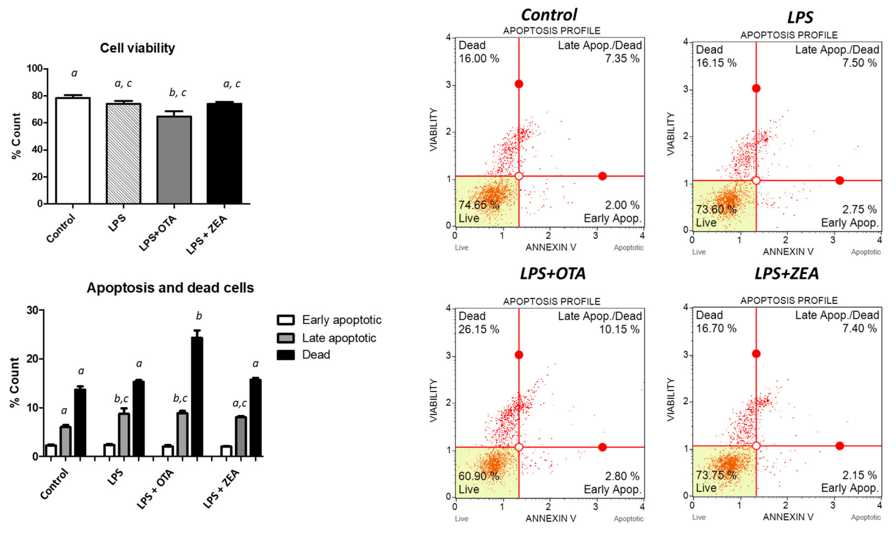
3.1. The Effect of Co-Exposure to LPS and Mycotoxins on Cell Proliferation, Apoptosis, and Necrosis

As shown in Figure 1, the LPS stimulation, as well as the exposure to both LPS and ZEA, did not affect the cell viability as compared with control. However, the simultaneous exposure to LPS and OTA induced a significant decrease (p = 0.015) in cell viability (17.5% compared with that of non-treated cells). Treatments had no effect in the early stages of apoptosis, but the exposure to LPS or to LPS and OTA significantly increased the percentage of cells in late apoptosis (28.5% and 51.3%, respectively, compared with that of the control cells). Only the exposure to the combination of LPS and OTA significantly increased (p = 0.0002) the percentage of dead cells (24.3% vs. 13.7% in the control cells or 15.3% in the LPS-treated cells), suggesting a higher cytotoxic effect of this treatment, compared with the untreated cells or PBMCs exposed only to bacterial challenge.

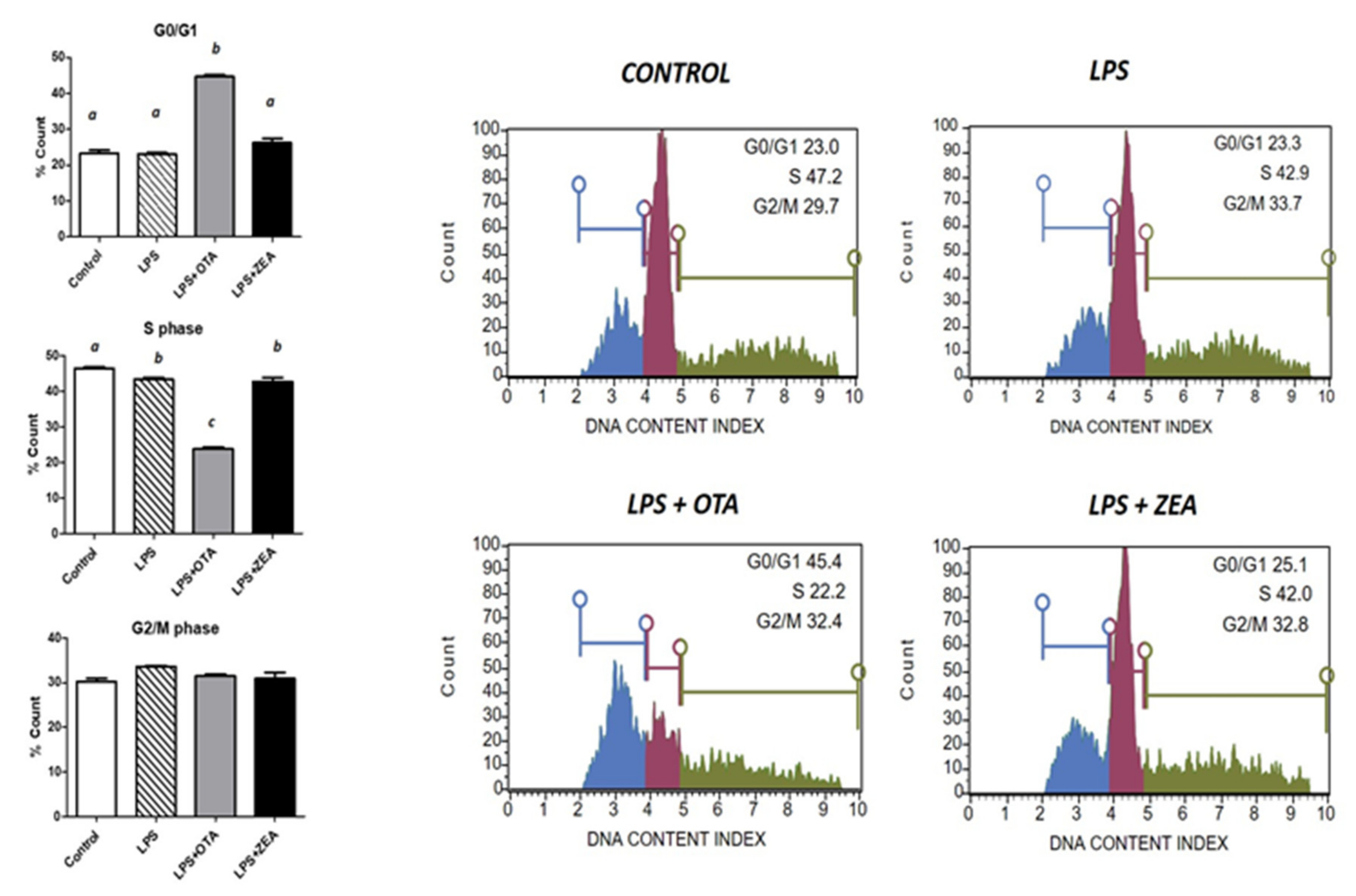
3.2. The Effect of Co-Exposure to LPS and Mycotoxins on Cell Cycle

The main effect of different treatments was observed in the S phase of the cell cycle (Figure 2). The LPS induced a significant decrease in the number of cells in the S phase (p = 0.031). The concomitant exposure to both bacteria and mycotoxin resulted in a more accentuated significant decrease in the S phase for OTA+LPS as compared with both the control group (p < 0.0001) and the LPS group (p < 0.0001), or for ZEA+LPS, but only compared with the control group (p = 0.0162). These results suggest that co-exposure to both LPS and mycotoxins modify the cell cycle by arresting cell proliferation and progression in the G2/M phase, the effect being more important than the simple exposure to LPS in the presence of OTA. The results show a significant increase in the G0/G1 phase induced by LPS+OTA; however, there was no effect from the treatments in the G2/M phase (Figure 2).

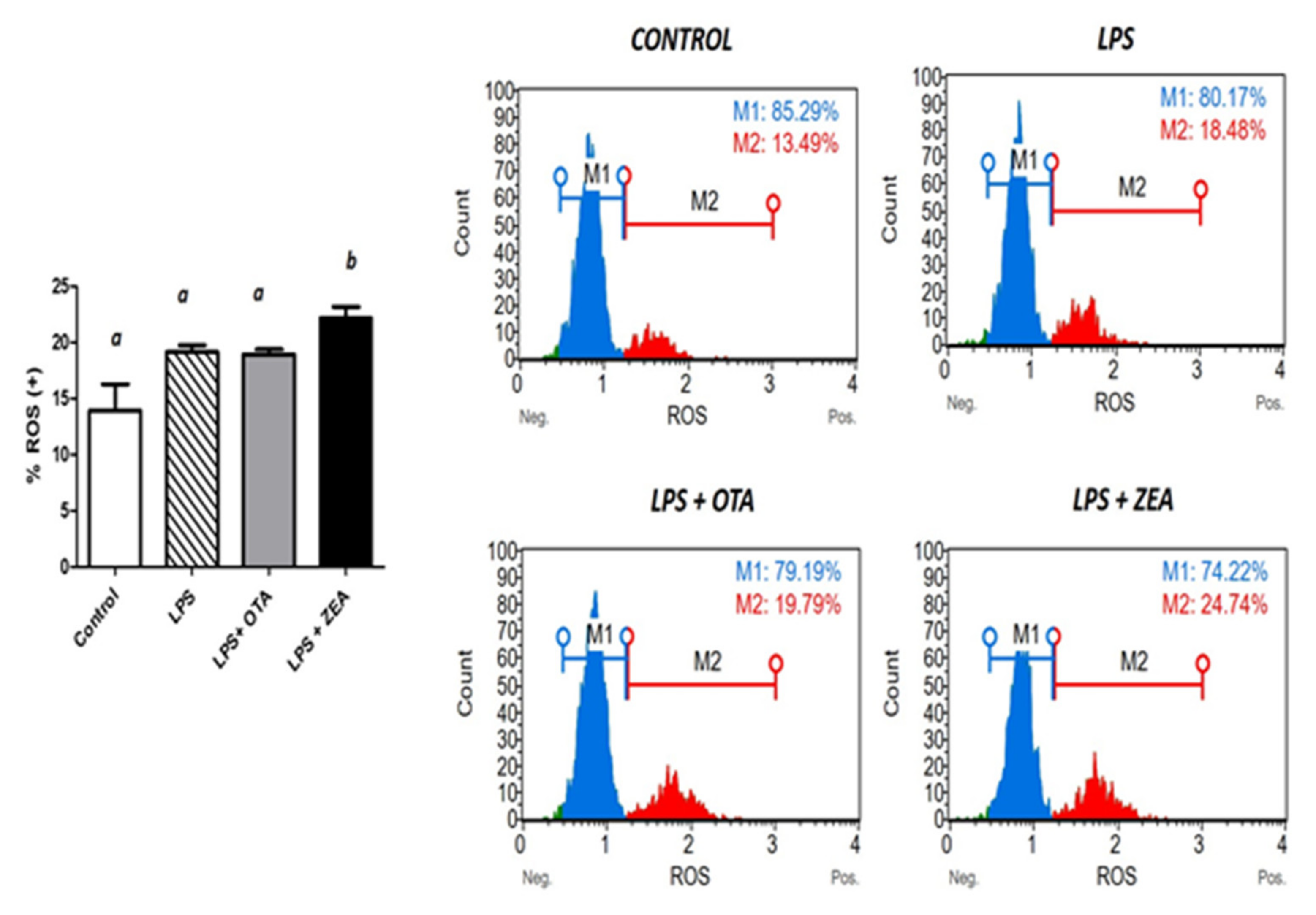
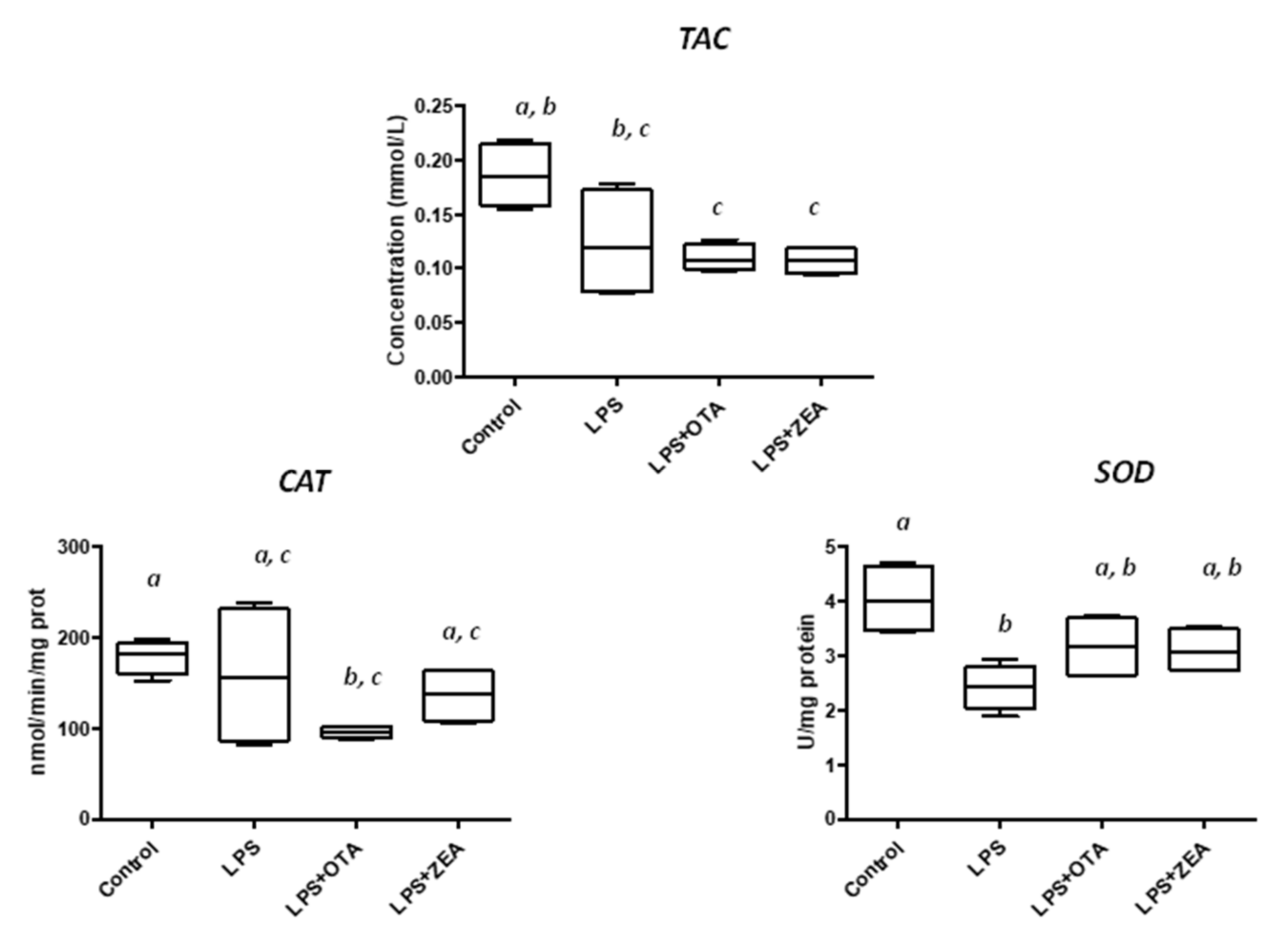
3.3. The Effect of Co-Exposure to LPS and Mycotoxins on ROS Production and on the Activity of the Enzymes Involved in Oxidative Defence

In comparison with the control group, the exposure of cells to LPS and LPS+OTA tended to increase the ROS concentration, as was the result in flowcytometry studies (p = 0.0764 and p = 0.0807), while LPS and ZEA treatment significantly increased the ROS concentration (22.14% ROS positive cells vs. 13.9% in untreated cells, p = 0.0183, or vs. 19.1% in LPS-challenged cells, p = 0.044) (Figure 3), showing a higher capacity of both LPS and ZEA to induce oxidative stress.
When cells were challenged with LPS, the total antioxidant status tended to decrease (p = 0.081), while the exposure to both combinations of LPS+OTA and LPS+ZEA induced a decrease in total antioxidant capacity in PBMCs. This decrease was statistically significant, compared that for untreated cells (p = 0.003 for LPS+OTA treatment and p = 0.0032 for LPS+ZEA treatment), but not compared with LPS-stimulated cells (p > 0.05) (Figure 4).
Exposure to LPS led to a significant decrease in SOD activity (40.2% from control), while SOD activity in LPS+OTA cells was not different from control or LPS. In LPS+ZEA group the SOD activity tended to decrease, compared with both untreated cells (p = 0.051) or LPS-challenged cells (p = 0.062). LPS exposure had no effect on catalase activity. However, the co-exposure to both LPS and mycotoxins significantly decreased catalase activity for LPS+OTA treatment (96.21 vs. 178 nmol/min/mg protein in control cells, p = 0.0002) or tended to decrease the enzyme activity for LPS+ZEA contamination (137.0 vs. 178 nmol/min/mg protein in control cells, p = 0.059).

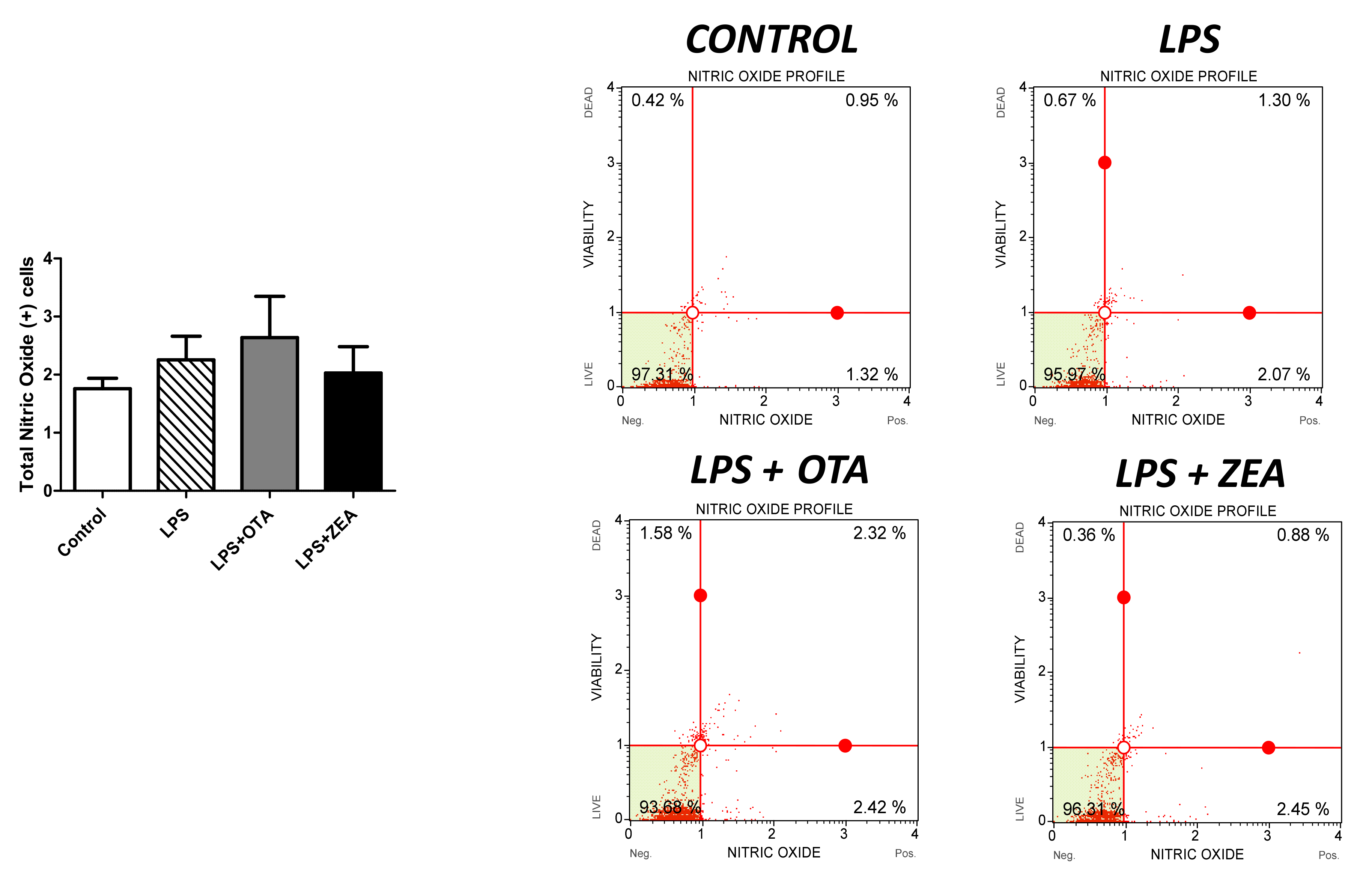
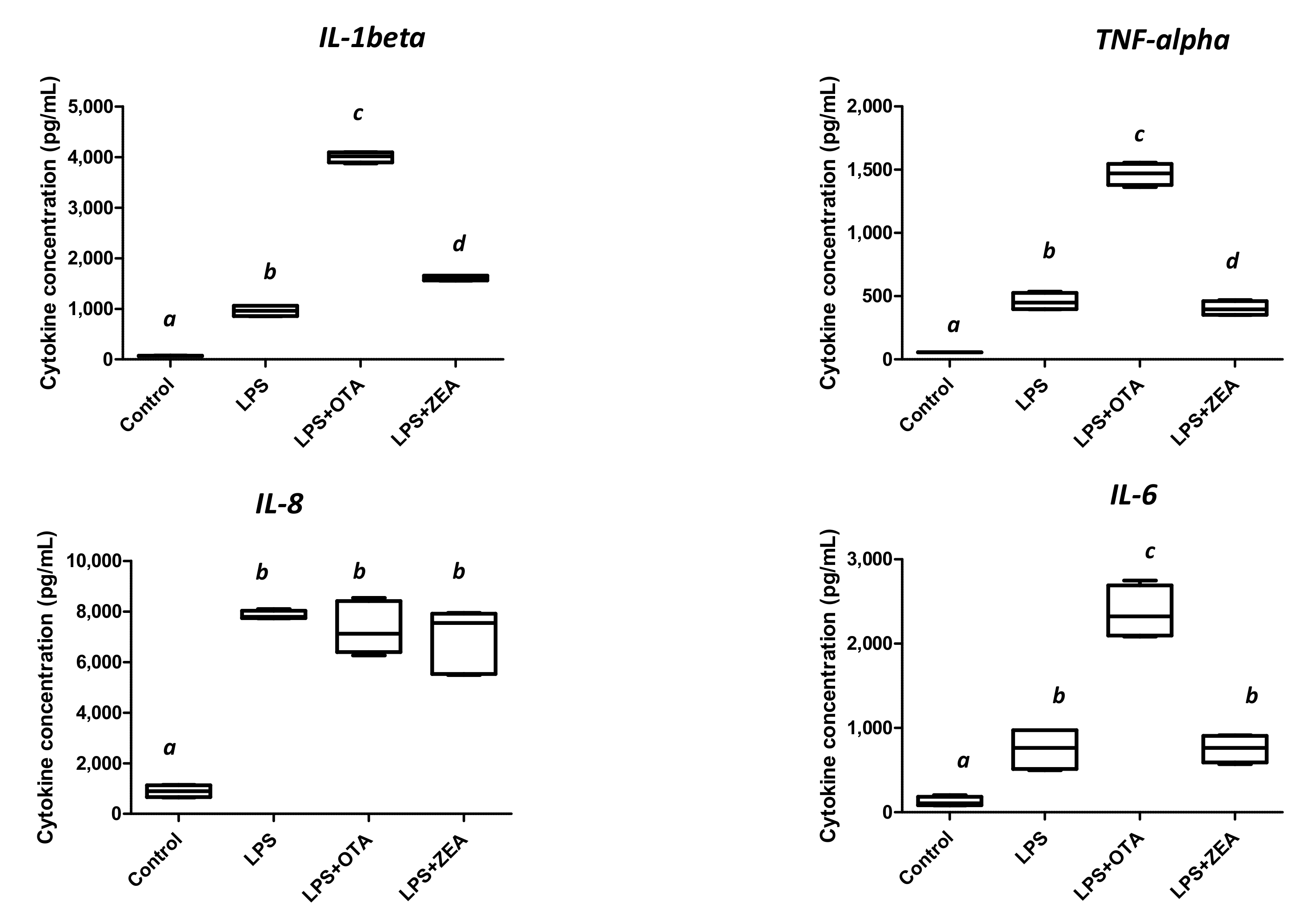
3.4. The Effect of Co-Exposure to LPS and Mycotoxins on Nitric Oxide and Inflammatory Cytokine Synthesis

We next examined the ability of LPS and LPS in combination with mycotoxin to modulate the synthesis of nitric oxide and of cytokines involved in the inflammatory response. While no effect on nitric oxide synthesis was observed for all of the treatments (Figure 5),
LPS challenge induced a significant increase in inflammatory cytokines IL-1β (13.9 times, p< 0.0001), TNF-α β (8.2 times, p < 0.0001), IL-8 β (8.7 times, p < 0.0001), and IL-6 (6.06 times, p = 0.003), compared with the untreated cells (Figure 6).
The exposure to both LPS and OTA induced the highest stimulation of inflammatory cytokines: IL-1β (57.9 times increase compared with control and 4.1 times increase compared with LPS), TNF-α (26.3 times increase compared with control and 3.2 times increase compared with LPS), IL-6 (19.1 times increase compared with control and 3.1 times increase compared with LPS). This suggested that the pro-inflammatory effect of LPS was augmented by the presence of OTA. When LPS+ZEA treatment was administered to PBMCs, only the IL-1β synthesis was increased, compared with LPS (1.67 times increase, p < 0.0001) while for TNF-α and IL-6, there were no differences between LPS and LPS+ZEA treatments. Similarly, the IL-8 synthesis in the cells treated with both LPS+OTA or LPS+ZEA was not different when compared with control (p > 0.05).

4. Discussion

Recent studies have shown that co-contamination of food/feed with bacteria and fungi is the rule rather than an exception, impacting all segments of society, from farmers and feed producers to the general public, government, industry, and academia [34] Food/feed may represent a carrier for harmful bacteria, viruses, parasites, or chemical substances that are responsible for different ranges of diseases, from diarrhoea to cancers [35]. Simultaneous exposure to more than one food/feed contaminant can produce a diverse range of interactions, such as antagonistic, synergistic, commensal, or symbiotic, that are different than the effect of single contaminant [22]. In this context, we investigated contamination with a mycotoxin arising at the same time as a bacterial contamination and whether the effects of the co-contamination were more important than the individual effect of bacterial contamination.
First, we investigated the effect of exposure to LPS or the co-exposure to both LPS and mycotoxins (OTA or ZEA) on cell proliferation, apoptosis, and necrosis. Our results showed that although exposure to LPS was not able to decrease cell viability, the addition of OTA significantly decrease cell proliferation. Many other studies have shown that OTA inhibits cell proliferation in different cell lines, such as human embryonic stem cells [36], neuronal cells [37], and kidney epithelial cells [38], while the cytotoxic effect of ZEA is still controversial, with the toxin decreasing or increasing cell proliferation depending on the cell type, the time of exposure, or the concentration [10,39].
Studies have shown that LPS induces apoptosis in cells of different origin, including immune cells. This process is involved in multiorgan failure during septic shock [40]. Apoptosis is also one of the most important processes involved in cytotoxicity induced by mycotoxins. OTA or ZEA exposure led to apoptosis through the activation of caspase-9 and caspase-3 and a decrease in Bcl2 protein accompanied by a loss of mitochondria membrane potential [37,41,42,43]. In our study, LPS alone or in combination with OTA significantly increased the percentage of cells in late apoptosis. Only the exposure to the combination of LPS and OTA significantly increased the percentage of dead cells, indicating a higher toxicity of the exposure to both bacteria and mycotoxin, compared with both untreated or LPS-challenged cells.
Food/feed contaminants, such as bacteria or mycotoxins, can affect the cell cycle by interacting with the events through which a cell duplicates its genome, grows, and divides [44,45]. In our study, exposure to LPS endotoxin was associated with a decrease in the number of S-phase cells. Other studies have shown that, in contrast to other mitogens, such as concanavalin A, which induces a high stimulation of the cells, LPS stimulation of PBMCs can decrease the percentages of S-phase cells in unstimulated cells [45]. We also showed that the combination of both mycotoxin and LPS induced a significant decrease in the S phase of the cell cycle, inducing an arrest of cell proliferation and progression in the G2/M phase. Indeed, OTA was also described as responsible for cell cycle arrest in human PBMCs, inducing an increase in the proportion of cells in the G1 phase, a decrease in the S phase, and a decrease in the expression of proteins involved in cell-cycle control, such as cyclin D1 and cyclin-dependent kinase [46]. Our results showed a correlation between cell-cycle arrest and apoptosis induced by the exposure to both bacteria and OTA, resulting in higher toxicity than the single exposure to bacterial endotoxin.
An important role in cell-cycle arrest and apoptosis is attributed to the excessive formation of free radicals that induce the oxidation of macromolecular structures, such as polyunsaturated lipids, proteins, and DNA, modifying their biological functions and causing oxidative stress and cellular damage [47]. In a study using human PBMCs, Liu et al. established a correlation between the oxidative DNA damage induced by OTA and the induction of cell-cycle-phase arrest and apoptosis [46]. Exposure to mycotoxins in general and to OTA and ZEA, in particular, has been found to induce an increase in ROS levels and oxidative damage in a large diversity of cell lines [11,48]. In addition, increasing ROS production as a response to LPS challenge helps cells destroy bacteria by oxidative-induced damage [49]. In our study, the ROS concentration tended to increase in cells exposed only to LPS or to LPS+OTA and significantly increased after LPS+ZEA treatment, compared with both control and LPS. The increase in ROS production significantly decreased the total antioxidant status and the catalase activity in the PBMCs and tended to decrease the SOD activity in cells exposed to the LPS+OTA and LPS+ZEA mixtures. Except for ROS production, it appears that the effect of co-exposure to the mycotoxin and LPS combination had no effect on PBMC antioxidant defence, compared with the exposure to bacterial endotoxin, and that in the particular case of oxidative stress there were no additive or synergic effects of the simultaneous exposure to bacteria and mycotoxins.
Inflammation represents a complex response of an organism to harmful stimuli, such as infections, injuries, and toxins, involving immune cells, blood vessels, and molecular mediators [50]. After TLR4 activation of human PBMCs, LPS can cause an acute inflammatory response by triggering the release of a vast number of inflammatory cytokines, leading to the activation of the NF-κB pathway [51]. Our results showed a substantial increase in pro-inflammatory cytokines IL-1β, TNF-α, and IL-6, and chemokine IL-8 after LPS exposure. Moreover, the exposure to both OTA and bacterial endotoxin led to an extraordinary increase in pro-inflammatory cytokines, compared with LPS-treated cells, but not for the IL-8 chemokine. While OTA induced pro-inflammatory effects in target organs (kidney, liver, gut, etc.) leading to toxicity and cell death [9], the effect of ZEA on inflammation is still controversial [10]. Indeed, our results showed that, except for IL-1β, the exposure to ZEA in addition to LPS did not induce a pro-inflammatory effect higher than that obtained after bacterial endotoxin challenge. Moreover, the concentration of TNF-α significantly decreased after LPS+ZEA treatment, compared with LPS. In a previous study, we demonstrated that the exposure of porcine PBMCs to 10 μM ZEA significantly decreased the TNF-α synthesis in the supernatant, but were not able to affect the IL-8 synthesis [23]. Considering this, we can assume that ZEA can interfere with the LPS pro-inflammatory response, with different effects on inflammation: an antagonist effect for TNF-α, no effect for IL-6 and IL-8, and an additive/synergic effect for IL-1 beta. These effects are in contrast with the pro-inflammatory, additive/synergic effect of OTA for both pro-inflammatory cytokines and chemokine. TNF-α is a major pro-inflammatory mediator but also a critical regulator of immune responses, cell proliferation, and survival [52]. Disruption of signalling events stimulated by TNF-α can trigger a switch from inflammatory gene signalling to cell death via apoptosis or necroptosis, via sequential formation of different signalling complexes upon the binding of TNF-α to its receptor, TNF-RI [53]. In addition, LPS-induced apoptosis results mainly through the autocrine secretion of TNF-α [54]. Thus, the important augmentation in the percentage of cells in late apoptosis and necrosis observed after the exposure to LPS and OTA can be due to the exceptional increase in TNF-α concentration.

5. Conclusions

Exposure to both bacteria (LPS) and fungal (mycotoxins) toxins is a common trait in toxin contamination of feed and food. Our results showed important additive/synergistic effects of the co-exposure to fungal and bacterial toxins on cell viability, the cell cycle, oxidative stress, and inflammation. These effects were more important when the cells were co-exposed to LPS and OTA, showing an exacerbation of inflammation accompanied by a significant increase in the percentage of cells undergoing apoptosis and necrosis, compared with the simple bacterial challenge. In the case of the co-exposure to ZEA+LPS. the effects were less important and concerned mainly the parameters related to the oxidative stress. The present study provides important data for risk assessment, as the concomitant contamination with bacteria and mycotoxins can lead to a higher toxicity than that which resulted after an infection with Gram-negative bacteria. Our results suggest that the synergistic effect of LPS and mycotoxins must be considered during risk assessment studies. In the near future, a reconsideration of the guidance levels for contaminants is required for the reduction of the health risk associated with the possible ingestion of food or feed co-contaminated with both bacteria and mycotoxins.

Author Contributions

Conceptualization, D.E.M. and I.T.; flow cytometry analysis, G.C.P.; analyses of oxidative stress and cytokines assessment, C.V.P.; data analysis and statistics, D.E.M.; writing—original draft preparation, D.E.M.; writing—review and editing; funding acquisition, D.E.M. All authors have read and agreed to the published version of the manuscript.

Funding

This work was financed through the projects PCE 42/2022 and 8PFE/2021, financed by Romanian Ministry of Research, Innovation, and Digitalization.

Institutional Review Board Statement

The present study did not involve humans or animals.

Data Availability Statement

The datasets used and analyzed during the current study are available from the corresponding author on reasonable request.

Conflicts of Interest

The authors declare no conflict of interest.

References

  1. Lorenzo, J.M.; Munekata, P.; Domínguez, R.; Pateiro, M.; Saraiva, J.; Franco Ruiz, D. Chapter 3—Main Groups of Microorganisms of Relevance for Food Safety and Stability: General Aspects and Overall Description. In Innovative Technologies for Food Preservation: Inactivation of Spoilage and Pathogenic Microorganisms; Academic Press: Cambridge, MA, USA, 2018; pp. 53–107. [Google Scholar]
  2. Alegbeleye, O.; Odeyemi, O.A.; Strateva, M.; Stratev, D. Microbial spoilage of vegetables, fruits and cereals. Appl. Food Res. 2022, 2, 100122. [Google Scholar] [CrossRef]
  3. Marroquín-Cardona, A.G.; Johnson, N.M.; Phillips, T.D.; Hayes, A.W. Mycotoxins in a changing global environment—A review. Food Chem. Toxicol. 2014, 69, 220–230. [Google Scholar] [CrossRef] [PubMed]
  4. Awuchi, C.G.; Ondari, E.N.; Ogbonna, C.U.; Upadhyay, A.K.; Baran, K.; Okpala, C.O.R.; Korzeniowska, M.; Guiné, R.P.F. Mycotoxins affecting animals, foods, humans, and plants: Types, occurrence, toxicities, action mechanisms, prevention, and detoxification strategies—A revisit. Foods 2021, 10, 1279. [Google Scholar] [CrossRef] [PubMed]
  5. El Khoury, A.; Atoui, A. Ochratoxin A: General overview and actual molecular status. Toxins 2010, 2, 461–493. [Google Scholar] [CrossRef] [Green Version]
  6. Khoi, C.S.; Chen, J.H.; Lin, T.Y.; Chiang, C.K.; Hung, K.Y. Ochratoxin a-induced nephrotoxicity: Up-to-date evidence. Int. J. Mol. Sci. 2021, 22, 11237. [Google Scholar] [CrossRef]
  7. Gaffoor, I.; Trail, F. Characterization of two polyketide synthase genes involved in zearalenone biosynthesis in Gibberella zeae. Appl. Environ. Microbiol. 2006, 72, 1793–1799. [Google Scholar] [CrossRef] [Green Version]
  8. Minervini, F.; Dell’Aquila, M.E. Zearalenone and reproductive function in farm animals. Int. J. Mol. Sci. 2008, 9, 2570–2584. [Google Scholar] [CrossRef] [Green Version]
  9. Marin, D.E.; Taranu, I. Ochratoxin a and its effects on immunity. Toxin Rev. 2015, 34, 11–20. [Google Scholar] [CrossRef]
  10. Bulgaru, C.V.; Marin, D.E.; Pistol, G.C.; Taranu, I. Zearalenone and the immune response. Toxins 2021, 13, 248. [Google Scholar] [CrossRef]
  11. Tao, Y.; Xie, S.; Xu, F.; Liu, A.; Wang, Y.; Chen, D.; Pan, Y.; Huang, L.; Peng, D.; Wang, X.; et al. Ochratoxin A: Toxicity, oxidative stress and metabolism. Food Chem. Toxicol. 2018, 112, 320–331. [Google Scholar] [CrossRef]
  12. Qin, X.; Cao, M.; Lai, F.; Yang, F.; Ge, W.; Zhang, X.; Cheng, S.; Sun, X.; Qin, G.; Shen, W.; et al. Oxidative stress induced by zearalenone in porcine granulosa cells and its rescue by curcumin in vitro. PLoS ONE 2015, 10, e0127551. [Google Scholar] [CrossRef] [PubMed] [Green Version]
  13. Crump, J.A.; Griffin, P.M.; Angulo, F.J. Bacterial contamination of animal feed and its relationship to human foodborne illness. Clin. Infect. Dis. 2002, 35, 859–865. [Google Scholar] [CrossRef] [PubMed] [Green Version]
  14. Yeleliere, E.; Cobbina, S.J.; Abubakari, Z.I. Review of microbial food contamination and food hygiene in selected capital cities of Ghana. Cogent Food Agric. 2017, 3, 1395102. [Google Scholar] [CrossRef]
  15. Maciorowski, K.G.; Herrera, P.; Jones, F.T.; Pillai, S.; Ricke, S. Effects on poultry and livestock of feed contamination with bacteria and fungi. Anim. Feed Sci. Technol. 2007, 133, 109–136. [Google Scholar] [CrossRef]
  16. Zhang, G.; Meredith, T.C.; Kahne, D. On the essentiality of lipopolysaccharide to gram-negative bacteria. Curr. Opin. Microbiol. 2013, 16, 779–785. [Google Scholar] [CrossRef] [PubMed] [Green Version]
  17. Rossol, M.; Heine, H.; Meusch, U.; Quandt, D.; Klein, C.; Sweet, M.J.; Hauschildt, S. Lps-induced cytokine production in human monocytes and macrophages. Crit. Rev. Immunol. 2011, 31, 379–446. [Google Scholar] [CrossRef] [PubMed]
  18. Abou Elazab, M.F.; Nasr, N.E.; Ahmed, M.S.; Alrashdi, B.M.; Dahran, N.; Alblihed, M.A.; Elmahallawy, E.K. The effects of bacterial lipopolysaccharide (lps) on turkey poults: Assessment of biochemical parameters and histopathological changes. Vet. Sci. 2022, 9, 240. [Google Scholar] [CrossRef] [PubMed]
  19. Wallace, R.J.; Gropp, J.; Dierick, N.; Costa, L.G.; Martelli, G.; Brantom, P.G.; Bampidis, V.; Renshaw, D.W.; Leng, L. Risks associated with endotoxins in feed additives produced by fermentation. Environ. Health 2016, 15, 5. [Google Scholar] [CrossRef] [Green Version]
  20. Luchi, M.; Morrison, D.C. Comparable endotoxic properties of lipopolysaccharides are manifest in diverse clinical isolates of gram-negative bacteria. Infect. Immun. 2000, 68, 1899–1904. [Google Scholar] [CrossRef] [Green Version]
  21. Svensson, M.; Han, L.; Silfversparre, G.; Häggström, L.; Enfors, S.O. Control of endotoxin release in Escherichia coli fed-batch cultures. Bioprocess Biosyst. Eng. 2005, 27, 91–97. [Google Scholar] [CrossRef]
  22. Alassane-Kpembi, I.; Schatzmayr, G.; Taranu, I.; Marin, D.; Puel, O.; Oswald, I.P. Mycotoxins co-contamination: Methodological aspects and biological relevance of combined toxicity studies. Crit. Rev. Food Sci. Nutr. 2017, 57, 3489–3507. [Google Scholar] [CrossRef] [PubMed]
  23. Marin, D.E.; Pistol, G.C.; Bulgaru, C.V.; Taranu, I. Cytotoxic and inflammatory effects of individual and combined exposure of hepg2 cells to zearalenone and its metabolites. Naunyn-Schmiedeberg’s Arch. Pharmacol. 2019, 392, 937–947. [Google Scholar] [CrossRef] [PubMed]
  24. Moore, M.D.; Jaykus, L.A. Virus–bacteria interactions: Implications and potential for the applied and agricultural sciences. Viruses 2018, 10, 61. [Google Scholar] [CrossRef] [PubMed] [Green Version]
  25. Kew, M.C. Synergistic interaction between aflatoxin b1 and hepatitis b virus in hepatocarcinogenesis. Liver Int. 2003, 23, 405–409. [Google Scholar] [CrossRef]
  26. Granados-Chinchilla, F.; Valenzuela-Martínez, C.; García-Murillo, B.; Aguilar-Madrigal, D.; Redondo-Solano, M.; Molina, A. Microbiological safety and presence of major mycotoxins in animal feed for laboratory animals in a developing country: The case of Costa Rica. Animals 2021, 11, 2389. [Google Scholar] [CrossRef] [PubMed]
  27. Yunus, A.W.; Sulyok, M.; Böhm, J. Mycotoxin cocktail in the samples of oilseed cake from early maturing cotton varieties associated with cattle feeding problems. Toxins 2015, 7, 2188–2197. [Google Scholar] [CrossRef] [Green Version]
  28. Cegielska-Radziejewska, R.; Stuper-Szablewska, K.; Szablewski, T. Microflora and mycotoxin contamination in poultry feed mixtures from western Poland. Ann. Agric. Environ. Med. AAEM 2013, 20, 30–35. [Google Scholar]
  29. Gagiu, V.; Avram, M.; Iorga, E.; Belc, N.; Diaconu, M.; Diaconu, A.; Pricop, M. Microbial and mycotoxin contamination of maize during storage. Rom. Biotechnol. Lett. 2007, 12, 3389–3394. [Google Scholar]
  30. Taranu, I.; Marin, D.; Pistol, G.C.; Untea, A.; Vlassa, M.; Filip, M.; Gras, M.; Rotar, C.; Anghel, A.C. Assessment of the ability of dietary yeast-fermented rapeseed meal to modulate inflammatory and oxidative stress in piglets after weaning. J. Anim. Feed Sci. 2022, 31, 109–122. [Google Scholar] [CrossRef]
  31. Marin, D.E.; Taranu, I.; Burlacu, R.; Manda, G.; Motiu, M.; Neagoe, I.; Dragomir, C.; Stancu, M.; Calin, L. Effects of zearalenone and its derivatives on porcine immune response. Toxicol. Vitr. 2011, 25, 1981–1988. [Google Scholar] [CrossRef]
  32. Marin, D.E.; Pistol, G.C. Effect of sea buckthorn meal extract in alleviating the toxic effect of ochratoxin a and zearalenone in porcine peripheral blood mononuclear cells. Arch. Zootech. 2021, 24, 84–92. [Google Scholar] [CrossRef]
  33. Marin, D.E.; Bulgaru, C.V.; Anghel, C.A.; Pistol, G.C.; Dore, M.I.; Palade, M.L.; Taranu, I. Grape seed waste counteracts aflatoxin b1 toxicity in piglet mesenteric lymph nodes. Toxins 2020, 12, 800. [Google Scholar] [CrossRef] [PubMed]
  34. Abdolshahi, A.; Behdad Shokrollahi, Y. Chapter 2—Food contamination. In Mycotoxins and Food Safety; Suna, S., Ed.; IntechOpen: Rijeka, Croatia, 2020. [Google Scholar]
  35. Todd, E.C.D. Foodborne Diseases: Overview of Biological Hazards and Foodborne Diseases. In Encyclopedia of Food Safety; Academic Press: Cambridge, MA, USA, 2014; pp. 221–242. [Google Scholar] [CrossRef]
  36. Erceg, S.; Mateo, E.M.; Zipancic, I.; Rodríguez Jiménez, F.J.; Pérez Aragó, M.A.; Jiménez, M.; Soria, J.M.; Garcia-Esparza, M. Assessment of toxic effects of ochratoxin a in human embryonic stem cells. Toxins 2019, 11, 217. [Google Scholar] [CrossRef] [PubMed] [Green Version]
  37. Zhang, X.; Boesch-Saadatmandi, C.; Lou, Y.; Wolffram, S.; Huebbe, P.; Rimbach, G. Ochratoxin a induces apoptosis in neuronal cells. Genes Nutr. 2009, 4, 41–48. [Google Scholar] [CrossRef] [Green Version]
  38. Qian, G.; Liu, D.; Hu, J.; Gan, F.; Hou, L.; Chen, X.; Huang, K. Ochratoxin a-induced autophagy in vitro and in vivo promotes porcine circovirus type 2 replication. Cell Death Dis. 2017, 8, e2909. [Google Scholar] [CrossRef] [Green Version]
  39. Zheng, W.; Wang, B.; Li, X.; Wang, T.; Zou, H.; Gu, J.; Yuan, Y.; Liu, X.; Bai, J.; Bian, J.; et al. Zearalenone promotes cell proliferation or causes cell death? Toxins 2018, 10, 184. [Google Scholar] [CrossRef] [Green Version]
  40. Munshi, N.; Fernandis, A.Z.; Cherla, R.P.; Park, I.-W.; Ganju, R.K. Lipopolysaccharide-induced apoptosis of endothelial cells and its inhibition by vascular endothelial growth factor. J. Immunol. 2002, 168, 5860–5866. [Google Scholar] [CrossRef] [Green Version]
  41. Assaf, H.; Azouri, H.; Pallardy, M. Ochratoxin a induces apoptosis in human lymphocytes through down regulation of bcl-xl. Toxicol. Sci. 2004, 79, 335–344. [Google Scholar] [CrossRef] [Green Version]
  42. Lee, H.J.; Oh, S.Y.; Jo, I. Zearalenone induces endothelial cell apoptosis through activation of a cytosolic ca(2+)/erk1/2/p53/caspase 3 signaling pathway. Toxins 2021, 13, 187. [Google Scholar] [CrossRef]
  43. Zhu, Y.; Wang, H.; Wang, J.; Han, S.; Zhang, Y.; Ma, M.; Zhu, Q.; Zhang, K.; Yin, H. Zearalenone induces apoptosis and cytoprotective autophagy in chicken granulosa cells by pi3k-akt-mtor and mapk signaling pathways. Toxins 2021, 13, 199. [Google Scholar] [CrossRef]
  44. Cimbalo, A.; Alonso Garrido, M.; Font, G.; Manyes, L. Toxicity of mycotoxins in vivo on vertebrate organisms: A review. Food Chem. Toxicol. 2020, 137, 111161. [Google Scholar] [CrossRef] [PubMed]
  45. Amadori, M.; Filipe, J.; Riva, F.; Vitali, A.; Ruggeri, J.; Lacetera, N. Characterization of the blastogenic response to lps of bovine peripheral blood mononuclear cells. PLoS ONE 2018, 13, e0204827. [Google Scholar] [CrossRef] [PubMed] [Green Version]
  46. Liu, J.; Wang, Y.; Cui, J.; Xing, L.; Shen, H.; Wu, S.; Lian, H.; Wang, J.; Yan, X.; Zhang, X. Ochratoxin a induces oxidative DNA damage and g1 phase arrest in human peripheral blood mononuclear cells in vitro. Toxicol. Lett. 2012, 211, 164–171. [Google Scholar] [CrossRef] [PubMed]
  47. Jacobson, M.D. Reactive oxygen species and programmed cell death. Trends Biochem. Sci. 1996, 21, 83–86. [Google Scholar] [CrossRef]
  48. Feng, Y.-Q.; Zhao, A.-H.; Wang, J.-J.; Tian, Y.; Yan, Z.-H.; Dri, M.; Shen, W.; de Felici, M.; Li, L. Oxidative stress as a plausible mechanism for zearalenone to induce genome toxicity. Gene 2022, 829, 146511. [Google Scholar] [CrossRef]
  49. Bromfield, J.J.; Iacovides, S.M. Evaluating lipopolysaccharide-induced oxidative stress in bovine granulosa cells. J. Assist. Reprod. Genet. 2017, 34, 1619–1626. [Google Scholar] [CrossRef]
  50. Chen, L.; Deng, H.; Cui, H.; Fang, J.; Zuo, Z.; Deng, J.; Li, Y.; Wang, X.; Zhao, L. Inflammatory responses and inflammation-associated diseases in organs. Oncotarget 2018, 9, 7204–7218. [Google Scholar] [CrossRef] [Green Version]
  51. Ngkelo, A.; Meja, K.; Yeadon, M.; Adcock, I.; Kirkham, P.A. Lps induced inflammatory responses in human peripheral blood mononuclear cells is mediated through nox4 and giα dependent pi-3kinase signalling. J. Inflamm. 2012, 9, 1. [Google Scholar] [CrossRef] [Green Version]
  52. Kalliolias, G.D.; Ivashkiv, L.B. Tnf biology, pathogenic mechanisms and emerging therapeutic strategies. Nat. Rev. Rheumatol. 2016, 12, 49–62. [Google Scholar] [CrossRef]
  53. Xu, Q.; Choksi, S.; Liu, Z. Switching from tnf-induced inflammation to death signaling. Mol. Cell. Oncol. 2018, 5, e1392402. [Google Scholar] [CrossRef]
  54. Xaus, J.; Comalada, M.; Valledor, A.F.; Lloberas, J.; López-Soriano, F.; Argilés, J.M.; Bogdan, C.; Celada, A. Lps induces apoptosis in macrophages mostly through the autocrine production of tnf-alpha. Blood 2000, 95, 3823–3831. [Google Scholar] [CrossRef] [PubMed]
Figure 1. Effect of LPS or of the combination of LPS and mycotoxins on cell viability, apoptosis, and cell death. PBMCs were treated for 48 h with LPS, OTA, or ZEA and an apoptosis profile was performed using the Muse Annexin & Dead Cell Kit. The results are displayed as mean ± standard error (SE) for four independent experiments. a,b,c indicate significant differences between the different treatments (p < 0.05).
Agriculture 12 01970 g001
Figure 2. Effect of LPS or of the combination of LPS and mycotoxins on cell cycle. PBMCs were treated for 48 h with LPS, OTA, or ZEA and the percentage of cells in different phases of the cell cycle were assessed using the Muse Cell Cycle Kit. The results are displayed as mean ± standard error (SE) for four independent experiments. a,b,c indicate significant differences between different treatments (p < 0.05).
Agriculture 12 01970 g002
Figure 3. Effect of LPS or of the combination of LPS and mycotoxins on ROS synthesis. PBMCs were treated for 48 h with LPS, OTA, or ZEA and the percentage of (+) ROS cells were assessed using the Muse Oxidative Stress Kit. The results are displayed as mean ± standard error (SE) for four independent experiments. a,b indicate significant differences between different treatments (p < 0.05).
Agriculture 12 01970 g003
Figure 4. Effect of LPS or of the combination of LPS and mycotoxins on different parameters of the oxidative stress. Total antioxidant status (TAC), catalase (CAT), and superoxide dismutase activity (SOD) were assessed in PBMCs treated for 48 h with LPS, OTA, or ZEA. The results are displayed as mean ± standard error (SE) for four independent experiments. a,b,c indicate significant differences between different treatments (p < 0.05).
Agriculture 12 01970 g004
Figure 5. Effect of LPS or of the combination of LPS and mycotoxins on nitric oxide profile. PBMCs were treated for 48 h with LPS, OTA, or ZEA and total nitric oxide (+) cells were measured using the Muse Nitric Oxide Kit (Merck). The results are displayed as mean ± standard error (SE) for four independent experiments.
Agriculture 12 01970 g005
Figure 6. Effect of LPS or of the combination of LPS and mycotoxins on cytokine synthesis. The synthesis of proinflammatory cytokines (IL-1β, TNF-α, IL-6) and of IL-8 chemokine were assessed in PBMCs treated for 48 h with LPS, OTA, or ZEA.The results are displayed as mean ± standard error (SE) for four independent experiments. a,b,c,d indicate significant differences between different treatments (p < 0.05).
Agriculture 12 01970 g006
Publisher’s Note: MDPI stays neutral with regard to jurisdictional claims in published maps and institutional affiliations.

