Article

Effective Applications of Trichoderma spp. as Biofertilizers and Biocontrol Agents Mitigate Tomato Fusarium Wilt Disease

by
Eman F. A. Awad-Allah
1,*,
Amany H. M. Shams
2,
Amira A. Helaly
3 and
Entsar I. M. Ragheb
3
1
Soil and Water Sciences Department, Faculty of Agriculture, Alexandria University, Alexandria 21545, Egypt
2
Plant Pathology Department, Faculty of Agriculture, Alexandria University, Alexandria 21545, Egypt
3
Vegetable Crops Department, Faculty of Agriculture, Alexandria University, Alexandria 21545, Egypt
*
Author to whom correspondence should be addressed.
Agriculture 2022, 12(11), 1950; https://doi.org/10.3390/agriculture12111950
Submission received: 20 October 2022 / Revised: 10 November 2022 / Accepted: 16 November 2022 / Published: 19 November 2022
(This article belongs to the Section Crop Protection, Diseases, Pests and Weeds)

Abstract

:
Eco-friendly and sustainable plant disease management employing Trichoderma spp. as bioagents is an economically feasible and ecologically sustainable approach. Therefore, their use in agriculture should be encouraged. The two main goals of the present study were to evaluate the abilities of two Trichoderma isolates to prevent Fusarium wilt disease, which is caused by Fusarium solani, in vitro and under greenhouse conditions, as well as their potential as biofertilizers to enhance cherry tomato growth and development. The results of a dual culture test revealed that T. viride and T. harzianum are antagonistic against the F. solani pathogen. The antagonism mechanisms include competition for nutrients and space, mycoparasitism, and antibiosis, according to scanning electron microscopy (SEM) findings. Additionally, T. harzianum reduced the mycelial growth of F. solani by 78.0%, whereas T. viride inhibited the growth by 61.2%, 10 days post-inoculation. In a greenhouse experiment, cherry tomato plants treated with each of these antagonistic Trichoderma isolates separately or in combination significantly suppressed Fusarium wilt disease, improved plant growth parameters, increased macro- and micronutrients uptake, and increased the content of photosynthetic pigments and total phenols. In conclusion, effective applications of Trichoderma isolates have the potential to mitigate Fusarium wilt disease, which is caused by F. solani in cherry tomato plants, while simultaneously promoting the growth and development of cherry tomatoes.

1. Introduction

Fusarium wilt is a destructive disease caused by fungal soil-borne pathogens such as Fusarium oxysporum or Fusarium solani that causes significant losses for many important vegetable and crop plants, including tomatoes, all over the world [1]. In Egypt, F. solani isolates were the most common soil-borne pathogenic fungi isolated from tomato plants in different governorates, causing damping-off and root rot diseases [2]. Early infections are characterized by stunted seedlings and yellowing of older leaves, followed by wilting that progresses up the stem until the foliage is killed and the stem decays, as well as brown vascular discoloration in stem tissue cross sections near the soil line [1]. In the last stages of the disease, the fungal pathogen generates white mycelia and plenteous macroconidia, and under adverse environmental conditions, chlamydospores develop from pathogen structures and conjoin into the soil [3]. Fusarium wilt disease is primarily produced by the spread of chlamydospores, which is the pathogen’s primary mode of life and is the least controllable aspect of Fusarium wilt infection and can survive for more than 10–15 years [4]. Fusarium wilt does not transfer from plant to plant within a season since there is no spore generation above ground in the field, but it can be propagated via contaminated infected seeds [3,4]. Unfortunately, soil-borne fungi are challenging to manage because the soil system is a complex system with several paths that occur in a short period of time [5,6]. Additionally, traditional disease control strategies, such as antifungal chemicals and crop rotation, are ineffective because spores may survive for many years and pesticide residues are hazardous to human health; hence, new and efficient control strategies are required [7]. Biological control of plant diseases looks to be the most promising approach to managing agricultural phytopathogens, as well as the most ecologically safe, cost-effective, and viable alternative to chemical control [8,9].
In agro-ecosystems, Trichoderma species are beneficial fungi from the Hypocreaceae family that improve soil health and crop development [10]. The majority of Trichoderma spp. investigated colonize the root surface or live as endophytes within root tissues [11]. However, some species can be isolated from plant aerial parts [11,12,13]. They have the ability to create mutualistic endophytic relationships with a wide range of plant species, increasing host growth in the face of biotic and abiotic stresses, and enhancing micro- and macronutrient uptake and use efficiency [11,12,14]. As a result, they should be encouraged and employed in the field as effective biofungicides and biofertilizers, decreasing the need for dangerous synthetic fungicides and fertilizers while simultaneously supporting eco-friendly and sustainable farming practices [15,16]. However, before they can be used in the field to prevent the spread of phytopathogens, their characteristics and mechanisms must be well understood. Furthermore, additional research is required to increase the efficacy and safety of these fungi. Therefore, the objectives of the current study were to (1) assess Trichoderma isolates’ ability to control Fusarium wilt disease, which is caused by Fusarium solani, in vitro and under greenhouse conditions, and (2) assess their potential biofertilizers’ effects on cherry tomato (Solanum lycopersicum L.) growth and development.

2. Materials and Methods

Assessment of the potential biocontrol of Trichoderma isolates against Fusarium wilt disease, caused by Fusarium solani, was performed in vitro and greenhouse conditions at the Plant Pathology Department, Faculty of Agriculture, Alexandria University, Egypt. Additionally, the evaluation of Trichoderma isolates on the growth promotion of cherry tomatoes was conducted in pot experiments.

2.1. Isolation of Pathogen and Antagonists

Cherry tomato plant samples cv. “Golden Cherry Hybrid” showing typical symptoms of Fusarium wilt disease were collected from a private farm in Borg El-Arab, Alexandria governorate, Egypt during the 2020 season as shown in Figure 1.
Different infected plant organs (wilted leaves and rotten roots) were chopped into small pieces (5 mm), their surface sterilized for 5 min in sodium hypochlorite (2%), rinsed three times with sterile distilled water for 5 min, and dried with a sterile filter paper, then transferred onto PDA plates (Potato-Dextrose Agar, HiMedia Laboratories MH096-500 G, India), and incubated at 25 ± 1 °C for five days. Preliminary identification of the fungal isolates was carried out by morphological and microscopic examinations according to Leslie and Summerell [17]. The isolates identified as F. solani based on appearance, shape, and size of macro- and micro-conidia, were used in all experiments. F. solani identity was confirmed by using the polymerase chain reaction (PCR) technique, and nucleotide sequences were determined according to the method described below.
As antagonistic fungi, Trichoderma isolates were isolated from soil rhizosphere samples collected from a healthy cherry tomato at a production farm in Borg El-Arab, Alexandria governorate, Egypt. For the isolation of antagonistic Trichoderma spp., the serial dilution plate technique was used, and soil samples were diluted into concentrations and vortexed. The supernatant was placed onto PDA plates with a 10 mg/mL stock solution of chloramphenicol (used to suppress bacterial growth in the medium) and incubated at 28 °C for 4 days before being transferred to a new plate containing PDA for another 5–7 days at 28 °C [18]. Trichoderma isolates were identified based on morphological characteristics and molecular typing employing the rDNA internal transcribed spacer (ITS), RNA polymerase II subunit (Rpb2), and translation elongation factor 1-α (Tef1-α) genes [19,20].

2.2. Molecular Identification of Pathogen and Antagonists

2.2.1. DNA Isolation and ITS Amplification of the Pathogen

The genomic DNA was isolated from fungal mycelium by the CTAB extraction method using standard protocols and the DNA concentration was adjusted to 50 ng [21]. The internal transcribed spacer (ITS) regions of rDNA were amplified using primers ITS1 and ITS4, PCR reactions containing a total volume of 25 μL containing 3 μL of template DNA, 12.5 μL PCR Green Master Mix (Thermo Scientific™), 0.5 μL of each forward and reverse primers (10 pmol) and 8.5 μL molecular grade water. Cycling was completed using a Techne Prime Thermal Cycler (Cole-Parmer, Staffordshire, UK) as follows: an initial denaturation at 95 °C for 1 min followed by 35 cycles at 94 °C for 30 s, 55 °C for 2 min, and 72 °C for 1 min and a final extension at 72 °C for 10 min. Amplified PCR products were electrophoresed on 1.5% agarose gel having 5 µL ethidium bromide, in 0.5X Tris Acetate buffer at 140 Volt for 15 min, then UV light was used for visualization of results, and bands were photographed [22].
The amplified fragment of ITS1-5.8 s and ITS2 region (600–700 bp) of selected isolates were sent for sequencing (lab Technology, Scientific Services Company). Identification of isolates was confirmed by applying Basic Local Alignment Search Tool (BLAST search) on National Center for Biotechnology Information (NCBI) site (http://www.ncbi.nlm.nih.gov (accessed on 29 July 2022)) using the obtained sequences of the amplified regions. Alignments were completed by using Molecular Evolutionary Genetics Analysis (MEGA X) software. Phylogenetic tree was constructed using neighbor-joining (NJ) method by MEGA X software [23]. The obtained sequences were compared with different international fungal strains obtained from GenBank.

2.2.2. PCR-Amplification of (Tef1-α) and (Rpb2) Genes of Trichoderma spp.

The PCR-amplification reactions were used to amplify the translation elongation factor 1-α (Tef1-α) and RNA polymerase II subunit (Rpb2) fragments. Details of the primer names and sequences are shown in Table 1. PCR was performed on a PCR machine, and consisted of 0.5 µL (10 μM) for each primer (forward and reverse primers), 12.5 µL Taq 2x ReadyMix (Enzynomics Inc., Daejeon, Korea), and 1 µL template DNA, with Milli-Q water added up to a total volume of 25 µL. Cycling was completed using Techne’s extensive range of Prime Thermal Cyclers (Cole-Parmer, UK), the PCR settings for amplification of the Tef1-α gene using specific primers were as follows: initial denaturation at 94 °C for 1 min, followed by 35 cycles each of 1 min at 94 °C, 1 min at 59 °C, 50 sec at 72 °C and, finally, 7 min at 72 °C [24]. The following protocol was used for the Rpb2 gene fragment: 3 min at 94 °C, 5 cycles each of 45 sec at 94 °C, 45 sec at 60 °C, and 2 min at 72 °C, followed by 5 cycles with the temperature decreasing by 1.0 °C per cycle from 58 °C to 54 °C, followed by 30 cycles at 54 °C, and finally, 10 min at 72 °C [25]. Trichoderma isolates were identified as Trichoderma viride (T1) and Trichoderma harzianum (T2).

2.3. Antagonistic Potential of Trichoderma Isolates against F. solani In Vitro

2.3.1. Dual Culture Assay

The in vitro antagonistic activity was checked for T. harzianum and T. viride isolates against F. solani using the dual-culture test, according to Nofal et al. [26]. For a week, Petri dishes (9 cm) containing PDA medium were inoculated with 0.5 cm diameter mycelial discs of three isolates individually. Then, on a PDA plate, 0.5 cm mycelial plugs of the biocontrol Trichoderma isolates and F. solani pathogen were put opposite each other, while F. solani was inoculated alone as a control treatment. For each treatment, three replicates were kept and incubated for 5 days at 27 ± 2 °C, after that the antagonistic ability was assessed. Mycelia growth of pathogen was recorded, and the percentage inhibition was calculated with respect to the control. The percentage of radial growth inhibition (% RGI) was calculated using the formula: % RGI = (C − T)/C × 100, where C = radial growth of the tested pathogen without the antagonist, and T = radial growth of the tested pathogen in dual culture with the antagonist [27].

2.3.2. Interaction Zones in Dual Cultures by Scanning Electron Microscope

A scanning electron microscope (SEM) was used to study the interaction zone between the F. solani hyphae and the biocontrol agents T. harzianum and T. viride using dual culture, according to Nofal et al. [28]. At the Electron Microscope Unit, Faculty of Science, Alexandria University, Alexandria, Egypt, samples were examined for mycoparasitism and photographed at different magnifications using the JSM-IT200 SEM series (JEOL Ltd., Tokyo, Japan).

2.4. Effect of Trichoderma Isolates to Manage Tomato Fusarium wilt Disease under Greenhouse Conditions

2.4.1. Inoculum of the Pathogen and Antagonists

All the fungal strains were individually cultured on PDA plates for 10 days at 25 °C. After incubation, sterile distilled water (SDW) was placed into the plates, and the conidia were scraped with a sterile glass rod, according to El-Komy et al. [29]. The spore suspensions were filtered to remove fungal hyphae, and the conidial suspension concentration was adjusted to 1 × 10⁷ conidia mL−1 SDW using a hemocytometer. The inoculum concentrations were adjusted to 103 conidia g−1 soil for F. solani and 106 conidia g−1 of soil for Trichoderma isolates.

2.4.2. Plant Materials, Experimental Conditions, and Design

A controlled greenhouse experiment was carried out to investigate the ability of T. viride (T1) and T. harzianum (T2), either alone or in mixture (TMix1+2), to suppress F. solani and boost cherry tomato growth. The compatibility of the two Trichoderma isolates was determined in vitro using the dual culture technique, with no inhibitory halos or overgrowth observed (data not shown).
Tomato “Golden Cherry” hybrid seeds were surface sterilized with 1% sodium hypochlorite (NaOCl) for 1 min, rinsed three times for 5 min in sterile distilled water, and sown in polystyrene germination trays with 200 cavities on September 15, 2021. Seedlings were cared for with regular irrigation and practices for tomato production in a greenhouse. During the experiment, the growth conditions were average temperature of 25 ± 2 °C; relative humidity of 77 ± 5%; and photoperiod of 14/10 h of light/dark. Plastic pots (20 cm inner diameter) were filled with a mixture of sterilized sandy loam–peat moss (2:1 v/v). The soil was artificially infested with F. solani by adding (103 conidia g−1 soil) conidial suspension and kept for 7 days with regular irrigation for pathogen establishment.
At the time of transplant, 35-day-old cherry tomato seedlings with three true leaves were treated with the Trichoderma by using the root-dip method. Control cherry tomato plants (T0) were root-dipped in sterile water. A factorial combination of eight experimental treatments was arranged in a randomized complete block split-plot design (with and/or without F. solani infections as the main factor and four Trichoderma inoculations as sub-factor) with five replicates for a total of 40 experimental plots. Cherry tomato roots were dipped in 1 × 10⁷ conidia mL−1 for 15 min with different Trichoderma treatments (T0, T1, T2, and TMix1+2) to allow the isolates to interact with the cherry tomato roots before being transplanted into soil pots (i.e., either with and/or without F. solani infested soil pots). The different Trichoderma treatments were repeated 15 and 30 days after transplanting (DAT), by watering each plant at the base with the spore suspension (106 conidia g−1 of soil). During the experiment, a surface drip irrigation system was employed in the greenhouse with a half-strength modified nutrient solution of Hoagland and Arnon as a base solution at a pH of 5.5~6.0, according to Hewitt [30]. Cherry tomato plants per treatment were grown for 45 DAT.

2.5. Measurements

2.5.1. Plant Disease Incidence (%)

Cherry tomato plants transplanted in soil previously infected with F. solani had clear disease symptoms, such as leaf yellowing and wilting, and then, plant disease incidence and severity were evaluated after 45 DAT.
The plant disease severity was recorded on a scale of 0–4 based on the degree of wilt as stated by Song et al. [31], where zero means no wilt symptoms, 1, 2, and 3 refer to varying degrees of wilt that reflect the scale of disease severity, and 4 means completely wilted plants as an indication of a complete infection. According to the following formula, plant disease incidence (%) is a parameter that incorporates both disease percentage and disease severity.
Disease   incidence   ( % ) = [ ( Σ   scale × number   of   plants   infected ) ( highest   scale   × total   number   of   plants ) ] × 100 .

2.5.2. Growth Parameters

Five cherry tomato plants were chosen randomly from each treatment at 45 DAT, for the fresh weights measurement of both roots and shoots. The plant materials were oven-dried at 65 °C for 48 h, and the dry weights were measured, and then ground in a stainless-steel mill and stored for analysis according to Chapman and Pratt [32]. The root-to-shoot ratio (R/S) was based on the ratio of root dry weight to shoot dry weight.

2.5.3. Leaves Chemical Composition

For mineral nutrient analysis, plant leaves were oven dried at 65 °C for 48 h, and N, P, K, Ca, Mg, Fe, Zn, and Cu contents were estimated. Total nitrogen was determined according to the method described by Jones Jr [33]. Total phosphorus was measured using the vanadate–molybdate method according to Page et al. [34]. Additionally, total potassium was measured according to the method described by Jones Jr [33]. In addition, calcium and magnesium contents were measured according to Chapman and Pratt [32]. Iron, zinc, and copper analysis were determined according to Jones Jr [33].

2.5.4. Total Phenolic Contents

Total phenolic contents in cherry tomato leaves were determined using the Folin–Ciocalteu reagent as reported by Malik and Singh [35]. One gram of leaf sample was ground in 80% ethanol, and the homogenate was centrifuged at 10,000 rpm for 30 min. The supernatant was kept, and the residue was re-extracted 5 times with the required volume of 80% ethanol, centrifuged, and the supernatants were collected together. The supernatant was evaporated nearly to dryness, and the residue was dissolved in 5 mL of distilled water. A 0.2 mL aliquot was pipetted into a clean test tube. The volume was made up to 3 mL with distilled water, and then 0.5 mL of Folin–Ciocalteu reagent was added. After 3 min, a 20% Na2CO3 solution was added to each tube, which was then thoroughly and immediately mixed. The tubes were then placed in boiling water for one min, cooled, and the absorbance at 650 nm was measured against a blank. The total phenolic content was determined, and the results were expressed as mg of gallic acid per gram of fresh weight (mg g−1 F.W.) using a spectrophotometric method.

2.5.5. Analysis of Photosynthetic Pigments

Photosynthetic pigments (chlorophyll a, b, and total carotenoids) were determined in fresh leaves by using a Shimadzu spectrophotometer (Model UV-1208) according to the methods outlined by Horowitz [36]. Concentrations of chlorophyll a, b, and total carotenoids were expressed as mg/g, of fresh weight (FW).

2.6. Statistical Analysis

Using CoStat computer software [37] (CoHort Software version 6.303, Monterey, CA, USA), experimental data were statistically analyzed by two-way analysis of variance according to Gomez and Gomez [38], and LSD at 0.05 level of significance was used for mean comparison.

3. Results

3.1. Identification of Pathogenic fungi F. solani and Biocontrol Agents Trichoderma Isolates

Three fungal species were isolated, one from infected plants identified as F. solani, and two Trichoderma isolates were isolated from soil samples collected from the rhizospheres’ area. The sequences of amplified ITS regions were submitted to GenBank and given accession numbers: OP106576, OP106577, and OP106578 of F. solani, T. harzianum, and T. viride, respectively. The sequence identification was made considering results obtained from the NCBI Blast search engine (BLAST-N), depending on ≥97% sequence identity to assign species identification.
The phylogenetic analysis of the ITS region for F. solani and other comparable GenBank isolates (data not shown) reveals a distinct clade with a bootstrap value of 91% between F. solani (OP106576) and the isolate of F. solani from tomatoes in Egypt (MT032385), while there was a low bootstrap value (78%) with F. oxysporum and Fusarium spp. isolates from India, Pakistan, and China.
Phylogenetic analysis of the T. harzianum, T. viride, and other Trichoderma reference isolates from GenBank (data not shown) indicates that clustering in one group of T. harzianum (OP106577) and (MW789612) from Egypt they share a high level of similarity in their genomes. Species T. viride (OP106578) was located within one clade with the isolates T. viride (MW646450 and MH333256) from Austria and India grouped in a distinct clade with a bootstrap value of 89%. In accordance with identification with the ITS region, we observed the PCR amplification of (Tef1-α) and (Rpb2) genes of Trichoderma spp. successfully amplified approximately 689 bp, and 1050 bp, respectively.

3.2. Antagonistic potential of Trichoderma Isolates against F. solani In Vitro

3.2.1. Dual Culture Assay

Through a dual culture test, the in vitro mycoparasitic activity of T. viride and T. harzianum against F. solani was demonstrated. The mycoparasitic nature of the Trichoderma isolates shows the growth over the F. solani pathogen, 10 days after inoculation at 27 ± 2 °C. Additionally, the results revealed that Trichoderma isolates were antagonistic to the F. solani pathogen that causes the fusarium wilt disease of cherry tomatoes, as shown in Figure 2.
Figure 3 shows the efficacy of Trichoderma isolates as biocontrol agents in significantly suppressing the mycelial growth of the F. solani pathogen. Through a dual culture assay, T. harzianum showed the maximum inhibitory action against the F. solani pathogen 5 and 10 days post-inoculation. T. harzianum reduced the mycelial growth of F. solani by 78.0%, whereas T. viride inhibited the growth by 61.2%, 10 days post-inoculation. Moreover, the T. harzianum isolate showed greater inhibition against the F. solani pathogen than the T. viride isolate.

3.2.2. Interaction Zone between F. Solani and Trichoderma by Scanning Electron Microscope

Scanning electron microscopy (SEM) images at different magnifications (500× and 1000×) show T. viride and T. harzianum mycoparasitism towards the pathogen F. solani (inoculated simultaneously). Figure 4A,B show higher growth of F. solani mycelium inoculated alone as control at different magnifications, and their mycelium growth is unchallenged by the biocontrol agents. Figure 4C–F shows overgrowth and heavy sporulation by both Trichoderma isolates on the colony of F. solani 10 days after inoculation at 27 ± 2 °C using dual culture assays on PDA. The mycoparasitic behavior of T. viride and T. harzianum was observed in the form of adhesion, coiling, penetration, degradation, and deformation of the F. solani mycelium, as shown in Figure 4. In addition to the parallel growth of these antagonist isolates that were closely linked to the pathogen, these two Trichoderma isolates were able to overgrow and sporulate on the colony of the F. solani pathogen. Based on SEM observations, the antagonism mechanisms of Trichoderma isolates towards the F. solani pathogen are competition for nutrients and space, mycoparasitism, and antibiosis.

3.3. Effect of Trichoderma Isolates to Manage Tomato Fusarium wilt Disease under Greenhouse Conditions

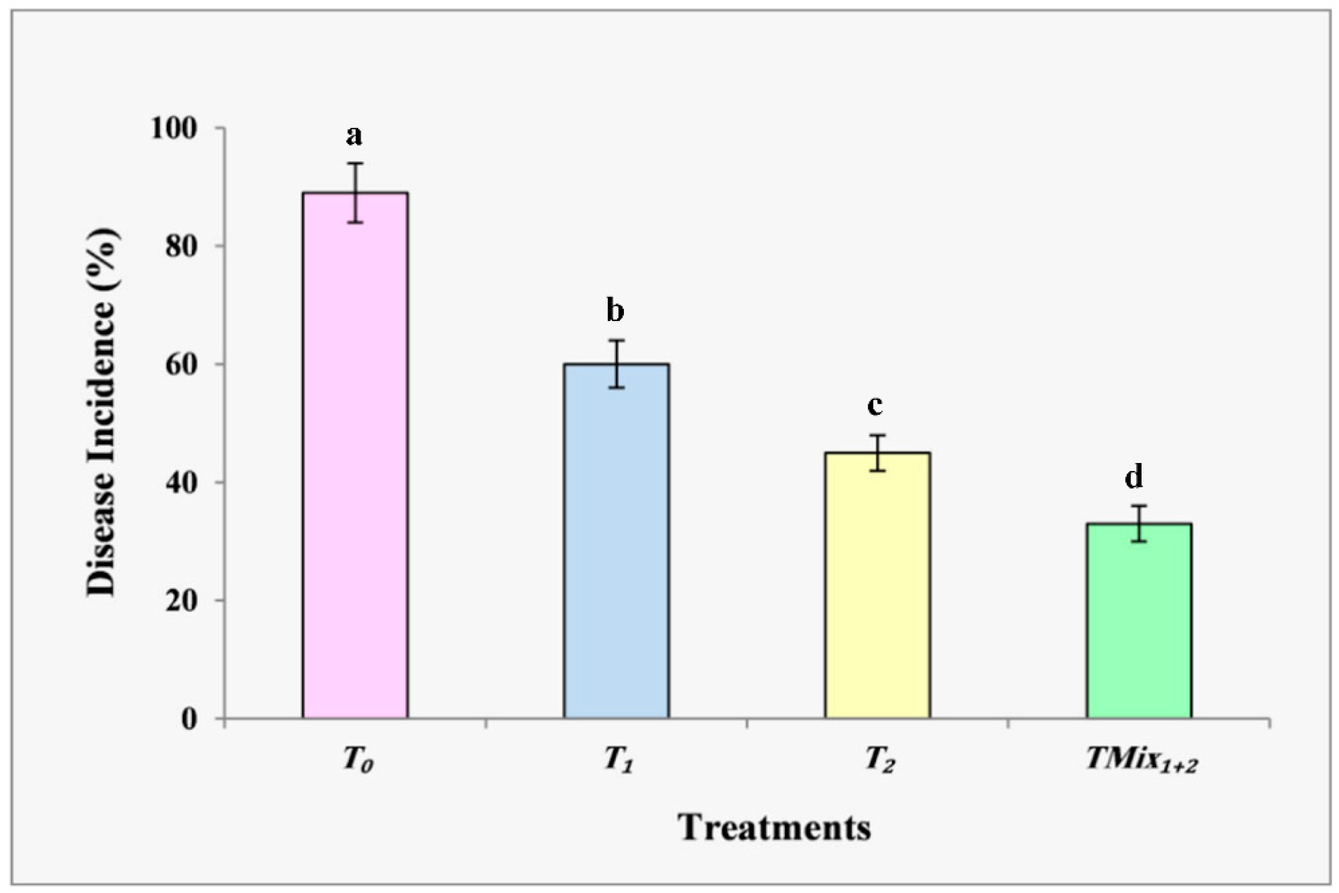
3.3.1. Plant Disease Incidence (%)

In this experiment, the biological control effects of the treatments on Fusarium wilt disease incidence (%) in cherry tomato plants grown in greenhouse conditions were demonstrated. According to Figure 5, cherry tomato plants with F. solani infection alone had the highest mean disease incidence value (95.2%). These infected plants displayed stunted seedlings, yellowing of older leaves, wilting, root rot, and a shorter main root, all of which might cause the plant serious harm or even death. The healthy control plants, on the other hand, showed no symptoms. In comparison to untreated plants grown on infected soil, plants treated with various Trichoderma treatments had less severe symptoms and slower development of the disease. Disease incidence (%) decreased significantly when T. viride (57.1%) and T. harzianum (42.3%) were incorporated into the pathogen-infested soil, indicating that the application of these biocontrol agents could minimize the occurrence of disease (Figure 5). The TMix1+2 treated plants, however, had the best control effectiveness of disease incidence (34%) overall. For both managing the fusarium wilt disease and promoting plant development in cherry tomato plants, it seems that a combination of the two Trichoderma isolates (TMix1+2) performs better than either isolate alone.

3.3.2. Cherry Tomato Growth Parameters

The fresh and dry weights, g plant−1, of cherry tomato plants growing in greenhouse conditions with and/or without F. solani-infested soil as affected by different Trichoderma isolates alone or in a combination are shown in Table 2. Cherry tomato plants treated with either T. harzianum alone (T2) or in combination (TMix1+2), displayed significant effects on plant fresh and dry weights as well as a better root-to-shoot ratio compared to non-infected controls, regardless of the presence or absence of the F. solani pathogen. It is obvious that a larger root system (better root-to-shoot ratio) is nearly always a guarantee of more stable performance across all species, crops, and cultivars. Under infection stress, treatment of a mixture of the two Trichoderma isolates (TMix1+2) showed the highest increase in plant fresh and dry weights as well as a better root-to-shoot ratio. The combination of these two biocontrol agents, T. viride and T. harzianum, seems to have synergistic effects.

3.3.3. Leaves Chemical Composition

Data in Table 3 shows the leaf chemical composition (%) of cherry tomato plants grown under greenhouse conditions with and/or without F. solani-infested soil and treated by different Trichoderma isolates alone or in a combination. It is obvious that the leaf chemical composition of cherry tomato plants, such as N, P, K, Mg, Ca, Fe, Zn, and Cu contents, was significantly affected by different Trichoderma isolates alone or in a mixture compared to non-infected controls. Regardless of the presence or absence of the F. solani infection, treatment of a mixture of the two Trichoderma isolates (TMix1+2) achieved the highest concentrations of N, P, K, Mg, Ca, Fe, Zn, and Cu in leaves of cherry tomato plants grown under greenhouse conditions.
The beneficial roles of Trichoderma isolates in cherry tomato nutrition, resistance, and development are graphically represented in Figure 6. Trichoderma isolates can stimulate cherry tomatoes' development while also protecting them from pathogenic attacks. Furthermore, different Trichoderma isolates can boost root growth and development, confer abiotic and biotic stress tolerance, and improve micro- and macronutrient absorption and utilization efficiency, resulting in higher crop productivity.

3.3.4. Total Phenolic Contents and Photosynthetic Pigments

Total phenolic content and photosynthetic pigments (chlorophyll a, b, and total carotenoids) of cherry tomato plants cultivated in greenhouse conditions with and/or without F. solani infected soil, as affected by different Trichoderma treatments, are shown in Table 4. We found that different Trichoderma treatments significantly improved chlorophyll content and total phenol content, regardless of the presence or absence of the F. solani infection. In general, total phenol levels were significantly higher in infected cherry tomato plants treated with various Trichoderma treatments than in healthy cherry tomato plants. Cherry tomato plants treated with a mixture of the two Trichoderma isolates (TMix1+2) showed the highest total phenolic content and photosynthetic pigments compared to non-infected controls.

4. Discussion

In the current study, we found that T. viride and T. harzianum were antagonistic to the F. solani pathogen that causes the Fusarium wilt disease in cherry tomatoes. A dual culture test revealed that Trichoderma isolates have in vitro mycoparasitic activity against F. solani. Within 10 days of inoculation, these two Trichoderma isolates were able to overgrow and sporulate on the colony of the F. solani pathogen, as well as parallel growth of these antagonist isolates closely associated with the pathogen. Furthermore, scanning electron microscopy revealed the coiling of Trichoderma isolates around F. solani hyphae, degradation and deformation of pathogen hyphae, and parallel growth of Trichoderma isolates closely connected with the fusarium wilt pathogen. Based on these findings, the antagonistic Trichoderma isolates investigated in the current study might be a source of novel biological fungicides, especially against the fusarium wilt pathogen F. solani, while avoiding the negative effects of chemical fungicides.
Several studies have revealed that the majority of root, shoot, and postharvest diseases are controlled by Trichoderma spp., which have antagonistic abilities based on the activation of a number of biocontrol mechanisms [16,39]. According to Benítez et al. [40], Trichoderma spp. either exert indirect biocontrol against fungal soil-borne pathogens by competing for nutrients and space, affecting environmental conditions, fostering plant development, enhancing plant defense mechanisms, and inducing antibiosis or direct biocontrol through mycoparasitism. Trichoderma spp. start the production of hydrolytic or lytic enzymes during mycoparasitic interactions, such as glucanase, chitinase, and protease, which break down the chitin polymers of the fungal pathogen cell wall [41,42]. In addition, Trichoderma may produce antibiotics or low-molecular-weight diffusible substances such tricholin, harzianic acid, peptaibols, viridin, 6-pentyl-pyrone, and heptelidic acid, all of which prevent the growth of other microorganisms [39]. These indirect and direct biocontrol mechanisms may work together, and their impact in the biocontrol process is controlled by Trichoderma spp., crop plant, and environmental factors such as nutrient availability, pH, temperature, and iron content [40]. For these reasons, Trichoderma spp. can be utilized as efficient biofungicides and alternative agents against fungal soil-borne pathogens [43].
Our present research in a greenhouse found that a combination of the two Trichoderma isolates (TMix1+2) showed greater antifungal activity as a result of their synergistic effect, resulting in slower growth of F. solani and better disease control compared to their separate application. Our findings also showed that T. viride and T. harzianum could stimulate cherry tomato development, but that their mixture was more efficient.
Trichoderma spp. are employed as successful biostimulants, biofertilizers, plant growth enhancers, and efficient biocontrol agents against a range of fungal diseases [44]. They are applied by foliar spray, soil application, seed treatment, bio-priming, and seedling dipping [45,46]. Additionally, for the control of agricultural plant pests and diseases as well as for the promotion of plant growth, mixtures of Trichoderma strains perform better than individual strains [45,47]. According to several studies, combining biocontrol agents with various modes of action (such as competition and the formation of antagonistic or parasitic/reparasitic chemicals) may be one explanation for the synergistic effects of these biocontrol agents [48,49].
The current study found that different Trichoderma isolates alone or in a combination significantly affected the leaf chemical composition of cherry tomato plants, such as N, P, K, Mg, Ca, Fe, Zn, and Cu contents, when compared to non-infected controls. Biofertilizers increase soil nutrients, making them more accessible to crops; consequently, utilizing microorganisms as bio-inoculants is seen as the best eco-friendly alternative to chemical fertilizers for plant development and soil fertility [50,51]. Scientists reported that mineral nutrients have a substantial impact on plant diseases and in many cases, they are the first line of defense against disease, and balanced nutrition is a factor that promotes plant disease resistance [52,53,54]. The effects of mineral nutrients on plant disease may be linked to effects on plant development, cell walls and tissues, biochemical composition, host growth rate, and pathogen growth rate [55]. As successful biofertilizers, Trichoderma spp. have the ability to provide important nutrients to crops in sufficient quantities to enhance crops without negatively impacting the environment [51,56]. Trichoderma spp. can also help to accelerate the composting process and improve compost humification [57]. Furthermore, root colonization by Trichoderma spp. increases root growth and development, which immediately leads to improved nutrient absorption and translocation in the shoots, resulting in greater plant biomass through the effective use of N, P, K, and micronutrients [57,58,59].
Regardless of the presence or absence of F. solani infection, our research found that different Trichoderma treatments significantly enhanced chlorophyll content and total phenol content. In response to pathogenic microbes, plants create a wide range of secondary metabolites, including phenolics, which are generated via the shikimate and phenylpropanoid pathways [60]. Phenolics can be deposited in the cell wall as an important first line of defense against fungal penetration and infection [61]. In addition, the accumulation of phenolic compounds at the site of infection has been proven to limit fungal activity by interacting with proteins and causing enzymatic dysfunction, resulting in pathogen restriction and disease spread inhibition to surrounding plant tissues [62,63]. According to Yedidia et al. [64], plant roots colonized by T. harzianum exhibited alterations in the synthesis of antimicrobial phenolics associated with plant defense against plant diseases. When pathogens attack plant tissues, accumulated phenolic compounds act as electron and hydrogen donors, protecting plant tissues from oxidative stress [65]. Trichoderma also creates peptides, proteins, and low molecular weight compounds that are involved in biochemical resistance to infections and cause resistance in plants [66].
In summary, our findings showed that two Trichoderma isolates significantly suppressed F. solani in vitro. The greenhouse experiment revealed that Trichoderma isolates, applied separately or as a mixture, were either directly protective against the F. solani pathogen or indirectly related to the plant’s defense mechanism. As a result, this study suggests employing two Trichoderma isolates as biocontrol agents for Fusarium wilt disease and to promote the development of cherry tomato plants. Additionally, the efficacy of Trichoderma isolates in managing Fusarium wilt disease in field conditions should be further investigated and explored. In addition, further study is needed to identify undiscovered microbial communities for specific applications, since plant growth-promoting microbial treatments are helpful for promoting plant growth and development as well as providing disease resistance.

5. Conclusions

Our current research revealed that two Trichoderma isolates, T. viride and T. harzianum, are antagonistic to the F. solani pathogen. The antagonism mechanisms include competition for nutrients and space, mycoparasitism, and antibiosis. In the greenhouse experiment, we found that treating cherry tomato plants with each of these antagonistic Trichoderma isolates separately was effective in preventing Fusarium wilt disease incidence. However, when the two Trichoderma isolates (TMix1+2) were mixed, they showed a synergistic impact and enhanced disease management as compared to when they were used separately. Additionally, two Trichoderma isolates applied separately or in combination significantly improved plant growth parameters, increased macro- and micronutrient uptake, and increased the content of photosynthetic pigments and total phenols. As a result, using Trichoderma isolates as effective biofungicides and biofertilizers can minimize the need for dangerous synthetic fungicides and chemical fertilizers while simultaneously supporting ecologically friendly and sustainable farming practices.

Author Contributions

Conceptualization, E.F.A.A.-A. and A.H.M.S.; methodology, A.H.M.S., E.I.M.R. and A.A.H.; software, E.F.A.A.-A. and E.I.M.R.; validation, E.F.A.A.-A., A.H.M.S., E.I.M.R. and A.A.H.; formal analysis, E.F.A.A.-A., A.H.M.S., E.I.M.R. and A.A.H.; investigation, E.F.A.A.-A., A.H.M.S., E.I.M.R. and A.A.H.; resources, A.A.H. and E.I.M.R.; data curation, A.H.M.S., E.I.M.R. and A.A.H.; writing—original draft preparation, E.F.A.A.-A. and A.A.H.; writing—review and editing, E.F.A.A.-A. and A.H.M.S.; visualization, E.F.A.A.-A., A.H.M.S. and A.A.H.; supervision, E.F.A.A.-A. and A.H.M.S. All authors have read and agreed to the published version of the manuscript.

Funding

This research received no external funding.

Institutional Review Board Statement

Not applicable.

Data Availability Statement

Data are contained within the article.

Conflicts of Interest

The authors declare that they have no conflicts of interest regarding the publication of this paper.

References

  1. Ajilogba, C.F.; Babalola, O.O. Integrated Management Strategies for Tomato Fusarium Wilt. Biocontrol Sci. 2013, 18, 117–127. [Google Scholar] [CrossRef] [Green Version]
  2. Abou-Zeid, N.M.; Mahmoud, N.A.; Eid, H.T.; Abbas, M.S.; Soliman, A.S. Induction of systematic resistance against tomato root rot disease under greenhouse conditions. Zagazig J. Agric. Res. 2018, 45, 931–944. [Google Scholar] [CrossRef]
  3. Rahman, M.Z.; Ahmad, K.; Bashir Kutawa, A.; Siddiqui, Y.; Saad, N.; Geok Hun, T.; Hata, E.M.; Hossain, M.I. Biology, Diversity, Detection and Management of Fusarium oxysporum f. sp. niveum Causing Vascular Wilt Disease of Water-melon (Citrullus lanatus): A Review. Agronomy 2021, 11, 1310. [Google Scholar] [CrossRef]
  4. Egel, D.S.; Martyn, R.D. Fusarium wilt of watermelon and other cucurbits. Plant Health Instr. 2007, 10, 1094. [Google Scholar] [CrossRef]
  5. Attia, M.S.; Abdelaziz, A.M.; Al-Askar, A.A.; Arishi, A.A.; Abdelhakim, A.M.; Hashem, A.H. Plant Growth-Promoting Fungi as Biocontrol Tool against Fusarium Wilt Disease of Tomato Plant. J. Fungi 2022, 8, 775. [Google Scholar] [CrossRef]
  6. Niu, B.; Wang, W.; Yuan, Z.; Sederoff, R.R.; Sederoff, H.; Chiang, V.L.; Borriss, R. Microbial Interactions within Multiple-Strain Biological Control Agents Impact Soil-Borne Plant Disease. Front. Microbiol. 2020, 11, 585404. [Google Scholar] [CrossRef]
  7. Attia, M.S.; El-Wakil, D.A.; Hashem, A.H.; Abdelaziz, A.M. Antagonistic Effect of Plant Growth-Promoting Fungi Against Fusarium Wilt Disease in Tomato: In vitro and In vivo Study. Appl. Biochem. Biotechnol. 2022, 194, 5100–5118. [Google Scholar] [CrossRef]
  8. Kriaa, M.; Hammami, I.; Sahnoun, M.; Azebou, M.C.; Triki, M.A.; Kammoun, R. Biocontrol of tomato plant diseases caused by Fusarium solani using a new isolated Aspergillus tubingensis CTM 507 glucose oxidase. Comptes Rendus. Biol. 2015, 338, 666–677. [Google Scholar] [CrossRef]
  9. Awad-Allah, E.F.A.; Shams, A.H.M.; Helaly, A.A. Suppression of Bacterial Leaf Spot by Green Synthesized Silica Nanoparticles and Antagonistic Yeast Improves Growth, Productivity and Quality of Sweet Pepper. Plants 2021, 10, 1689. [Google Scholar] [CrossRef]
  10. Chen, J.; Zhou, L.; Din, I.U.; Arafat, Y.; Li, Q.; Wang, J.; Wu, T.; Wu, L.; Wu, H.; Qin, X.; et al. Antagonistic activity of Trichoderma spp. against Fusarium oxysporum in rhizosphere of Radix pseudostellariae triggers the expression of host defense genes and improves its growth under long-term monoculture system. Front. Microbiol. 2021, 12, 579920. [Google Scholar] [CrossRef]
  11. Tseng, Y.-H.; Rouina, H.; Groten, K.; Rajani, P.; Furch, A.C.U.; Reichelt, M.; Baldwin, I.T.; Nataraja, K.N.; Uma Shaanker, R.; Oelmüller, R. An endophytic Trichoderma strain promotes growth of its hosts and defends against pathogen attack. Front. Plant Sci. 2020, 11, 573670. [Google Scholar] [CrossRef] [PubMed]
  12. Samolski, I.; Rincón, A.M.; Pinzón, L.M.; Viterbo, A.; Monte, E. The qid74 gene from Trichoderma harzianum has a role in root architecture and plant biofertilization. Microbiology 2012, 158, 129–138. [Google Scholar] [CrossRef] [PubMed]
  13. Ruano-Rosa, D.; Prieto, P.; Rincón, A.M.; Gómez-Rodríguez, M.V.; Valderrama, R.; Barroso, J.B.; Mercado-Blanco, J. Fate of Trichoderma harzianum in the olive rhizosphere: Time course of the root colonization process and interaction with the fungal pathogen Verticillium dahliae. BioControl 2016, 61, 269–282. [Google Scholar] [CrossRef]
  14. Prakash, O.; Sharma, R.; Rahi, P.; Karthikeyen, N. Role of microorganisms in plant nutrition and health. In Nutrient Use Efficiency: From Basics to Advances; Rakshit, A., Singh, H.B., Sen, A., Eds.; Springer India: New Delhi, India, 2015; pp. 125–161. [Google Scholar] [CrossRef]
  15. Rajamanikyam, M.; Vadlapudi, V.; Amanchy, R.; Upadhyayula, S.M. Endophytic fungi as novel resources of natural therapeutics. Braz. Arch. Biol. Technol. 2017, 60, e17160542. [Google Scholar] [CrossRef] [Green Version]
  16. Zin, N.A.; Badaluddin, N.A. Biological functions of Trichoderma spp. for agriculture applications. Ann. Agric. Sci. 2020, 65, 168–178. [Google Scholar] [CrossRef]
  17. Leslie, J.F.; Summerell, B.A. The Fusarium Laboratory Manual; Blackwell Publishing Professional: Hoboken, NJ, USA, 2006; p. 212. [Google Scholar] [CrossRef]
  18. Sallam, N.M.A.; Eraky, A.M.I.; Sallam, A. Effect of Trichoderma spp. on Fusarium wilt disease of tomato. Mol. Biol. Rep. 2019, 46, 4463–4470. [Google Scholar] [CrossRef] [PubMed]
  19. Liu, Y.J.; Whelen, S.; Hall, B.D. Phylogenetic relationships among ascomycetes: Evidence from an RNA polymerse II subunit. Mol. Biol. Evol. 1999, 16, 1799–1808. [Google Scholar] [CrossRef] [PubMed] [Green Version]
  20. Heflish, A.A.; Abdelkhalek, A.; Al-Askar, A.A.; Behiry, S.I. Protective and Curative Effects of Trichoderma asperelloides Ta41 on Tomato Root Rot Caused by Rhizoctonia solani Rs33. Agronomy 2021, 11, 1162. [Google Scholar] [CrossRef]
  21. Sambrook, J.; Fritsch, E.F.; Maniatis, T. Molecular Cloning: A Laboratory Manual, 2nd ed.; Cold Spring Harbor Laboratory, ColdSpring: Berlin/Heidelberg, Germany, 1989. [Google Scholar]
  22. White, T.J.; Bruns, T.; Lee, S.; Taylor, J. PCR Protocols: A Guide to Methods and Applications; Innis, M.A., Gelfand, D.H., Shinsky, J.J., White, T.J., Eds.; Academic Press: San Diego, CA, USA, 1990; pp. 315–322. [Google Scholar] [CrossRef]
  23. Kumar, S.; Stecher, G.; Li, M.; Knyaz, C.; Tamura, K. MEGA X: Molecular evolutionary genetics analysis across computing platforms. Mol. Biol. Evol. 2018, 35, 1547–1549. [Google Scholar] [CrossRef]
  24. Jaklitsch, W.M.; Samuels, G.J.; Dodd, S.L.; Lu, B.; Druzhinina, I.S. Hypocrea rufa/Trichoderma viride: A reassessment, and description of five closely related species with and without warted conidia. Stud. Mycol. 2006, 55, 135–177. [Google Scholar] [CrossRef] [Green Version]
  25. Oskiera, M.; Szczech, M.; Bartoszewski, G. Molecular identification of Trichoderma strains collected to develop plant growth-promoting and biocontrol agents. J. Hortic. Res. 2015, 23, 75–86. [Google Scholar] [CrossRef] [Green Version]
  26. Nofal, A.M.; Abdelghany, T.M.; Abd-EL-Hamed, W.F.M. Significance of local Trichoderma isolates in controlling Pythium ultimum and Rhizoctonia solani on bean in Egypt. Egypt. J. Phytopathol. 2021, 49, 131–140. [Google Scholar] [CrossRef]
  27. Aamir, M.; Kashyap, S.P.; Zehra, A.; Dubey, M.K.; Singh, V.K.; Ansari, W.A.; Upadhyay, R.S.; Singh, S. Trichoderma erinaceum Bio-Priming Modulates the WRKYs Defense Programming in Tomato Against the Fusarium oxysporum f. sp. lycopersici (Fol) Challenged Condition. Front. Plant Sci. 2019, 10, 911. [Google Scholar] [CrossRef] [PubMed] [Green Version]
  28. Nofal, A.M.; Abd El-Rahman, M.; Abdelghany, T.M.; Abd El-Mongy, M. Mycoparasitic nature of Egyptian Trichoderma isolates and their impact on suppression Fusarium wilt of tomato. Egypt. J. Biol. Pest Control 2021, 31, 103. [Google Scholar] [CrossRef]
  29. El-Komy, M.H.; Al-Qahtani, R.M.; Ibrahim, Y.E.; Almasrahi, A.A.; Al-Saleh, M.A. Soil application of Trichoderma asperellum strains significantly improves Fusarium root and stem rot disease management and promotes growth in cucumbers in semi-arid regions. Eur. J. Plant Pathol. 2022, 162, 637–653. [Google Scholar] [CrossRef]
  30. Hewitt, E.J. Sand and Water Culture Methods Used in the Study of Plant Nutrition; Technical Communication No.22; Commonwealth Bureau: London, UK, 1966. [Google Scholar]
  31. Song, W.; Zhou, L.; Yang, C.; Cao, X.; Zhang, L.; Liu, X. Tomato Fusarium Wilt and its chemical control strategies in a hydroponic system. Crop Prot. 2004, 23, 243–247. [Google Scholar] [CrossRef]
  32. Chapman, H.D.; Pratt, P.F. Methods of Analysis for soils, Plants and Waters; Division of Agricultural Science, University of California: Los Angeles, CA, USA, 1961. [Google Scholar]
  33. Jones, J.B., Jr. Laboratory Guide for Conducting Soil Tests and Plant Analysis; CRC Press: Boca Raton, FL, USA, 2001. [Google Scholar]
  34. Page, A.L.; Miller, R.H.; Keeney, D.R. Methods of Soil Analysis. Part 2: Chemical and Microbiological Properties, 2nd ed.; Agron. No. 9 (Part 2) in the Agronomy Series; ASA, SSSA, Inc.:: Madison, WI, USA, 1982. [Google Scholar]
  35. Malik, C.P.; Singh, M.B. Plant Enzymology and Histo-Enzymology, 1st ed.; Kalyani Publishers: New Delhi, India, 1980; p. 286. [Google Scholar]
  36. Horowitz, W. Official Methods of Analysis of the Association of Official Analytical Chemists, 12th ed.; Association of Official Analytical Chemists: Washington, DC, USA, 1975. [Google Scholar]
  37. CoStat Software, version 6.303; Microcomputer Program Analysis; CoHort Software: Monterey, CA, USA, 2004.
  38. Gomez, K.A.; Gomez, A.A. Statistical Procedures for Agricultural Research, 2nd ed.; A Wiley−Interscience Publication; John Wiley and Sons: New York, NY, USA, 1984. [Google Scholar]
  39. Abdel-lateif, K.S. Trichoderma as biological control weapon against soil borne plant pathogens. Afr. J. Biotechnol. 2017, 16, 2299–2306. [Google Scholar] [CrossRef]
  40. Benítez, T.; Rincón, A.M.; Limón, M.C.; Codon, A.C. Biocontrol mechanisms of Trichoderma strains. Int. Microbiol. 2004, 7, 249–260. [Google Scholar] [CrossRef] [PubMed]
  41. Mukhopadhyay, R.; Kumar, D. Trichoderma: A beneficial antifungal agent and insights into its mechanism of biocontrol potential. Egypt. J. Biol. Pest Control 2020, 30, 1–8. [Google Scholar] [CrossRef]
  42. Parmar, H.J.; Bodar, N.P.; Lakhani, H.N.; Patel, S.V.; Umrania, V.V.; Hassan, M.M. Production of lytic enzymes by Trichoderma strains during in vitro antagonism with Sclerotium rolfsii, the causal agent of stem rot of groundnut. Afr. J. Microbiol. Res. 2015, 9, 365–372. [Google Scholar] [CrossRef] [Green Version]
  43. Srivastava, M.; Kumar, V.; Shahid, M.; Pandey, S.; Singh, A. Trichoderma- a potential and effective bio fungicide and alternative source against notable phytopathogens: A review. Afr. J. Agric. Res. 2016, 11, 310–316. [Google Scholar] [CrossRef] [Green Version]
  44. Lorito, M.; Woo, S. Trichoderma: A Multi-Purpose Tool for Integrated Pest Management. In Principles of Plant-Microbe Interactions; Lugtenberg, B., Ed.; Springer: Cham, Switzerland, 2015. [Google Scholar] [CrossRef]
  45. Meher, J.; Rajput, R.S.; Bajpai, R.; Teli, B.; Sarma, B.K. Trichoderma: A globally dominant commercial biofungicide. In Trichoderma: Agricultural Applications and Beyond; Soil Biology; Manoharachary, C., Singh, H.B., Varma, A., Eds.; Springer Nature: Cham, Switzerland, 2020; Volume 61. [Google Scholar] [CrossRef]
  46. Abdullah, N.S.; Doni, F.; Mispan, M.S.; Saiman, M.Z.; Yusuf, Y.M.; Oke, M.A.; Suhaimi, N.S.M. Harnessing Trichoderma in agriculture for productivity and sustainability. Agronomy 2021, 11, 2559. [Google Scholar] [CrossRef]
  47. Puyam, A. Advent of Trichoderma as a bio-control agent—A review. J. Appl. Nat. Sci. 2016, 8, 1100–1109. [Google Scholar] [CrossRef]
  48. Zhou, Y.; Yang, L.; Wang, J.; Guo, L.; Huang, J. Synergistic Effect between Trichoderma virens and Bacillus velezensis on the Control of Tomato Bacterial Wilt Disease. Horticulturae 2021, 7, 439. [Google Scholar] [CrossRef]
  49. Xu, X.M.; Jeger, M.J. Combined use of two biocontrol agents with different biocontrol mechanisms most likely results in less than expected efficacy in controlling foliar pathogens under fluctuating conditions: A modeling study. Phytopathology 2013, 103, 108–116. [Google Scholar] [CrossRef] [PubMed]
  50. Singh, S.P.; Pandey, S.; Mishra, N.; Giri, V.P.; Mahfooz, S.; Bhattacharya, A.; Kumari, M.; Chauhan, P.; Verma, P.; Nautiyal, C.S.; et al. Supplementation of Trichoderma improves the alteration of nutrient allocation and transporter genes expression in rice under nutrient deficiencies, Plant Physiol. Biochem. 2019, 143, 351–363. [Google Scholar] [CrossRef]
  51. Kour, D.; Rana, K.L.; Yadav, A.N.; Yadav, N.; Kumar, M.; Kumar, V.; Vyas, P.; Dhaliwal, H.S.; Saxena, A.K. Microbial biofertilizers: Bioresources and eco-friendly technologies for agricultural and environmental sustainability. Biocatal. Agric. Biotechnol. 2020, 23, 101487. [Google Scholar] [CrossRef]
  52. Datnoff, L.E.; Elmer, W.H.; Huber, D.M. Mineral Nutrition and Plant Disease; APS Press, The American Phytopathological Society: St. Paul, MN, USA, 2007. [Google Scholar]
  53. Fageria, N.K. The Use of Nutrients in Crop Plants; CRC Press: Boca Raton, FL, USA; Taylor and Francis Group: London, UK, 2009. [Google Scholar]
  54. Mostafa, D.M.; Awd Allah, S.F.A.; Awad-Allah, E.F.A. Potential of Pleurotus sajor-caju compost for controlling Meloidogyne incognita and improve nutritional status of tomato plants. J. Plant Sci. Phytopathol. 2019, 3, 118–127. [Google Scholar] [CrossRef] [Green Version]
  55. Chowdhury, M.S.R.; Rahman, M.A.; Nahar, K.; Dastogeer, K.M.G.; Hamim, I.; Mohiuddin, K.M. Mineral nutrient content of infected plants and allied soils provide insight into wheat blast epidemics. Heliyon 2022, 8, e08966. [Google Scholar] [CrossRef]
  56. Zhang, F.; Huo, Y.; Cobb, A.B.; Luo, G.; Zhou, J.; Yang, G.; Wilson, G.W.; Zhang, Y. Trichoderma biofertilizer links to altered soil chemistry, altered microbial communities, and improved grassland biomass. Front. Microbiol. 2018, 9, 848. [Google Scholar] [CrossRef] [PubMed]
  57. Mehetre, S.T.; Mukherjee, P.K. Trichoderma improves nutrient use efficiency in crop plants. In Nutrient Use Efficiency: From Basics to Advances; Rakshit, A., Singh, H.B., Sen, A., Eds.; Springer: New Delhi, India, 2015; pp. 173–180. [Google Scholar] [CrossRef]
  58. Bláha, L. Importance of root-shoot ratio for crops production. J. Agron. Agri. Sci. 2019, 2, 12. [Google Scholar] [CrossRef]
  59. Awad-Allah, E.F.; Elsokkary, I.H. Influence of potassium nutrition and exogenous organic acids on iron uptake by monocot and dicot plants. Open J. Soil Sci. 2020, 10, 486–500. [Google Scholar] [CrossRef]
  60. Lattanzio, V.; Lattanzio, V.M.; Cardinali, A. Role of phenolics in the resistance mechanisms of plants against fungal pathogensand insects. Phytochem. Adv. Res. 2006, 661, 23–67. [Google Scholar]
  61. Mikulic-Petkovsek, M.; Schmitzer, V.; Stampar, F.; Veberic, R.; Koron, D. Changes in phenolic content induced by infection with Didymella applanata and Leptosphaeria coniothyrium, the causal agents of raspberry spur and cane blight. Plant Pathol. 2014, 63, 185–192. [Google Scholar] [CrossRef]
  62. Hijaz, F.; Al-Rimawi, F.; Manthey, J.A.; Killiny, N. Phenolics, flavonoids and antioxidant capacities in Citrus species with different degree of tolerance to Huanglongbing. Plant Signal. Behav. 2020, 15, 1752447. [Google Scholar] [CrossRef] [PubMed]
  63. Ortega-García, J.G.; Montes-Belmont, R.; Rodríguez-Monroy, M.; Ramírez-Trujillo, J.A.; Suárez-Rodríguez, R.; Sepúlveda-Jiménez, G. Effect of Trichoderma asperellum applications and mineral fertilization on growth promotion and the content of phenolic compounds and flavonoids in onions. Sci. Hortic. 2015, 195, 8–16. [Google Scholar] [CrossRef]
  64. Yedidia, I.; Shoresh, M.; Kerem, Z.; Benhamou, N.; Kapulnik, Y.; Chet, I. Concomitant induction of systemic resistance to Pseudomonas syringae pv. lachrymans in cucumber by Trichoderma asperellum (T-203) and accumulation of phytoalexins. Appl. Environ. Microbiol. 2003, 69, 7343–7353. [Google Scholar] [CrossRef] [PubMed] [Green Version]
  65. Singh, B.N.; Singh, A.; Singh, S.P.; Singh, H.B. Trichoderma harzianum- mediated reprogramming of oxidative stress response in root apoplast of sunflower enhances defiance against Rhizoctonia solani. Eur. J. Plant Pathol. 2011, 131, 121–134. [Google Scholar] [CrossRef]
  66. Shahriar, S.A.; Islam, M.N.; Chun, C.N.W.; Kaur, P.; Rahim, M.A.; Islam, M.M.; Uddain, J.; Siddiquee, S. Microbial Metabolomics Interaction and Ecological Challenges of Trichoderma Species as Biocontrol Inoculant in Crop Rhizosphere. Agronomy 2022, 12, 900. [Google Scholar] [CrossRef]
Figure 1. Natural infection of tomato plants with Fusarium wilt disease under open field conditions at a private farm in Borg El-Arab, Alexandria governorate, Egypt.
Agriculture 12 01950 g001
Figure 2. The dual culture assay showing in vitro mycoparasitic and antagonistic activity of Trichoderma spp. (T. viride and T. harzianum) against the pathogen F. solani (inoculated simultaneously). (A,B) F. solani inoculated alone as control, (C,D) mycoparasitic activity of T. viride against F. solani, (E,F) mycoparasitic behavior of T. harzianum against F. solani, (A,C,E) pictures taken 5 days post-inoculation, and (B,D,F) pictures taken 10 days post-inoculation.
Agriculture 12 01950 g002
Figure 3. Bar graph represents the percentage of growth inhibition of the pathogenic mycelia of F. solani by Trichoderma spp. (T. viride and T. harzianum) 5 and 10 days post-inoculation, through dual culture assay. Error bars represent standard deviation of the mean.
Agriculture 12 01950 g003
Figure 4. Mycoparasitic activity of Trichoderma isolates against F. solani in dual culture (inoculated simultaneously). Scanning electron microscope (SEM) images at different magnifications (500× and 1000×). (A,B) F. solani inoculated alone as control, (C,D) T. viride against F. solani, (E,F) mycoparasitic behavior of T. harzianum against F. solani. (Agriculture 12 01950 i001
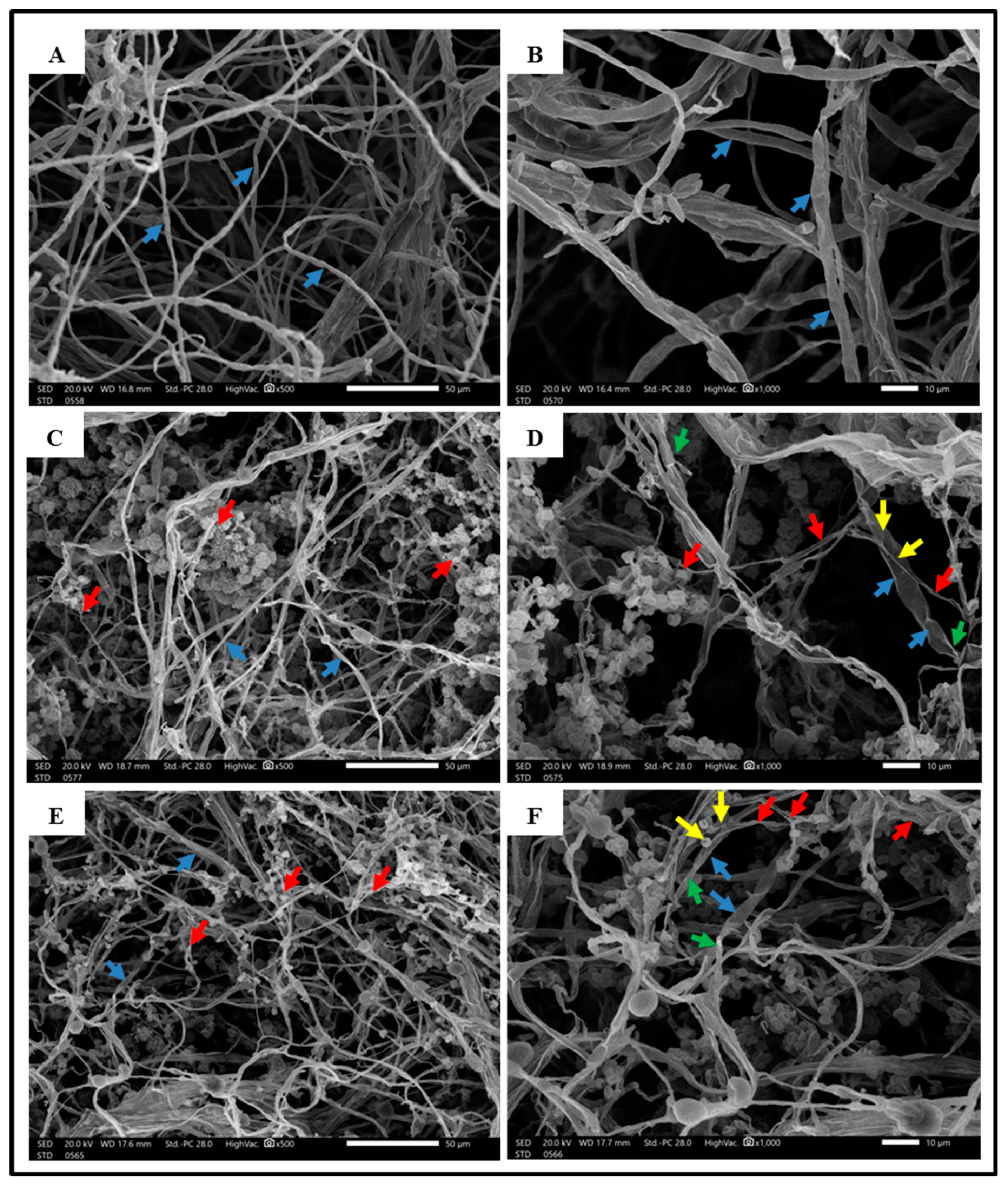
) F. solani hyphae, (Agriculture 12 01950 i002) Trichoderma spp. hyphae, (Agriculture 12 01950 i003) the Trichoderma hyphae coiled or attach to the F. solani hyphae, (Agriculture 12 01950 i004) degradation and deformation of F. solani hyphae are caused by Trichoderma spp. activity.
Agriculture 12 01950 g004
Figure 5. Disease incidence (%) at 45 DAT under greenhouse conditions after treating infected cherry tomato plants with different Trichoderma treatments, (T0), infected control with F. solani, (T1), T. viride, (T2), T. harzianum, and (TMix1+2), a mixture of the two Trichoderma isolates. Error bars represent the standard deviation of the mean. Different letter(s) above the error bars indicate statistically significant differences at (p ≤ 0.05).
Agriculture 12 01950 g005
Figure 6. Graphical diagram representing the beneficial roles of Trichoderma spp. in cherry tomato nutrition and growth.
Agriculture 12 01950 g006
Table 1. The primer names and sequences used in PCR amplification reactions.
Primer NameGenePrimer DirectionSequence (5′-3′)
Internal Transcribed Spacer ITSITS1/ITS4TCCGTAGGTGAACCTGCGG/
TCCTCCGCTTATTGATATGC
RNA polymerase II subunit 2Rpb2fRPB2-5f/fRPB2-7crGAYGAYMGWGATCAYTTYGG/
CCCATRGCTTGYTTRCCCAT
Translation elongation factor 1-αTef1-αEF1-728F/TEF1LLErevCATCGAGAAGTTCGAGAAGG/
AACTTGCAGGCAATGTGG
ITS region used in this study.
Table 2. Plant growth promoting effects of different Trichoderma treatments on cherry tomato plants cultivated in greenhouse conditions either with and/or without F. solani infested soil.
TreatmentsFresh Weight (g Plant−1)Dry Weight (g Plant−1)
F. solani InfectionTrichoderma IsolatesShootsRootsWhole PlantShootsRootsWhole PlantRoot-Shoot Ratio
WithoutT0132 ± 1.67 e78 ± 0.71 e210 ± 1.52 e64 ± 2.07 f40 ± 1.78 e103 ± 1.95 f0.63 ± 0.01 d
T1155 ± 1.68 d103 ± 1.82 d257 ± 3.42 d70 ± 0.84 e50 ± 0.70 d120 ± 1.48 e0.71 ± 0.02 c
T2184 ± 1.67 b128 ± 1.67 b311 ± 1.79 b86 ± 2.51 b69 ± 1.67 b154 ± 1.30 b0.80 ± 0.01 b
TMix1+2199 ± 2.51 a136 ± 1.52 a334 ± 3.35 a95 ± 1.82 a85 ± 2.50 a179 ± 2.39 a0.89 ± 0.03 a
WithT0105 ± 1.82 f55 ± 1.81 f159 ± 3.11 f50 ± 0.71 h25 ± 1.79 g75 ± 2.17 h0.50 ± 0.02 f
T1130 ± 1.87 e80 ± 1.82 e210 ± 1.82 e59 ± 0.70 g36 ± 1.67 f95 ± 2.07 g0.61 ± 0.01 e
T2156 ± 1.81 d103 ± 0.71 d260 ± 0.89 d73 ± 0.71 d51 ± 0.71 d124 ± 0.71 d0.70 ± 0.01 c
TMix1+2175 ± 1.82 c120 ± 1.87 c295 ± 2.19 c84 ± 2.61 c66 ± 2.51 c150 ± 3.67 c0.79 ± 0.02 b
(T0): control; (T1): T. viride; (T2): T. harzianum; and (TMix1+2): a mixture of the two Trichoderma isolates. Means in each column, followed by the same alphabetical letter(s) in common, are not significantly different at p ≤ 0.05. Values are expressed as mean ± SD (n = 5).
Table 3. Leaf chemical composition (%) of cherry tomato plants grown under greenhouse conditions with and/or without F. solani infested soil, as affected by different Trichoderma treatments.
TreatmentsLeaf Chemical Composition of Cherry Tomato Plants
F. solani InfectionTrichoderma Isolates(%)(µg/g), of D.W.
NPKCaMgFeZnCu
WithoutT03.58 ± 0.02 f0.31 ± 0.01 f3.00 ± 0.01 f0.97 ± 0.02 f0.43 ± 0.01 e63 ± 0.71 e35 ± 0.71 e6.24 ± 0.09 f
T13.99 ± 0.02 d0.37 ± 0.01 d3.38 ± 0.01 d1.38 ± 0.02 d0.64 ± 0.02 c75 ± 1.79 c47 ± 1.82 c7.12 ± 0.08 d
T24.47 ± 0.08 b0.46 ± 0.01 ab3.78 ± 0.04 a1.76 ± 0.04 a0.71 ± 0.01 b86 ± 2.51 b52 ± 1.79 b8.33 ± 0.15 b
TMix1+24.71 ± 0.03 a0.47 ± 0.02 a3.80 ± 0.03 a1.78 ± 0.04 a0.75 ± 0.03 a98 ± 3.65 a58 ± 2.86 a9.15 ± 0.26 a
WithT03.21 ± 0.02 g0.22 ± 0.03 g2.81 ± 0.02 g0.82 ± 0.02 g0.37 ± 0.01 f47 ± 0.70 g22 ± 0.70 g5.13 ± 0.09 g
T13.79 ± 0.01 e0.33 ± 0.01 e3.22 ± 0.01 e1.12 ± 0.02 e0.46 ± 0.02 e56 ± 0.71 f30 ± 1.87 f6.59 ± 0.06 e
T24.25 ± 0.02 c0.41 ± 0.02 c3.51 ± 0.02 c1.57 ± 0.01 c0.55 ± 0.02 d71 ± 3.19 d41 ± 1.79 d7.55 ± 0.07 c
TMix1+24.42 ± 0.07 b0.45 ± 0.01 b3.73 ± 0.04 b1.69 ± 0.03 b0.69 ± 0.04 b84 ± 3.17 b50 ± 1.78 b8.49 ± 0.23 b
(T0): control; (T1): T. viride; (T2): T. harzianum; and (TMix1+2): a mixture of the two Trichoderma isolates. Means in each column, followed by the same alphabetical letter(s) in common, are not significantly different at p ≤ 0.05. Values are expressed as mean ± SD (n = 5).
Table 4. Total phenolic content and photosynthetic pigments (chlorophyll a, b, and total carotenoids), of cherry tomato plants grown under greenhouse conditions with and/or without F. solani infested soil, as affected by different Trichoderma treatments.
TreatmentsPhotosynthetic PigmentsTotal Phenolic Content
(mg/g), of F.W.
F. solani InfectionTrichoderma IsolatesChlorophyll (mg/g), of F.W.Carotenoids
(mg/g), of F.W.
abTotal
Chlorophyll
WithoutT01.63 ± 0.09 d0.70 ± 0.03 d2.33 ± 0.11 d0.30 ± 0.01 e0.67 ± 0.04 h
T11.85 ± 0.10 c0.86 ± 0.02 c2.71 ± 0.13 c0.47 ± 0.01 c1.51 ± 0.10 f
T22.10 ± 0.10 b1.12 ± 0.03 b3.22 ± 0.12 b0.59 ± 0.03 b2.10 ± 0.05 e
TMix1+22.35 ± 0.09 a1.30 ± 0.04 a3.64 ± 0.13 a0.68 ± 0.03 a2.41 ± 0.10 c
WithT01.25 ± 0.10 f0.50 ± 0.04 f1.74 ± 0.12 f0.23 ± 0.01 f1.20 ± 0.06 g
T11.54 ± 0.06 e0.67 ± 0.03 e2.20 ± 0.06 e0.35 ± 0.02 d2.27 ± 0.08 d
T21.90 ± 0.07 c0.88 ± 0.03 c2.78 ± 0.06 c0.46 ± 0.01 c2.65 ± 0.10 b
TMix1+22.15 ± 0.10 b1.10 ± 0.02 b3.25 ± 0.13 b0.57 ± 0.03 b3.12 ± 0.19 a
(T0): control; (T1): T. viride; (T2): T. harzianum; and (TMix1+2): a mixture of the two Trichoderma isolates. Means in each column, followed by the same alphabetical letter(s) in common, are not significantly different at p ≤ 0.05. Values are expressed as mean ± SD (n = 5).
Publisher’s Note: MDPI stays neutral with regard to jurisdictional claims in published maps and institutional affiliations.

