Article

Unraveling the Lipidome and Antioxidant Activity of Native Bifurcaria bifurcata and Invasive Sargassum muticum Seaweeds: A Lipid Perspective on How Systemic Intrusion May Present an Opportunity

1
Mass Spectrometry Centre, LAQV-REQUIMTE, Department of Chemistry, University of Aveiro, Santiago University Campus, 3810-193 Aveiro, Portugal
2
CESAM—Centre for Environmental and Marine Studies, Department of Chemistry, University of Aveiro, Santiago University Campus, 3810-193 Aveiro, Portugal
*
Author to whom correspondence should be addressed.
Both authors share first co-authorship.
Antioxidants 2020, 9(7), 642; https://doi.org/10.3390/antiox9070642
Submission received: 8 June 2020 / Revised: 16 July 2020 / Accepted: 17 July 2020 / Published: 21 July 2020
(This article belongs to the Special Issue Bioactive Extract Derived Marine Algae in Antioxidants)

Abstract

:
Brown seaweeds are known to present components with appealing bioactive properties eliciting great interest for industrial applications. However, their lipid content is generally disregarded beyond their fatty acid (FA) composition. This study thoroughly characterized the lipid profile of two brown seaweeds collected from Portuguese coast, the native Bifurcaria bifurcata and the invasive Sargassum muticum species, and bioprospecting for antioxidant activity. An integrated state-of-the-art approach including gas chromatography-mass spectrometry (GC–MS) and liquid chromatography-mass spectrometry (HILIC–ESI-MS/MS), allowed a comprehensive picture of FA and polar lipid content. Polar lipid profile of B. bifurcata and S. muticum included 143 and 217 lipid species respectively, distributed between glycolipids, phospholipids, and betaine lipids. Some of the lipid species found have been assigned biological activity and contain of n-3 and n-6 FA. Sargassum muticum presented the highest n-3 FA content. Low concentrations of extracts of both seaweeds displayed antioxidant activity, with S. muticum presenting more promising results. These findings contribute to the nutritional and industrial exploitation of both seaweeds, highlighting their relevance as viable sources of bioactive and added-value compounds. Sargassum muticum presented interesting lipid composition and bioactivity, which may represent an accessible opportunity for the exploitation of this invasive seaweed, especially taking advantage of Sargassum blooms.

1. Introduction

Over the last decades consumer priority turned its focus to lifestyle, healthiness, and well-being, without neglecting environmental and sustainability concerns [1]. These concerns, along with the increasing demand for natural compounds and functional foods, justified a new look at the composition of seaweeds, since it is well recognized that these marine resources are a natural and sustainable source of natural compounds [2,3]. This new paradigm led to an increasing interest in seaweed utilization for food, cosmetic, agricultural, pharmaceutical, biomedical, and nutraceutical applications [4,5]. The Portuguese coast hosts 1909 different marine algae, 243 of which belong to Phaeophyceae group [6], where native seaweeds like Bifurcaria bifurcata (R. Ross, 1958) and invasive species like Sargassum muticum ((Yendo) Fensholt, 1955) share the same habitats. Bifurcaria bifurcata inhabits Atlantic Ocean and can be found from southern limit of Morocco to north-western Ireland [7,8]. As for S. muticum, from the shorelines of Gulf of Mexico in the Atlantic Ocean, it was pushed by maritime currents into the North Atlantic, composing the so-called Sargasso Sea [9]. In 2018 the total amount of S. muticum biomass was estimated to attain a whopping amount of 20 million tones, impacting activities like sailing or fishing and causing great environmental disturbances in beaches and reef lagoons [9,10]. Sargassum muticum invasion into new territories brought subsequent changes to local native species, causing the decrease in the occurrence of some native perennial seaweeds [11,12], or even leading to the loss of native genotypes [13,14]. Moreover, it has been suggested that invasive seaweeds have the potential to change epifaunal communities, and therefore alter the dynamics of entire ecosystems [15]. Until now there is no permanent method to remove this invasive species, nevertheless several efforts to fully take advantage of this biomass have been proposed [16,17]. In fact, invasive seaweeds such as S. muticum are potentially interesting and a viable natural resource for a global marine-derived drugs market, predicted to represent $21,955.6 billion by 2025 [18].
Brown seaweeds have been studied mostly due to the intrinsic properties of their polysaccharides, which are largely used by the hydrocolloid industry [19,20], but they also have been explored in the cosmetic and textile industries, and probed for biomedical and pharmaceutical applications [17,21]. Other compounds such as proteins, minerals, pigments, polyphenols, and polyunsaturated fatty acids (PUFA) justified the interest for distinct applications [22,23]. The total lipid content in brown seaweeds accounts for ca. 8% of dry weigh (DW) biomass and is commonly characterized in terms of fatty acid composition and recognized for its high content in PUFA. Lipid fractions from brown seaweeds, such as B. bifurcata and S. muticum, include an interesting content in PUFA eicosapentaenoic acid (20:5 n-3), docosahexaenoic acid (22:6 n-3), octadecatetraenoic acid (18:4 n-3), α-linolenic acid (18:3 n-3), and eicosatetraenoic acid (20:4 n-6) [7,24,25,26,27]. This is especially important because n-3 PUFA are claimed to be beneficial for the prevention of cardiovascular diseases and other chronic diseases [21,28,29].
Antioxidant effects are among the most searched bioactivities for natural products, including lipids [30,31]. Natural antioxidant products, with radical scavenging activity and capacity to neutralize reactive oxygen species (ROS) protect cells from ROS-induced cellular damage [30,31,32,33], thereby reducing the risk of diseases associated with oxidative stress [34,35]. Seaweed antioxidants are sustainable alternatives to synthetic antioxidants, in line with consumer preference towards natural substances, and can be use as functional ingredients in food or in cosmetics products [25]. In general, in vitro studies have been mainly devoted to the survey of lipophilic compounds from seaweeds such as carotenoids, some polyphenols, and flavonoids exhibiting antioxidant activity [21,31]. Particularly, B. bifurcata dichloromethane extract, mainly composed of diterpenes, fatty acids (FA), and sterols, showed antioxidant, anti-inflammatory, and antibacterial activities [24] and provided interesting information for the further exploitation of this seaweed. Moreover, active components of the ethyl acetate, ethanol, and methanol extracts of S. serratifolium showed radical scavenging activities [31], while chloroform/methanol (1/1, v/v) polar lipids rich extract from S. muticum demonstrated radical scavenging activity [33]. Lipid extracts from brown algae containing glycolipids (GL) showed the capacity to suppress ROS in lipopolysaccharide (LPS)-stimulated RAW 264.7 macrophages [36], that were positively correlated with the oxidative stability of eicosapentaenoic acid (20:5 n-3) and stearidonic acid (18:4 n-3) PUFA in their GL forms, which encouraged further studies in seaweed polar lipids as source functional lipids.
The lipid content of brown seaweeds has usually been addressed by FA profiling. However, FA in seaweeds are mainly included in polar lipids related to GL, phospholipids (PL), and betaine lipids, that have been pinpointed by using low tech methods [37,38]. The characterization of detailed lipidomic signatures using omics approaches for brown seaweeds was only performed for Fucus vesiculosus [39] and Saccharina latissima [40] and has never been performed for these species.
The goal of this work was to characterize lipid extracts from of B. bifurcata and S. muticum in terms of polar lipid molecular components, the polar lipdome, and antioxidant activity, and to attempt to relate composition with bioactivity, by signaling components with previously reported biological activity. Therefore, we performed an in-depth analysis of the lipid composition of B. bifurcata and S. muticum through a liquid chromatography coupled to high resolution mass spectrometry approach. The survey of antioxidant activity of total lipids extracts from B. bifurcata and S. muticum was conducted through their free radical scavenging potential against 2,2-diphenyl-1-picrylhydrazyl (DPPH) and 2,2′-azino-bis-3-ethylbenzothiazoline-6-sulfonic acid (ABTS) radicals. Overall, this work will contribute to the valorization of these two species, highlighting the potential of turning S. muticum biomass in particular into natural high-value products and commercially viable resources.

2. Materials and Methods

2.1. Reagents

Dichloromethane (CH2Cl2), methanol (MeOH), and acetonitrile (high-performance liquid chromatography—HPLC, grade) were purchased from Fisher Scientific Ltd. (Loughborough, UK). Milli-Q water (Synergy, Millipore Corporation, Billerica, MA, USA) was used. Phospholipid standards: 1,2-dimyristoyl-sn-glycero-3- phosphocholine (dMPC), 1,2-dimyristoyl-sn-glycero-3-phosphoethanolamine (dMPE), 1,2-dimyristoyl-sn-glycero-3-phospho-(1′-rac-)glycerol (dMPG), 1,2-dipalmitoyl-sn-glycero-3-phosphatidylinositol (dPPI), 1–nonadecanoyl-2-hydroxy-sn-glycero-3-phosphocholine (LPC), dimyristoyl phosphatidic acid (dMPA), and N-heptadecanoyl-D-erythro-sphingosine (Cer (d18:1/17:0)) were purchased from Avanti Polar Lipids, Inc. (Alabaster, AL).
2,2-Diphenyl-1-picrylhydrazyl (DPPH) was purchased from Aldrich (Milwaukee, WI). 2,2’-Azino-bis(3-ethylbenzothiazoline-6-sulfonic acid) diammonium salt (ABTS) was obtained from Fluka (Buchs, Switzerland). Ammonium acetate and 6-hydroxyl-2,5,7,8-tetramethylchromane-2-carboxylic acid (Trolox) were purchased from Sigma-Aldrich (St Louis, MO, USA). All other reagents and chemicals used were of the highest grade of purity and were purchased from major commercial sources.

2.2. Sampling

Bifurcaria bifurcata and S. muticum samples were collected in Aguda beach, Porto, Portugal (Portugal, 41°2′38″ N, 8°39′10″ W) on 18 April 2018 (spring). Biomass samples (composite sampling of at least five specimens were used) were washed thoroughly with fresh water to remove epiphytes, eliminate salt, sand, or shells and freeze-dried. Samples were maintained at −80 °C for posterior analyses.

2.3. Lipid Extraction

The biomass samples were extracted using a modified Bligh and Dyer method [41]. Samples were weighed (250 mg, a total of five replicates for each seaweed) and transferred to glass tubes with Teflon-lined screw caps. Total lipid extraction was performed by adding 3.75 mL of a mixture methanol:dichloromethane (2:1) to each replicate sample. After individual homogenization during 2 min using a vortex and 1 min of sonication, samples were incubated on ice on a rocking platform shaker (Stuart Scientific STR6, Bibby, UK) for 2 h and 30 min. The mixture was centrifuged at 392× g for 10 min (Pro-Analytical series, UK), and the organic phase was collected to a new glass tube. The remaining biomass residue was re-extracted three times with 3 mL of a mixture methanol:dichloromethane (2:1). Water (2 mL) was then added to the total collected organic phase and tubes were centrifuged again at 392× g for 10 min, and the lower organic phase was recovered. The remaining organic solvent was dried under a nitrogen gas stream and the total lipid extract content was estimated by gravimetry. Lipid extracts were stored at −20 °C, under nitrogen atmosphere until their use in liquid chromatography-mass spectrometry (LC–MS), gas chromatography-mass spectrometry (GC–MS) and bioactivity analyses [39,42].

2.4. Phospholipid and Glycolipid Quantification

Phospholipids quantification was performed using modified Bartlett and Lewis method as previously described [43]. At least, five replicates of 100 μL of lipid extract in dichloromethane (1 mg mL−1) were used. Absorbance of standards and samples was measured on a microplate UV-Vis spectrophotometer (Multiskan GO, Thermo Scientific, Hudson, NH, USA). Phospholipids were calculated as P x 25. Glycolipids quantification was performed by calculating the hexose content (% glucose) by the orcinol colorimetric method as described previously (CyberLipids, [43]). The amount of sugar was estimated from a calibration curve prepared by performing the reaction with defined amounts of glucose (up to 40 μg, from an aqueous solution containing 2 mg mL−1 of sugar) At least five replicates of 50 μL of lipid extract in dichloromethane (1 mg mL−1) were used. The factor 100/35 was chosen to convert glucose to GL [43,44].

2.5. Fatty Acid Analysis by Gas Chromatography-Mass Spectrometry (GC–MS)

Fatty acid methyl esters (FAME) were prepared from total lipid extracts by alkaline transesterification using a methanolic solution of potassium hydroxide (2.0 M), according to the Aued-Pimentel-based methodology, as described for seaweeds [43]. Therefore, 1 mL of internal standard (1 μg mL−1 of methyl nonadecanoate (C19:0) in n-hexane) was added to 30 µg of total lipid extract in dichloromethane (1 mg mL−1), followed by 200 µL of potassium hydroxide (2.0 M), prepared in methanol. After 2 min of vortexing, 2 mL of NaCl aqueous solution (10 g L−1) was added. The mixture was centrifuged at 392× g for 5 min, and 600 µL of organic phase were collected and dried under a nitrogen gas stream. For GC–MS analysis, the derivatized extract was diluted in 60 µL of hexane. Sample volumes of 2.0 µL of the hexane solution containing FAME were analyzed by GC-MS on an Agilent Technologies 6890 N Network (Santa Clara, CA, USA) equipped with a DB-FFAP column with the following specifications: 30 m of length, 0.32 mm internal diameter, and 0.25 µm film thickness (123-3232, J&W Scientific, Folsom, CA, USA). The GC equipment was connected to an Agilent 5973 Network Mass Selective Detector operating with an electron impact mode at 70 eV and scanning the range m/z 50–550 in a 1 s cycle in a full scan mode acquisition. The oven temperature was programed at an initial temperature of 80 °C for 3 min followed by a linear increase to 160 °C and 10 min at this temperature. Helium was used as carrier gas at flow rate of 1.4 mL min−1. Two analytical replicates of four lipid extracts (total of eight replicates, n = 2 × 4) were injected into the equipment. The identification of each FA was performed considering the retention times (Figure S1) and similarity to MS spectra of FA standards (Supelco 37 Component Fame Mix, Sigma-Aldrich, St. Louis, MO, USA) and available spectra in the Wiley 275 library and AOCS Lipid Library. The relative amounts of FA were calculated by the percent relative area method with proper normalization using C19:0 as internal standard, considering the sum of all relative areas of the identified FA. Results were expressed as means ± standard deviation (SD).

2.6. Polar Lipid Analysis by Hydrophilic Interaction Liquid Chromatography-High Resolution Mass Spectrometry (HILIC−MS) and Tandem Mass Spectrometry (MS/MS)

Lipid extracts analysis was performed in a HPLC Ultimate 3000 Dionex (Thermo Fisher Scientific, Bremen, Germany) system with an autosampler and coupled online to the Q-Exactive® hybrid quadrupole Orbitrap® mass spectrometer (Thermo Fisher Scientific, Waltham, MA, USA). The solvent system consisted of two mobile phases as follows: mobile phase A (acetonitrile:methanol 60:40 (per volume) with 2.5 mM ammonium acetate) and mobile phase B (acetonitrile:methanol:water 50:25:25 (per volume) with 2.5 mM ammonium acetate). Initially, 90% of mobile phase A was held isocratically for 2 min, followed by a linear decrease to 10% of A within 13 min, after which settings were maintained for 2 min and then returned to the initial conditions in 3 min, followed by a re-equilibration period of 10 min prior to the next analysis. A volume of 5 µL of each sample, containing 5 µg of lipid extract, a volume of 4 µL of PL standards mix (dMPC—0.02 µg, dMPE—0.02 µg, lysophosphatidylcholine (LPC)—0.02 µg, dPPI—0.08 µg, dMPG—0.012 µg, dMPA—0.08 µg, Cer—0.04 µg) and 91 µL of eluent (10% of mobile phase A and 90% of mobile phase B) were mixed and introduced into the Ascentis®Si column (10 cm × 1 mm, 3 µm, Sigma-Aldrich) with a flow rate of 50 µL min−1 and at 35 °C.
The mass spectrometer was operated simultaneously in positive (electrospray voltage 3.0 kV) and negative (electrospray voltage −2.7 kV) modes with a resolution of 70,000 and automatic gain control (AGC) target of 1 × 10⁶, the capillary temperature was 250 °C and the sheath gas flow was 15 U. In MS/MS experiments, a resolution of 17,500 and AGC target of 1 × 10⁵ were used. Cycles consisted of one full scan mass spectrum and 10 data-dependent MS/MS scans were repeated continuously throughout the experiments with the dynamic exclusion of 60 s and intensity threshold of 2 × 10⁴ [39,42]. Normalized collision energy (CE) ranged between 20, 25, and 30 eV. Four replicates (corresponding to four lipid extracts, n = 4) were analyzed.

2.7. Data Analysis

Data acquisition was carried out using the Xcalibur data system (V3.3, Thermo Fisher Scientific, Waltham, MA, USA). Peak integration and assignments of HPLC–MS data were performed using MZmine 2.39. The software was used for filtering and smoothing, peak detection, peak processing, and assignment against an in-house database. The validated peaks were within the time range of a MS full run. All the peaks with intensity lower than 1 × 10⁴ were excluded. All the information originating from the MZmine software was confirmed based on the assignment of the molecular ions observed in the LC–MS spectra, typical retention time, exact mass accuracy, and MS/MS spectra information. Only exact mass accuracy with an error of less than 5 ppm was considered. MS/MS spectra were performed to confirm the identity of the molecular species as previously described by Marine Lipidomics Laboratory group [39,45,46]. The identification of molecular species of polar lipids was based on the LC retention time (Tables S1 and S2, Figure S2). The normalization of the identified lipid species was performed by exporting integrated peak areas values (.csv file) and dividing the peak area value of each species by the peak area value of a standard lipid species with the closest retention time. The resulting data matrix with normalized areas of all species was used to relative quantitation of each lipid species per class, by dividing each normalized peak area by the sum of all normalized peak areas of the lipid species (ions) assigned in each class and multiplying by 100 to get the relative abundance in percentage. Data was exported into software GraphPad Prism 8.0 to design bar graphs. Identified lipid species were subjected to further analysis for determination of common and unique species through Venn diagram representation using the open source jvenn [47]. We defined “unique” when a lipid species was found only in B. bifurcata or S. muticum datasets. “Common” lipid species are those that have been found in samples from both seaweeds (Table S3).
2.8. 2.2´-Azino-bis-3-Ethylbenzothiazoline-6-Sulfonic Acid Radical Cation (ABTS) Assay Radical Scavenging Activity
The antioxidant scavenging activity against 2,2´-azino-bis-3-ethylbenzothiazoline-6-sulfonic acid radical cation (ABTS●+) was evaluated using a previously described method [42] with some modifications. The ABTS radical solution (3.5 mmol L−1) was prepared by mixing 10 mL of ABTS stock solution (7 mmol L−1 in water) with 10 mL of potassium persulfate K2S2O8 (2.45 mmol L−1 in water). This mixture was kept for 12–16 h in the dark at room temperature and was diluted in ethanol to obtain an absorbance value of ≈0.9 measured at 734 nm using a UV-vis spectrophotometer (Multiskan GO 1.00.38, Thermo Scientific, Hudson, NH, USA). For an evaluation of the radical stability, a volume of 150 µL of ethanol was added to 12 microplate wells followed by addition of 150 µL of ABTS●+ diluted solution and an incubation period of 120 min, with absorbance measured at 734 nm every 5 min. For an evaluation of the radical scavenging potential, a volume of 150 µL of S. muticum (5–250 µmol L−1 in ethanol) and B. bifurcata (25–250 µmol L−1 in ethanol) lipid extracts and 150 µL of Trolox standard solution (10–75 µmol L−1) were placed in each well followed by addition of 150 µL of ABTS●+ diluted solution, and absorbance was measured at 734 nm every 5 min during a total incubation period of 120 min. Control lipid assays were performed by replacing 150 µL of ABTS●+ diluted solution by 150 µL of ethanol. Radical reduction was monitored by measuring the decrease in absorbance during the reaction, thereby quantifying radical scavenging activity. All measurements were performed in triplicate. The % of ABTS radical remaining was determined according to Equation (1):
% ABTS remaining = (Abs samples after incubation time/Abs sample at the beginning of reaction) × 100
The free radical-scavenging activity of samples was determined as the percentage of inhibition of ABTS radical according to Equation (2):
% Inhibition = ((Abs ABTS − (Abs samples − Abs control))/Abs ABTS) × 100
The concentration of samples reducing 50% of ABTS radical after 120 min (IC50) were calculated by linear regression using the concentration of samples and the percentage of the inhibition curve. The activity, expressed as Trolox Equivalents (TE, µmol Trolox/g of sample), was calculated according to Equation (3):
TE = IC50 Trolox (µmol L−1) × 1000/IC50 of samples (µg mL−1)

2.9. 2.2-Diphenyl-1-Picrylhydrazyl Radical Assay (DPPH) Radical Scavenging Activity

The antioxidant scavenging activity against ⍺,⍺-diphenyl-𝛽-picrylhydrazyl radical (DPPH) was evaluated using a previously described method applied with some modifications [42,47]. A stock solution of DPPH in ethanol (250 µmol L−1) was prepared and diluted to provide a working solution with an absorbance value of ≈0.9 measured at 517 nm using a UV-Vis spectrophotometer (Multiskan GO 1.00.38, Thermo Scientific, Hudson, NH, USA). To evaluate the radical stability, a volume of 150 µL of ethanol was added to 12 microplate wells followed by addiction of 150 µL of DPPH diluted solution and an incubation period of 120 min, with absorbance measured at 517 nm every 5 min. For evaluation of the radical scavenging potential, a volume of 150 µL of S. muticum and B. bifurcata lipid extracts (25–250 µmol L−1) and 150 µL of Trolox standard solution (10–75 µmol L−1) were placed in each well followed by addition of 150 µL of DPPH diluted solution, and again an incubation period of 120 min was followed with absorbance being measured at 517 nm every 5 min. Control lipid assays were performed by replacing 150 µL of DPPH diluted solution by 150 µL of ethanol. Radical reduction was monitored by measuring the decrease in absorbance during the reaction, thereby quantifying radical scavenging activity. All measurements were performed in triplicate. The % of DPPH radical remaining was determined according to Equation (4):
% DPPH remaining = (Abs samples after incubation time/Abs sample at the beginning of reaction) × 100
The free radical-scavenging activity of samples was determined as the percentage of inhibition of DPPH radical according to Equation (5):
% Inhibition = ((Abs DPPH − (Abs samples − Abs control))/Abs DPPH) × 100
The concentration of samples reducing 50% of DPPH radical after 120 min (IC50) was calculated by linear regression using the concentration of samples and the percentage of the inhibition curve. For B. bifurcata the IC25 was calculated instead of IC50. The activity expressed, as TE (µmol Trolox/g of sample), was calculated according to Equation (3) (for S. muticum) and Equation (6) (for B. bifurcata):
TE = IC25 Trolox (µmol L−1) × 1000/IC25 of samples (µg mL−1)

3. Results

3.1. Total Lipid Content

The total lipid content of the two brown seaweeds, B. bifurcata and S. muticum, was estimated by gravimetry after lipid extraction procedure. The average lipid content (expressed as g/100 g of dry weigh biomass, DW) of B. bifurcata was 7.20 ± 0.31 g/100 g while that of S. muticum was lower, representing 2.80 ± 0.06 g/100 g. Bifurcaria bifurcata presented a total of 0.12 ± 0.03 g/100 g1 of PL and 1.01 ± 0.07 g/100 g of GL. Sargassum muticum presented higher content of both PL and GL in lipid extracts, with 0.25 ± 0.03 g/100 g and 1.31 ± 0.04 g/100 g, respectively.

3.1.1. Bifurcaria bifurcata and S. muticum Fatty Acid Profiles

The FA profile of the extracts obtained from B. bifurcata and S. muticum were determined by GC–MS analysis of the FAME obtained after alkaline transmethylation [45] (Table 1). In the case of B. bifurcata, the fatty acid profile included 16 FA species, and the most abundant was 16:0 (30.07% ± 2.78%), followed by 20:4 n-6 (14.28% ± 0.72%) and 18:1 n-9 (12.12% ± 1.22%). The lipid fraction of this seaweed is rich in saturated fatty acids (SFA) (46.09% ± 4.30%), with 16:0 and 18:0 being the major contributors for total SFA. The monounsaturated fatty acids (MUFA) included 16:1, 18:1 n-9, and 20:1, and the PUFA included FAs 18:2 n-6, 18:3 n-3, 18:4 n-3, 20:3, 20:4 n-6, and 20:5 n-3 (Table 1). The results obtained for the S. muticum allowed identifying 20 different FA (Table 1). The most abundant was 16:0 with a relative content of 24.18% ± 0.48%, followed by 20:4 n-6 (12.53% ± 0.72%) and 20:5 n-3 (9.71% ± 0.34%). This brown seaweed exhibited a FA profile rich in PUFA, with 16:2, 18:2 n-6, 18:3 n-3, 18:4 n-3, 20:2, 20:3, 20:4 n-6, and 20:5 n-3 as the major contributors for total PUFA abundance. MUFA such as 16:1, 18:1 n-9, 20:1, and 22:1, and SFA such as 14:0, 15:0, 16:0, 18:0, 20:0, 22:0 were also identified (Table 1). PUFA content of B. bifurcata was lower than that of S. muticum.

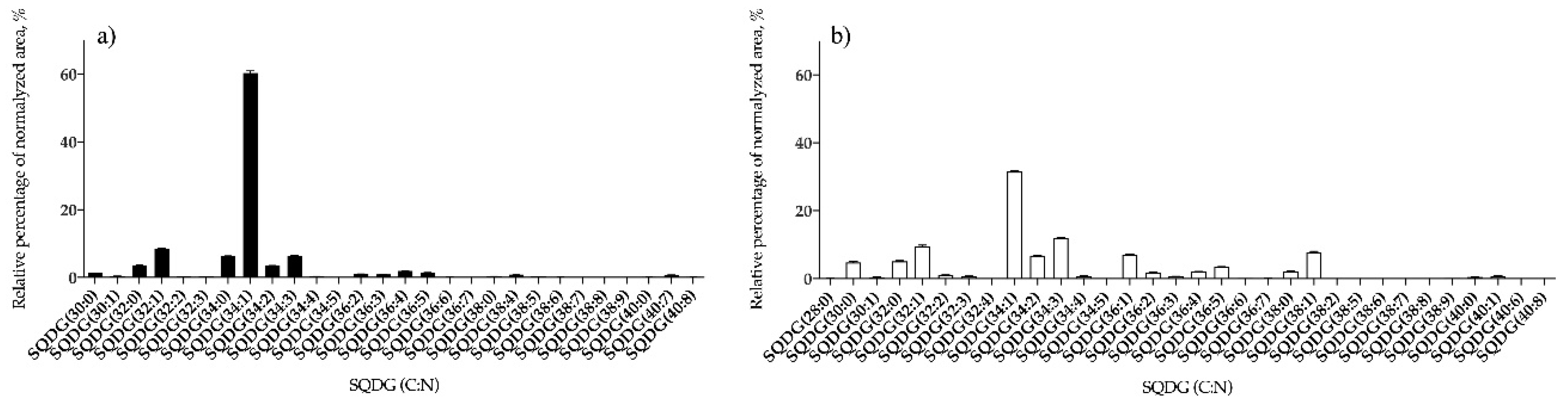
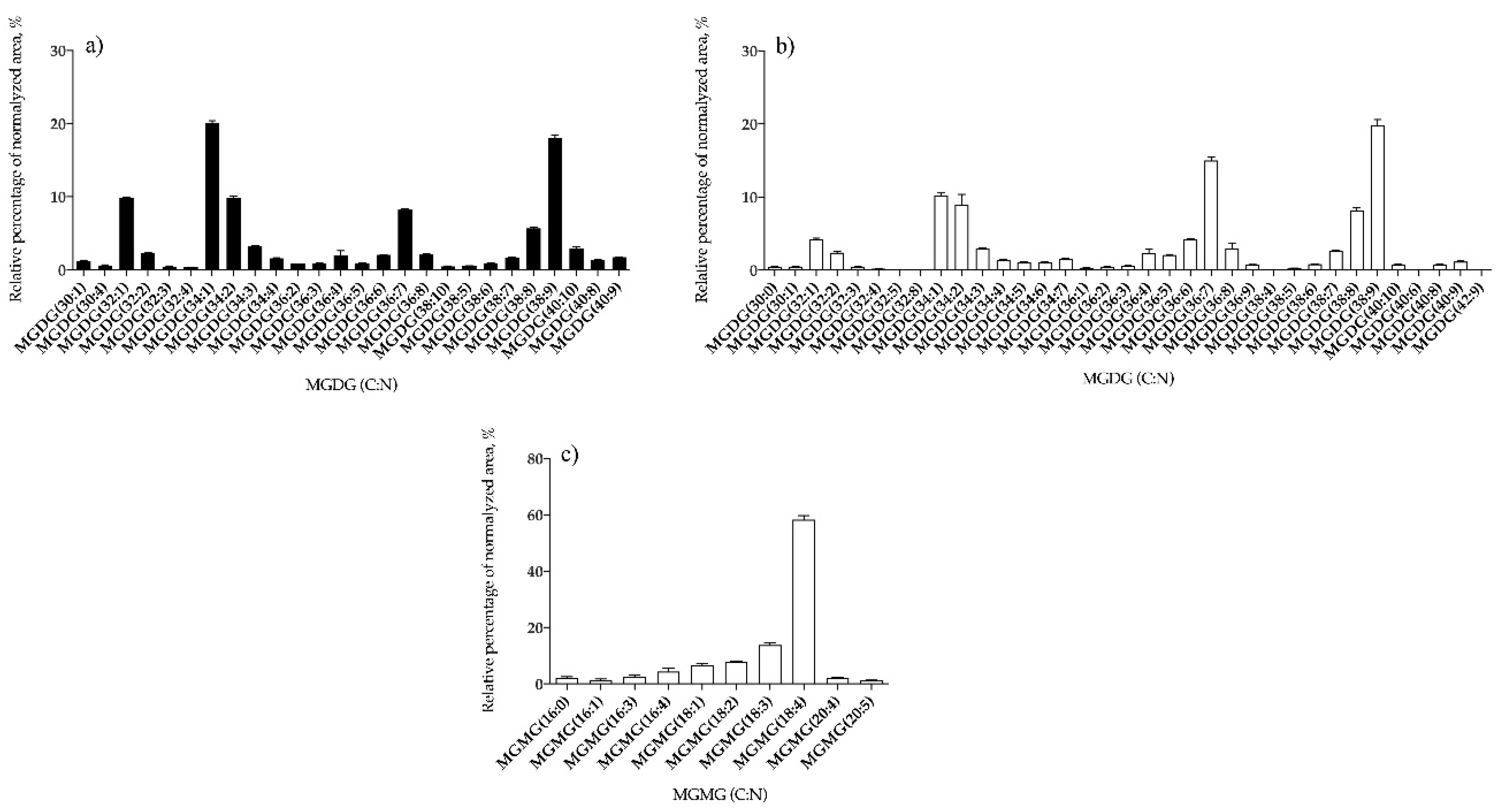
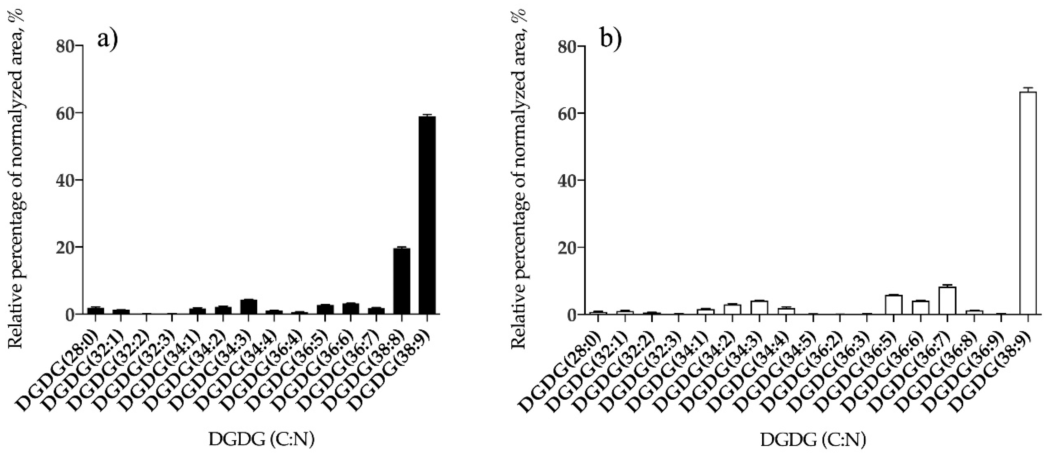
3.1.2. Bifurcaria bifurcata and S. muticum Polar Lipid Profiles

The polar lipid profiles of B. bifurcata and S. muticum were characterized at the molecular level by high resolution HILIC-LC–MS and MS/MS analysis. In the case of B. bifurcata, this approach allowed identifying 143 lipid species, from nine classes of polar lipids. Polar lipid profile included 68 GL species distributed into three GL classes, 44 PL species distributed into four PL classes and 31 betaine lipids species distributed into two classes (Table 2). Considering GL profile, in B. bifurcata, the GL classes identified included the acidic glycolipid sulfoquinovosyl diacylglycerol class (SQDG), as well as the neutral glycolipids digalactosyl diacylglycerol (DGDG) and monogalactosyl diacylglycerol (MGDG) classes (Table 2 and Table S1 from Supplementary material) but no lyso-forms were identified.
In the S. muticum lipid extracts a total of 217 polar lipid species were identified, distributed into 13 lipid classes. A total of 94 species of GL distributed into four different classes were identified, as well as 73 PL species distributed into seven classes, and 50 betaine lipid species distributed in two classes (Table 2). The criteria for identifying the lipid species included accuracy of the mass measurements (< 5 ppm), the LC retention time and the characteristics of the MS/MS spectra for each lipid class.
In what concerned GL profile, the classes of SQDG, of DGDG, and MGDG were identified in both algae, while lyso forms monogalactosyl monoacylglycerol (MGMG) were only seen in S. muticum (Table 2 and Table S2 from Supplementary material).
These acidic SQDGs were identified as [M − H] and [M + NH4]+ in LC-MS spectra and typical fragmentation observed in LC–MS/MS spectra is shown in Figure S3a,b in the Supplementary material, respectively. Overall, 28 and 32 lipid species of SQDG were identified for B. bifurcata and S. muticum, respectively (Figure 1, Tables S1 and S2). The most abundant SQDG was assigned as SQDG (34:1) at m/z 819.5, for both seaweed species, being identified as SQDG (14:0/20:1), SQDG (16:0/18:1), and SQDG (16:1/18:0) in B. bifurcata (Figure 1a, Table S1), and as SQDG (16:0/18:1) in S. muticum (Figure 1b, Table S2).
The neutral GL were detected in the positive LC–MS spectra as [M + NH4]+ ions (Tables S1 and S2) and the typical fragmentation obtained in the LC–MS/MS spectra of MGDG, MGMG, and DGDG species as [M + NH4]+ are shown in Figure S4 for MGDG and MGMG and Figure S5 for DGDG species. Considering the neutral GL, 26 species of MGDG (Figure 2a and Table S1) and 14 of DGDG (Figure 3a and Table S1) were identified in B. bifurcata and 35 lipid species of MGDG (Figure 2b and Table S2), 10 of MGMG (Figure 2c and Table S2), and 17 of DGDG (Figure 3b and Table S2) were identified in S. muticum. In B. bifurcata the most abundant MGDG was MGDG (34:1), at m/z 774.6, and assigned as MGDG (16:0/18:1), while in S. muticum the most abundant was MGDG (38:9) at m/z 814.5, assigned as MGDG (20:5/18:4) (Figure 2, Tables S1 and S2). The DGDG (38:9) at m/z 976.6 was the most abundant DGDG in both seaweed species and was identified as DGDG (20:5/18:4) also for both (Figure 3, Tables S1 and S2). The most abundant MGMG in S. muticum was detected at m/z 530.3 and was assigned as MGMG (18:4) (Figure 2c and Table S2). The profile for each GL class is represented in Figure 2 and Figure 3, showing the relative abundance of the lipid species per class.
Betaine lipids identified in B. bifurcata and S. muticum were assigned to the diacylglyceroltrimethylhomoserine (DGTS) and diacylglyceroltrimethyl-β-alanine (DGTA) classes, which were identified in the LC–MS spectra in positive ion mode, as [M + H]+ ions. The DGTA class is a structural isomer of DGTS and both were discriminated on the basis of having different retention times [39]: DGTA molecular species eluted at RT 9 min while DGTS molecular species are eluted at RT 6 min. Overall, five lipid species of DGTS (Figure 4a, Table S1) and 26 lipid species of DGTA (Figure 4c and Table S1) were found in B. bifurcata, and 13 lipid species of DGTS (Figure 4b, Table S2) and 37 lipid species of DGTA (Figure 4d, Table S1) were present in S. muticum. The predominant DGTS species in B. bifurcata was detected at m/z 738.6, corresponding to DGTS (34:1) (Figure 4a and Table S1) while in S. muticum the predominant DGTS was detected at m/z 736.6, corresponding to DGTS (34:2), (Figure 4b, Table S2). DGTA assigned as DGTA (36:4) at m/z 760.6 had similar predominance for both seaweed species (Figure 4c,d, and Tables S1 and S2). The structural features of betaine lipids were confirmed through the identification of the typical product ions and fragment pathways observed in the LC–MS/MS spectra (Figure S6).
Phospholipid classes identified in B. bifurcata and S. muticum included phosphatidylcholine (PC), phosphatidylethanolamine (PE), phosphatidylglycerol (PG), and phosphatidylinositol (PI). Sargassum muticum also contained lysoPC, lysoPE, and lysoPG species, while no lyso-forms of PL were identified in B. bifurcata (Table 2). Overall, 10 lipid species of PG (Figure 5a, Table S1), seven PI (Figure 6a, Table S1), three PC (Figure 7a, Table S1), and 24 PE (Figure 8a, Table S1) were identified in B. bifurcata. On the other hand, in S. muticum, 15 species of PG (Figure 5b, Table S2), one LPG (Table S2), eight PI (Figure 6b, Table S2), 10 PC (Figure 7b, Table S2), three LPC (Table S2), 34 of PE (Figure 8b, Table S2), and two of lysophosphatidylethanolamine (LPE) (Table S2) were identified. The predominant PG of B. bifurcata was assigned as PG (34:1) at m/z 747.5 (Figure 5a and Table S1) while the predominant PG of S. muticum was assigned as PG (34:4) at m/z 741.5 (Figure 5b and Table S2). PG and lysophosphatidylglycerol (LPG) classes were identified in negative LC−MS spectra as [M − H] ions and respective typical MS/MS spectra are represented in Figure S7a,b. The predominant PI of B. bifurcata was assigned as PI (38:8) at m/z 877.5 (Figure 6a and Table S1) while the predominant PI of S. muticum was assigned as PI (34:2) at m/z 833.5 (Figure 6b and Table S2). The PI class was identified in negative LC–MS spectra as [M − H] ions, and a characteristic MS/MS spectrum is shown in Figure S5c. LPC, PC, LPE, and PE molecular species were identified in positive LC–MS spectra as [M + H]+ ions. The predominant PC for B. bifurcata was assigned as PC (30:3) at m/z 700.5 (Figure 7a, Table S1), while the predominant PC species for S. muticum were PC (30:3) at m/z 700.5 and PC (36:4) at m/z 782.6 (Figure 7b and Table S2). The predominant PE for both seaweed species was assigned as PE (40:8) at m/z 788.5 (Figure 8a,b, Tables S1 and S2). Figure S8 shows the typical fragmentation pathways observed in the LC-MS/MS spectra of PC, LPC, PE, and LPE.
We compared the common and contrasting lipid species in the lipidome of these two brown seaweeds, members of the Fucales order. We found 124 lipid species that were common (52.5% of total number of lipid species), 19 lipid species found only in the lipidome of B. bifurcata (8.1%), and 93 lipid species identified only in the lipidome of S. muticum (39.4%). The common lipid species included 24 MGDG, 25 SQDG, 12 DGDG species, three PC, 22 PE, eight PG, and two PI species and five DGTS and 23 DGTA species (Table S3). In the case of B. bifurcata, there were two MGDG, two DGDG, three SQDG, two PE, two PG, five PI, and three DGTA unique species. Many lipid species were only assigned to S. muticum lipidome and included 11 MGMG, five DGDG, seven SQDG, seven PC, 12 PE, seven PG, six PI, and betaine lipids (eight DGTS, and 14 DGTA). Moreover, 10 MGMG, three LPC, two LPE, and one LPG were only found in S. muticum lipidome and not in B. bifurcata, since these classes were not detected in the latter.

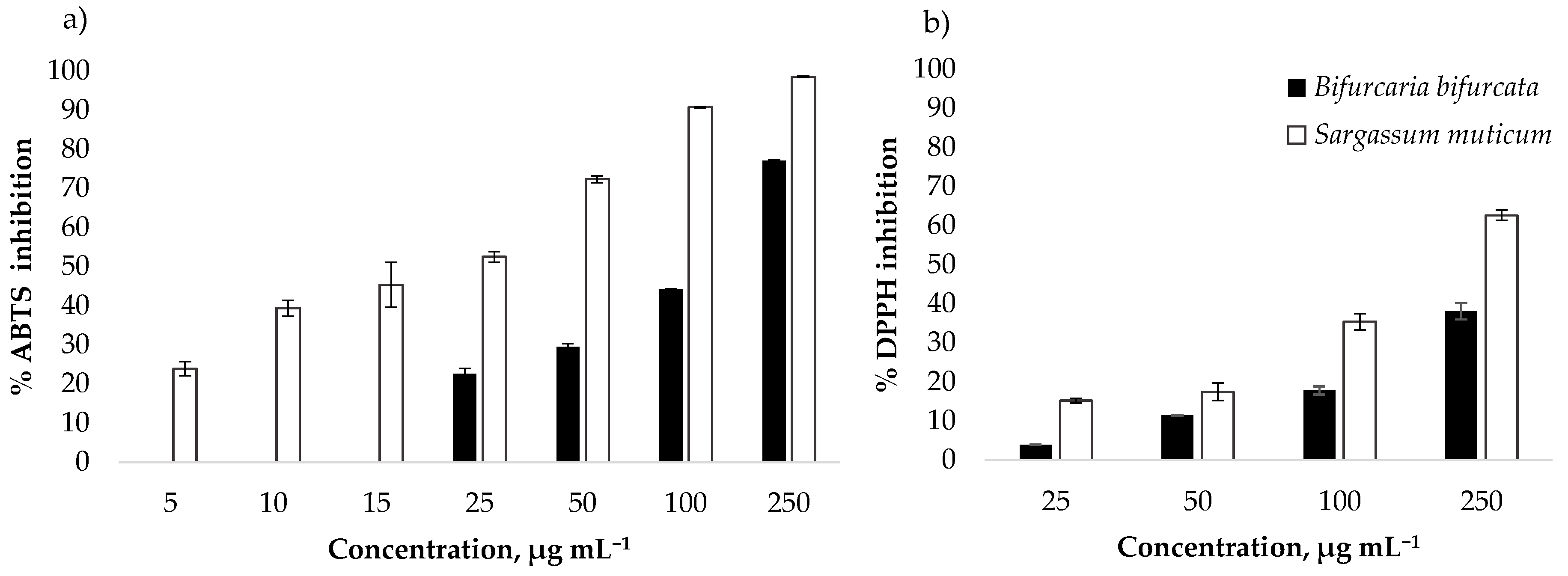
3.2. Antioxidant Activity

The antioxidant activity of B. bifurcata and S. muticum lipid extracts was evaluated by 2,2´-azino-bis-3-ethylbenzothiazoline-6-sulfonic acid radical cation assay (ABTS●+) and 2,2-diphenyl-1-picrylhydrazyl radical assay (DPPH) that measured the free radical scavenging capacity of the lipid extracts. The percentage of radical inhibition in the presence of lipid extracts was calculated after 120 min. Seven varying concentrations of lipid extracts ranging from 5 to 250 μg mL−1 of S. muticum and four varying concentrations ranging from 25 to 250 μg mL−1 of B. bifurcata were tested for ABTS antioxidant activity, while concentrations ranging from 25 to 250 μg mL−1 of B. bifurcata and S. muticum were tested for DPPH antioxidant activity. A dose-dependent increase in the scavenging capacity was observed for all concentrations tested (Figure 9). The 250 μg mL−1 extracts showed the best antioxidant activity, where among them, the S. muticum extract showed the highest antioxidant potential both in ABTS (98.4% ± 0.19%) and DPPH (62.59% ±1.29%).
For B. bifurcata, 50% of inhibition (IC50) of ABTS●+ radical was attained at a concentration of 134.19 ± 2.12 μg mL−1(IC50), representing a TE of 100.33 ± 1.57 µmol g−1, while for DPPH, only 25% of inhibition (IC25) was attained at a concentration of 155.00 ± 3.96 μg mL−1 representing a TE of 74.89 ± 1.93 µmol g−1 (Table 3). Sargassum muticum lipid extracts showed antioxidant activity against both DPPH and ABTS●+ radicals (Table 3). For ABTS●+ radical scavenging activity, IC50 was attained at 23.42 ± 0.79 μg mL−1, representing a TE of 648.35 ± 21.43 µmol g−1 and for DPPH assay the IC50 was attained at a concentration of 188.43 ± 2.55 μg mL−1 representing a TE of 124.19 ± 14.39 µmol g−1 (Table 3).

4. Discussion

Seaweeds are currently recognized as promising sources of different bioactive metabolites, making them attractive for prospection efforts in the search of new functional ingredients and pharmacophores/drugs [21]. In this work we studied the lipid composition and antioxidant activity of S. muticum, an invasive species of eastern European shores. The invasive S. muticum species may represent an especially attractive prospective resource because of its abundance and wide availability, and also because of the prospect of removing its biomass from the invaded habitats. In order to study the promise and pertinence of using S. muticum as an industry resource, we compared its composition and antioxidant potential to those of a native species of the Iberian Atlantic coast inhabiting the same geographic niche for some decades, in B. bifurcata. Commercially viable solutions such as the use of lipids from brown seaweeds as source of functional lipids for food and cosmetic, pharmaceutical products could encourage their harvesting, which became particularly relevant in the case of invasive species. To our knowledge, the present study represents the first detailed characterization of the lipidomic signature of these two brown seaweeds, B. bifurcata and S. muticum, from the Portuguese coast. The lipid content obtained for B. bifurcata was 7.29% ± 0.51% DW, while for S. muticum it was 2.84% ± 0.16% DW. These values were similar with the ones reported in the literature for both seaweeds, B. bifurcata [7,25] and S. muticum [27,33,48].
Both seaweeds showed the presence of SFA in their FA profile, namely 16:0, MUFA and PUFA, namely 18:1 n-9, 18:4 n-3, 20:4 n-6, and 20:5 n-3, respectively, in accordance with the available literature for the FA composition of B. bifurcata [7,24,25] and S. muticum [26,27]. Both seaweeds attract specific interest as edible seaweeds. The low n-6/n-3 ratio found in both algae guarantees appealing nutritional and health characteristics, since omega-3 FA are proposed to be beneficial for the prevention of cardiovascular diseases and other chronic diseases [21,49], while also being associated with antimicrobial, antiviral, anti-inflammatory, and antitumoral properties [29,50,51]. Sargassum muticum displayed a higher PUFA content and n-3 FA, unveiling a profitable avenue for further research in the nutraceutical/dietary supplements scope.
Polar lipids identified in the two seaweeds include GL, phospholipids, and betaine lipids. Sargassum muticum presented a much more varied and complex polar lipidome (217 vs. 143 different lipid species detected) making it a much more attractive target if searching for bioactive lipids. That happens in the case of GL, with S. muticum presenting 62 different lipid species from the class, while B. bifurcata only presented 40. GL from algae have especially been assigned bioactivities such as antioxidant, anti-inflammatory, antiviral, antibacterial, and antitumoral activities [46,52]. Besides glycolipids, phospholipids also represented a major component of the lipidome of B. bifurcata and S. muticum. Phospholipids are major components of cell membranes and can be important signaling molecules in living organisms, as well as in seaweeds [53]. In addition, considering seaweeds as foods or as raw materials for other biotechnological applications, the PL rich in PUFA can be potentially used as ingredients for supplements or for food fortification in n-3 FA, since there is evidence that PLs are better at delivering PUFA than the triglycerides from diet [42,46].
Betaines are also important components of brown seaweeds, although far less studied [39,40]. It was suggested that DGTS species have the same biological function as PC due to their similar zwitterionic structure, with both lipid classes being able to interchange their roles within the cell [54]. Some species of Phaeophyta can have DGTS and/or DGTA, the latter formed by conversion of DGTS into DGTA [55]. The Fucales order, to which B. bifurcata and S. muticum belong, are known to accumulate high amounts of DGTA/DGTS, while displaying reportedly low amounts of PC [39,56]. However, the bioactivities and nutritional value of the betaine lipids are yet to be unraveled.
The biological potential of polar lipids present in both lipidomes enhance the commercial viability to turn these seaweeds into components with value for different biotechnological applications and industries, as well as explored for compounds with bioactivities, representing viable options in the prevention and management of modern lifestyle diseases [57]. In this work, we explored the antioxidant activity of lipid extracts from both seaweeds, fostering their valorization. Total lipid extracts from B. bifurcata and S. muticum showed antioxidant activity potential in the µg mL−1 range. The values of IC50 for ABTS free radical scavenging assays obtained for the B. bifurcata lipid extract (Table 3), are comparable to ones reported by Santos et al. in dichloromethane extract [24] (ABTS●+, IC50 116.25 ± 2.54 µg mL−1). Otherwise, slightly higher DPPH radical scavenging activity has been reported in methanol extract [7] and dichloromethane extracts [58], revealing the impact of extraction solvents selection in antioxidant activities, with different solvent polarities altering their efficacy to extract specific antioxidant components [31]. Therefore, our study contributes for the valorization B. bifurcata as a promising source of bioactive compounds and functional ingredients. B. bifurcata may be produced in integrated multi-trophic aquaculture setups, allowing taking advantage of lipid compounds of interest and, otherwise, promoting the aquaculture sector [24]. Sargassum muticum lipid extracts showed higher antioxidant activity than the ones from B. bifurcata in both ABTS and DPPH radical scavenging assays. The IC50 values presented by S. muticum for ABTS●+ assay (Table 3) are also lower than those presented in the literature for S. serratifolium ethyl acetate (IC50, 34.6 ± 0.47 μg mL−1), or methanol and ethanol extracts, mentioned as effective antioxidants [59]. These results concur to the realization of the great antioxidant potential of S. muticum extracts. The results of the DPPH assay in this survey also unveil a higher antioxidant capacity than that demonstrated for S. muticum chloroform/methanol extracts (1/1, v/v) meaning the extraction procedure used in our study is efficient in the capture of lipid compounds with antioxidant properties [33]. The reported findings also demonstrated that polar lipids can be relevant active lipid players in the antioxidant activity survey of the total lipid extract [33]. Overall, the results from antioxidant assays are more promising for S. muticum than for the native B. bifurcata, supporting the pertinence of using the biomass of this invasive species for production of bioactive liquid extracts, for further bioactivity prospection. Interestingly, from a structural point of view, a distinct lipid profile between both seaweeds highlighted by lipid species rich in PUFA and also the higher PL and GL content obtained in S. muticum extract (PL and GL content, 0.25 ± 0.03 g/100 g DW and 1.31 ± 0.04 g/100 g DW, respectively), may justify the greatest antioxidant performance in this seaweed. The degree of inhibition of ROS was found to be correlated to the amounts of PUFA, the n−3 and n−6 PUFA ratios in the extracts, and with the GL classes MGDG and DGDG, showing greater antioxidant activities than SGDG [60]. Generally, PUFA and n-3 FA have been suggested to hold increased antioxidant potential with regard to other FA [61], and lipid extracts from S. muticum are significantly more abundant in these than those of B. bifurcata, what might concur to the increased antioxidant activity in extracts from the invasive species. Moreover, it was reported that MGDG containing 20:5, 18:3, and 18:4 [42,53,54,55] and DGDG rich in 20:5 [56,61], which are all FA with increased amounts in S. muticum, present biological activity. Synergic effects of the whole lipid compounds contributing to the effective antioxidant outcome cannot be excluded. Sargassum muticum lipid extracts are differentiated from those of B. bifurcata by the specific presence of a huge number of galactolipids, phospholipids, and betaine lipids found in the lipidome of this invasive seaweed, some not found in B. bifurcata, which could contribute to its higher antioxidant potential.
Oxidative stress is thought to play a pivotal role in a significant range of diseases, including cardiovascular and neurodegenerative diseases, diabetes, autoimmune processes, and cancer [62]. Moreover, inflammation is being positively correlated with chronic metabolic and inflammatory diseases associated with modern lifestyle [63], and a close relationship between oxidative stress and inflammation is established [64]. All this makes the screening for antioxidant properties a good place to start in terms of bioactivity, while also justifying a constant search for novel compounds with antioxidant and anti-inflammatory properties. This study has justified the prospect of deeper research of the scavenging activity in lipid extract of seaweeds. These lipid extracts can be eventually used as a natural antioxidant in the food industry as well as dietary supplements with antioxidant upside or be used in the cosmetic industry.

5. Conclusions

This work meaningfully contributed to the valorization of local natural resources in the brown seaweeds B. bifurcata and S. muticum, by providing a thorough characterization of their lipidome and highlighting their unique lipidomics features. Sargassum muticum, an invasive seaweed of the Portuguese coast, revealed a wide diversity in its polar lipid profile, displaying 13 different classes with 217 lipid species. Either way, either this high lipid variability is correlated to the adaptation to environmental and geographic changes experienced by S. muticum while intruding different settlements, or it contributes to the invasive potential of this species. In its turn, B. bifurcata, a native seaweed restricted to the environmental conditions particular to a given natural area, revealed a more narrow polar lipid profile, totaling nine different classes with 143 lipid species. Both seaweeds of the Portuguese coast present an interesting PUFA content with interesting nutritional value, namely a high n-3 FA content. These features highlight further possible uses in the nutraceutical/supplement food scope. The lipid extracts of the studied seaweeds presented lipid molecular species that have been specifically assigned biological activities, and displayed promising antioxidant properties, especially those of S. muticum. This may open new perspectives for the use of these species of algae for the chemical exploration of novel and more effective natural antioxidant compounds representing innovative functional ingredients in foods or as resources for the pharmaceutical and cosmetic industries. Generally, S. muticum presented a more varied lipid profile and more enticing results in terms of FA composition and antioxidant activity, which may concur for its further exploitation as a natural resource. The prospect of using an invasive species such as S. muticum for further nutritional purposes and as a source of polar lipids with antioxidants activity seems especially attractive, given the great biomass availability and the fact that its use would mean its gradual removal from the invaded habitats.

Supplementary Materials

The following are available online at https://www.mdpi.com/2076-3921/9/7/642/s1; Figure S1. LC−MS/MS spectra of SQDG (34:1), Figure S2. LC−MS/MS spectra of [M + NH4]+ ions of MGDG (36:2), Figure S3. LC−MS/MS spectrum of [M + NH4]+ ion of DGDG (32:2), Figure S4. LC−MS/MS spectra of [M + H]+ ion at m/z 736.6 of DGTS (34:2), Figure S5. LC−MS/MS spectra of [M − H] ions of PG (34:4) at m/z 741.5, Figure S6. LC-MS/MS spectra of the [M + H]+ ion of LPE (20:4) at m/z 502.3, Table S1. Molecular species identified by HILIC-MS in B. bifurcate, Table S2. Molecular species identified by HILIC-MS and MS/MS in S. muticum, Table S3 List of common and unique lipid species in the lipidome of B. bifurcata and S. muticum.

Author Contributions

Conceptualization, F.S.; J.P.M.; E.d.C.; M.R.D.; Methodology and formal analysis, F.S., J.P.M., D.D., T.M., E.d.C.; Investigation, and writing—original draft preparation, F.S., J.P.M., E.d.C., MRD.; Data curation, T.M., E.d.C., M.R.D.; Statistic, software and validation, D.L.; E.d.C.; M.R.D. Resources, T.M.; D.L.; E.d.C., M.R.D.; Coordination and supervision, J.P.M.; M.R.D.; Review and editing, all authors. All authors have read and agreed to the published version of the manuscript.

Funding

This work was financially supported by project OMICS 4ALGAE: Lipidomic tools for chemical phenotyping, traceability and valorisation of seaweeds from aquaculture as a sustainable source of high added-value compounds (POCI-01-0145-FEDER-030962), funded by Centro2020, through FEDER and PT2020, as well as by project SmartBioR (Centro-01-0145-FEDER-000018), co-funded by the Centro 2020 program, Portugal 2020, European Union, through the European Regional Development Fund.

Acknowledgments

The authors are also grateful to FCT/MCTES (Portugal) for the financial support to CESAM (UIDB/50017/2020+UIDP/50017/2020), QOPNA (FCT UID/QUI/00062/2019), LAQV/REQUIMTE (UIDB/50006/2020) and RNEM (LISBOA-01-0145-FEDER-402-022125), through national funds, and the co-funding by the FEDER, within the PT2020 Partnership Agreement and Compete 2020. João P. Monteiro and Elisabete da Costa (BPD/UI51/5042/2018) acknowledge the scholarship within the framework of Project GENIALG. Tânia Melo thanks the research contract under the project OMICS 4ALGAE (POCI-01-0145-FEDER-030962). Diana Lopes (SFRH/BD/119027/2016) is grateful to FCT (Fundação para a Ciência e Tecnologia), Programa Operacional do Capital Humano (POCH) and European Union through European Social Fund (FSE) for her grant.

Conflicts of Interest

The authors declare no conflict of interest.

References

  1. Nie, C.; Zepeda, L. Lifestyle segmentation of US food shoppers to examine organic and local food consumption. Appetite 2011, 57, 28–37. [Google Scholar] [CrossRef] [PubMed]
  2. Anand, N.; Rachel, D.; Thangaraju, N.; Anantharaman, P. Potential of marine algae (seaweeds) as source of medicinally important compounds. Plant Genet. Resour. Characterisation Util. 2016, 14, 303–313. [Google Scholar] [CrossRef]
  3. Cardozo, K.H.M.; Guaratini, T.; Barros, M.P.; Falcão, V.R.; Tonon, A.P.; Lopes, N.P.; Campos, S.; Torres, M.A.; Souza, A.O.; Colepicolo, P.; et al. Metabolites from algae with economical impact. Comp. Biochem. Physiol. C. Toxicol. Pharmacol. 2007, 146, 60–78. [Google Scholar] [CrossRef] [PubMed]
  4. Cornish, M.L.; Garbary, D.J. Antioxidants from macroalgae: Potential applications in human health and nutrition. ALGAE 2010, 25, 155–171. [Google Scholar] [CrossRef]
  5. Zubia, M.; Robledo, D.; Freile-Pelegrin, Y. Antioxidant activities in tropical marine macroalgae from the Yucatan Peninsula, Mexico. J. Appl. Phycol. 2007, 19, 449–458. [Google Scholar] [CrossRef]
  6. Guiry, M.D.; Guiry, G.M. AlgaBase. 2019. Available online: https://www.algaebase.org/about/ (accessed on 1 May 2019).
  7. Alves, C.; Pinteus, S.; Simões, T.; Horta, A.; Silva, J.; Tecelão, C.; Pedrosa, R. Bifurcaria bifurcata: A key macro-alga as a source of bioactive compounds and functional ingredients. Int. J. Food Sci. Technol. 2016, 51, 1638–1646. [Google Scholar] [CrossRef]
  8. Le Lann, K.; Rumin, J.; Cérantola, S.; Culioli, G.; Stiger-Pouvreau, V. Spatiotemporal variations of diterpene production in the brown macroalga Bifurcaria bifurcata from the western coasts of Brittany (France). J. Appl. Phycol. 2014, 26, 1207–1214. [Google Scholar] [CrossRef]
  9. Briggs, H. Sargassum: The biggest seaweed bloom in the world. BBC News, 4 July 2019. [Google Scholar]
  10. Gower, J.F.R.; King, S.A. Distribution of floating Sargassum in the Gulf of Mexico and the Atlantic ocean mapped using MERIS. Int. J. Remote Sens. 2011, 32, 1917–1929. [Google Scholar] [CrossRef]
  11. Sánchez, Í.; Fernández, C.; Arrontes, J. Long-term changes in the structure of intertidal assemblages after invasion by Sargassum muticum (Phaeophyta). J. Phycol. 2005, 41, 942–949. [Google Scholar] [CrossRef]
  12. Stæhr, P.A.; Pedersen, M.F.; Thomsen, M.S.; Wernberg, T.; Krause-Jensen, D. Invasion of Sargassum muticum in Limfjorden (Denmark) and its possible impact on the indigenous macroalgal community. Mar. Ecol. Prog. Ser. 2000, 207, 79–88. [Google Scholar] [CrossRef]
  13. Britton-Simmons, K.H. Direct and indirect effects of the introduced alga Sargassum muticum on benthic, subtidal communities of Washington State, USA. Mar. Ecol. Prog. Ser. 2004, 277, 61–78. [Google Scholar] [CrossRef] [Green Version]
  14. Critchley, A.T.; Farnham, W.F.; Morrell, S.L. An account of the attempted control of an introduced marine alga, Sargassum muticum, in Southern England. Biol. Conserv. 1986, 35, 313–332. [Google Scholar] [CrossRef]
  15. DeAmicis, S.; Foggo, A. Long-term field study reveals subtle effects of the invasive alga Sargassum muticum upon the epibiota of Zostera marina. PLoS ONE 2015, 10, 1–12. [Google Scholar] [CrossRef] [PubMed]
  16. Pérez-López, P.; Balboa, E.M.; González-García, S.; Domínguez, H.; Feijoo, G.; Moreira, M.T. Comparative environmental assessment of valorization strategies of the invasive macroalgae Sargassum muticum. Bioresour. Technol. 2014, 161, 137–148. [Google Scholar] [CrossRef] [PubMed] [Green Version]
  17. Milledge, J.J.; Nielsen, B.V.; Bailey, D. High-value products from macroalgae: The potential uses of the invasive brown seaweed, Sargassum muticum. Rev. Environ. Sci. Biotechnol. 2016, 15, 67–88. [Google Scholar] [CrossRef]
  18. Infinium Global Research, Marine-derived Drugs Market (Type - Phenol, Steroid, Ether, Peptide, and Other; Source - Algae, Invertebrates, and Microorganisms; Mode of Delivery - Anti-microbial, Anti-tumor, Anti-cardiovascular, Anti-viral, Anti-inflammatory, and Others): Global Industry Analysis, Trends, Size, Share and Forecasts to 2025. 2019. Available online: https://www.medgadget.com/2019/07/marinederived-dr (accessed on 4 July 2019).
  19. Rupérez, P.; Ahrazem, O.; Leal, J.A. Potential antioxidant capacity of sulfated polysaccharides from the edible marine brown seaweed Fucus vesiculosus. J. Agric. Food Chem. 2002, 50, 840–845. [Google Scholar] [CrossRef]
  20. Yoza, B.A.; Masutani, E.M. The analysis of macroalgae biomass found around Hawaii for bioethanol production. Environ. Technol. 2013, 34, 1859–1867. [Google Scholar] [CrossRef]
  21. Rengasamy, K.R.; Mahomoodally, M.F.; Aumeeruddy, M.Z.; Zengin, G.; Xiao, J.; Kim, D.H. Bioactive compounds in seaweeds: An overview of their biological properties and safety. Food Chem. Toxicol. 2020, 135, 1–27. [Google Scholar] [CrossRef]
  22. Chojnacka, K. Biologically Active compounds in seaweed extracts - the prospects for the application. Open Conf. Proc. J. 2012, 3, 20–28. [Google Scholar] [CrossRef] [Green Version]
  23. Gomez-Zavaglia, A.; Prieto Lage, M.A.; Jimenez-Lopez, C.; Mejuto, J.C.; Simal-Gandara, J. The potential of seaweeds as a source of functional ingredients of prebiotic and antioxidant value. Antioxidants 2019, 8, 406. [Google Scholar] [CrossRef] [Green Version]
  24. Santos, S.; Trindade, S.; Oliveira, C.; Parreira, P.; Rosa, D.; Duarte, M.; Ferreira, I.; Cruz, M.; Rego, A.; Abreu, M.; et al. Lipophilic Fraction of Cultivated Bifurcaria bifurcata R. Ross: Detailed composition and In vitro prospection of current challenging bioactive properties. Mar. Drugs 2017, 15, 340. [Google Scholar] [CrossRef] [Green Version]
  25. Lorenzo, J.M.; Agregán, R.; Munekata, P.E.S.; Franco, D.; Carballo, J.; Şahin, S.; Lacomba, R.; Barba, F.J. Proximate composition and nutritional value of three macroalgae: Ascophyllum nodosum, Fucus vesiculosus and Bifurcaria bifurcata. Mar. Drugs 2017, 15, 360. [Google Scholar] [CrossRef] [PubMed] [Green Version]
  26. Plouguerné, E.; Ioannou, E.; Georgantea, P.; Vagias, C.; Roussis, V.; Hellio, C.; Kraffe, E.; Stiger-Pouvreau, V. Anti-microfouling activity of lipidic metabolites from the invasive brown alga Sargassum muticum (Yendo) Fensholt. Mar. Biotechnol. 2010, 12, 52–61. [Google Scholar] [CrossRef] [PubMed]
  27. Conde, E.; Moure, A.; Domínguez, H. Supercritical CO2 extraction of fatty acids, phenolics and fucoxanthin from freeze-dried Sargassum muticum. J. Appl. Phycol. 2015, 27, 957–964. [Google Scholar] [CrossRef]
  28. Simopoulos, A.P. Omega-3 fatty acids in wild plants, nuts and seeds. Asia Pac. J. Clin. Nutr. 2002, 11, S163–S173. [Google Scholar] [CrossRef] [Green Version]
  29. Newton, I.S. Long chain fatty acids in health and nutrition. J. Food Lipids 1996, 233–249. [Google Scholar] [CrossRef]
  30. Miyashita, K.; Mikami, N.; Hosokawa, M. Chemical and nutritional characteristics of brown seaweed lipids: A review. J. Funct. Foods 2013, 5, 1507–1517. [Google Scholar] [CrossRef]
  31. Lim, S.; Choi, A.H.; Kwon, M.; Joung, E.J.; Shin, T.; Lee, S.G.; Kim, N.G.; Kim, H.R. Evaluation of antioxidant activities of various solvent extract from Sargassum serratifolium and its major antioxidant components. Food Chem. 2019, 278, 178–184. [Google Scholar] [CrossRef] [PubMed]
  32. Silva, J.; Alves, C.; Pinteus, S.; Horta, A.; Pedrosa, R. High antioxidant activity of Sargassum muticum and Padina pavonica collected from Peniche coast (Portugal). Curr. Opin. Biotechnol. 2013, 24, S116. [Google Scholar] [CrossRef]
  33. Terme, N.; Boulho, R.; Kucma, J.-P.; Bourgougnon, N.; Bedoux, G.; Terme, N.; Bourgougnon, N.; Kucma, J.-P.; Bedoux, G. Radical scavenging activity of lipids from seaweeds isolated by solid-liquid extraction and supercritical fluids. OCL 2018, 25, D505. [Google Scholar] [CrossRef]
  34. Airanthi, M.K.W.-A.; Sasaki, N.; Iwasaki, S.; Baba, N.; Abe, M.; Hosokawa, M.; Miyashita, K. Effect of brown seaweed lipids on fatty acid composition and lipid hydroperoxide levels of mouse liver. J. Agric. Food Chem. 2011, 59, 4156–4163. [Google Scholar] [CrossRef]
  35. Sachindra, N.M.; Airanthi, M.K.; Hosokawa, M.; Miyashita, K. Radical scavenging and singlet oxygen quenching activity of extracts from Indian seaweeds. J. Food Sci. Technol. 2010, 47, 94–99. [Google Scholar] [CrossRef] [PubMed] [Green Version]
  36. Lehtimaa, T.; Tarvo, V.; Mortha, G.; Kuitunen, S.; Vuorinen, T. Reactions and kinetics of Cl(III) decomposition. Ind. Eng. Chem. Res. 2008, 47, 5284–5290. [Google Scholar] [CrossRef]
  37. Dembitsky, V.M.; Rozentsvet, O.A.; Pechenkina, E.E. Glycolipids, phospholipids and fatty acids of brown algae species. Phytochemistry 1990, 29, 3417–3421. [Google Scholar] [CrossRef]
  38. Salehi, B.; Sharifi-Rad, J.; Seca, A.M.L.; Pinto, D.C.G.A.; Michalak, I.; Trincone, A.; Mishra, A.P.; Nigam, M.; Zam, W.; Martins, N. Current trends on seaweeds: Looking at chemical composition, phytopharmacology, and cosmetic applications. Molecules 2019, 24, 4182. [Google Scholar] [CrossRef] [PubMed] [Green Version]
  39. Da Costa, E.; Domingues, P.; Melo, T.; Coelho, E.; Pereira, R.; Calado, R.; Abreu, M.H.; Domingues, M.R. Lipidomic signatures reveal seasonal shifts on the relative abundance of high-valued lipids from the brown algae Fucus vesiculosus. Mar. Drugs 2019, 17, 335. [Google Scholar] [CrossRef] [Green Version]
  40. Rey, F.; Lopes, D.; Maciel, E.; Monteiro, J.; Skjermo, J.; Funderud, J.; Raposo, D.; Domingues, P.; Calado, R.; Domingues, M.R. Polar lipid profile of Saccharina latissima, a functional food from the sea. Algal Res. 2019, 39, 1–8. [Google Scholar] [CrossRef]
  41. da Costa, E.; Ricardo, F.; Melo, T.; Mamede, R.; Abreu, M.H.; Domingues, P.; Domingues, M.R.; Calado, R. Site-specific lipidomic signatures of sea lettuce (Ulva spp., chlorophyta) hold the potential to trace their geographic origin. Biomolecules 2020, 10, 489. [Google Scholar] [CrossRef] [Green Version]
  42. Lopes, D.; Melo, T.; Meneses, J.; Abreu, M.H.; Pereira, R.; Domingues, P.; Lillebø, A.I.; Calado, R.; Rosário Domingues, M. A new look for the red macroalga Palmaria palmata: A seafood with polar lipids rich in EPA and with antioxidant properties. Mar. Drugs 2019, 17, 533. [Google Scholar] [CrossRef] [Green Version]
  43. Da Costa, E.; Melo, T.; Moreira, A.S.P.A.; Bernardo, C.; Helguero, L.; Ferreira, I.; Cruz, M.T.M.; Rego, A.M.A.; Domingues, P.; Calado, R.; et al. Valorization of lipids from Gracilaria sp. through lipidomics and decoding of antiproliferative and anti-Inflammatory activity. Mar. Drugs 2017, 15, 62. [Google Scholar] [CrossRef] [Green Version]
  44. Bell, B.M.; Daniels, D.G.H.; Fearn, T.; Stewart, B.A. Lipid compositions, baking qualities and other characteristics of wheat varieties grown in the U.K. J. Cereal Sci. 1987, 5, 277–286. [Google Scholar] [CrossRef]
  45. Melo, T.; Alves, E.; Azevedo, V.; Martins, A.S.; Neves, B.; Domingues, P.; Calado, R.; Abreu, M.H.; Domingues, M.R. Lipidomics as a new approach for the bioprospecting of marine macroalgae—Unraveling the polar lipid and fatty acid composition of Chondrus crispus. Algal Res. 2015, 8, 181–191. [Google Scholar] [CrossRef]
  46. Lopes, D.; Moreira, A.S.P.; Rey, F.; da Costa, E.; Melo, T.; Maciel, E.; Rego, A.; Abreu, M.H.; Domingues, P.; Calado, R.; et al. Lipidomic signature of the green macroalgae Ulva rigida farmed in a sustainable integrated multi-trophic aquaculture. J. Appl. Phycol. 2018, 1369–1381. [Google Scholar] [CrossRef] [Green Version]
  47. Magalhães, L.M.; Segundo, M.A.; Reis, S.; Lima, J.L.F.C. Automatic method for determination of total antioxidant capacity using 2,2-diphenyl-1-picrylhydrazyl assay. Anal. Chim. Acta 2006, 558, 310–318. [Google Scholar] [CrossRef]
  48. Balboa, E.M.; Gallego-fábrega, C.; Moure, A.; Domínguez, H. Study of the seasonal variation on proximate composition of oven-dried Sargassum muticum biomass collected in Vigo Ria, Spain. J. Appl. Phycol. 2016, 28, 1943–1953. [Google Scholar] [CrossRef]
  49. Simopoulos, A.P. The importance of the omega-6/omega-3 fatty acid ratio in cardiovascular disease and other chronic diseases. Exp. Biol. Med. 2008, 233, 674–688. [Google Scholar] [CrossRef]
  50. Dawczynski, C.; Schubert, R.; Jahreis, G. Amino acids, fatty acids, and dietary fibre in edible seaweed products. Food Chem. 2007, 103, 891–899. [Google Scholar] [CrossRef]
  51. Kamat, S.Y.; Wahidulla, S.; D’Souza, L.; Naik, C.G.; Ambiye, V.; Bhakuni, D.S.; Goel, A.K.; Garg, H.S.; Srimal, R.C. Bioactivity of Marine Organisms: VI. Antiviral evaluation of marine algal extracts from the Indian Coast. Bot. Mar. 1992, 35, 161–164. [Google Scholar] [CrossRef]
  52. Plouguerné, E.; da Gama, B.A.P.; Pereira, R.C.; Barreto-Bergter, E. Glycolipids from seaweeds and their potential biotechnological applications. Front. Cell. Infect. Microbiol. 2014, 4, 1–3. [Google Scholar] [CrossRef]
  53. da Costa, E.; Azevedo, V.; Melo, T.; Rego, A.M.; Evtuguin, D.V.; Domingues, P.; Calado, R.; Pereira, R.; Abreu, M.H.; Domingues, M.R. High-resolution lipidomics of the early life stages of the red seaweed Porphyra dioica. Molecules 2018, 23, 187. [Google Scholar] [CrossRef] [Green Version]
  54. Murakami, H.; Nobusawa, T.; Hori, K.; Shimojima, M.; Ohta, H. Betaine lipid is crucial for adapting to low temperature and phosphate deficiency in Nannochloropsis. Plant Physiol. 2018, 177, 181–193. [Google Scholar] [CrossRef] [PubMed] [Green Version]
  55. Kunzler, K.; Eichenberger, W. Betaine lipids and zwitterionic phospholipids in plants and fungi. Phytochemistry 1997, 46, 883–892. [Google Scholar] [CrossRef]
  56. Sato, N. Betaine Lipids. Bot. Mag. Tokyo 1992, 1, 185–197. [Google Scholar] [CrossRef]
  57. Pal, A.; Kamthania, M.C.; Kumar, A. Bioactive compounds and properties of seaweeds—A Review. Open Access Libr. J. 2014, e752, 1–17. [Google Scholar] [CrossRef]
  58. Silva, J.; Alves, C.; Freitas, R.; Martins, A.; Pinteus, S.; Ribeiro, J.; Gaspar, H.; Alfonso, A.; Pedrosa, R. Antioxidant and neuroprotective potential of the brown seaweed Bifurcaria bifurcata in an In vitro Parkinson’s disease model. Mar. Drugs 2019, 17, 85. [Google Scholar] [CrossRef] [PubMed] [Green Version]
  59. Pinteus, S.; Silva, J.; Alves, C.; Horta, A.; Fino, N.; Rodrigues, A.I.; Mendes, S.; Pedrosa, R. Cytoprotective effect of seaweeds with high antioxidant activity from the Peniche coast (Portugal). Food Chem. 2017, 218, 591–599. [Google Scholar] [CrossRef]
  60. Gerasimenko, N.; Menchinskaya, E.; Esipov, A.; Aminin, D.; Logvinov, S.; Pislyagin, E. Application of glyceroglycolipids, photosynthetic pigments and extracts of brown algae for suppression ROS. Open J. Mar. Sci. 2016, 6, 371–385. [Google Scholar] [CrossRef] [Green Version]
  61. Richard, D.; Kefi, K.; Barbe, U.; Bausero, P.; Visioli, F. Polyunsaturated fatty acids as antioxidants. Pharmacol. Res. 2008, 57, 451–455. [Google Scholar]
  62. Matschke, V.; Theiss, C.; Matschke, J. Oxidative stress: The lowest common denominator of multiple diseases. Neural Regen. Res. 2019, 14, 238–241. [Google Scholar] [CrossRef]
  63. Christ, A.; Latz, E. The Western lifestyle has lasting effects on metaflammation. Nat. Rev. Immunol. 2019, 19, 267–268. [Google Scholar] [CrossRef]
  64. Chatterjee, S. Oxidative Stress, Inflammation, and Disease. In Oxidative Stress and Biomaterials; Academic Press: Cambridge, MA, USA, 2016; pp. 35–58. [Google Scholar]
Figure 1. Relative abundance (RA) of lipid species of the sulfoquinovosyl diacylglycerol (SQDG) class identified in B. bifurcata (a) and S. muticum (b). The results are expressed as relative percentages after dividing the normalized peak area of each lipid species to the sum of normalized peak areas for all the lipid species within the class obtained after LC–MS analysis. Numbers in parentheses (C:N) indicate the number of carbon atoms (C) and double bounds (N) in the fatty acid side chains. The species with higher RA was SQDG (34:1) for both seaweeds.
Antioxidants 09 00642 g001
Figure 2. Relative abundances (RA) of lipid species of monogalactosyl diacylglycerol (MGDG) identified in B. bifurcata (a) and S. muticum (b) and of monogalactosyl monoacylglycerol (MGMG) identified in S. muticum (c). The results are expressed as relative percentage after dividing the normalized peak area of each lipid species to the sum of normalized peak areas for all the lipid species within the class obtained after LC–MS analysis. Numbers in parentheses (C:N) indicate the number of carbon atoms (C) and double bounds (N) in the fatty acid side chains. The species with higher RA were MGDG (34:1) for B. bifurcata and MGDG (38:9) and MGMG (18:4) for S. muticum.
Antioxidants 09 00642 g002
Figure 3. Relative abundances (RA) of lipid species of digalactosyl diacylglycerol (DGDG) glycolipids identified in B. bifurcata (a) and S. muticum (b). The results are expressed as relative percentage after dividing the normalized peak area of each lipid species to the sum of normalized peak areas for all the lipid species within the class obtained after LC–MS analysis. Numbers in parentheses (C:N) indicate the number of carbon atoms (C) and double bounds (N) in the fatty acid side chains. The species with higher RA was DGDG (38:9) for both seaweeds.
Antioxidants 09 00642 g003
Figure 4. Relative abundance (RA) of betaine lipid species identified as diacylglyceroltrimethylhomoserine (DGTS) (a) and diacylglyceroltrimethyl-β-alanine (DGTA) (c) in B. bifurcata, and DGTS (b) and DGTA (d) in S. muticum. The results are expressed as relative percentage after dividing the normalized peak area of each lipid species to the sum of normalized peak areas for all the lipid species within the class obtained after LC–MS analysis. Numbers in parentheses (C:N) indicate the number of carbon atoms (C) and double bounds (N) in the fatty acid side chains. The species with higher RA were DGTS (34:1) and DGTA (36:4) for B. bifurcata and DGTS (34:2) and DGTA (36:4) for S. muticum.
Antioxidants 09 00642 g004
Figure 5. Relative abundance (RA) of lipid species of the phosphatidylglycerol (PG) class identified in B. bifurcata (a) and S. muticum (b). The results are expressed as relative percentage after dividing the normalized peak area of each lipid species to the sum of normalized peak areas for all the lipid species within the class obtained after LC–MS analysis. Numbers in parentheses (C:N) indicate the number of carbon atoms (C) and double bounds (N) in the fatty acid side chains. The species with higher RA were PG (34:1) for B. bifurcata and PG (34:4) for S. muticum.
Antioxidants 09 00642 g005
Figure 6. Relative abundance (RA) of lipid species of the phosphatidylinositol (PI) class identified in B. bifurcata (a) and S. muticum (b). The results are expressed as relative percentage after dividing the normalized peak area of each lipid species to the sum of normalized peak areas for all the lipid species within the class obtained after LC–MS analysis. Numbers in parentheses (C:N) indicate the number of carbon atoms (C) and double bounds (N) in the fatty acid side chains. The species with higher RA were PG (38:8) for B. bifurcata and PI (34:2) for S. muticum.
Antioxidants 09 00642 g006
Figure 7. Relative abundance (RA) of lipid species of the phosphatidylcholine (PC) class identified in B. bifurcata (a) and S. muticum (b). The results are expressed as relative percentage after dividing the normalized peak area of each lipid species to the sum of normalized peak areas for all the lipid species within the class obtained after LC–MS analysis. Numbers in parentheses (C:N) indicate the number of carbon atoms (C) and double bounds (N) in the fatty acid side chains. The species with higher RA were PC (30:3) for B. bifurcata, and PC (30:3) and PC (36:4) for S. muticum.
Antioxidants 09 00642 g007
Figure 8. Relative abundance (RA) of lipid species of the phosphatidylethanolamine (PE) class identified in B. bifurcata (a) and S. muticum (b). The results are expressed as relative percentage after dividing the normalized peak area of each lipid species to the sum of normalized peak areas for all the lipid species within the class obtained after LC–MS analysis. Numbers in parentheses (C:N) indicate the number of carbon atoms (C) and double bounds (N) in the fatty acid side chains. The species with higher RA was PC (40:8) for both seaweeds.
Antioxidants 09 00642 g008
Figure 9. Free radical-scavenging activity (%) on 2,2´-azino-bis-3-ethylbenzothiazoline-6-sulfonic acid radical cation (ABTS) (a) and 2,2-diphenyl-1-picrylhydrazyl radical (DPPH) assays (b) radicals of B. bifurcata (a) and S. muticum lipid extracts. Each value is expressed as mean ± standard deviation (n = 3).
Antioxidants 09 00642 g009
Table 1. Fatty acid profile of Bifurcaria bifurcata and Sargassum muticum determined by gas chromatography-mass spectrometry (GC–MS) analysis fatty acid methyl esters (FAME) and expressed as relative abundance mean (%) + SD, n = 8.
Fatty AcidB. bifurcata (%)S. muticum (%)
14:04.04 ± 1.18%3.00 ± 0.17%
15:00.90 ± 0.36%0.77 ± 0.05%
16:030.07 ± 2.78%24.18 ± 0.48%
16:12.84 ± 0.56%2.34 ± 0.43%
16:21.65 ± 0.86%
18:010.42 ± 7.06%3.29 ± 0.33%
18:1 n-912.12 ± 1.22%7.91 ± 0.14%
18:2 n-63.22 ± 0.45%5.61 ± 0.26%
18:3 n-60.37 ± 0.26%0.67 ± 0.09%
18:3 n-33.84 ± 0.51%7.07 ± 0.07%
18:4 n-35.02 ± 0.73%8.03 ± 0.15%
20:00.67 ± 0.18%0.64 ± 0.12%
20:12.23 ± 0.33%1.56 ± 0.10%
20:21.46 ± 0.14%
20:3 4.08 ± 1.31% (a)3.57 ± 0.33% (a)
20:4 n-31.19 ± 0.20%0.72 ± 0.09%
20:4 n-614.28 ± 0.72%12.53 ± 0.72%
20:5 n-34.72 ± 0.26%9.71 ± 0.34%
22:00.74 ± 0.10%
22:14.56 ± 1.05%
∑ SFA46.09 ± 4.30%32.62 ± 0.92%
∑ MUFA17.19 ± 1.95%16.36 ± 0.49%
∑ PUFA36.72 ± 2.55%51.02 ± 4.79%
∑ (n-3)14.77 ± 1.28%25.53 ± 0.49%
∑ (n-6)17.87 ± 0.74%18.81 ± 0.94%
n-6/n-31.22 ± 0.090.74 ± 0.03
(a)With contribution of unknown compound. (–) not detected.
Table 2. Polar lipid classes identified by hydrophilic interaction liquid chromatography-high resolution mass spectrometry (HILIC LC–MS) in total lipid extracts of B. bifurcata and S. muticum with the indication of the total number of lipid species identified in each class and the major lipid species per class.
Lipid ClassesBifurcaria bifurcataSargassum muticum
Lipid Species NumberMajor SpeciesLipid Species NumberMajor Species
MGDG26MGDG (34:1)35MGDG (38:9)
MGMG10MGMG (18:4)
DGDG14DGDG (38:9)17DGDG (38:9)
SQDG28SQDG (34:1)32SQDG (34:1)
LPC3LPC (16:0)
PC3PC (30:3)10PC (30:3)
LPE2LPE (20:4)
PE24PE (40:8)34PE (40:8)
LPG1LPG (16:0)
PG10PG (34:1)15PG (34:4)
PI7PI (38:8)8PI (34:2)
DGTS5DGTS (34:1)13DGTS (34:2)
DGTA26DGTA (36:4)37DGTA (36:4)
Glycolipids6894
Phospholipids4473
Betaine lipids3150
Total143217
(–) not detected, MGDG: monogalactosyl diacylglycerol; MGMG: monogalactosyl monoacylglycerol; DGDG: digalactosyl diacylglycerol; SQDG: sulfoquinovosyl diacylglycerol; LPC: lysophosphatidylcholine; PC: phosphatidylcholine; LPE: lysophosphatidylethanolamine; PE: phosphatidylethanolamine; LPG: lysophosphatidylglycerol; PG: phosphatidylglycerol; PI: phosphatidylinositol; DGTS: diacylglyceroltrimethylhomoserine; DGTA: diacylglyceroltrimethyl-β-alanine.
Table 3. Antioxidant activity of B. bifurcata and S. muticum total lipid extracts expressed as inhibitory concentration (IC) values (μg mL−1) for ABTS and DPPH assays, and Trolox Equivalents (TE) (µmol g−1) for each assay.
SpeciesABTS AssayDPPH Assay
IC50
(μg mL−1)
TE
(µmol g−1)
IC *
(μg mL−1)
TE
(µmol g−1)
B. bifurcata134.19 ± 2.12100.33 ± 1.57155.00 ± 3.9674.89 ± 1.93
S. muticum23.42 ± 0.79648.35 ± 21.43188.43 ± 20.55124.19 ± 14.39
* IC25 for B. bifurcata and IC50 for S. muticum.

