Article

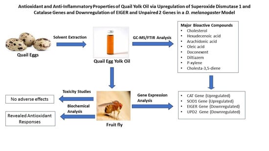
Antioxidant and Anti-Inflammatory Properties of Quail Yolk Oil via Upregulation of Superoxide Dismutase 1 and Catalase Genes and Downregulation of EIGER and Unpaired 2 Genes in a D. melanogaster Model

by
Muhammad Sani Ismaila
1,
Kamaldeen Olalekan Sanusi
2,
Uwaisu Iliyasu
3,
Mustapha Umar Imam
2,
Karla Georges
1,
Venkatesan Sundaram
1 and
Kegan Romelle Jones
1,*
1
Department of Basic Veterinary Sciences, School of Veterinary Medicine, The University of the West Indies, St. Augustine 999183, Trinidad and Tobago
2
Centre for Advanced Medical Research and Training (CAMRET), Usmanu Danfodiyo University, Sokoto 840004, Nigeria
3
Department of Pharmacognosy and Drug Development, Faculty of Pharmaceutical Sciences, Kaduna State University, Kaduna 800283, Nigeria
*
Author to whom correspondence should be addressed.
Antioxidants 2024, 13(1), 75; https://doi.org/10.3390/antiox13010075
Submission received: 29 November 2023 / Revised: 23 December 2023 / Accepted: 28 December 2023 / Published: 5 January 2024
(This article belongs to the Collection Advances in Antioxidant Ingredients from Natural Products)

Abstract

:
Quail egg yolk oil (QEYO) has a rich history of medicinal use, showcasing heightened antioxidant and bioactive properties in our prior studies. This positions QEYO as a promising candidate for therapeutic and cosmetic applications. In this investigation, QEYO was extracted using ethanol/chloroform and 2-propanol/hexane solvents. GC–MS and FTIR analyses quantified 14 major bioactive compounds in the ethanol/chloroform fraction and 12 in the 2-propanol/hexane fraction. Toxicity evaluations in fruit flies, spanning acute, sub chronic, and chronic exposures, revealed no adverse effects. Negative geotaxis assays assessed locomotor activity, while biochemical assays using fly hemolymph gauged antioxidant responses. Real-time PCR revealed the relative expression levels of the antioxidant and anti-inflammatory genes. FTIR spectra indicated diverse functional groups, and the GC–MS results associated bioactive compounds with the regulation of the anti-inflammatory genes EIGER and UPD2. While no significant change in SOD activities was noted, male flies treated with specific QEYO doses exhibited increased catalase activity and total antioxidant capacity, coupled with a significant decrease in their malondialdehyde levels. This study offers valuable insights into the bioactive compounds of QEYO and their potential regulatory roles in gene expression.

Graphical Abstract

1. Introduction

Quails and their products, especially eggs, have long been recognized for their medicinal potential [1,2]. Quail meat and eggs are gaining popularity worldwide as awareness of their medicinal benefits has increased [3,4,5]. It has been discovered that quail meat is becoming a popular delicacy in Europe, where around 9 million wild quails are harvested annually from Turkey, Portugal, and Spain and transported to France, where over 20 million tons of quail meat is processed annually for human consumption [6]. The Japanese quail egg has been reported to contain nutrients four times the value of chicken eggs [7] and contain a higher amount of crude protein, fat, and mineral ash per unit egg weight than the traditionally consumed chicken egg and other poultry species. Functional proteins such as ovomucoid, ovotransferrin, and lysozymes have also been documented [8,9].
Oils from natural products have widely been known for their use as medicines, excipients, or carriers of various medicinal substances used in the treatment of various ailments. Pisseri et al. [10] reported that essential oils contain a mixture of compounds that need to be properly quantified to define the standards for their safety and efficacy. These essential compounds have various medicinal and therapeutic activities, ranging from spasmolytic, revulsive, anti-inflammatory, decongestant, immunomodulatory, antimicrobial, antimitotic, expectorant, mucolytic, antioxidant, psychotropic, analgesic, and acaricidal activities. Chicken egg yolk oil (CEYO) has demonstrated anti-inflammatory and wound-healing activities [5,11]. The analgesic and anti-inflammatory effects of duck and chicken egg yolk have been previously reported [12,13]. This anti-inflammatory effect was postulated to regulate the Nrf2/NF-κB pathway in Caco3 cell lines in vitro [14]. It has been documented that quail eggs have better cosmetic effects than chicken eggs due to their high contents of egg yolk, dry matter, and vitamin A, and have higher stability, making them more resistant to decomposition [15]. Our previous study showed that quail egg yolk oil contains more antioxidants and other chemical components such as saponins, anthraquinones, and other bioactive substances, which are higher compared to other products [16]. Hen egg is known for its hemostatic effect in the treatment of acute and chronic eczema [12,17,18].
In this study, our aim was to analyze the different compounds in QEYO via both GC–MS and FTIR, investigate their antioxidant activity, and quantify their toxicity. A further mechanistic study will also reveal the effects of QEYO on the expression of antioxidant and anti-inflammatory genes in the fruit fly model.

2. Materials and Methods

2.1. Source and Extraction of Oil from Liquid Quail Yolk

The quail eggs were procured from a local vendor in St. Augustine, Trinidad and Tobago. The oil extraction from liquid egg yolk involved a combination of two solvents, as previously outlined [19]. Specifically, a solvent mixture of ethanol/chloroform (30/70, v/v) was used for the first extraction group, while 2-propanol/hexane (30/70, v/v) was employed for the other extraction group.

2.2. Fourier-Transform Infrared Spectroscopy (FTIR) Analysis

The FTIR analysis was conducted utilizing the Agilent Cary 630 FTIR spectrometer (Agilent Technologies, Santa Clara, CA, USA) with a base system featuring high emissions of infrared radiation. This base system operates in conjunction with a 5-bounce diamond ATR sampling accessory equipped with an internal reflection element (IRE) crystal. This analysis was executed in transmission mode, and the signal was recorded within the wavelength range of 4000–650 cm−1. The sample spectra were generated using MicroLab Software version 5.6 (Agilent Technologies, Santa Clara, CA, USA), as reported previously [20].

2.3. Gas Chromatography/Mass Spectrometry (GC–MS) of QEYO Bioactives

The GC–MS analysis was conducted using the Agilent Technologies Intuvo 9000 GC system and the Agilent Technologies 5977B mass selective detector (MSD) coupled with the 4513A automatic liquid sampler (ALS) (Agilent Technologies, Santa Clara, CA, USA). The column that was used had the part number Agilent 1909IS-483UI-INT capillary column with the specification HP-5MS UI 30 m, 0.25 mm, and 0.25 µm (Intuvo, Scotts Valley, CA, USA). Helium served as the carrier gas with a flow rate of 1.2 mL/min. The injection volume was 1 µL. The inlet temperature was maintained at 300 °C. The oven temperature was initially programmed at 50 °C for 5 min at a rate of 5 °C. A total run time of 58 min was employed. The MSD transfer line was held at a temperature of 250 °C. The source temperature was 230 °C, and the MS quad was set at 150 °C. Electron ionization at 70 eV was employed as the ionization mode. The total ion count (TIC) was utilized for the identification and quantification of compounds. The spectrum of the separated compound was compared with the database of spectra of known compounds stored in the NIST05 reference spectra library. Data analyses and peak area measurement were carried out using Agilent Mass Hunter software version 12.0 (Agilent Technologies, Santa Clara, CA, USA) [21].

2.4. Fly Culture

The fruit flies that were utilized in this study belonged to the Drosophila melanogaster w1118 strain, obtained from the Drosophila laboratory at the Centre for Advanced Medical Research and Training (CAMRET), Usmanu Danfodiyo University, Sokoto, Nigeria. These flies were kept on a standard cornmeal diet comprising white corn flour, yeast, agar, methylparaben, and distilled water. They were cultured in 50 mL of tube media at room temperature (ranging from 22 to 25 °C) with a relative humidity of 50–60%. All the flies that were included in this study were aged between 1 day old and 3 days old, unless explicitly mentioned otherwise.

2.5. Experimental Design

The flies were categorized into three study groups (groups A, B, and C) based on the extraction solvent. Group A comprised flies that were fed a diet supplemented with QEYO, extracted using a combination of ethanol and chloroform solvents. Group B included the flies receiving a diet supplemented with QEYO, extracted using a combination of 2-propanol and hexane solvents. Group C, designated as the control group, consisted of male and female flies on a standard diet with no supplementation. Within groups A and B, further divisions were made into eight subgroups based on sex and the dose of the added extract. Figure 1 illustrates these group divisions. Each treatment group utilized two media vials, with 20 flies per vial.

2.6. Toxicity Studies

The extracts underwent testing for acute (24 h), sub-chronic (7 days), and chronic (4 weeks) toxicity in fruit flies, conducted in two batches. In the first batch, doses of 100 mg, 50 mg, 25 mg, and 12.5 mg per gram of the standard diet were tested. The second batch involved doses of 10 mg, 5 mg, 2.5 mg, and 1.25 mg per gram of the prepared standard diet. The flies were transferred into fresh medium after each three-day period.

2.7. The Negative Geotaxis Assay

The negative geotaxis ability was assessed to evaluate locomotor activity following the conclusion of the treatment period before sacrifice. Ten flies per treatment group were briefly anesthetized on ice and then transferred into empty 50-mL graduated cylinders. The cylinder was marked 6 cm from the bottom. The flies were given 10 min to acclimate at room temperature. Subsequently, the cylinder was gently tapped to ensure that all the flies were at the bottom of the tube, and the count of flies passing the 6 cm mark within ten seconds was recorded. This process was repeated three times for each treatment group, with a 2-min break between repetitions.

2.8. The Biochemical Assay

Ten flies per treatment group were anesthetized on ice and promptly placed in a dish, followed by rinsing with 100 µL of cold PBS. Subsequently, the flies were transferred into a 1.5-mL tube, and 200 µL of cold PBS was added before rapid homogenization using a pellet pestle on ice. The homogenized samples were then centrifuged at 14,000 rpm for 3 min in a refrigerated centrifuge. The resulting supernatant containing the hemolymph was carefully collected into a new 1.5-mL tube for subsequent biochemical analyses [22].

2.9. The Superoxide Dismutase (SOD) Assay

The SOD level in the hemolymph was determined using a colorimetric assay kit from Solarbio Life Sciences, Beijing, China. Its principle is based on the dismutation of superoxide anions into molecular oxygen or H2O2. The superoxide anion, generated via the xanthine and xanthine oxidase reaction system, produced a blue formazan, and the absorbance was detected at 560 nm. The reagents were added to the sample, control, and blank samples 1 and 2, mixed, and incubated at room temperature for 30 min. Subsequently, the absorbance was measured at 560 nm, and the level of SOD activity was calculated according to the manufacturer’s protocol.

2.10. The Total Antioxidant Capacity (TAOC) Assay

The total antioxidant and antioxidant enzyme contents of hemolymph were determined using a colorimetric kit from Solarbio Life Sciences, Beijing, China. The reaction principle involves the reduction of Fe3+-TPTZ to blue Fe2+-TPTZ. The reaction mixture was prepared, the absorbance was read at 593 nm, and the total antioxidant capacity was then calculated according to the manufacturer’s protocol.

2.11. The Malondialdehyde (MDA) Assay

The MDA levels in the hemolymph were determined using a lipid peroxidation assay kit from Solarbio Life Sciences, Beijing, China. This reaction generates MDA and thiobarbituric acid (TBA) under acidic conditions and high temperatures. Briefly, 60 µL of the MDA working reagent was added to the sample tube and blank, followed by the addition of 20 µL of the sample to the sample well alone. Then, 20 µL of double-distilled water was added to the blank tube alone, and 20 µL of reagent III was added to both tubes. The mixtures were incubated at 100 °C for 1 h and cooled in an ice bath. Subsequently, they were centrifuged at 10,000× g for 10 min under room temperature. The supernatant was collected for absorbance measurements. The difference in absorbance at 532 nm, 450 nm, and 600 nm was used to calculate the level of MDA using the following formula:
M D A   ( n m o l / g ) = 5   ( 6.45   × Δ A 532 Δ A 600   1.29 ×   Δ A 450 ) s a m p l e   w e i g h t

2.12. The Catalase (CAT) Assay

The CAT levels in the hemolymph were determined using a colorimetric kit from Solarbio Life Sciences, Beijing, China. Its principle is based on the action of CAT in decomposing H2O2 into H2O and O2. The sample was mixed with a preheated CAT working solution according to the manufacturer’s protocol, and the absorbance was measured at 240 nm. The activities of CAT were calculated based on the rate of change in absorbance according to the following formula:
C A T   a c t i v i t y   ( U / m L )   = ( Δ A × E x t r a c t i o n   v o l u m e ) ( Ɛ   ×   d )   ×   1 0 9 ÷ S a m p l e   V o l u m e   R e a c t i o n   t i m e
where ε—molar coefficient, and d—light path.

2.13. Gene Expression Study

Based on the biochemical changes exhibited by the high dose of the extracts, the mRNA levels of the flies that received the high dose were subsequently quantified to evaluate their mechanistic basis.

2.13.1. RNA Extraction

Ten fruit flies from each group were anesthetized on ice and transferred on ice into a 1.5-mL centrifuge tube. RNA was extracted using the spin column-based nucleic acid isolation kit (Daan Gene Co., Ltd., SunYat-sen University, Guangdong, China), according to the manufacturer’s instructions. In brief, the flies were homogenized on ice in lysis buffer using a micro pestle. Chloroform was added to the mixture, vortexed, and centrifuged at 12,000× g for 3 min at 40 °C in a refrigerated centrifuge (MX-301 Highspeed, Tomy Kogyo Co., Ltd., Tagara, Japan). The aqueous supernatant was withdrawn into a new tube and precipitated with absolute ethanol. This mixture was passed through the spin column after incubation at 720 °C for 10 min. The column was spun at 12,000× g for 1 min at 40 °C, and the flow-through was discarded. The pellet was washed with a deionized solution and inhibitor remover at 12,000× g for 1 min at 40 °C. Elution was performed with 50 µL of an eluent. RNA quality was checked using a BioSpec-nano spectrophotometer (Shimadzu Biotech, Kawasaki, Japan). RNA was considered acceptable at purities of 1.8 and 2.0 for A260/230 and A260/280, respectively.

2.13.2. Primer Design

Primers for SOD1, CAT, EIGER, UPD2, and a housekeeping gene (RPL-32) were designed using the PrimerQuest qPCR assay design tool from Integrated DNA Technologies (IDT) after obtaining the FASTA format of the mRNA sequences from the GenBank database (http://www.ncbi.nlm.nih.gov/nucleotide/) accessed on 3 November 2020. The primer sequences have been listed in the following table (Table 1).

2.13.3. Quantitative Real-Time PCR Analysis

Quantitative reverse transcription polymerase chain reaction (qRT-PCR) was conducted using a one-step qRT-PCR Sybr reagent (Toroivd Technology Company Ltd., Shanghai, China) with the Rotor-Gene Q thermal cycler (Qiagen, Hilden, Germany). The reaction mixture was prepared by combining RNA samples (2 µL each), 10 µL of qRT-PCR master mix, 1 µL of manganese (Mn), 0.4 µL of forward/reverse primer, and 6.2 µL of DNAse/RNase-free water (PCR-grade water). This mixture was gently vortexed and loaded into the thermal cycler set to the following cycling conditions: reverse transcription at 95 °C for 12 min, followed by 40 cycles of denaturation at 95 °C for 15 s, annealing at 65 °C for 30 s, and extension at 72 °C for 30 s. The fold change of each gene was determined using the comparative CT method (2−ΔΔCT).

2.14. Statistical Analysis

All statistical analyses were conducted using SPSS version 20.0 (IBM, Armonk, NY, USA). All data were expressed as the mean ± SEM from three independent experiments (n = 3). A test for normality on the data was performed, and the data were found to be normally distributed. The data were analyzed using the one-way ANOVA statistical test followed by the Tukey–Kramer post-hoc test to assess for the difference between the means of the treated groups, with a p-value of <0.05 considered significant. The gene expression data were also normally distributed and were analyzed via the one-way ANOVA in JMP Statistical Software Version 17.2 (SAS Institute Inc., Cary, NC, USA).

3. Results

3.1. Quail Egg Oil (QEYO) Yield
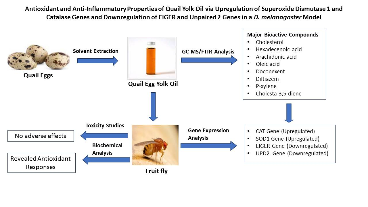
The percentage yield for the fraction of quail egg extracted using the ethanol/chloroform solvent was 25.7 ± 1.32%, while the percentage yield for the quail egg yolk extracted using the 2-propanol/hexane combination was 29.04 ± 1.72%.

3.2. FTIR Analysis

The FTIR bands in the QEYO fraction extracted with both the ethanol/chloroform and the 2-propanol/hexane combination showed different functional groups, indicating different compounds.

3.2.1. Ethanol/Chloroform QEYO Extract

The observed absorption band in the range of 2855–2922 cm−1 indicated C-H stretching, a typical vibrational pattern of a methyl group showing a fatty acid chain. The band that was observed at 1742 cm−1 (C=O stretching) indicated a saturated aldehyde. Other bands included C-O stretching at 1381–1463 cm−1, P=O stretching (1237 cm−1), S=O stretching (1162 cm−1), C-N stretching (1094 cm−1), and Si-H deformation at 805 cm−1 (Figure 2A).

3.2.2. 2-Propanol/Hexane Quail Egg Yolk Oil Extract
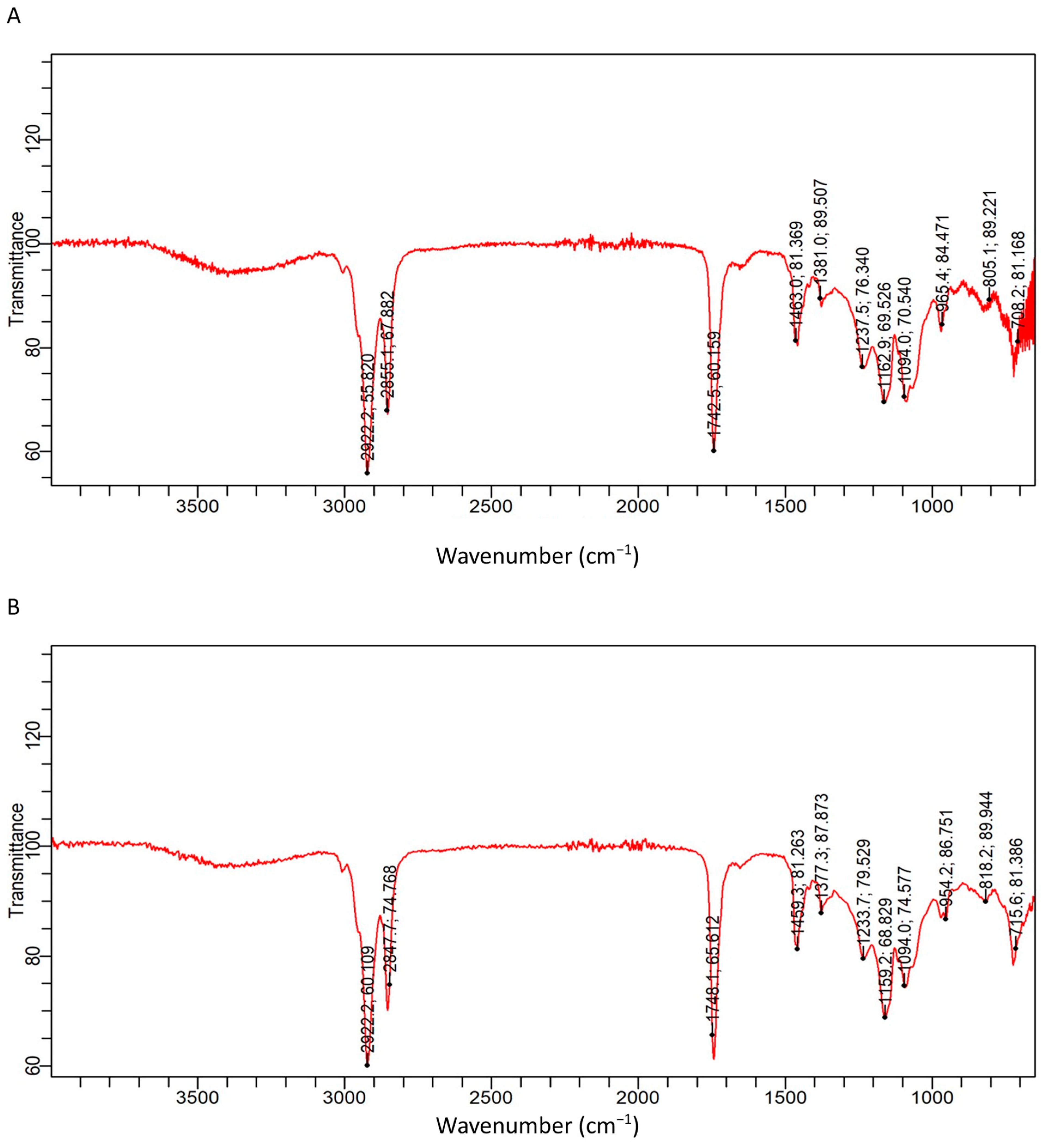
The bands that were obtained in the 2-propanol/hexane fraction of the extract were C-H stretching (2847–2922 cm−1), C=O stretching at 1784 cm−1, S=O stretching (1459.3 cm−1), C-O stretching at 1377 cm−1, Si methyl group vibration (1233 cm−1), C-N stretching (1094 cm−1), and Si-H deformation at 715–818 cm−1 (Figure 2B).

3.3. GC–MS Analysis

3.3.1. Ethanol/Chloroform Quail Egg Yolk Oil Extract

GC–MS analysis of the QEYO fraction extracted with ethanol and chloroform revealed eight major compounds, with cholesterol showing the highest percentage (41.64%), followed by cis-13-octadecenoic acid, methyl ester (15.01%), hexadecanoic acid, methyl ester (10.58%), methyl stearate (7.42%), methyl 10-trans,12-cis-octadecadienoate (5.67%), arachidonic acid (2.51%), 9-octadecenoic acid (Z)-, 2-hydroxyethyl ester (1.22%), and desmosterol (1.04%) (Figure 3A). Other important compounds that were extracted include doconexent (0.82%) and diltiazem (0.46%).

3.3.2. 2-Propanol/Hexane Quail Egg Yolk Oil Extract

The propanol/hexane GC–MS results showed six major compounds with cholesterol (50.32%); cholesta-3,5-diene (7.03%); cholesta-4,6-diene-3-ol (5.36%); ethylbenzene (4.22%); p-xylene (3.24%); cholesta-4,6-diene-3-ol (3. beta.) (2.94%); and benzene, 1,3-dimethyl (2.80%) (Figure 3B).

3.4. Toxicity Study

The flies were transferred to the control diet and different doses of the interventional diet. They were then observed for 24 h.

3.4.1. Acute Toxicity

The flies were observed for 24 h, and no mortality was observed across all the treatment and control groups.

3.4.2. Sub-Chronic Toxicity

The flies were observed for 7 days, and no mortality was observed across all the treatment and control groups.

3.4.3. Chronic Toxicity

The flies were observed for 28 days, and no mortality was observed across all the treatment and control groups.

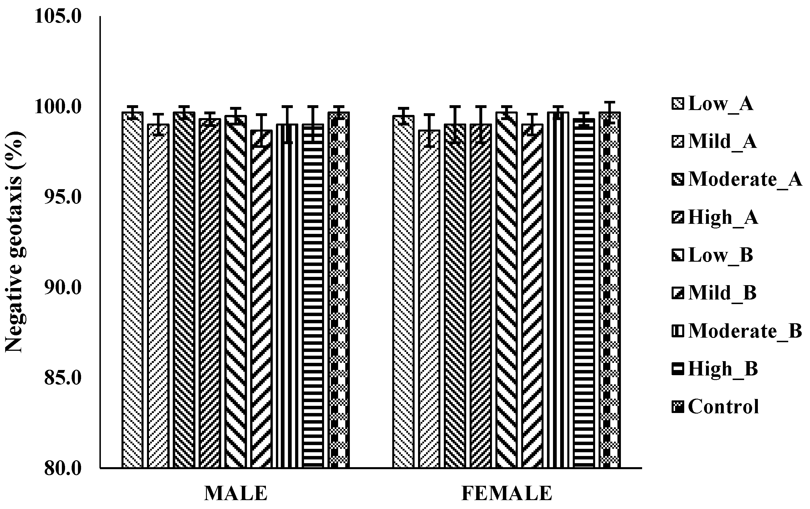
3.4.4. The Negative Geotaxis Assay

There were no significant differences in the locomotor activities of male and female flies treated with low (1.25 mg), mild (2.5 mg), moderate (5 mg), and high (10 mg) doses of ethanol/chloroform and 2-propanol/methanol extracts compared to the control group after 28 days of exposure (Figure 4).

3.5. Biochemical Assays

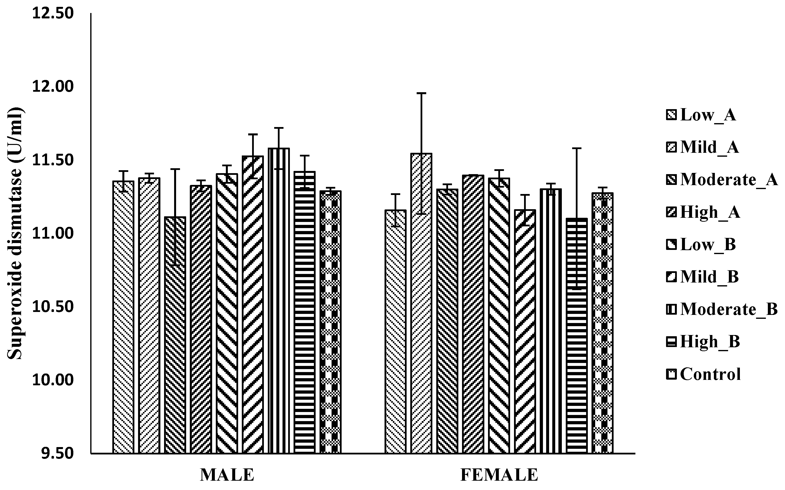
3.5.1. Superoxide Dismutase (SOD) Activity

No significant differences were observed in the SOD activities across all the treatment groups compared to the control group in both male and female flies (Figure 5).

3.5.2. Catalase Activity

In male flies, the treatment with 10 mg of ethanol/chloroform extract and 5 mg and 10 mg of 2-propanol/hexane extract resulted in a significant increase in catalase activities. Conversely, in female flies, the ethanol/chloroform extract, at doses of 5 mg and 10 mg, led to increased catalase activities, whereas no significant difference was observed in the flies treated with the 2-propanol/hexane extract compared to the control (Figure 6).

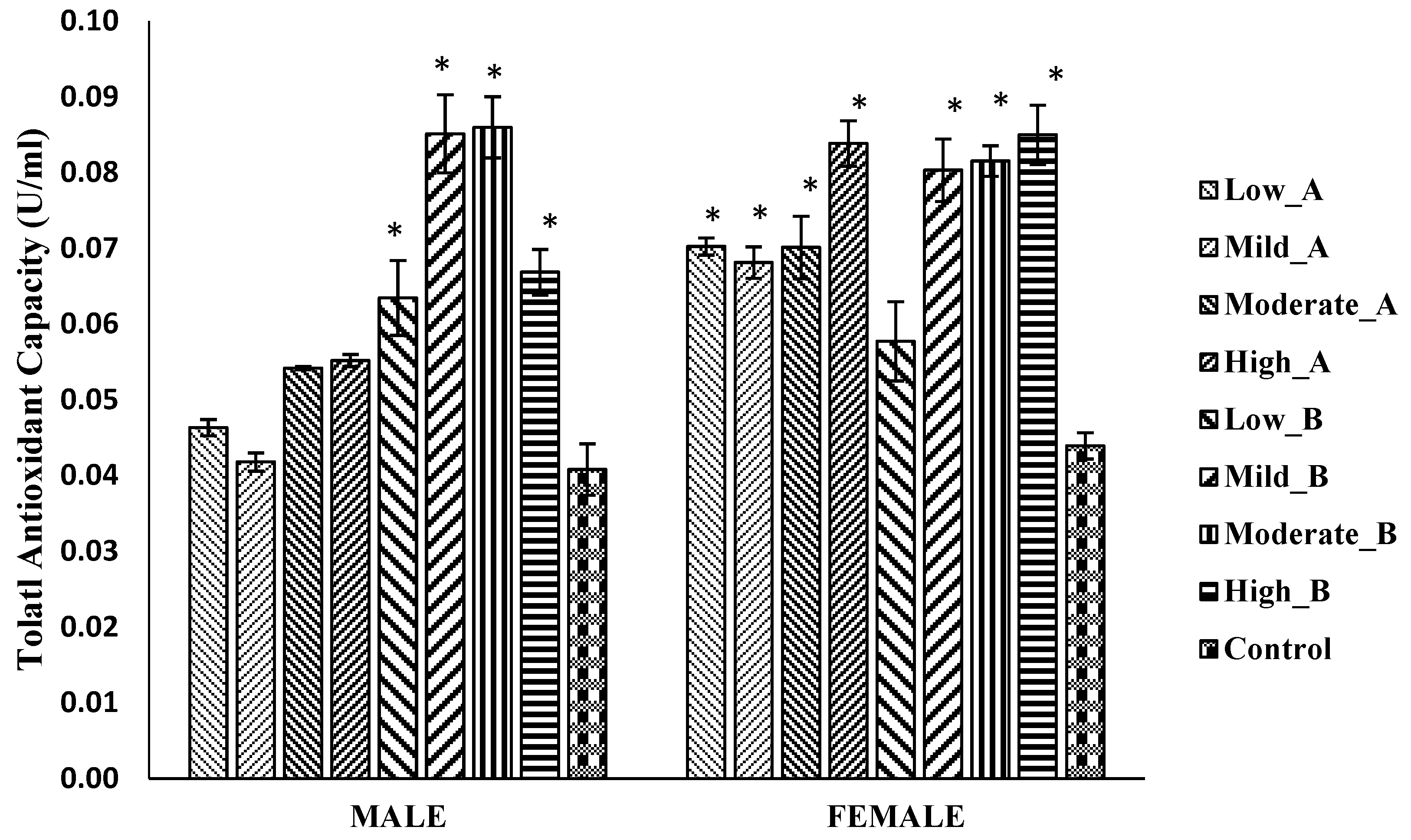
3.5.3. Total Antioxidant Capacity

Male flies treated with all doses of the 2-propanol/hexane extract exhibited a significant increase in total antioxidant capacity, while no significant difference was observed in the flies treated with the ethanol/chloroform extract. In female flies, both the ethanol/chloroform and 2-propanol/hexane extracts significantly increased total antioxidant capacity at all doses, except for the low (1.25 mg) dose of 2-propanol/hexane compared to the control (Figure 7).

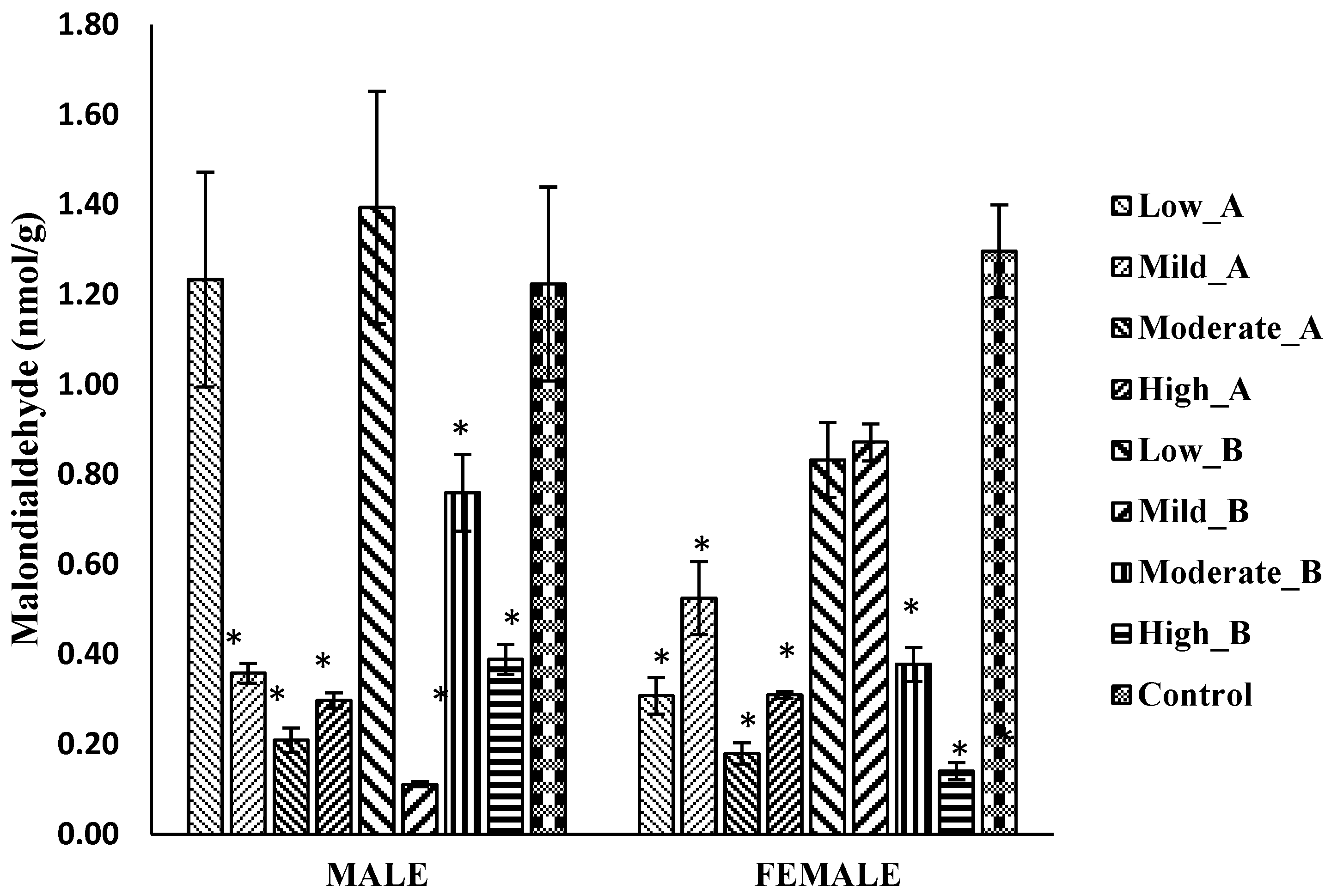
3.5.4. Malondialdehyde Levels

In male flies, the administration of mild (2.5 mg), moderate (5 mg), and high (10 mg) doses of the ethanol/chloroform extract, as well as mild and high doses of the 2-propanol/hexane extract, led to a significant reduction in malondialdehyde (MDA) levels. Similarly, in female flies, all doses of the ethanol/chloroform extract, along with moderate and high doses of the 2-propanol/hexane extract, exhibited a significant decrease in MDA levels compared to the control (Figure 8).

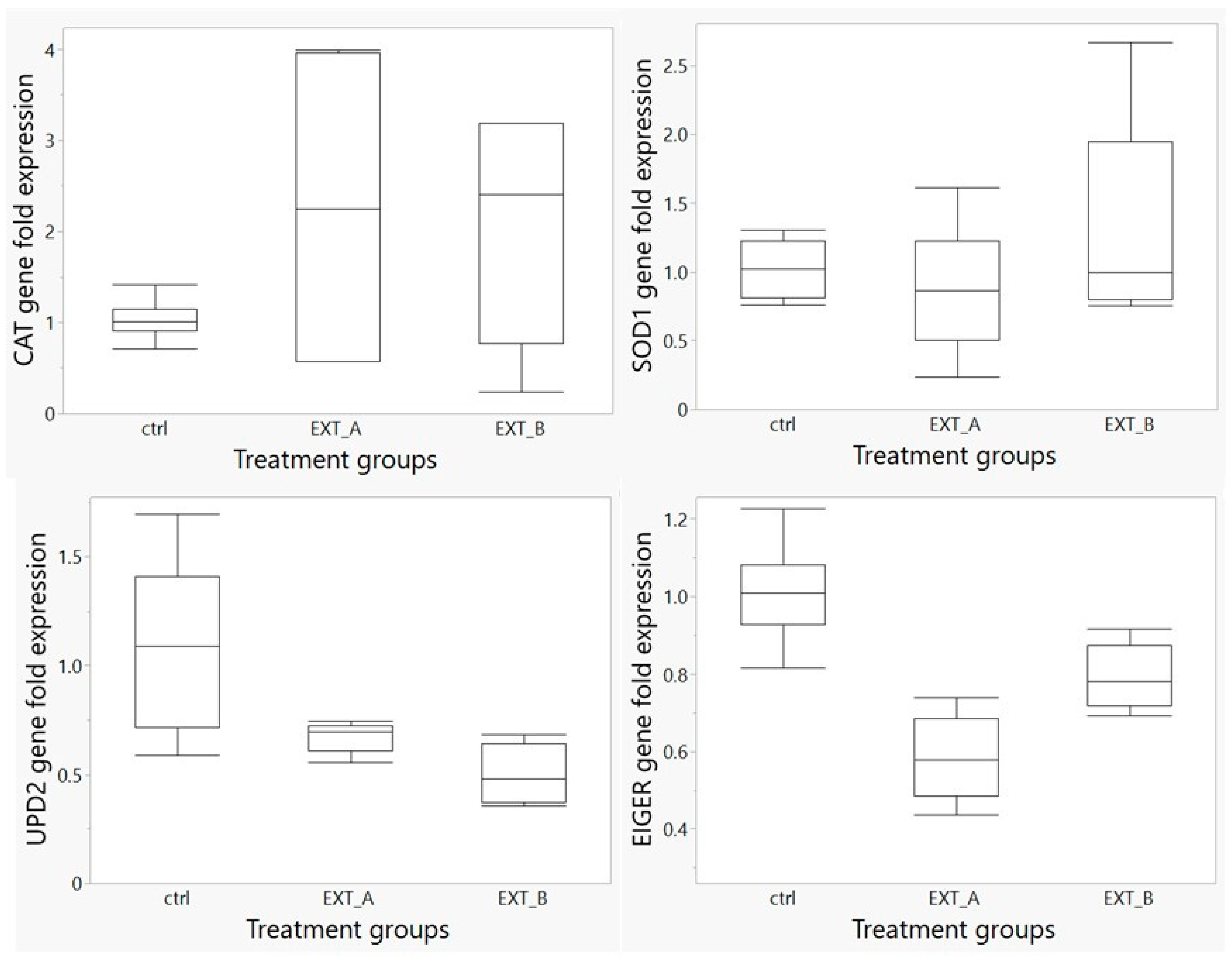
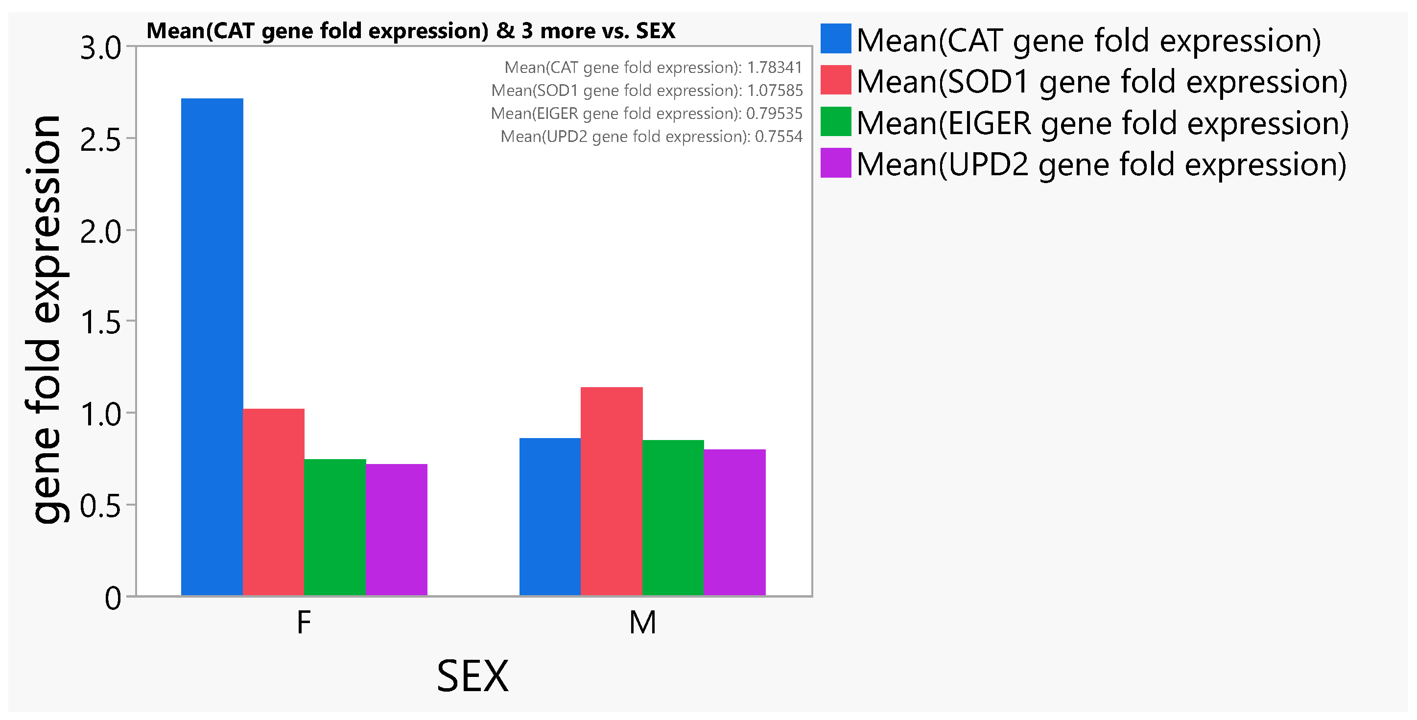
3.6. Gene Expression Study

A total of four genes were analyzed, including two antioxidant genes (CAT and SOD1) and two inflammatory genes (UPD2 and EIGER). No significant difference was observed in the expression of the CAT gene in the treated groups (p > 0.05) when compared to the control non-treated group (Figure 9). The SOD1 gene showed an increase in its level of expression, although not statistically significant (p > 0.05), in the flies in the group treated with extract B (EXT_B) when compared to the group treated with extract A and the control group (Figure 9).
The EIGER gene was significantly downregulated (p < 0.0001) in the flies treated with the QEYO extracted with the ethanol/chloroform combination compared to the control group. A significant decrease was also observed in the expression of this gene in female flies with the QEYO extracted using both the 2-propanol/hexane and ethanol/chloroform combinations (p < 0.05) (Figure 9). Furthermore, a significant decrease was observed in the expression of the UPD2 gene in the flies treated with the two QEYO extracts compared to the control non-treated group (p < 0.0002) (Figure 9).
When considering the sexes, a significant (p < 0.05) difference was observed in the CAT gene of both sexes. It was noted that females expressed the CAT genes more than their male counterparts (Figure 10).

4. Discussion

Natural food ingredients are considered richer and safer sources of antioxidants compared to synthetic sources, and edible oils have been reported to contain high levels of antioxidants [23]. Additionally, oils have been utilized in the treatment of diseases [23,24,25]. Our findings suggest the presence of various bioactive compounds with antioxidant and anti-inflammatory effects in the QEYO. The bands in the FTIR spectra (Figure 2) indicate various essential fatty acids involved in the modulation of metabolic and non-metabolic diseases, such as diabetes, atopic eczema, cancer, inflammatory diseases, and cardiovascular diseases [26,27,28,29].
The GC–MS spectra obtained from the chloroform/hexane fraction of the QEYO revealed about eight compounds with anti-inflammatory effects, while three compounds were obtained in the 2-propanol/hexane fraction (Table 2 and Table 3). Notably, various octadecanoic acids (OOAs) were extracted, such as methyl 10-trans, 12-cis-octadecadienoate, cis-13-octadecenoic acid, methyl ester, and methyl stearate, which are monounsaturated fatty acids with antimicrobial, antioxidant, anticancer, and anti-aging properties (Table 2). OOA inhibits LPS-induced nuclear factor (NF)-κB signaling by reducing the phosphorylation of the IκB-α and p50 proteins, significantly reducing pro-inflammatory signaling and resulting in a reduction in the levels of pro-inflammatory mediators [30].
Arachidonic acid (ARA), one of the major n-6 LCPUFAs, exerts its well-studied anti-inflammatory effect by blocking prostaglandin synthesis through the lipoxygenase and cyclooxygenase pathways [31,32]. Desmosterol has been reported to suppress macrophage inflammation activation and protect against vascular inflammation and atherosclerosis (Table 2) [33]. Cholesta-3,5-diene promotes wound healing by enhancing fibroblast migration, increasing extracellular matrix production and capillary formation, and recruiting immune cells. Its cytotoxic effect has also been documented (Table 3).
Our previous study demonstrated that oils extracted from quail egg yolk exhibit higher antioxidant levels and other bioactive compounds than those extracted from some plant sources. The propanol/hexane fraction contains p-xylene, which has been shown to have antipsoriatic, antimicrobial, antioxidant, and antifungal activity (Table 3). In another study, Lee et al. [34] reported the synthesis of ibuprofen from p-xylene, which may indicate the analgesic and anti-inflammatory activity of the compound extracted from quail egg oil.
Doconexent, an omega-3 fatty acid identified at 0.82% in the present study, is a high-docosahexaenoic acid (DHA) supplement with anti-inflammatory effects. It acts as a ligand at PPARs that has an anti-inflammatory effect and regulates inflammatory gene expression and NF-κB activation [35] (Table 2). It serves as the primary structural component of the human brain, cerebral cortex, skin, and retina, playing an important role in their functions [35].
Table 2. The GC–MS peaks, retention time, area, name, and molecular formula of the compounds in QEYO samples that were extracted using the ethanol/chloroform solvent combination.
PeakRT (min)AreaCompound NameMolecular FormulaPharmacological Use of the Most Important Compounds
2035.08210.58Hexadecanoic acid, methyl esterC17H34O2Antibacterial [36], antifungal [37], anti-inflammation, antifibrosis, and peripheral vasodilation effects [38]
2335.7170.68n-Hexadecanoic acidC16H32O2Anti-inflammatory [39], antibacterial, and antioxidant activities [40]
2436.3350.321-TetracoseneC24H48Cytotoxic effect [41]
2537.9260.09Gamolenic acidC18H30O2Anti-inflammatory, antithrombotic, antiproliferative, and lipid-lowering effects [42]
2638.2235.67Methyl 10-trans,12-cis-octadecadienoateC19H34O2Antifungal and antioxidant [43]
2738.34915.01cis-13-Octadecenoic acid, methyl esterC19H36O2Anti-inflammatory [44]
2938.7967.42Methyl stearateC19H38O2Antibacterial, antioxidant, antifungal [45], antidiarrheal, cytotoxic, and anti-inflammatory [46]
3038.9790.93Oleic AcidC18H34O2Decreases myocardial infarction rate, platelet aggregation and secretion of TXA2, reduces systolic blood pressure, and improves immunity [47]
3741.0902.51Arachidonic acidC20H32O2An integral constituent of the biological cell membrane that is necessary for the function of all cells, especially in the nervous system, skeletal muscle, and immune system. It modulates the function of ion channels, several receptors, and enzymes via activation as well as inhibition [48]
4544.2260.82DoconexentC22H32O2DHA acts as a ligand at PPARs that has an anti-inflammatory effect and regulates inflammatory gene expression and NFκB activation [35]
4644.3060.46DiltiazemC22H26N2O4SA calcium channel blocker that is clinically used as an antihypertensive, anti-arrhythmic, and anti-anginal agent for the management of cardiovascular conditions, such as hypertension, chronic stable angina, atrial fibrillation, and atrial flutter [49]
5547.7511.229-Octadecenoic acid (Z)-, 2-hydroxyethyl esterC20H38O3Antimicrobial and antifungal [50]
6053.63941.64CholesterolC27H46OHelps build new tissue and repairs damage to existing tissue, produces steroid hormones, including estrogen, helps create bile in the liver, and aids in the production of vitamin D [51]
6154.1251.04DesmosterolC27H44OSuppresses macrophage inflammasome activation and protects against vascular inflammation and atherosclerosis [33]
Table 3. The GC–MS peaks, retention time, area, name, and molecular formula of the compounds in QEYO samples that were extracted using a 2-propanol/hexane solvent combination.
PeakRTAreaCompoundMolecular FormulaPharmacological Use
67.2163.24p-xyleneC8H10Antipsoriatic, antimicrobial, antioxidant, and antifungal activity [52,53]
87.9942.80Benzene, 1,3-dimethylC8H10Anti-tumor [54]
3814.9750.77UndecaneC11H24Antiallergic and anti-inflammatory [55]
8743.7910.47Oleic acid, 3-hydroxypropyl esterC21H34O3Protection against cardiovascular diseases [56]
8844.1860.12Octadecanoic acid, 2,3-dihydroxypropyl esterC21H42O4Antimicrobial and anticancer effects [57]
9149.6790.30SqualeneC30H50In animals, supplementation of the diet with squalene can reduce cholesterol and triglyceride levels. In humans, it potentiates the effects of some cholesterol-lowering drugs. It functions in the skin as a quencher of singlet oxygen, protecting the skin from lipid peroxidation due to exposure to UV and other radiations. Its primary therapeutic use is as an adjunctive therapy in a variety of cancers [58]
9450.3662.94Cholesta-4,6-dien-3-ol, (3.beta.)C27H44OAnticancer effect [59]
9550.6467.03Cholesta-3,5-dieneC27H44Promotes wound healing [60] and exhibits a cytotoxic effect [52]
9652.5570.27Silicic acid, diethyl bis (trimethylsilyl) esterC10H28O4Si3Anti-inflammatory, antibacterial, and antioxidant effects [61]
9753.55950.32CholesterolC27H46OHelps build new tissue and repairs damage to existing tissue, produces steroid hormones, including estrogen, helps create bile in the liver, and aids in the production of vitamin D [51]
9854.0910.904-Dehydroxy-N-(4,5-methylenedioxy-2-nitrobenzylidene) tyramineC16H14N2O4Antibacterial, antidepressant, antidiarrheal, and anti-inflammatory effects [62]
9955.4531.86Cholest-4-en-3-oneC27H44OA novel drug candidate for amyotrophic lateral sclerosis [63]. An oxime with neuroprotective and antinociceptive activity [64]. It also has an anti-obesity effect [65]
Diltiazem, extracted at 0.46%, is a well-known calcium channel blocker used in the management of hypertension and angina [66]. Squalene, a free radical scavenger, has an anticarcinogenic effect, especially when combined with oleic acid, and is found in many edible oils, such as olive and palm oil [67]. Silicic acid was also obtained from the 2-propanol/hexane fraction. This antioxidant compound is a powerful radical scavenger that has been reported to have a strong free radical quenching effect in several essential oils and other bioactive products [68,69,70]. It was also reported to have an anti-inflammatory effect (Table 2).
Other compounds that were reported to have anti-inflammatory effects in the propanol/hexane fraction of the QEYO extract include undecane (0.77%), which was recently reported to have antiallergic and anti-inflammatory effects, especially in inflammatory conditions related to the skin, where it increases intracellular cAMP levels in mast cells and keratinocytes and inhibits degranulation and the secretion of histamine and tumor necrosis factor α (TNF-α) in sensitized mast cells. It reversed p38 phosphorylation and nuclear factor kappa B (NF-κB) transcriptional activity, and targeted cytokine/chemokine genes [44]. 4-Dehydroxy-N-(4,5-methylenedioxy-2-nitrobenzylidene) tyramine (0.90%) interacts with monoamine oxidase, serotonin, in its effects on depression and diarrhea [71].
It has been observed in this study that cholesterol has the highest peak in both fractions with 41.64% and 50.32%, respectively. The Hellenic National Nutrition and Health Survey (HNNHS) reported that eggs, a main source of dietary cholesterol, do not increase the risk of dyslipidemia; in contrast, they indicated that the risk of dyslipidemia could decrease with moderate egg consumption [72]. Cholesterol and its metabolites influence bone homeostasis by modulating the differentiation and activation of osteoblasts and osteoclasts [73,74,75].
The toxicity study with fruit flies confirmed the safety of our extracts in acute and chronic toxicity tests, including the negative geotaxis assay. The flies that were treated with QEYO exhibited an increase in their total antioxidant and catalase levels. The gene expression mechanism provides a clear picture of the effects of our extraction, stimulating antioxidant genes and downregulating anti-inflammatory genes.
The mRNA expression levels of the SOD1 antioxidant genes increased in the treated group compared to the control group, although not significantly. Meanwhile, the catalase gene was significantly upregulated in female flies treated with the extracts. These enzymes play a crucial role in the inactivation of superoxide and hydrogen peroxide. The upregulation of antioxidant genes, such as SOD1, using quail egg oil extracts confirms the antioxidant effects of the bioactive compounds present in this extract.
There was a significant increase in the expression of the CAT gene in female Drosophila flies compared to what was observed in males. Singh et al. [76] reported sex-biased gene expression (SBGE), where the sexes differ in the amount a gene is expressed in Drosophila due to sexual dimorphism. SBGE represents sex differences in the abundance of all transcripts mapping to a given gene. In addition to dimorphism, in the total expression of a gene, the sexes may differ in other aspects of the transcriptome. It has also been stated that SBGE is associated with the chromatin state in Drosophila melanogaster and Drosophila simulans [77]. Furthermore, it has been suggested that sex-related characters evolve rapidly relative to non-sex-related characters in gene expression [78].
The EIGER gene in Drosophila is an inflammatory gene that is a homologous gene to TNF. It serves as a ligand of the TNF superfamily, triggering the JNK signaling pathway in Drosophila, which is a powerful genetic model to study the role of cell death in vivo and its physiological regulation [79,80]. Moreover, mRNA expression of the pro-inflammatory UPD2 (IL-6) was downregulated via the QEYO. In mammals, injuries activate Toll-like receptor/NF-κB signaling in macrophages, which then express and secrete secondary cytostatic, pro-inflammatory cytokines. In Drosophila, distal puncture injury to the body causes the activation of Toll and Jun kinase (JNK) signaling via the hematopoietic system, and Toll and JNK signaling are coupled in their activation [81,82].
The effects of QEYO on these two important inflammatory cytokines further explain the anti-inflammatory effects of the bioactive compounds extracted with the various solvents. Based on our observations, we can tentatively postulate that the anti-inflammatory and antioxidant effects of QEYO result from the synergistic effect of these compounds present in interfering with the JNK signaling pathway. This interference significantly reduces the effects of pro-inflammatory mediators, partly due to the powerful free radical scavenging activities of these compounds.

5. Conclusions

In this study, compounds extracted from QEYO demonstrated a broad and diverse range of bioactive activities, including antioxidant, anti-inflammatory, antimicrobial, analgesic, anti-eczematic, and psoriatic effects. These pharmacological effects stem from the diverse array of compounds isolated using two distinct extraction solvents. Importantly, our research highlights the ability of these bioactive compounds from quail egg oil to modulate the regulation of anti-inflammatory genes, showcasing robust antioxidative effects. The observed effects can be attributed to the targeted regulation of specific genes, providing valuable insights into the potential therapeutic applications of QEYO-derived compounds.

Author Contributions

Conceptualization, M.S.I.; Methodology, M.S.I., K.O.S., U.I. and M.U.I.; Software, K.O.S. and K.R.J.; Validation, M.S.I., V.S. and K.R.J.; Formal Analysis, M.S.I., K.O.S., U.I. and M.U.I.; Investigation, M.S.I., K.O.S., U.I., M.U.I., V.S. and K.R.J. Resources, K.O.S., U.I. and M.U.I.; Writing—Original Draft Preparation, M.S.I., K.O.S., V.S. and K.R.J.; Writing—Review and Editing, M.S.I., V.S., K.G. and K.R.J.; Funding Acquisition, M.S.I. All authors have read and agreed to the published version of the manuscript.

Funding

This research received no external funding.

Institutional Review Board Statement

This study was conducted according to the guidelines of the Declaration of Helsinki and approved by the Institutional Review Board of Usmanu Danfodiyo University research and ethical committee (UDUS/UREC/2021/036).

Informed Consent Statement

Not applicable.

Data Availability Statement

Data are contained within this article.

Acknowledgments

We wish to acknowledge the contribution of Beverly Moore for partaking in the extraction of the quail egg oil.

Conflicts of Interest

The authors declare no conflict of interest.

References

  1. Tunsaringkarn, T.; Tungjaroenchai, W.; Siriwong, W. Nutrient benefits of quail (Coturnix coturnix japonica) eggs. Int. J. Sci. Res. Publ. 2013, 3, 1–8. [Google Scholar]
  2. Truffier, J. Approche therapeutique de la maladie allergique par ingestion d’oeufs de caille. La Clin. 1978, 22, 2–4. [Google Scholar]
  3. Jeke, A.; Phiri, C.; Chitindingu, K.; Taru, P. Ethnomedicinal use and pharmacological potential of Japanese quail (Coturnix coturnix japonica) birdsmeat and eggs, and its potential implications on wild quail conservation in Zimbabwe: A review. Cogent Food Agric. 2018, 4, 1507305. [Google Scholar] [CrossRef]
  4. Chang, G.B.; Chang, H.; Liu, X.P.; Xu, W.; Wang, H.Y.; Zhao, W.M.; Olowofeso, O. Developmental research on the origin and phylogeny of quails. World’s Poult. Sci. J. 2005, 61, 105–112. [Google Scholar] [CrossRef]
  5. Genchev, A. Quality, and composition of Japanese quail eggs (Coturnix japonica). Trakia J. Sci. 2012, 10, 91–101. [Google Scholar]
  6. Perennou, C. European Union-Management Plan 2009–2011. Common Quail, Coturnix Coturnix; Office for Official Publications of the European Communities: Luxembourg, 2009. [Google Scholar]
  7. Chepkemoi, M.; Sila, D.; Oyier, P.; Malaki, P.; Ndiema, E.; Agwanda, B.; Obanda, V.; Ngeiywa, K.J.; Lichoti, J.; Ommeh, S. Nutritional diversity of meat and eggs of five poultry species in Kenya. In Proceedings of the Scientific Conference Proceedings, Bursa, Turkey, 4 May 2016. [Google Scholar]
  8. Song, K.T.; Choi, S.H.; Oh, H.R. A comparison of egg quality of pheasant, chukar, quail and guinea fowl. Asian-Australas. J. Anim. Sci. 2000, 13, 986–990. [Google Scholar] [CrossRef]
  9. Pisseri, F.; Bertoli, A.; Pistelli, L. Essential oils in medicine: Principles of therapy. Parassitologia 2008, 50, 89–91. [Google Scholar]
  10. Shen, Q.; Riedl, K.M.; Cole, R.M.; Lehman, C.; Xu, L.; Alder, H.; Belury, M.A.; Schwartz, S.J.; Ziouzenkova, O. Egg yolks inhibit activation of NF-κB and expression of its target genes in adipocytes after partial delipidation. J. Agric. Food Chem. 2015, 63, 2013–2025. [Google Scholar] [CrossRef]
  11. Rastegar, F.; Azarpira, N.E.; Amiri, M.; Azarpira, A. The effect of egg yolk oil in the healing of third degree burn wound in rats. Iran. Red Crescent Med. J. 2011, 13, 739. [Google Scholar]
  12. Mahmoudi, M.; Ebrahimzadeh, M.A.; Pourmorad, F.; Rezaie, N.; Mahmoudi, M.A. Anti-inflammatory and analgesic effects of egg yolk: A comparison between organic and machine made. Eur. Rev. Med. Pharmacol. Sci. 2013, 17, 472–476. [Google Scholar]
  13. Ganley, O.H.; Graessle, O.E.; Robinson, H.J. Anti-inflammatory activity of compounds obtained from egg yolk, peanut oil, and soybean lecithin. J. Lab. Clin. Med. 1958, 51, 709–714. [Google Scholar] [PubMed]
  14. Xiao, N.; Zhao, Y.; He, W.; Yao, Y.; Wu, N.; Xu, M.; Du, H.; Tu, Y. Egg yolk oils exert anti-inflammatory effect via regulating Nrf2/NF-κB pathway. J. Ethnopharmacol. 2021, 274, 114070. [Google Scholar] [CrossRef] [PubMed]
  15. nee Kricsfalussy, M.N.; nee Szabo, A.Z.; Rakoczi, J.; Halmos, J. Quail Egg Based Stabilized Foam Compositions for Cosmetic Purposes. U.S. Patent 4,661,340, 28 April 1987. [Google Scholar]
  16. Sani, I.M.; Balogun, S.O.; Khalid, M.; Agaie, B.M.; Sani, A.A.; Salisu, B. Physicochemical characteristics and antioxidant activities of Japanese quail (Coturnix coturnix japonica) egg yolk oil (Qeyo) extracted using two different methods. Pol. J. Nat. Sci. 2019, 34, 515–529. [Google Scholar]
  17. Zhao, Y.; Zhang, Y.; Liu, X.; Kong, H.; Wang, Y.; Qin, G.; Cao, P.; Song, X.; Yan, X.; Wang, Q.; et al. Novel carbon quantum dots from egg yolk oil and their hemostatic effects. Sci. Rep. 2017, 7, 4452. [Google Scholar] [CrossRef] [PubMed]
  18. Wu, P.; Pan, Y.; Yan, J.; Huang, D.; Li, S. Assessment of egg yolk oil extraction methods of for ShiZhenKang oil by pharmacodynamic index evaluation. Molecules 2016, 21, 106. [Google Scholar] [CrossRef] [PubMed]
  19. Kovalcuks, A.; Duma, M. Solvent extraction of egg oil from liquid egg yolk. In Proceedings of the 9th Baltic Conference on Food Science and Technology “Food for Consumer Well-Being, Jelgava, Latvia, 8–9 May 2014; p. 253. [Google Scholar]
  20. Agatonovic-Kustrin, S.; Ristivojevic, P.; Gegechkori, V.; Litvinova, T.M.; Morton, D.W. Essential oil quality and purity evaluation via ft-ir spectroscopy and pattern recognition techniques. Appl. Sci. 2020, 10, 7294. [Google Scholar] [CrossRef]
  21. Ng, F.; Basri, N.; Wu, W.; Thong, A.; Thong, G.; Chew, W.; Dharmawan, J. Characterization of volatile compounds in Ylang-Ylang essential oils from Comoros and Madagascar by gas chromatography and principal component analysis. Flavour Fragr. J. 2021, 36, 159–166. [Google Scholar] [CrossRef]
  22. Semaniuk, U.V.; Gospodaryov, D.V.; Feden’Ko, K.M.; Yurkevych, I.S.; Vaiserman, A.M.; Storey, K.B.; Simpson, S.J.; Lushchak, O. Insulin-like peptides regulate feeding preference and metabolism in Drosophila. Front. Physiol. 2018, 9, 1083. [Google Scholar] [CrossRef]
  23. Yanishlieva, N.V.; Marinova, E.M. Stabilisation of edible oils with natural antioxidants. Eur. J. Lipid Sci. Technol. 2001, 103, 752–767. [Google Scholar] [CrossRef]
  24. Amorati, R.; Foti, M.C.; Valgimigli, L. Antioxidant activity of essential oils. J. Agric. Food Chem. 2013, 61, 10835–10847. [Google Scholar] [CrossRef]
  25. Aluyor, E.O.; Ori-Jesu, M. The use of antioxidants in vegetable oils—A review. Afr. J. Biotechnol. 2008, 7, 4836–4842. [Google Scholar]
  26. Lindley, M. The impact of food processing on antioxidants in vegetable oils, fruits and vegetables. Trends Food Sci. Technol. 1998, 9, 336–340. [Google Scholar] [CrossRef]
  27. Horrobin, D.F. Medical uses of essential fatty acids (EFAs). Vet. Dermatol. 1993, 4, 161–166. [Google Scholar] [CrossRef]
  28. Hansen, A.E. Serum lipid changes and therapeutic effects of various oils in infantile eczema. Proc. Soc. Exp. Biol. Med. 1933, 31, 160–161. [Google Scholar] [CrossRef]
  29. Hasen, A.E.I. Role of Unsaturated Dietary Fat in Infant Nutrition. Am. J. Public Health Nations Health 1957, 47 Pt 1, 1367–1370. [Google Scholar] [CrossRef] [PubMed]
  30. Kang, M.C.; Ham, Y.M.; Heo, S.J.; Yoon, S.A.; Cho, S.H.; Kwon, S.H.; Jeong, M.S.; Jeon, Y.J.; Sanjeewa, K.K.; Yoon, W.J.; et al. Anti-inflammation effects of 8-oxo-9-octadecenoic acid isolated from Undaria peterseniana in lipopolysaccharide-stimulated macrophage cells. EXCLI J. 2018, 17, 775. [Google Scholar] [PubMed]
  31. Kawashima, H. Intake of arachidonic acid-containing lipids in adult humans: Dietary surveys and clinical trials. Lipids Health Dis. 2019, 18, 101. [Google Scholar] [CrossRef]
  32. Higgins, A.J.; Lees, P. The acute inflammatory process, arachidonic acid metabolism and the mode of action of anti-inflammatory drugs. Equine Vet. J. 1984, 16, 163–175. [Google Scholar] [CrossRef]
  33. Zhang, X.; McDonald, J.G.; Aryal, B.; Canfrán-Duque, A.; Goldberg, E.L.; Araldi, E.; Ding, W.; Fan, Y.; Thompson, B.M.; Singh, A.K.; et al. Desmosterol suppresses macrophage inflammasome activation and protects against vascular inflammation and atherosclerosis. Proc. Natl. Acad. Sci. USA 2021, 118, e2107682118. [Google Scholar] [CrossRef]
  34. Lee, H.J.; Kim, H.; Kim, D.P. From p-Xylene to Ibuprofen in Flow: Three-Step Synthesis by a Unified Sequence of Chemoselective C–H Metalations. Chem. Eur. J. 2019, 25, 11641–11645. [Google Scholar] [CrossRef]
  35. Cederholm, T.; Salem, N., Jr.; Palmblad, J. omega-3 fatty acids in the prevention of cognitive decline in humans. Adv. Nutr. 2013, 4, 672–676. [Google Scholar] [CrossRef] [PubMed]
  36. Shaaban, M.T.; Ghaly, M.F.; Fahmi, S.M. Antibacterial activities of hexadecanoic acid methyl ester and green-synthesized silver nanoparticles against multidrug-resistant bacteria. J. Basic Microbiol. 2021, 61, 557–568. [Google Scholar] [CrossRef] [PubMed]
  37. Abubacker, M.N.; Deepalakshmi, T. In vitro antifungal potential of bioactive compound methyl ester of hexadecanoic acid isolated from Annona muricata linn (annonaceae) leaves. Biosci. Biotechnol. Res. Asia 2013, 10, 879–884. [Google Scholar] [CrossRef]
  38. Wang, N.; Kuczmanski, A.; Dubrovska, G.; Gollasch, M. Palmitic acid methyl ester and its relation to control of tone of human visceral arteries and rat aortas by perivascular adipose tissue. Front. Physiol. 2018, 9, 583. [Google Scholar] [CrossRef] [PubMed]
  39. Aparna, V.; Dileep, K.V.; Mandal, P.K.; Karthe, P.; Sadasivan, C.; Haridas, M. Anti-inflammatory property of n-hexadecanoic acid: Structural evidence and kinetic assessment. Chem. Biol. Drug Des. 2012, 80, 434–439. [Google Scholar] [CrossRef] [PubMed]
  40. Ganesan, T.; Subban, M.; Christopher Leslee, D.B.; Kuppannan, S.B.; Seedevi, P. Structural characterization of n-hexadecanoic acid from the leaves of Ipomoea eriocarpa and its antioxidant and antibacterial activities. Biomass Convers. Biorefinery 2022, 1–12. [Google Scholar] [CrossRef]
  41. Paudel, M.R.; Chand, M.B.; Pant, B.; Pant, B. Assessment of antioxidant and cytotoxic activities of extracts of Dendrobium crepidatum. Biomolecules 2019, 12, 478. [Google Scholar] [CrossRef]
  42. Dobryniewski, J.; Szajda, S.D.; Waszkiewicz, N.; Zwierz, K. Kwas gamma-linolenowy (GLA)—znaczenie terapeutyczne [The gamma-linolenic acid (GLA)—the therapeutic value]. Przegl. Lek. 2007, 64, 100–102. (In Polish) [Google Scholar]
  43. Adekoyeni, O.O.; Ajayi, F.; Adegoke, A. GC-MS analysis and identification of pharmacological Components of doum palm nuts. Niger. J. Sci. Res. 2019, 18, 571–578. [Google Scholar]
  44. Diab, T.A.; Donia, T.; Saad-Allah, K.M. Characterization, antioxidant, and cytotoxic effects of some Egyptian wild plant extracts. Beni-Suef Univ. J. Basic Appl. Sci. 2021, 10, 13. [Google Scholar] [CrossRef]
  45. Pinto, M.E.; Araujo, S.G.; Morais, M.I.; Sá, N.P.; Lima, C.M.; Rosa, C.A.; Siqueira, E.P.; Johann, S.; Lima, L.A. Antifungal and antioxidant activity of fatty acid methyl esters from vegetable oils. An. Acad. Bras. Ciências 2017, 89, 1671–1681. [Google Scholar] [CrossRef] [PubMed]
  46. Basa’ar, O.; Fatema, S.; Alrabie, A.; Mohsin, M.; Farooqui, M. Supercritical carbon dioxide extraction of Triognella foenum graecum Linn seeds: Determination of bioactive compounds and pharmacological analysis. Asian Pac. J. Trop. Biomed. 2017, 7, 1085–1091. [Google Scholar] [CrossRef]
  47. Karacor, K.; Cam, M. Effects of oleic acid. Med. Sci. Discov. 2015, 2, 125–132. [Google Scholar] [CrossRef]
  48. Tallima, H.; El Ridi, R. Arachidonic acid: Physiological roles and potential health benefits–a review. J. Adv. Res. 2018, 11, 33–41. [Google Scholar] [CrossRef] [PubMed]
  49. Talreja, O.; Cassagnol, M. Diltiazem; StatPearls Publishing: St. Petersburg, FL, USA, 2023. [Google Scholar] [PubMed]
  50. Rajasekar, G.; Ebenezar, E.; Thiruvudainambi, S.; Vanniarajan, C.; Vellaikumar, M.S.S. Exploitation and identification of antifungal compounds of botanicals through gas chromatography-mass spectrometry (GC-MS) against Bipolaris oryzae in rice. J. Entomol. Zool. Stud. 2020, 8, 1509–1515. [Google Scholar]
  51. Zampelas, A.; Magriplis, E. New insights into cholesterol functions: A friend or an enemy? Nutrients 2019, 11, 1645. [Google Scholar] [CrossRef] [PubMed]
  52. Javaid, A.; Naqvi, S.F.; Khan, I.H. Ethyl acetate extract of Chenopodium murale root, a source of bioactive compounds. Pak. J. Weed Sci. Res. 2021, 27, 93. [Google Scholar] [CrossRef]
  53. Tiwari, S.; Mishra, S.; Misra, D.R.; Upadhyay, R. Identification of new bioactive compounds from fruit of Abutilon indicum through GCMS analysis. Biol. Forum–Int. J. 2016, 8, 548–554. [Google Scholar]
  54. Caruso, F.; Monti, E.; Matthews, J.; Rossi, M.; Gariboldi, M.B.; Pettinari, C.; Pettinari, R.; Marchetti, F. Synthesis, characterization, and antitumor activity of water-soluble (arene) ruthenium (II) derivatives of 1, 3-Dimethyl-4-acylpyrazolon-5-ato ligands. First example of Ru (arene)(ligand) antitumor species involving simultaneous Ru–N7 (guanine) bonding and ligand intercalation to DNA. Inorg. Chem. 2014, 53, 3668–3677. [Google Scholar]
  55. Choi, D.; Kang, W.; Park, T. Anti-allergic and anti-inflammatory effects of undecane on mast cells and keratinocytes. Molecules 2020, 25, 1554. [Google Scholar] [CrossRef]
  56. Damayanti, A.; Hakim, A.R.; Saputri, R. GC-MS Analysis of Metabolite Composition in Edible Bird’s Nest From Jenamas Central Kalimantan. Int. J. Pharm. Appl. Health Sci. 2023, 1, 6–9. [Google Scholar]
  57. Arora, S.; Kumar, G. Phytochemical screening of root, stem and leaves of Cenchrus biflorus Roxb. J. Pharmacogn. Phytochem. 2018, 7, 1445–1450. [Google Scholar]
  58. Kelly, G.S. Squalene and its potential clinical uses. Altern. Med. Rev. J. Clin. Ther. 1999, 4, 29–36. [Google Scholar]
  59. Shen, Y.; Sun, Z.; Shi, P.; Wang, G.; Wu, Y.; Li, S.; Zheng, Y.; Huang, L.; Lin, L.; Lin, X.; et al. Anticancer effect of petroleum ether extract from Bidens pilosa L and its constituent’s analysis by GC-MS. J. Ethnopharmacol. 2018, 217, 126–133. [Google Scholar] [CrossRef] [PubMed]
  60. Al-Hassan, J.M.; Hinek, A.; Renno, W.M.; Wang, Y.; Liu, Y.F.; Guan, R.; Wen, X.Y.; Litvack, M.L.; Lindenmaier, A.; Afzal, M.; et al. Potential mechanism of dermal wound treatment with preparations from the skin gel of Arabian Gulf catfish: A unique furan fatty acid (F6) and cholesta-3, 5-diene (S5) Recruit neutrophils and fibroblasts to promote wound healing. Front. Pharmacol. 2020, 11, 899. [Google Scholar] [CrossRef]
  61. Kumar, S.V.; Rajeshkumar, S. Anti-inflammatory, Antioxidant, Antibacterial effect and Phytochemical Analysis of Mucuna pruriens seed extract. Int. J. ChemTech Res. 2017, 10, 273–283. [Google Scholar]
  62. Chatterjee, A.; Acherjee, M.; Das, B.K.; Chakraborty, S.; Pal, H. Multi-target Inhibitory Potency of Active Metabolites Dictates the Antimicrobial Activity of Indigenous Medicinal Plant Leucas biflora: GC-MS Analysis, Biological Evaluations, and Molecular Docking Studies. J. Herbs Spices Med. Plants 2023, 29, 134–144. [Google Scholar] [CrossRef]
  63. Bordet, T.; Buisson, B.; Michaud, M.; Drouot, C.; Galéa, P.; Delaage, P.; Akentieva, N.P.; Evers, A.S.; Covey, D.F.; Ostuni, M.A.; et al. Identification and characterization of cholest-4-en-3-one, oxime (TRO19622), a novel drug candidate for amyotrophic lateral sclerosis. J. Pharmacol. Exp. Ther. 2007, 322, 709–720. [Google Scholar] [CrossRef]
  64. Xiao, W.H.; Zheng, F.Y.; Bennett, G.J.; Bordet, T.; Pruss, R.M. Olesoxime (cholest-4-en-3-one, oxime): Analgesic and neuroprotective effects in a rat model of painful peripheral neuropathy produced by the chemotherapeutic agent, paclitaxel. PAIN® 2009, 147, 202–209. [Google Scholar] [CrossRef]
  65. Nagao, K.; Inoue, N.; Suzuki, K.; Shimizu, T.; Yanagita, T. The cholesterol metabolite cholest-5-en-3-one alleviates hyperglycemia and hyperinsulinemia in obese (db/db) mice. Metabolites 2021, 12, 26. [Google Scholar] [CrossRef]
  66. Chaffman, M.; Brogden, R.N. Diltiazem: A review of its pharmacological properties and therapeutic efficacy. Drugs 1985, 29, 387–454. [Google Scholar] [CrossRef] [PubMed]
  67. Mateos, R.; Sarria, B.; Bravo, L. Nutritional and other health properties of olive pomace oil. Crit. Rev. Food Sci. Nutr. 2020, 60, 3506–3521. [Google Scholar] [CrossRef] [PubMed]
  68. Khan, M.S.; Yusufzai, S.K.; Kaun, L.P.; Shah, M.D.; Idris, R. Chemical composition and antioxidant activity of essential oil of leaves and flowers of Alternanthera sessilis red from Sabah. J. Appl. Pharm. Sci. 2016, 6, 157–161. [Google Scholar] [CrossRef]
  69. Kang, K.S.; Kim, H.Y.; Pyo, J.S.; Yokozawa, T. Increase in the free radical scavenging activity of ginseng by heat-processing. Biol. Pharm. Bull. 2006, 29, 750–754. [Google Scholar] [CrossRef] [PubMed]
  70. Kalaivani, K.; Senthil-Nathan, S.; Stanley-Raja, V.; Vasantha-Srinivasan, P. Physiological and biochemical alterations in Vigna rdiate L. triggered by sesame derived elicitors as defense mechanism against Rhizoctonia and Macrophomina infestation. Sci. Rep. 2023, 13, 13884. [Google Scholar] [CrossRef]
  71. Nasrin, S.; Islam, M.N.; Tayab, M.A.; Nasrin, M.S.; Siddique, M.A.; Emran, T.B.; Reza, A.A. Chemical profiles and pharmacological insights of Anisomeles indica Kuntze: An experimental chemico-biological interaction. Biomed. Pharmacother. 2022, 149, 112842. [Google Scholar] [CrossRef]
  72. Magriplis, E.; Mitsopoulou, A.; Karageorgou, D.; Bakogianni, I.; Dimakopoulos, I.; Micha, R.; Michas, G.; Chourdakis, M.; Chrousos, G.; Roma, E.; et al. Frequency and Quantity of Egg Intake Is Not Associated with Dyslipidemia: The Hellenic National Nutrition and Health Survey (HNNHS). Nutrients 2019, 11, 1105. [Google Scholar] [CrossRef]
  73. Yin, W.; Li, Z.; Zhang, W. Modulation of bone and marrow niche by cholesterol. Nutrients 2019, 11, 1394. [Google Scholar] [CrossRef]
  74. Akhmetshina, A.; Kratky, D.; Rendina-Ruedy, E. Influence of Cholesterol on the Regulation of Osteoblast Function. Metabolites 2023, 13, 578. [Google Scholar] [CrossRef]
  75. Suzuki, A.; Minamide, M.; Iwaya, C.; Ogata, K.; Iwata, J. Role of metabolism in bone development and homeostasis. Int. J. Mol. Sci. 2020, 21, 8992. [Google Scholar] [CrossRef]
  76. Singh, A.; Agrawal, A.F. Two forms of sexual dimorphism in gene expression in Drosophila melanogaster: Their coincidence and evolutionary genetics. Mol. Biol. Evol. 2023, 40, msad091. [Google Scholar] [CrossRef] [PubMed]
  77. Nanni, A.V.; Martinez, N.; Graze, R.; Morse, A.; Newman, J.R.; Jain, V.; Vlaho, S.; Signor, S.; Nuzhdin, S.V.; Renne, R.; et al. Sex-Biased Expression Is Associated With Chromatin State in Drosophila melanogaster and Drosophila simulans. Mol. Biol. Evol. 2023, 40, msad078. [Google Scholar] [CrossRef] [PubMed]
  78. Zhang, Z.; Hambuch, T.M.; Parsch, J. Molecular evolution of sex-biased genes in Drosophila. Mol. Biol. Evol. 2004, 21, 2130–2139. [Google Scholar] [CrossRef] [PubMed]
  79. Igaki, T.; Kanda, H.; Yamamoto-Goto, Y.; Kanuka, H.; Kuranaga, E.; Aigaki, T.; Miura, M. Eiger, a TNF superfamily ligand that triggers the Drosophila JNK pathway. EMBO J. 2002, 21, 3009–3018. [Google Scholar] [CrossRef]
  80. Jo, J.; Im, S.H.; Babcock, D.T.; Iyer, S.C.; Gunawan, F.; Cox, D.N.; Galko, M.J. Drosophila caspase activity is required independently of apoptosis to produce active TNF/Eiger during nociceptive sensitization. Cell Death Dis. 2017, 8, e2786. [Google Scholar] [CrossRef]
  81. Evans, C.J.; Liu, T.; Girard, J.R.; Banerjee, U. Injury-induced inflammatory signaling and hematopoiesis in Drosophila. Proc. Natl. Acad. Sci. USA 2022, 119, e2119109119. [Google Scholar] [CrossRef]
  82. Azam, S.; Jakaria, M.; Kim, I.S.; Kim, J.; Haque, M.E.; Choi, D.K. Regulation of toll-like receptor (TLR) signaling pathway by polyphenols in the treatment of age-linked neurodegenerative diseases: Focus on TLR4 signaling. Front. Immunol. 2019, 10, 1000. [Google Scholar] [CrossRef]
Figure 1. Experimental design.
Antioxidants 13 00075 g001
Figure 2. FTIR spectrum of the ethanol/chloroform (A) and 2-propanol/hexane (B) quail egg yolk oil extracts.
Antioxidants 13 00075 g002
Figure 3. GC–MS spectrum analysis of the ethanol/chloroform (A) and 2-propanol/hexane (B) quail egg yolk oil extracts.
Antioxidants 13 00075 g003
Figure 4. Negative geotaxis assay to evaluate locomotion in D. melanogaster flies treated with 2.5 mg/g (low); 5 mg/g (moderate); and 10 mg/kg (high) doses of A: ethanol/chloroform extract and B: 2-propanol/hexane extract of QEYO. The values have been given as the mean ± SEM (n = 3). These data were analyzed using the one-way ANOVA statistical test followed by Tukey’s post-hoc test using IBM SPSS v20.
Antioxidants 13 00075 g004
Figure 5. Levels of superoxidase dismutase (SOD) in D. melanogaster flies treated with low: 1.25 mg/g; mild: 2.5 mg/g; moderate: 5 mg/g; and high: 10 mg/g doses of A: ethanol/chloroform extract and B: 2-propanol/hexane extract of QEYO. The values have been expressed as the mean ± SEM (n = 3). These data were analyzed using the one-way ANOVA statistical test followed by Tukey’s post-hoc test using IBM SPSS v20.
Antioxidants 13 00075 g005
Figure 6. Catalase levels of D. melanogaster flies treated with low (1.25 mg/g), mild (2.5 mg/g), moderate (5 mg/g), and high (10 mg/g) doses of A: ethanol/chloroform extract and B: 2-propanol/hexane extract of quail egg yolk oil (QEYO). Values are expressed as the mean ± SEM (n = 3). These data were analyzed with the one-way ANOVA followed by Tukey’s post-hoc test using IBM SPSS v20. Bars with asterisks indicate significant differences from the control at p < 0.05.
Antioxidants 13 00075 g006
Figure 7. Total antioxidant capacity in D. melanogaster flies treated with low (1.25 mg/g), mild (2.5 mg/g), moderate (5 mg/g), and high (10 mg/g) doses of A: ethanol/chloroform extract and B: 2-propanol/hexane extract of quail egg yolk oil (QEYO). Values are expressed as the mean ± SEM (n = 3). These data were analyzed using the one-way ANOVA followed by Tukey’s post-hoc test with IBM SPSS v20. Bars with asterisks indicate significant differences from the control at p < 0.05.
Antioxidants 13 00075 g007
Figure 8. Malondialdehyde levels in D. melanogaster flies treated with different doses (low: 1.25 mg/g; mild: 2.5 mg/g; moderate: 5 mg/g; and high: 10 mg/g) of A: ethanol/chloroform extract and B: 2-propanol/hexane extract of quail egg yolk oil (QEYO). Values are expressed as the mean ± SEM (n = 3). These data were analyzed using the one-way ANOVA followed by Tukey’s post-hoc test with IBM SPSS v20. Bars with asterisks indicate a significant difference from the control at p < 0.05.
Antioxidants 13 00075 g008
Figure 9. Gene expression analysis of the CAT, SOD1, UPD2, and EIGER genes in D. melanogaster flies treated with quail egg yolk oil (QEYO) extracts. The mean ± SEM fold change in the gene expression of the CAT, SOD1, UPD2, and EIGER genes in D. melanogaster flies treated with two different extracts of quail egg yolk oil (QEYO), namely EXT_A (ethanol/chloroform) and EXT_B (2-propanol/hexane), compared to the control (ctrl) non-treated group (n = 6) using a moderate dose of 5 mg/g. The gene expression levels of EIGER and UPD2 showed a significant decrease (p < 0.05), while no significant change was observed in the expression of the CAT and SOD1 genes (p > 0.05).
Antioxidants 13 00075 g009
Figure 10. Means of the gene expression levels of CAT, SOD1, EIGER, and UPD2 of D. melanogaster flies of both sexes.
Antioxidants 13 00075 g010
Table 1. The Drosophila melanogaster gene name, annotation symbol, fly base ID, accession number, and primer sequences were used.
GeneAnnotation SymbolFly Base IDAccessionForward (5′ to 3′)Reverse (5′ to 3′)
SOD1CG11793FBgn0003462NM_057387.5CGGTCACACCATAGAAGATACCCAGACAGCTTTAACCACCATTTC
CATCG6871FBgn0000261NM_080483.3TGGTCGTCTGTTCTCCTACTCCGCTGGAAGTTCTCAATCT
UPD2CG5988FBgn0030904NM_001370039.1TGAGGCAACTTCCAAAGAGAGCGGATCTGGCTGAAAGAAGAG
EIGERCG12919FBgn0033483NM_165735.4TTGACCATAAACGCCTCCTATCGTGAAAGTTGAGACGCTCCT
RPL32CG7939FBgn0002626NM_170460.2GTCGTCGCTTTGTCATCTGCAGGTTGTAGCCCTTCTT
SOD1: superoxide dismutase 1, CAT: catalase, UPD2: unpaired 2, EGR: tissue necrotic factor Eiger, and RPL-32 = 60S ribosomal protein large subunit-32.
Disclaimer/Publisher’s Note: The statements, opinions and data contained in all publications are solely those of the individual author(s) and contributor(s) and not of MDPI and/or the editor(s). MDPI and/or the editor(s) disclaim responsibility for any injury to people or property resulting from any ideas, methods, instructions or products referred to in the content.

