
The Current State of Knowledge in Biological Properties of Cirsimaritin
 , ,
, ,  , ,
, ,  ,
,  ,
,  , , and
Abstract
:1. Introduction
2. Materials and Methods
3. Results and Discussion
3.1. Sources of Cirsimaritin
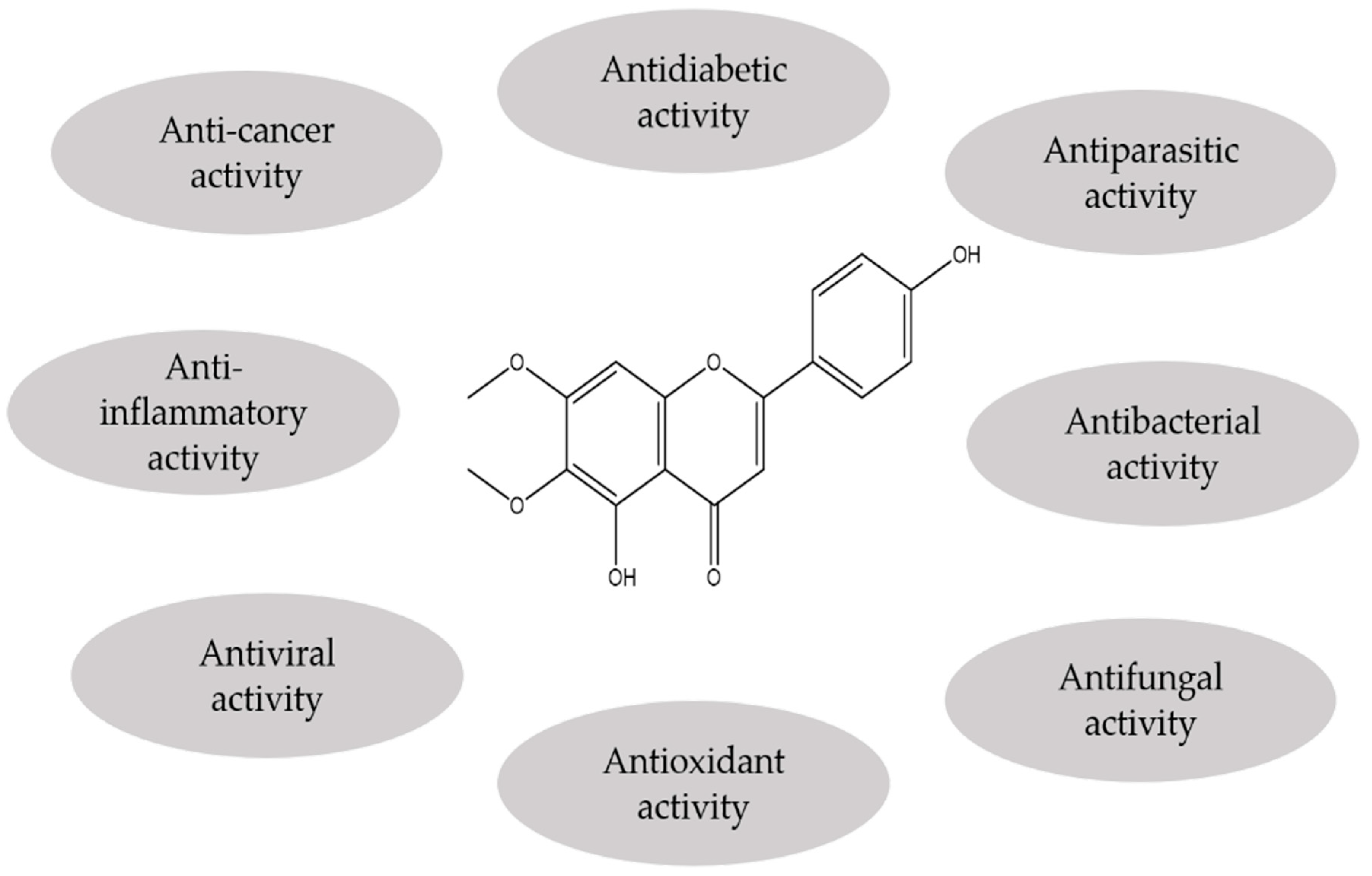
3.2. Biological and Pharmacological Properties
3.2.1. Antibacterial and Anti-Fungal Activities
3.2.2. Antiviral and Antiparasitic Activities
3.2.3. Antioxidant Activity
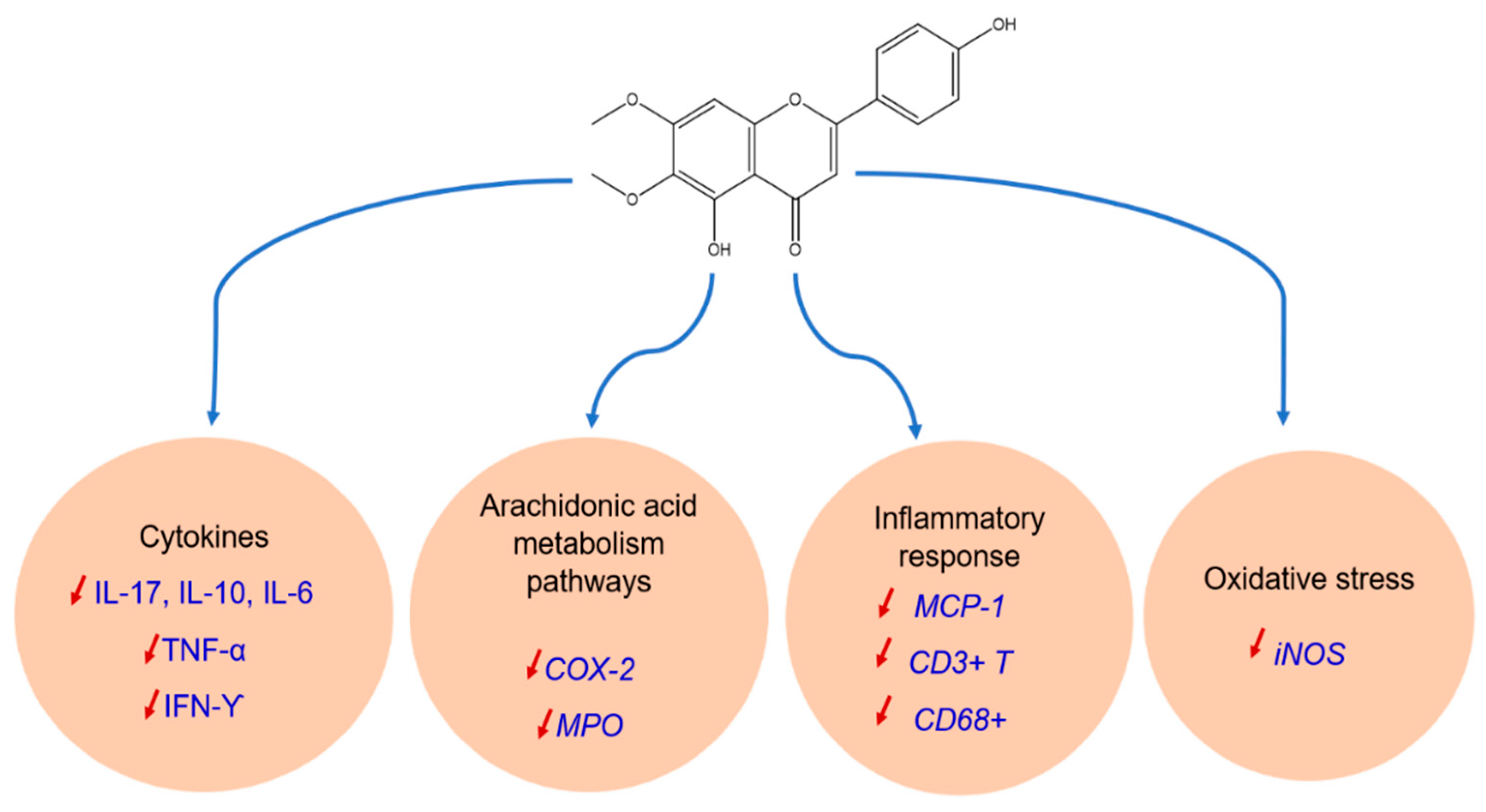
3.2.4. Anti-Inflammatory Activity
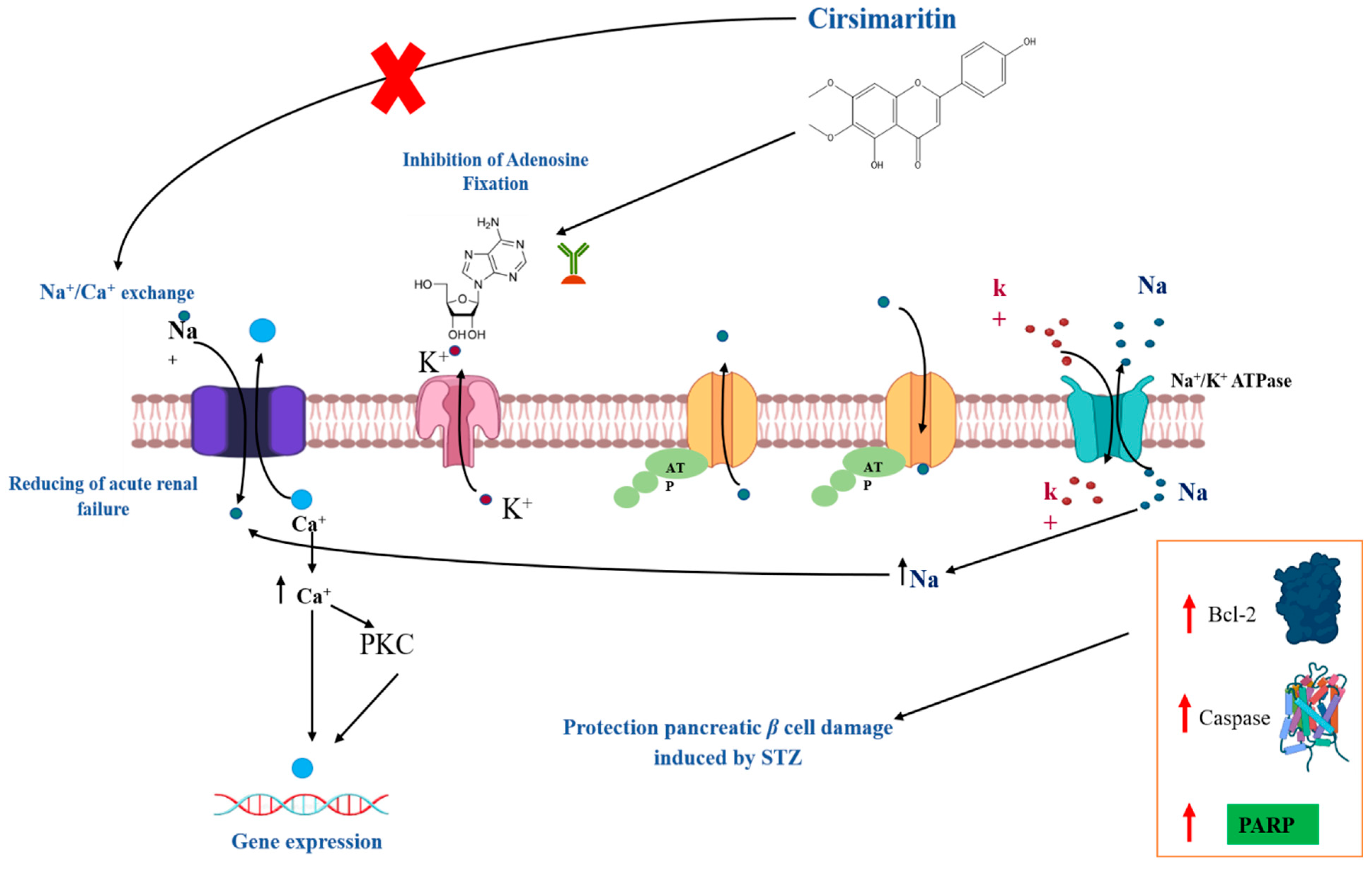
3.2.5. Antidiabetic Activity
3.2.6. Anti-Cancer Activity
3.2.7. Other Biological Activities
4. Conclusions and Perspectives
Author Contributions
Funding
Institutional Review Board Statement
Informed Consent Statement
Data Availability Statement
Conflicts of Interest
References
- Bouyahya, A.; El Omari, N.; Elmenyiy, N.; Guaouguaou, F.-E.; Balahbib, A.; Belmehdi, O.; Salhi, N.; Imtara, H.; Mrabti, H.N.; El-Shazly, M.; et al. Moroccan Antidiabetic Medicinal Plants: Ethnobotanical Studies, Phytochemical Bioactive Compounds, Preclinical Investigations, Toxicological Validations and Clinical Evidences; Challenges, Guidance and Perspectives for Future Management of Diabetes Worldwide. Trends Food Sci. Technol. 2021, 115, 147–254. [Google Scholar] [CrossRef]
- Bouyahya, A.; El Omari, N.; Hakkur, M.; El Hachlafi, N.; Charfi, S.; Balahbib, A.; Guaouguaou, F.-E.; Rebezov, M.; Maksimiuk, N.; Shariati, M.A.; et al. Sources, Health Benefits, and Biological Properties of Zeaxanthin. Trends Food Sci. Technol. 2021, 118, 519–538. [Google Scholar] [CrossRef]
- Bouyahya, A.; Guaouguaou, F.-E.; El Omari, N.; El Menyiy, N.; Balahbib, A.; El-Shazly, M.; Bakri, Y. Anti-Inflammatory and Analgesic Properties of Moroccan Medicinal Plants: Phytochemistry, in Vitro and in Vivo Investigations, Mechanism Insights, Clinical Evidences and Perspectives. J. Pharm. Anal. 2021, 12, 35–37. [Google Scholar] [CrossRef] [PubMed]
- Chamkhi, I.; Benali, T.; Aanniz, T.; El Menyiy, N.; Guaouguaou, F.-E.; El Omari, N.; El-Shazly, M.; Zengin, G.; Bouyahya, A. Plant-Microbial Interaction: The Mechanism and the Application of Microbial Elicitor Induced Secondary Metabolites Biosynthesis in Medicinal Plants. Plant Physiol. Biochem. 2021, 167, 269–295. [Google Scholar] [CrossRef] [PubMed]
- Rauf, A.; Akram, M.; Semwal, P.; Mujawah, A.A.H.; Muhammad, N.; Riaz, Z.; Munir, N.; Piotrovsky, D.; Vdovina, I.; Bouyahya, A.; et al. Antispasmodic Potential of Medicinal Plants: A Comprehensive Review. Oxid. Med. Cell. Longev. 2021, 2021, e4889719. [Google Scholar] [CrossRef] [PubMed]
- Pina, L.T.; Serafini, M.R.; Oliveira, M.A.; Sampaio, L.A.; Guimarães, J.O.; Guimarães, A.G. Carvone and Its Pharmacological Activities: A Systematic Review. Phytochemistry 2022, 196, 113080. [Google Scholar] [CrossRef]
- Aboulaghras, S.; Sahib, N.; Bakrim, S.; Benali, T.; Charfi, S.; Guaouguaou, F.-E.; Omari, N.E.; Gallo, M.; Montesano, D.; Zengin, G. Health Benefits and Pharmacological Aspects of Chrysoeriol. Pharmaceuticals 2022, 15, 973. [Google Scholar] [CrossRef]
- El Omari, N.; Jaouadi, I.; Lahyaoui, M.; Benali, T.; Taha, D.; Bakrim, S.; El Menyiy, N.; El Kamari, F.; Zengin, G.; Bangar, S.P. Natural Sources, Pharmacological Properties, and Health Benefits of Daucosterol: Versatility of Actions. Appl. Sci. 2022, 12, 5779. [Google Scholar] [CrossRef]
- Bakrim, S.; Machate, H.; Benali, T.; Sahib, N.; Jaouadi, I.; Omari, N.E.; Aboulaghras, S.; Bangar, S.P.; Lorenzo, J.M.; Zengin, G. Natural Sources and Pharmacological Properties of Pinosylvin. Plants 2022, 11, 1541. [Google Scholar] [CrossRef]
- Bouyahya, A.; Chamkhi, I.; Benali, T.; Guaouguaou, F.-E.; Balahbib, A.; El Omari, N.; Taha, D.; Belmehdi, O.; Ghokhan, Z.; El Menyiy, N. Traditional Use, Phytochemistry, Toxicology, and Pharmacology of Origanum majorana L. J. Ethnopharmacol. 2021, 265, 113318. [Google Scholar] [CrossRef]
- Bouyahya, A.; Mechchate, H.; Benali, T.; Ghchime, R.; Charfi, S.; Balahbib, A.; Burkov, P.; Shariati, M.A.; Lorenzo, J.M.; Omari, N.E. Health Benefits and Pharmacological Properties of Carvone. Biomolecules 2021, 11, 1803. [Google Scholar] [CrossRef] [PubMed]
- Bouyahya, A.; Chamkhi, I.; Guaouguaou, F.-E.; Benali, T.; Balahbib, A.; El Omari, N.; Taha, D.; El-Shazly, M.; El Menyiy, N. Ethnomedicinal Use, Phytochemistry, Pharmacology, and Food Benefits of Thymus Capitatus. J. Ethnopharmacol. 2020, 259, 112925. [Google Scholar] [CrossRef] [PubMed]
- El Omari, N.; Bakha, M.; Imtara, H.; Guaouguaoua, F.-E.; Balahbib, A.; Zengin, G.; Bouyahya, A. Anticancer Mechanisms of Phytochemical Compounds: Focusing on Epigenetic Targets. Environ. Sci. Pollut. Res. 2021, 28, 47869–47903. [Google Scholar] [CrossRef]
- El Omari, N.; Bakrim, S.; Bakha, M.; Lorenzo, J.M.; Rebezov, M.; Shariati, M.A.; Aboulaghras, S.; Balahbib, A.; Khayrullin, M.; Bouyahya, A. Natural Bioactive Compounds Targeting Epigenetic Pathways in Cancer: A Review on Alkaloids, Terpenoids, Quinones, and Isothiocyanates. Nutrients 2021, 13, 3714. [Google Scholar] [CrossRef] [PubMed]
- El Omari, N.; El Menyiy, N.; Zengin, G.; Goh, B.H.; Gallo, M.; Montesano, D.; Naviglio, D.; Bouyahya, A. Anticancer and Anti-Inflammatory Effects of Tomentosin: Cellular and Molecular Mechanisms. Separations 2021, 8, 207. [Google Scholar] [CrossRef]
- El Omari, N.; Ezzahrae Guaouguaou, F.; El Menyiy, N.; Benali, T.; Aanniz, T.; Chamkhi, I.; Balahbib, A.; Taha, D.; Shariati, M.A.; Zengin, G.; et al. Phytochemical and Biological Activities of Pinus halepensis Mill., and Their Ethnomedicinal Use. J. Ethnopharmacol. 2021, 268, 113661. [Google Scholar] [CrossRef]
- Cheriet, T.; Ben-Bachir, B.; Thamri, O.; Seghiri, R.; Mancini, I. Isolation and Biological Properties of the Natural Flavonoids Pectolinarin and Pectolinarigenin—A Review. Antibiotics 2020, 9, 417. [Google Scholar] [CrossRef]
- Williamson, G.; Barron, D.; Shimoi, K.; Terao, J. In Vitro Biological Properties of Flavonoid Conjugates Found in Vivo. Free Radic. Res. 2005, 39, 457–469. [Google Scholar] [CrossRef]
- Caltagirone, S.; Rossi, C.; Poggi, A.; Ranelletti, F.O.; Natali, P.G.; Brunetti, M.; Aiello, F.B.; Piantelli, M. Flavonoids Apigenin and Quercetin Inhibit Melanoma Growth and Metastatic Potential. Int. J. Cancer 2000, 87, 595–600. [Google Scholar] [CrossRef]
- Xing, N.; Meng, X.; Wang, S. Isobavachalcone: A Comprehensive Review of Its Plant Sources, Pharmacokinetics, Toxicity, Pharmacological Activities and Related Molecular Mechanisms. Phytother. Res. 2022, 36, 3120–3142. [Google Scholar] [CrossRef]
- Ferreira, A.; Pousinho, S.; Fortuna, A.; Falcão, A.; Alves, G. Flavonoid Compounds as Reversal Agents of the P-Glycoprotein-Mediated Multidrug Resistance: Biology, Chemistry and Pharmacology. Phytochem. Rev. 2015, 14, 233–272. [Google Scholar] [CrossRef]
- Dey, D.; Biswas, P.; Paul, P.; Mahmud, S.; Ema, T.I.; Khan, A.A.; Ahmed, S.Z.; Hasan, M.M.; Saikat, A.S.M.; Fatema, B. Natural Flavonoids Effectively Block the CD81 Receptor of Hepatocytes and Inhibit HCV Infection: A Computational Drug Development Approach. Mol. Divers. 2022, 1–14. [Google Scholar] [CrossRef] [PubMed]
- Thapa, A.; Chi, E.Y. Biflavonoids as Potential Small Molecule Therapeutics for Alzheimer’s Disease. In Natural Compounds as Therapeutic Agents for Amyloidogenic Diseases; Springer: Berlin/Heidelberg, Germany, 2015; pp. 55–77. [Google Scholar]
- Bouzid, N.; Moulis, C.; Fouraste, I. Flavones Libres de Artemisia Mesatlantica. Planta Med. 1982, 44, 157–158. [Google Scholar] [CrossRef] [PubMed]
- Saleh, N.A.M.; El-Negoumy, S.I.; Abou-zaid, M.M. Flavonoids of Artemisia Judaica, A. Monosperma and A. Herba-Alba. Phytochemistry 1987, 26, 3059–3064. [Google Scholar] [CrossRef]
- Hasrat, J.A.; De Bruyne, T.; De Backer, J.-P.; Vauquelin, G.; Vlietinck, A.J. (Phytolaccaceae) with Adenosine Antagonistic Properties in Rats: Leads for New Therapeutics in Acute Renal Failure. J. Pharm. Pharmacol. 1997, 49, 1150–1156. [Google Scholar] [CrossRef]
- Kelm, M.A.; Nair, M.G.; Strasburg, G.M.; De Witt, D.L. Antioxidant and Cyclooxygenase Inhibitory Phenolic Compounds from Ocimum Sanctum Linn. Phytomedicine 2000, 7, 7–13. [Google Scholar] [CrossRef]
- Ragasa, C.; Pendon, Z.; Veronica, S.; Rideout, J.A. Antimicrobial flavones from Coleus amboinicus. Philipp. J. Sci. 1999, 128, 347–352. [Google Scholar]
- Rijo, P.; Simões, M.F.; Duarte, A.; Rodríguez, B. Isopimarane Diterpenoids from Aeollanthus Rydingianus and Their Antimicrobial Activity. Phytochemistry 2009, 70, 1161–1165. [Google Scholar] [CrossRef]
- Marino, A.; Zengin, G.; Nostro, A.; Ginestra, G.; Dugo, P.; Cacciola, F.; Miceli, N.; Taviano, M.F.; Filocamo, A.; Bisignano, G.; et al. Antimicrobial Activities, Toxicity and Phenolic Composition of Asphodeline anatolica E. Tuzlaci Leaf Extracts from Turkey. Nat. Prod. Res. 2016, 30, 2620–2623. [Google Scholar] [CrossRef]
- Cazzola, R.; Cestaro, B. Antioxidant Spices and Herbs Used in Diabetes. In Diabetes: Oxidative Stress and Dietary Antioxidants; Elsevier: Amsterdam, The Netherlands, 2014; pp. 89–97. [Google Scholar]
- Shin, M.-S.; Park, J.Y.; Lee, J.; Yoo, H.H.; Hahm, D.-H.; Lee, S.C.; Lee, S.; Hwang, G.S.; Jung, K.; Kang, K.S. Anti-Inflammatory Effects and Corresponding Mechanisms of Cirsimaritin Extracted from Cirsium japonicum var. maackii Maxim. Bioorgan. Med. Chem. Lett. 2017, 27, 3076–3080. [Google Scholar] [CrossRef]
- Bower, A.M.; Real Hernandez, L.M.; Berhow, M.A.; De Mejia, E.G. Bioactive Compounds from Culinary Herbs Inhibit a Molecular Target for Type 2 Diabetes Management, Dipeptidyl Peptidase IV. J. Agric. Food Chem. 2014, 62, 6147–6158. [Google Scholar] [CrossRef] [PubMed]
- Yan, H.; Wang, H.; Ma, L.; Ma, X.; Yin, J.; Wu, S.; Huang, H.; Li, Y. Cirsimaritin Inhibits Influenza A Virus Replication by Downregulating the NF-ΚB Signal Transduction Pathway. Virol. J. 2018, 15, 88. [Google Scholar] [CrossRef] [PubMed]
- Ibañez, E.; Kubátová, A.; Señoráns, F.J.; Cavero, S.; Reglero, U.; Hawthorne, S.B. Subcritical Water Extraction of Antioxidant Compounds from Rosemary Plants. J. Agric. Food Chem. 2003, 51, 375–382. [Google Scholar] [CrossRef]
- Jipa, S.; Zaharescu, T.; Kappel, W.; Dǎneţ, A.F.; Popa, C.V.; Bumbac, M.; Gorghiu, L.M.; Maris, A.M. The Effects of γ-Irradiation on the Antioxidant Activity of Rosemary Extract. Optoelectron. Adv. Mater. Rapid Commun. 2009, 3, 1315–1320. [Google Scholar]
- Tasdemir, D.; Kaiser, M.; Brun, R.; Yardley, V.; Schmidt, T.J.; Tosun, F.; Rüedi, P. Antitrypanosomal and Antileishmanial Activities of Flavonoids and Their Analogues: In Vitro, in Vivo, Structure-Activity Relationship, and Quantitative Structure-Activity Relationship Studies. Antimicrob. Agents Chemother. 2006, 50, 1352–1364. [Google Scholar] [CrossRef]
- Al Ati, H.Y.; Fawzy, G.A.; El Gamal, A.A.; Khalil, A.T.; El Din El Tahir, K.; Abdel-Kader, M.S.; Gilani, A.-H. Phytochemical and Biological Evaluation of Buddleja Polystachya Growing in Saudi Arabia. Pak. J. Pharm. Sci. 2015, 28, 1533–1540. [Google Scholar]
- Morita, N.; Shimizu, M. Naokata Orita and Ineo Shimizu: Of Irsium of the Leaves Iavonoids Components Resources. XXI Runts (Compositae) in Japan. ÝCirsium MAKINO 1963, 83, 615–618. [Google Scholar]
- Shilin, Y.; Roberts, M.F.; Phillipson, J.D. Methoxylated Flavones and Coumarins from Artemisia Annua. Phytochemistry 1989, 28, 1509–1511. [Google Scholar] [CrossRef]
- Namba, T.; Hattori, M.; Takehana, Y.; Tsunezuka, M.; Tomimori, T.; Kizu, H.; Miyaichi, Y.; Medical, T. A flavone Artemisia capillaris. Phytochemistry 1983, 22, 1057–1058. [Google Scholar] [CrossRef]
- Sanz, J.F.; Barbera, O.; Alberto, M.J. Sesquiterpene lactones from Artemisia hispanica. Phytochemistry 1989, 28, 2163–2167. [Google Scholar] [CrossRef]
- Zhang, W.; Zhao, D.B.; Li, M.J.; Liu, X.H.; Wang, H.Q. Studies on Flavonoid Constituents from Herbs of Artemisia Ordosica II. Zhongguo Zhong Yao Za Zhi Zhongguo Zhongyao Zazhi China J. Chin. Mater. Medica 2006, 31, 1959–1961. [Google Scholar]
- Chandrasekharan, I.; Khan, H.A.; Ghanim, A. Flavonoids from Artemisia scoparia. Planta Med. 1981, 43, 310–311. [Google Scholar] [CrossRef]
- Chemesova, I.I.; Belenovskaya, L.M.; Markova, L.P. Flavonoids of Artemisia xanthochroa. Chem. Nat. Compd. 1985, 20, 748. [Google Scholar] [CrossRef]
- de Sousa Andrade, L.M.; de Oliveira, A.B.M.; Leal, A.L.A.B.; de Alcântara Oliveira, F.A.; Portela, A.L.; de Sousa Lima Neto, J.; de Siqueira-Júnior, J.P.; Kaatz, G.W.; da Rocha, C.Q.; Barreto, H.M. Antimicrobial Activity and Inhibition of the NorA Efflux Pump of Staphylococcus Aureus by Extract and Isolated Compounds from Arrabidaea brachypoda. Microb. Pathog. 2020, 140, 103935. [Google Scholar] [CrossRef] [PubMed]
- Weimann, C.; Göransson, U.; Pongprayoon-Claeson, U.; Claeson, P.; Bohlin, L.; Rimpler, H.; Heinrich, M. Spasmolytic Effects of Baccharis Conferta and Some of Its Constituents. J. Pharm. Pharmacol. 2001, 54, 99–104. [Google Scholar] [CrossRef] [PubMed]
- Graver, R.E.J.; Veitch, N.C. An 8-hydroxylated external flavone and its 8-O-glucoside from Becium grandiflorum. Phytochemistry 1998, 47, 7–10. [Google Scholar]
- Szoka, L.; Nazaruk, J.; Stocki, M.; Isidorov, V. Santin and Cirsimaritin from Betula Pubescens and Betula Pendula Buds Induce Apoptosis in Human Digestive System Cancer Cells. J. Cell. Mol. Med. 2021, 25, 11085–11096. [Google Scholar] [CrossRef]
- Sen, A.; Turan, S.O.; Bitis, L. Bioactivity-Guided Isolation of Anti-Proliferative Compounds from Endemic Centaurea kilaea. Pharm. Biol. 2016, 55, 541–546. [Google Scholar] [CrossRef]
- Al-Wahaibi, L.H.; Mahmood, A.; Khan, M.; Alkhathlan, H.Z. Phytochemical Analysis and Bioactivity Screening of Three Medicinal Plants of Saudi Arabia. Trop. J. Pharm. Res. 2020, 19, 371–376. [Google Scholar] [CrossRef]
- Diaa, Y.; August, W.F. Constituents of the Aerial Parts of Ruta Montana. Planta Med. 1995, 61, 279. [Google Scholar]
- Lee, D.; Kim, K.H.; Lee, J.; Hwang, G.S.; Lee, H.L.; Hahm, D.H.; Huh, C.K.; Lee, S.C.; Lee, S.; Kang, K.S. Protective Effect of Cirsimaritin against Streptozotocin-Induced Apoptosis in Pancreatic Beta Cells. J. Pharm. Pharmacol. 2017, 69, 875–883. [Google Scholar] [CrossRef] [PubMed]
- Lee, J.S.; Paje, L.A.; Rodriguez, J.P.; Kang, K.S.; Hahm, D.H.; Shim, J.S.; Choi, Y.J.; Lee, S. Validation of an HPLC/UV Analysis Method for Cirsimaritin in Cirsium japonicum var. Maackii. Korean J. Pharmacogn. 2020, 51, 217–221. [Google Scholar] [CrossRef]
- Zhu, M.; Phillipson, J.D.; Greengrass, P.M.; Bowery, N.G. Chemical and Biological Investigation of the Root Bark of Clerodendrum mandarinorum. Planta Med. 1996, 62, 393–396. [Google Scholar] [CrossRef] [PubMed]
- Dawé, A.; Mbiantcha, M.; Yakai, F.; Jabeen, A.; Ali, M.S.; Lateef, M.; Ngadjui, B.T. Flavonoids and Triterpenes from Combretum Fragrans with Anti-Inflammatory, Antioxidant and Antidiabetic Potential. Z. Nat.-Sect. C J. Biosci. 2018, 73, 211–219. [Google Scholar] [CrossRef] [PubMed]
- Fattahi, M.; Nazeri, V.; Torras-Claveria, L.; Sefidkon, F.; Cusido, R.M.; Zamani, Z.; Palazon, J. Identification and Quantification of Leaf Surface Flavonoids in Wild-Growing Populations of Dracocephalum kotschyi by LC-DAD-ESI-MS. Food Chem. 2013, 141, 139–146. [Google Scholar] [CrossRef]
- Tahtah, Y.; Wubshet, S.G.; Kongstad, K.T.; Heskes, A.M.; Pateraki, I.; Møller, B.L.; Jäger, A.K.; Staerk, D. High-Resolution PTP1B Inhibition Profiling Combined with High-Performance Liquid Chromatography-High-Resolution Mass Spectrometry-Solid-Phase Extraction-Nuclear Magnetic Resonance Spectroscopy: Proof-of-Concept and Antidiabetic Constituents in Crude Extra. Fitoterapia 2016, 110, 52–58. [Google Scholar] [CrossRef] [PubMed]
- Liu, Y.; Ho, D.K.; Cassady, J.M. Isolation of Potential Cancer Chemopreventive Agents. J. Nat. Prod. 1992, 5, 7–13. [Google Scholar]
- Lin, S.; Zhang, Q.W.; Zhang, N.N.; Zhang, Y.X. Determination of Flavonoids in Buds of Herba Artemisiae Scopariae by HPLC. Zhongguo Zhong Yao Za Zhi Zhongguo Zhongyao Zazhi China J. Chin. Mater. Medica 2005, 30, 591–594. [Google Scholar]
- Isobe, T.; Doe, M.; Morimoto, Y.; Nagata, K.; Ohsaki, A. The Anti-Helicobacter Pylori Flavones in a Brazilian Plant, Hyptis fasciculata, and the Activity of Methoxyflavones. Biol. Pharm. Bull. 2006, 29, 1039–1041. [Google Scholar] [CrossRef] [Green Version]
- Yu, Z.; Zhu, H.; Yang, X.; Sun, Q.; Hao, X. Study on Chemical Constituents from Incarvillea Arguta and Their Accelerating PC-12 Cell Differentiation. Zhongguo Zhong Yao Za Zhi Zhongguo Zhongyao Zazhi China J. Chin. Mater. Medica 2005, 30, 1335–1338. [Google Scholar]
- Bai, N.; He, K.; Roller, M.; Lai, C.S.; Shao, X.; Pan, M.H.; Bily, A.; Ho, C.T. Flavonoid Glycosides from Microtea debilis and Their Cytotoxic and Anti-Inflammatory Effects. Fitoterapia 2011, 82, 168–172. [Google Scholar] [CrossRef] [PubMed]
- Berim, A.; Park, J.J.; Gang, D.R. Unexpected Roles for Ancient Proteins: Flavone 8-Hydroxylase in Sweet Basil Trichomes Is a Rieske-Type, PAO-Family Oxygenase. Plant J. 2014, 80, 385–395. [Google Scholar] [CrossRef] [PubMed]
- Vieira, R.F.; Grayer, R.J.; Paton, A.; Simon, J.E. Genetic Diversity of Ocimum gratissimum L. Based on Volatile Oil Constituents, Flavonoids and RAPD Markers. Biochem. Syst. Ecol. 2001, 29, 287–304. [Google Scholar] [CrossRef]
- Bosabalidis, A.; Gabrieli, C.; Niopas, I. Flavone Aglycones in Glandular Hairs of Origanum × intercedens. Phytochemistry 1998, 49, 1549–1553. [Google Scholar] [CrossRef]
- Khaliq, S.; Volk, F.J.; Frahm, A.W. Phytochemical Investigation of Perovskia abrotanoides. Planta Med. 2007, 73, 77–83. [Google Scholar] [CrossRef] [PubMed]
- Zhong, J.; Huang, C.G.; Yu, Y.J.; Li, Z.Q.; Wang, W.; Huang, X.Z.; Liu, W.X.; Yuan, Y.; Jiang, Z.Y. Chemical Constituents from Perovskia Atriplicifolia. Zhongguo Zhong Yao Za Zhi Zhongguo Zhongyao Zazhi China J. Chin. Mater. Medica 2015, 40, 1108–1113. [Google Scholar] [CrossRef]
- De Azevedo Maia, G.L.; Dos Santos Falcão-Silva, V.; Aquino, P.G.V.; De Araújo-Júnior, J.X.; Tavares, J.F.; Da Silva, M.S.; Rodrigues, L.C.; De Siqueira-Júnior, J.P.; Barbosa-Filho, J.M. Flavonoids from Praxelis clematidea R.M. King and Robinson Modulate Bacterial Drug Resistance. Molecules 2011, 16, 4828–4835. [Google Scholar] [CrossRef] [PubMed]
- Abdelhalim, A.; Karim, N.; Chebib, M.; Aburjai, T.; Khan, I.; Johnston, G.A.R.; Hanrahan, J.R. Antidepressant, Anxiolytic and Antinociceptive Activities of Constituents from Rosmarinus Officinalis. J. Pharm. Pharm. Sci. 2015, 18, 448–459. [Google Scholar] [CrossRef]
- Cavero, S.; Jaime, L.; Martín-Álvarez, P.J.; Señoráns, F.J.; Reglero, G.; Ibañez, E. In Vitro Antioxidant Analysis of Supercritical Fluid Extracts from Rosemary (Rosmarinus officinalis L.). Eur. Food Res. Technol. 2005, 221, 478–486. [Google Scholar] [CrossRef]
- Pérez-Sánchez, A.; Borrás-Linares, I.; Barrajón-Catalán, E.; Arráez-Román, D.; González-Álvarez, I.; Ibáñez, E.; Segura-Carretero, A.; Bermejo, M.; Micol, V. Evaluation of the Intestinal Permeability of Rosemary (Rosmarinus officinalis L.) Extract Polyphenols and Terpenoids in Caco-2 Cell Monolayers. PLoS ONE 2017, 12, e0172063. [Google Scholar] [CrossRef] [Green Version]
- Xu, J.-H.; Lo, Y.M.; Chang, W.-C.; Huang, D.-W.; Wu, J.S.-B.; Jhang, Y.-Y.; Huang, W.-C.; Ko, C.-Y.; Shen, S.-C. Identification of Bioactive Components from Ruellia tuberosa L. on Improving Glucose Uptake in TNF-α-Induced Insulin-Resistant Mouse FL83B Hepatocytes. Evid. Based Complement. Alternat. Med. 2020, 2020, 6644253. [Google Scholar] [CrossRef] [PubMed]
- Srivedavyasasri, R.; Hayes, T.; Ross, S.A. Phytochemical and Biological Evaluation of Salvia apiana. Nat. Prod. Res. 2017, 31, 2058–2061. [Google Scholar] [CrossRef] [PubMed]
- Kanetis, L.; Exarchou, V.; Charalambous, Z.; Goulas, V. Edible Coating Composed of Chitosan and Salvia fruticosa Mill. Extract for the Control of Grey Mould of Table Grapes. J. Sci. Food Agric. 2017, 97, 452–460. [Google Scholar] [CrossRef] [PubMed]
- Exarchou, V.; Kanetis, L.; Charalambous, Z.; Apers, S.; Pieters, L.; Gekas, V.; Goulas, V. HPLC-SPE-NMR Characterization of Major Metabolites in Salvia fruticosa Mill. Extract with Antifungal Potential: Relevance of Carnosic Acid, Carnosol, and Hispidulin. J. Agric. Food Chem. 2015, 63, 457–463. [Google Scholar] [CrossRef]
- Kavvadias, D.; Monschein, V.; Sand, P.; Riederer, P.; Schreier, P. Constituents of Sage (Salvia officinalis) with in Vitro Affinity to Human Brain Benzodiazepine Receptor. Planta Med. 2003, 69, 113–117. [Google Scholar] [CrossRef]
- Miski, M.; Ulubelen, A.; Johansson, C.; Mabry, T.J. Antibacterial Activity Studies of Flavonoids from Salvia palaestina. J. Nat. Prod. 1983, 46, 874–875. [Google Scholar] [CrossRef]
- Cottiglia, F.; Casu, L.; Bonsignore, L.; Casu, M.; Floris, C.; Sosa, S.; Altinier, G.; Della Loggia, R. Topical Anti-Inflammatory Activity of Flavonoids and a New Xanthone from Santolina insularis. Z. Nat.-Sect. C J. Biosci. 2005, 60, 63–66. [Google Scholar] [CrossRef]
- Malmir, M.; Gohari, A.R.; Saeidnia, S.; Silva, O. A New Bioactive Monoterpene-Flavonoid from Satureja khuzistanica. Fitoterapia 2015, 105, 107–112. [Google Scholar] [CrossRef]
- Shafiq, N.; Riaz, N.; Ahmed, S.; Ashraf, M.; Ejaz, S.A.; Ahmed, I.; Saleem, M.; Touseef, M.I.; Tareen, R.B.; Jabbar, A. Bioactive Phenolics from Seriphidium stenocephalum. J. Asian Nat. Prod. Res. 2013, 15, 286–293. [Google Scholar] [CrossRef]
- Beer, M.F.; Frank, F.M.; Germán Elso, O.; Ernesto Bivona, A.; Cerny, N.; Giberti, G.; Luis Malchiodi, E.; Susana Martino, V.; Alonso, M.R.; Patricia Sülsen, V.; et al. Trypanocidal and Leishmanicidal Activities of Flavonoids Isolated from Stevia satureiifolia var. Satureiifolia. Pharm. Biol. 2016, 54, 2188–2195. [Google Scholar] [CrossRef] [Green Version]
- Ren, X.; Bao, Y.; Zhu, Y.; Liu, S.; Peng, Z.; Zhang, Y.; Zhou, G. Isorhamnetin, Hispidulin, and Cirsimaritin Identified in Tamarix Ramosissima Barks from Southern Xinjiang and Their Antioxidant and Antimicrobial Activities. Molecules 2019, 24, 390. [Google Scholar] [CrossRef] [PubMed]
- Polatoğlu, K.; Karakoç, Ö.C.; Demirci, F.; Gökçe, A.; Gören, N. Chemistry and Biological Activities of Tanacetum chiliophyllum var. oligocephalum Extracts. J. AOAC Int. 2013, 96, 1222–1227. [Google Scholar] [CrossRef] [PubMed]
- Stefkov, G.; Kulevanova, S.; Miova, B.; Dinevska-Kjovkarovska, S.; Mølgaard, P.; Jäger, A.K.; Josefsen, K. Effects of Teucrium polium spp. Capitatum Flavonoids on the Lipid and Carbohydrate Metabolism in Rats. Pharm. Biol. 2011, 49, 885–892. [Google Scholar] [CrossRef]
- Ben Sghaier, M.; Skandrani, I.; Nasr, N.; Franca, M.G.D.; Chekir-Ghedira, L.; Ghedira, K. Flavonoids and Sesquiterpenes from Tecurium ramosissimum Promote Antiproliferation of Human Cancer Cells and Enhance Antioxidant Activity: A Structure-Activity Relationship Study. Environ. Toxicol. Pharmacol. 2011, 32, 336–348. [Google Scholar] [CrossRef] [PubMed]
- Wang, R.F.; Yang, X.W.; Ma, C.M.; Liu, H.Y.; Shang, M.Y.; Zhang, Q.Y.; Cai, S.Q.; Park, J.H. Trollioside, a New Compound from the Flowers of Trollius chinensis. J. Asian Nat. Prod. Res. 2004, 6, 139–144. [Google Scholar] [CrossRef] [PubMed]
- Nyiligira, E.; Viljoen, A.M.; Van Heerden, F.R.; Van Zyl, R.L.; Van Vuuren, S.F.; Steenkamp, P.A. Phytochemistry and in Vitro Pharmacological Activities of South African Vitex (Verbenaceae) Species. J. Ethnopharmacol. 2008, 119, 680–685. [Google Scholar] [CrossRef] [PubMed]
- Hussain, W.; Amir, A.; Rasool, N. Computer-Aided Study of Selective Flavonoids against Chikungunya Virus Replication Using Molecular Docking and DFT-Based Approach. Struct. Chem. 2020, 31, 1363–1374. [Google Scholar] [CrossRef]
- Kiran, G.; Karthik, L.; Devi, M.S.; Sathiyarajeswaran, P.; Kanakavalli, K.; Kumar, K.M.; Kumar, D.R. In Silico Computational Screening of Kabasura Kudineer-Official Siddha Formulation and JACOM against SARS-CoV-2 Spike Protein. J. Ayurveda Integr. Med. 2020, 13, 100324. [Google Scholar] [CrossRef]
- Tasdemir, D.; Lack, G.; Brun, R.; Rüedi, P.; Scapozza, L.; Perozzo, R. Inhibition of Plasmodium f Alciparum Fatty Acid Biosynthesis: Evaluation of FabG, FabZ, and FabI as Drug Targets for Flavonoids. J. Med. Chem. 2006, 49, 3345–3353. [Google Scholar] [CrossRef]
- Quintanilla-Licea, R.; Vargas-Villarreal, J.; Verde-Star, M.J.; Rivas-Galindo, V.M.; Torres-Hernández, Á.D. Antiprotozoal Activity against Entamoeba Histolytica of Flavonoids Isolated from Lippia Graveolens Kunth. Molecules 2020, 25, 2464. [Google Scholar] [CrossRef]
- Kolak, U.; Hacibekiroǧlu, I.; Öztürk, M.; Özgökçe, F.; Topçu, G.; Ulubelen, A. Antioxidant and Anticholinesterase Constituents of Salvia Poculata. Turk. J. Chem. 2009, 33, 813–823. [Google Scholar] [CrossRef]
- Liu, C.Z.; Murch, S.J.; El-Demerdash, M.; Saxena, P.K. Artemisia Judaica L.: Micropropagation and Antioxidant Activity. J. Biotechnol. 2004, 110, 63–71. [Google Scholar] [CrossRef] [PubMed]
- Burki, S.; Mehjabeen; Burki, Z.G.; Shah, Z.A.; Imran, M.; Khan, M. Phytochemical Screening, Antioxidant, and in Vivo Neuropharmacological Effect of Monotheca buxifolia (Falc.) Barks Extract. Pak. J. Pharm. Sci. 2018, 31, 1519–1528. [Google Scholar] [PubMed]
- Kuo, C.-F.; Su, J.-D.; Chiu, C.-H.; Peng, C.-C.; Chang, C.-H.; Sung, T.-Y.; Huang, S.-H.; Lee, W.-C.; Chyau, C.-C. Anti-Inflammatory Effects of Supercritical Carbon Dioxide Extract and Its Isolated Carnosic Acid from Rosmarinus officinalis Leaves. J. Agric. Food Chem. 2011, 59, 3674–3685. [Google Scholar] [CrossRef]
- Moghaddam, G.; Ebrahimi, S.A.; Rahbar-Roshandel, N.; Foroumadi, A. Antiproliferative Activity of Flavonoids: Influence of the Sequential Methoxylation State of the Flavonoid Structure: Antiproliferative flavonoids from dracocephalum kotschyi. Phytother. Res. 2012, 26, 1023–1028. [Google Scholar] [CrossRef]
- Pathak, G.; Singh, S.; Kumari, P.; Raza, W.; Hussain, Y.; Meena, A. Cirsimaritin, a Lung Squamous Carcinoma Cells (NCIH-520) Proliferation Inhibitor. J. Biomol. Struct. Dyn. 2020, 39, 3312–3323. [Google Scholar] [CrossRef]
- Quan, Z.; Gu, J.; Dong, P.; Lu, J.; Wu, X.; Wu, W.; Fei, X.; Li, S.; Wang, Y.; Wang, J.; et al. Reactive Oxygen Species-Mediated Endoplasmic Reticulum Stress and Mitochondrial Dysfunction Contribute to Cirsimaritin-Induced Apoptosis in Human Gallbladder Carcinoma GBC-SD Cells. Cancer Lett. 2010, 295, 252–259. [Google Scholar] [CrossRef]
- Patel, D.K. Health Beneficial Aspect and Therapeutic Potential of Cirsimaritin in the Medicine for the Treatment of Human Health Complications. Curr. Bioact. Compd. 2022, 18, 27–38. [Google Scholar] [CrossRef]
- Awad, B.M.; Abd-Alhaseeb, M.M.; Habib, E.S.; Ibrahim, A.K.; Ahmed, S.A. Antitumor Activity of Methoxylated Flavonoids Separated from Achillea fragrantissima Extract in Ehrlich’s Ascites Carcinoma Model in Mice. J. Herbmed Pharmacol. 2020, 9, 28–34. [Google Scholar] [CrossRef]
- Kim, H.; Kim, I.; Dong, Y.; Lee, I.-S.; Kim, J.; Kim, J.-S.; Woo, J.-T.; Cha, B.-Y. Melanogenesis-Inducing Effect of Cirsimaritin through Increases in Microphthalmia-Associated Transcription Factor and Tyrosinase Expression. Int. J. Mol. Sci. 2015, 16, 8772–8788. [Google Scholar] [CrossRef] [Green Version]
- Lee, D.; Jung, Y.; Baek, J.Y.; Shin, M.-S.; Lee, S.; Hahm, D.-H.; Lee, S.C.; Shim, J.S.; Kim, S.N.; Kang, K.S. Cirsimaritin Contributes to the Estrogenic Activity of Cirsium Japonicum var. Maackii through the Activation of Estrogen Receptor α: Estrogenic Compounds of Cirsium Japonicum var. Maackii. Bull. Korean Chem. Soc. 2017, 38, 1486–1490. [Google Scholar] [CrossRef]
- Manurung, K.; Sulastri, D.; Zubir, N.; Ilyas, S. In Silico Anticancer Activity and in Vitro Antioxidant of Flavonoids in Plectranthus amboinicus. Pharmacogn. J. 2020, 12, 1573–1577. [Google Scholar] [CrossRef]
- Plochmann, K.; Korte, G.; Koutsilieri, E.; Richling, E.; Riederer, P.; Rethwilm, A.; Schreier, P.; Scheller, C. Structure–Activity Relationships of Flavonoid-Induced Cytotoxicity on Human Leukemia Cells. Arch. Biochem. Biophys. 2007, 460, 1–9. [Google Scholar] [CrossRef] [PubMed]
- Yeon Park, J.; Young Kim, H.; Shibamoto, T.; Su Jang, T.; Cheon Lee, S.; Suk Shim, J.; Hahm, D.-H.; Lee, H.-J.; Lee, S.; Sung Kang, K. Beneficial Effects of a Medicinal Herb, Cirsium japonicum var. Maackii, Extract and Its Major Component, Cirsimaritin on Breast Cancer Metastasis in MDA-MB-231 Breast Cancer Cells. Bioorgan. Med. Chem. Lett. 2017, 27, 3968–3973. [Google Scholar] [CrossRef] [PubMed]
- Zehra, B.; Ahmed, A.; Sarwar, R.; Khan, A.; Farooq, U.; Abid Ali, S.; Al-Harrasi, A. Apoptotic and Antimetastatic Activities of Betulin Isolated from Quercus incana against Non-Small Cell Lung Cancer Cells. Cancer Manag. Res. 2019, 11, 1667–1683. [Google Scholar] [CrossRef]
- Abdalla, S.S.; Zarga, M.A. Effects of Cirsimaritin, a Flavone Isolated from Artemisia judaica, on Isolated Guinea-Pig Ileum. Planta Med. 1987, 53, 322–324. [Google Scholar] [CrossRef]
- González-Trujano, M.E.; Hernández-Sánchez, L.Y.; Muñoz Ocotero, V.; Dorazco-González, A.; Guevara Fefer, P.; Aguirre-Hernández, E. Pharmacological Evaluation of the Anxiolytic-like Effects of Lippia graveolens and Bioactive Compounds. Pharm. Biol. 2017, 55, 1569–1576. [Google Scholar] [CrossRef]
- Wang, J.-P.; Chang, L.-C.; Hsu, M.-F.; Chen, S.-C.; Kuo, S.-C. Inhibition of Formyl-Methionyl-Leucyl-Phenylalanine-Stimulated Respiratory Burst by Cirsimaritin Involves Inhibition of Phospholipase D Signaling in Rat Neutrophils. Naunyn. Schmiedebergs Arch. Pharmacol. 2002, 366, 307–314. [Google Scholar] [CrossRef]


| Plants | Part Used | Botanical Families | Type of Extract | Concentration/Fraction | References |
|---|---|---|---|---|---|
| Aeollanthus rydingianus | Aerial parts | Lamiaceae | Me2CO Extract | 58 mg | [29] |
| Artemisia annua | Leaves Stems | Asteraceae | Methanolic extract | 1.45 mg | [40] |
| Artemisia capillaris | Spikes | Asteraceae | Methanolic extract | not specified | [41] |
| Artemisia hispanica | Aerial parts | Asteraceae | Methanolic extract | not specified | [42] |
| Artemisia judaica | Leaves Stems | Asteraceae | Ethanolic extract | not specified | [25] |
| Artemisia meatlanticae | Aerial parts | Asteraceae | Ether extract | not specified | [24] |
| Artemisia monosperma | Leaves Stems | Asteraceae | Ethanolic extract | not specified | [25] |
| Artemisia ordosica II | not specified | Asteraceae | not specified | not specified | [43] |
| Artemisia scoparia | Dried inflorescence | Asteraceae | Chloroform extract | not specified | [44] |
| Artemisia xanthochroa | Epigeal parts | Asteraceae | Ethanolic extract | not specified | [45] |
| Arrabidaea brachypoda | Flowers | Bignoniaceae | Ethanolic extract | not specified | [46] |
| Asphodeline anatolica | Leaves | Liliaceae | Acetone and methanol extract | not specified | [30] |
| Baccharis conferta | Aerial parts | Asteraceae | Ethanolic extract | not specified | [47] |
| Becium grandiflorum | Leaves | Lamiaceae | Methanolic extract | not specified | [48] |
| Betula pendula | Buds | Betulaceae | Carbon dioxide supercritical extraction | 3.79 mg/g | [49] |
| Betula pubescens | Buds | Betulaceae | Carbon dioxide supercritical extraction | 4.21 mg/g | [49] |
| Buddleja polystachya | Aerial parts | Buddlejaceae | Ethanolic extract (cold maceration) | not specified | [38] |
| Centuarea kilaea | Aerial parts | Asteraceae | Chloroform extract | 10.2 mg | [50] |
| Centaurea pseudosinaica | Entire plant (leaves, flowers, stems) | Asteraceae | Ethanolic extract | 0.52 g | [51] |
| Centaurea scoparia | Aerial parts | Asteraceae | Ethanolic extract | 10 mg | [52] |
| Cirsium martimum | Leaves | Asteraceae | Methanolic extract | not specified | [39] |
| Cirsium japonicum | Aerial parts Leaves | Asteraceae | Ethanolic Extract | 6.24 mg/g | [32] |
| 6.24 mg/g | [53] | ||||
| 37.13 mg/g | [54] | ||||
| Clerodendrum mandarinorum | Root bark | Lamiaceae | Ethanolic Extract | 50 mg | [55] |
| Combretum fragrans | Leaves | Combretaceae | Methanolic extract | not specified | [56] |
| Dracocephalum kotschyi | Leaves | Lamiaceae | Diethyl Ether extract | 97.3–637.6 µg/g | [57] |
| Eremophila lucida | Leaves | Myoporaceae | Ethyl acetate extract | not specified | [58] |
| Eriodictyon californicum | Leaves | Hydrophyllaceae | Ethanol extract | 3.85 mg | [59] |
| Herba artemisiae Scopariae | Buds | Compositae | Ethyl acetate extract (ultrasonic) | not specified | [60] |
| Hyptis fasciculata | Aerial parts | Labiatae | Chloroform and methanol extract | 19 mg | [61] |
| Incarvillea arguta | not specified | Bignoniaceae | not specified | not specified | [62] |
| Microtea debilis | Whole plant Aerial parts | Microteaceae | Aqueous & ethanol extractEthanol extract | 0.7 mg/mL 65 mg | [26] [63] |
| Ocimum basilicum | Tricoms | Lamiaceae | Crude protein extract with HCl | not specified | [64] |
| Ocimum gratissimum | Above-ground biomass | Lamiaceae | Clevenger apparatus | 10% of the total flavonoids | [65] |
| Origanum intercedens | Leaves | Lamiaceae | Chloroform extract | not specified | [66] |
| Osimum sanctum | Leaves Stems | Labiatae | Chloroform extract | 1 mg | [27] |
| Perovskia abrotanoies | Aerial parts | Lamiaceae | Methanolic extract | 10 mg | [67] |
| Perovskia atriplicifolia | Leaves | Lamiaceae | Ethanolic Extract | not specified | [68] |
| Praxelis clematidea | Aerial parts | Asteraceae | Ethanolic Extract (exhaustive maceration) | not specified | [69] |
| Rosmarinus officinalis | Leaves | Lamiaceae | Ethyl acetate extract | not specified | [70] |
| Subcritical extraction with water | 1.72% | [35] | |||
| Supercritical fluid extraction | 0.54–17.59% | [71] | |||
| Ethanolic extract | not specified | [36] | |||
| Super critical fluid extraction | not specified | [72] | |||
| Ruellia tuberosa | Leaves & stems | Acanthaceae | Methanolic extract | 805 µg/g | [73] |
| Salvia apiana | Aerial parts | Lamiaceae | Aqueous ethanolic extract | not specified | [74] |
| Salvia fruticosa | Aerial parts | Lamiaceae | Acetonic extract (Soxtec system) | not specified | [75] |
| Ethyl acetate extract (Soxtec extraction) | 10.4 mg/g | [76] | |||
| Salvia officinalis | Leaves | Lamiaceae | Methanolic extraction (ultrasonic bath) | 194 mg | [77] [31] |
| Salvia palaestina | Leaves | Lamiaceae | Benzene extract (Soxhlet) | 30 mg | [78] |
| Santolina insularis | Leaves | Asteraceae | Methanolic extract | 6.9 mg | [79] |
| Satureja khuzistanica | Aerial parts | Lamiaceae | Ethyl acetate extraction | 5 mg | [80] |
| Seriphidium stenocephalum | not specified | Asteraceae | Methanolic extract | 15 mg | [81] |
| Stevia satureiifolia | Aerial parts | Asteraceae | Dichloromethane extract | 1.9% | [82] |
| Tamarix ramosissima | Bark | Tamaricaceae | Ethanolic extract | 13.35 µg/mg | [83] |
| Tanacetum chiliophyllum | Stems | Compositae | Ethyl acetate extract | 36 mg | [84] |
| Teucrium polium | Aerial parts | Lamiaceae | Alcohol extraction | not specified | [85] |
| Teucrium ramosissimum | Leaves | Lamiaceae | Chloroformic extract | not specified | [86] |
| Trollius chinensis | Flowers | Ranunculaceae | Ethanolic extract | 14 mg | [87] |
| Vitex rehmannii | Aerial parts | Verbenaceae | Acetone extract | 5 mg | [88] |
| Methods Used | Strains Tested | Key Results | References |
|---|---|---|---|
| Disk diffusion assay | Escherichia coli | MIC = 31.25 μg/mL, MBC = 125 μg/mL | [78] |
| Klebsiella pneumonia | MIC = 31.25 μg/mL, MBC = 125 μg/mL | ||
| Pseudomonas aeruginosa | MIC = 45 μg/mL, MBC = 90 μg/mL | ||
| Proteus vulgaris | MIC = 31.25 μg/mL, MBC = 125 μg/mL | ||
| Staphylococcus aureus | MIC = 31.25 μg/mL, MBC = 125 μg/mL | ||
| Staphylococcus epidermis | MIC = 62.5 μg/mL, MBC = 125 μg/mL | ||
| Disk diffusion method | Aspergillus niger | Φ = 13 mm at 40 µg | [28] |
| Basilus subtilis | Φ = 0 mm at 40 µg | ||
| Candida albicans | Φ = 12 mm at 40 µg | ||
| Escherichia coli | Φ = 0 mm at 40 µg | ||
| Pseudomonas aeruginosa | Φ = 13 mm at 40 µg | ||
| Staphylococcus aureus | Φ = 11 mm at 40 µg | ||
| Trichophyton mentagrophytes | Φ = 14 mm at 40 µg | ||
| Agar diffusion method | Candida albicans | No activity | [29] |
| Escherichia coli | No activity | ||
| Enterococcus hirae | Growth zone inhibition | ||
| Mycobacterium smegmatis | No activity | ||
| Pseudomonas aeruginosa | No activity | ||
| Staphylococcus aureus | Growth zone inhibition | ||
| Micro-dilution technique | Aspergilus fumigatus | MIC = 1.95 μg/mL | [51] |
| Bacillus subtilis | MIC = 0.03 μg/mL | ||
| Candida albicans | MIC = 1.95 μg/mL | ||
| Escherichia coli | MIC = 11.25 μg/mL | ||
| Geotrichum candidum | MIC = 0.48 μg/mL | ||
| Pseudomonas aeruginosa | MIC = 50.0 μg/mL | ||
| Streptococcus pneumoniae | MIC = 7.81 μg/mL | ||
| Syncephalastrum racemosum | MIC = 12.5 μg/mL | ||
| Micro-dilution method | Bacillus cereus | MIC = 5 mg/mL, MBC = 20 mg/mL | [83] |
| Escherichia coli | MIC = 10 mg/mL, MBC = 25 mg/mL | ||
| Listeria monocytogenes | MIC = 5 mg/mL, MBC = 10 mg/mL | ||
| Pseudomonas aeruginosa | MIC > 10 mg/mL, MBC = NA | ||
| Salmonella typhimurium | MIC > 10 mg/mL, MBC = NA | ||
| Shigella castellani | MIC = 5 mg/mL, MBC = 15 mg/mL | ||
| Staphylococcus aureus | MIC = 5 mg/mL, MBC = 15 mg/mL |
| Used Method | Key Results | References |
|---|---|---|
| DPPH radical scavenging activity | EC50 = 11.3 µg/mL | [35] |
| β-carotene bleaching, superoxide anion radical, and ABTS cation radical scavenging activity assays | No antioxidant activity | [93] |
| ABTS assay | TEAC (µM) = 2.04 | [86] |
| CUPRAC assay | TEAC (µM) = 4.7 | |
| RP (Reducing power) assay | TEAC (µM) = 0.95 | |
| FRAP assay | TEAC (µM) = 0.625 | |
| DPPH Scavenging activity | Inhibition efficiency (%) = 80–100 at a concentration of 100 µg/mL | [53] |
| FRAP assay | AC = 203.39 to 681.27 µmol Fe2+/100 g DW at a concentration of 97.38–637.66 µg/g DW | [57] |
| DPPH assay | Significantly higher capacity to detoxify oxygen radicals | [94] |
| DPPH scavenging | % inhibition (at 500 µg/mL) = 89.55 | [95] |
| Superoxide scavenging | % inhibition (at 500 µg/mL) = 82.10 | |
| Hydrogen peroxide scavenging | % inhibition (at 500 µg/mL) = 80.55 | |
| DPPH assay | IC50 = 55.9 µM | [56] |
| Origin | Biological Model (In Vitro or In Vivo) | Experimental Approach | Results and Mechanism of Action | References |
|---|---|---|---|---|
| Synthetic compound | human cancer cell lines namely COLO-205, MDA-MB-231, HaCaT, K562, A431, A549, MCF-7, PC-3, NCIH- 520, normal cell lines WRL-68, HEK 293 and L132 and in primary macrophages | MTT assay Inhibitory potential and binding interaction with the selected targets were analyzed through in vitro and in silico analysis | Inhibited the growth of NCIH-520 cell-line (IC50 23.29 μM) Induced apoptosis Inhibited the activity of ODC and CATD Exhibited a good binding in silico score with the selected targets and it non-mutagenic | [98] |
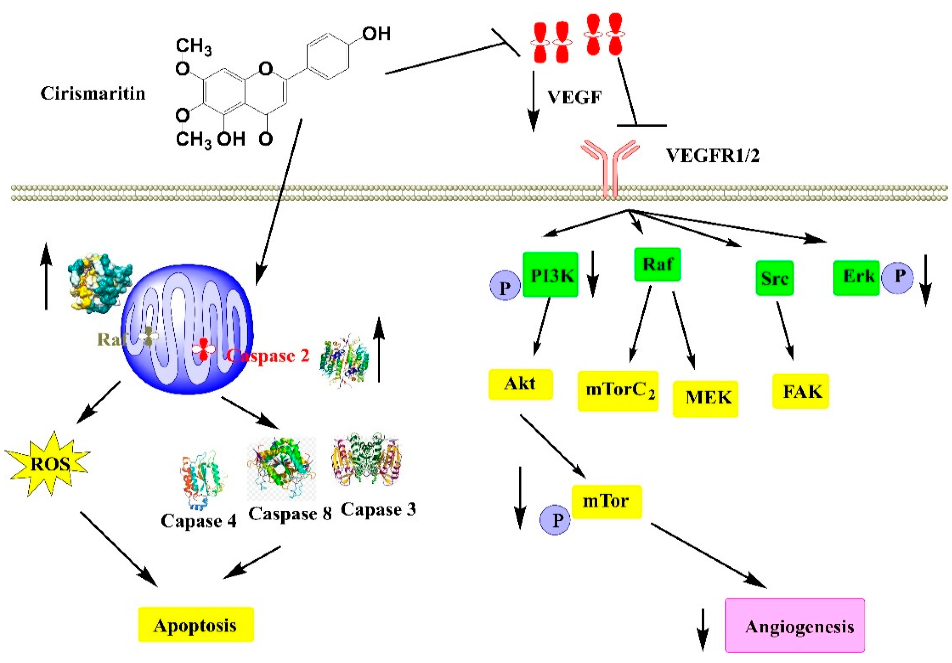
| Synthetic compound | gallbladder carcinoma cell lines GBC-SD and GBCSD18H cells, gastric carcinoma cell line BGC-823 cells, and hepatoma cell line SMMC-7721 cells | Cytotoxicity assay Cell apoptosis assay Cell mitochondrial membrane potential assay Subcellular fractionation Western blot Small interference RNA RT)-PCR Detection of intracellular ROS | Inhibited the growth of tumor cells Induced mitochondrial apoptosis in GBC-SD cells Triggered endoplasmic reticulum (ER) stress Down-regulated the phosphorylation of Akt | [99] |
| Centaurea kilaea | one normal cell line (L-929, mouse fibroblast) three human cancer cell lines (Hela, cervix carcinoma; MCF-7, breast carcinoma; PC-3, prostate carcinoma | MTT assay | Inhibited the growth of MCF-7 and PC-3 | [50] |
| Teucrium ramosissimum | Ehrlich’s ascites carcinoma model in mice | (5, 10, 20 mg/kg/d, orally) | Reduced tumor weight compared to EAC-control and cisplatin groups Induced tumor cell necrosis Reduced significantly the level of TNF-α in serum | [101] |
| Teucrium ramosissimum | human chronic myelogenous K562 cells | MTT assay | Exhibited an antiproliferative effect of human cancer cells IC50 = 1.015 × 10⁻⁷ mol/mL | [86] |
| Lithocarpus dealbatus | Murine melanoma B16F10 cells (CRL-6415) | Cell Morphology and Cell Viability Measurement Measurement of Cellular Tyrosinase Activity Melanin Content Measurement Western Blotting | Stimulated melanogenesis in B16F10 cells Activated of CREB as well as upregulation of MITF and tyrosinase expression activated by cAMP signaling | [102] |
| Cirsium japonicum var. maackii | human breast cancer (MCF-7) cell-based | Transactivation assay Proliferative activity | Exerted beneficial effects on MCF-7 cells Increased estrogenic activity | [103] |
| Plectranthus amboinicus | Cancer P-Glycoprotein-1, Cyclin Dependent Kinase-2, and Phosphoinositide-3-Kinase receptors | In silico anticancer Test | Exhibited an important strong anti-cancer effect | [104] |
| Dracocephalum kotschyi Boiss. | AGS, HT-29, HL60, SaOs-2, WEHI-164 and HFFF-P16 cells | MTT assay | Exhibited and antiproliferative activity of malignant cells | [97] |
| Isolated | Human T lymphoblasts (Jurkat Clone E6-1) | Cytotoxicity experiments Flow cytometry | Induced cytotoxicity EC50 = 66.8 µM (24 h) EC50 = 44.4 µM (48 h) | [105] |
| Cirsium japonicum | Breast cancer | Cell proliferation assay Tube-formation assay Western blot analysis | Inhibited the viability of HUVECs in a dose-dependent manner Inhibited angiogenesis by downregulation of VEGF, p-Akt and p-ERK in MDA-MB-231 cells | [106] |
| Betula pubescens and Betula pendula | gastric (AGS), colon (DLD-1) and liver (HepG2) cancer cells | Cell viability assay DNA biosynthesis Colony formation assay Apoptosis assay Western immunoblot Immunofluorescence microscopy | Induced apoptosis Activated caspase-3, caspase-7, caspase-8 and caspase-9 expression Upregulated p53 expression | [49] |
| Quercus incana | non-small cell lung carcinoma (NCI-H460) and normal mouse fibroblast (NIH-3T3) cell lines. | mRNA extraction and qRT-PCR Colony formation assay Flow cytometry analysis Cell cycle analysis Western blot analysis | Induced antiproliferative against NIH 3T3 (IC50 = 26.23 ± 0.053 μM) and in NCI-H460 (IC50 = 38.84 ± 0.037 μM) | [107] |
Publisher’s Note: MDPI stays neutral with regard to jurisdictional claims in published maps and institutional affiliations. |
© 2022 by the authors. Licensee MDPI, Basel, Switzerland. This article is an open access article distributed under the terms and conditions of the Creative Commons Attribution (CC BY) license (https://creativecommons.org/licenses/by/4.0/).
Share and Cite
Benali, T.; Jaouadi, I.; Ghchime, R.; El Omari, N.; Harboul, K.; Hammani, K.; Rebezov, M.; Shariati, M.A.; Mubarak, M.S.; Simal-Gandara, J.; et al. The Current State of Knowledge in Biological Properties of Cirsimaritin. Antioxidants 2022, 11, 1842. https://doi.org/10.3390/antiox11091842
Benali T, Jaouadi I, Ghchime R, El Omari N, Harboul K, Hammani K, Rebezov M, Shariati MA, Mubarak MS, Simal-Gandara J, et al. The Current State of Knowledge in Biological Properties of Cirsimaritin. Antioxidants. 2022; 11(9):1842. https://doi.org/10.3390/antiox11091842
Chicago/Turabian StyleBenali, Taoufiq, Imane Jaouadi, Rokia Ghchime, Nasreddine El Omari, Kaoutar Harboul, Khalil Hammani, Maksim Rebezov, Mohammad Ali Shariati, Mohammad S. Mubarak, Jesus Simal-Gandara, and et al. 2022. "The Current State of Knowledge in Biological Properties of Cirsimaritin" Antioxidants 11, no. 9: 1842. https://doi.org/10.3390/antiox11091842