Article

Tamarix articulata Induced Prevention of Hepatotoxicity Effects of In Vivo Carbon Tetrachloride by Modulating Pro-Inflammatory Serum and Antioxidant Enzymes to Reverse the Liver Fibrosis

by
Abdullah M. Alnuqaydan
1,*,
Abdulmajeed G. Almutary
1,
Mohammed A. Alsahli
2,
Sulaiman Alnasser
3 and
Bilal Rah
1,4,*
1
Department of Medical Biotechnology, College of Applied Medical Sciences, Qassim University, Buraydah 51452, Saudi Arabia
2
Department of Medical Laboratories, College of Applied Medical Sciences, Qassim University, Buraydah 51452, Saudi Arabia
3
Department of Pharmacology and Toxicology, Unaizah College of Pharmacy, Qassim University, Buraydah 51452, Saudi Arabia
4
Iron Biology Group, Sharjah Institute of Medical Research, University of Sharjah, Sharjah 27272, United Arab Emirates
*
Authors to whom correspondence should be addressed.
Antioxidants 2022, 11(9), 1824; https://doi.org/10.3390/antiox11091824
Submission received: 25 July 2022 / Revised: 29 August 2022 / Accepted: 13 September 2022 / Published: 15 September 2022
(This article belongs to the Special Issue Oxidative Stress in Inflammatory Skin and Tissue Disorders)

Abstract

:
This study evaluates the hepatoprotective activity of a Tamarix articulata extract against carbon tetrachloride-mediated hepatotoxicity in Wistar rats. Our results demonstrated that the oral administration of Tamarix articulata extract (50 mg/kg b.w.) significantly restored the serum levels of liver enzymes and antioxidant parameters (superoxide dismutase, catalase, glutathione reductase, and thiobarbituric reactive substances). Histopathology analysis revealed that Tamarix articulata extract significantly reduced hepatic fibrosis by inhibiting the necrosis of hepatocytes. Furthermore, serum pro-inflammatory (tumor necrosis factor-alpha, tumor growth factor-beta, and interleukin-6) markers were significantly restored. However, the anti-inflammatory cytokine adiponectin levels increased to normal levels in the group treated with Tamarix articulata extract. Additionally, we observed diminished reactive oxygen species production and the depolarization of mitochondrial membrane potential in hepatocytes extracted from animal livers treated with Tamarix articulata extract. Our findings suggest that Tamarix articulata extract prevents liver fibrosis induced by carbon tetrachloride and decreases the necrotic population of hepatocytes. These events restored the antioxidant enzymatic activity, serum levels of liver enzymes, and pro-inflammatory markers to their normal levels.

Graphical Abstract

1. Introduction

The liver performs a key role in the regulation of numerous biochemical functions associated with metabolism [1]. Liver injury is a multi-factorial disease caused by environmental and chemical toxins, drugs, and alcohol intake, which induce oxidative stress, leading to complicated pathophysiology. Typically, the end result of this is cirrhosis and hepatocellular carcinoma (HCC) [2,3]. The liver is rich in mitochondria that aid in aerobic metabolism through the electron transport chain (ETC). During the process of oxidative phosphorylation, reactive oxygen species (ROS) are produced, and this means that hepatocytes are highly susceptible to oxidative stress, which ultimately leads to hepatocellular damage. Owing to a number of side effects of the existing therapeutic modalities used for liver diseases, the management of hepatocellular diseases is often poor. Thus, the need to develop efficient, effective, and reliable hepatoprotective drugs from plant sources with fewer adverse effects is seemingly important [4] This notion is supported by various reports that suggest that vegetarian diets and fruits from plant sources are rich in antioxidants. The consumption of such a diet drastically curtails the risk of developing chronic hepatocellular disorders [5].
Environmental toxicants have been documented as inducing oxidative stress by promoting ROS production, which leads to liver injury, necrosis, and tissue damage. [6]. One of the potent environmental toxicants is carbon tetrachloride (CCl4). Upon the administration of CCl4into the body orally or through systemic circulation, the detoxifying enzyme cytochrome P-450 present in the liver converts CCl4into a more toxic form called the trichloromethyl radical (CCl3+) through a process called biotransformation [7]. In the presence of oxygen, CCl3+ is converted into a highly reactive radical form called trichloromethylperoxy (CCl3OO+) [8]. These highly reactive free radical species covalently bind with phospholipid membranes, which in turn induce lipid peroxidation that firstly impairs cell membrane integrity and secondly enhances the permeability of cells, thus causing severe damage to them [9]. Owing to its toxic effects, the use of CCl4is restricted, yet it is still one of the most widely used chemicals for the induction of hepatotoxicity in animal studies. It serves to evaluate the modulation of the pro-inflammatory and anti-scavenging effects of plant products abundant in antioxidants that could act as promising hepatoprotective agents [10].
Recently, antioxidants obtained from various plant remedies have been introduced in therapeutics for liver fibrosis [11]. Such anti-fibrotic therapies are used to modulate oxidative stress and have been entered into clinical trials [12]. Morin, a flavonoid compound with antioxidant activity, primarily isolated from Maclura pomifera, prevents CCl4-mediated liver fibrosis by attenuating the levels of nitric oxide (NO) and MDA to restore the GSR of hepatocytes to its normal levels [13]. Silymarin—a mixture of flavonolignan compounds—exhibits promising antioxidant activity. When administered orally, a 100 mg/kg silymarin dose prevents CCl4-mediated liver fibrosis by reducing the MDA levels, restoring the GSR activity of hepatocytes, and preventing the depolarization of MMP to counter oxidative stress [14]. Epigallocatechin (EGCG) is another polyphenolic compound with antioxidant activity that prevents liver fibrosis in a plethora of CCl4-mediated animal models [15]. Together, these studies suggest that natural antioxidant compounds show much potential to reverse fibrosis by attenuating oxidative stress, and preclinical investigations as part of clinical trials are required for future therapeutics.
Tamarix articulata (TA) is a halophytic plant (belongingto the family Tamaricaceae) commonly found in the deserts of Saudi Arabia. Traditionally called “Athal” in the Arabic language, TA grows extremely well in drought, harsh, and arid conditions. The plant grows to a height of 15 m and a girth of 2 m, as described in our previously published study [16]. Ethnobotanical studies have revealed that TA has been extensively used as folk medicine by the Tafilalet, a tribal population in the South-Eastern area of Morocco. Traditionally, the plant is used to treat various ailments, such as gastrointestinal, skin, heart diseases, and other ailments [17]. Phytochemical analysis revealed the major constituents of TA extract thatdisplay pharmacological activities, as mentioned in our previous study [18]. Crude extracts of the plant have been reported to exhibit anticancer activities by inhibiting cell viability in various types of cancer cells [19]. Owing to the large amount of polyphenolic and flavonoid compounds, the methanolic extract of TA is apromising antioxidant with antiproliferative activities, as stated in our previous research [16,20]. Although TA exhibits some promising pharmacological activities, including antioxidant, antiproliferative, and antilipidemic activities [16,18,19,20,
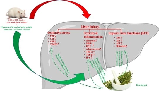
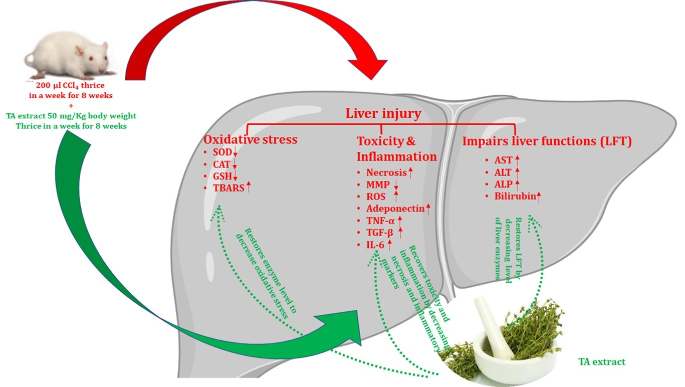
21], there is not a single report suggesting that TA extract exhibits hepatoprotective activity. Therefore, the current study evaluates its hepatoprotective activity against the well-established carbontetrachloride-induced hepatotoxicity in Wistar rats. We found that TA extract reverses liver fibrosis induced by CCl4in animals and restores pro-inflammatory as well as serum enzymes and antioxidant enzymes. Together, these results suggest that TA has a promising hepatoprotective effect and has much potential as a remedy against fibrosis and liver-associated diseases.

2. Materials and Methods

2.1. Chemicals, Reagents, and Kits

Carbon tetrachloride (CCl4) (#PHR1063), silymarin (#S0292), and other chemicals and reagents were purchased from Sigma Aldrich Chemicals Co., St. Louis, Missouri, United States. Kits for the pro-inflammatory (TGF-β #ab119558, TNF-α #ab236712, IL-6 #ab234570, and adiponectin #ab239421) and anti-inflammatory cytokine markers (SOD #ab65354, CAT #ab118184, GSR #ab65322, and TBARS/MDA #ab238537), liver function test (LFT) (AST #ab263882, ALT #234579, ALP #ab83369, and Bil #ab235627) enzymes to detect free radical scavenging activity, mitochondrial membrane potential (MMP) (MMP #ab113852), and ROS (ab #ab186027) detection were purchased from Abcam, Cambridge, United Kingdom.

2.2. Plant Material

TA plant was collected in August 2019 from Qassim Province in Saudi Arabia. The leaves of the TA plant were air-dried in the shade to remove all of the moisture [20]. The data from our previous work identified the plant extracts, while the phytochemical analysis of major constituents of TA extracts displayed various pharmacological activities. A comparative assessment of different parts of the plant was conducted as described in our recently published studies [18,20,21].

2.3. Preparation of Extract

Methanolic extract of TA was prepared as per the standard protocol [21]. Dry TA leaves were collected from the floor and washed with distilled water. After cleaning, the TA leaves were shade-dried for 10 days to ensure complete drying followed by grinding in a kitchen blender to produce a fine powder. The dried TA leaf (100 g) powder was soaked in methanol (300 mL). Using the manual method of extraction, the mixture of dry leafpowder in methanol was constantly stirred on amagnetic stirrer at room temperature for 5 days. The obtained extract was filtered through Whatman filter paper and was concentrated by evaporating the solvent to attain a fine residue powder. The yield of the methanolic extract of the dry leaves of TA amounted to 8.33% and the dry powder of the extract was stored at 4 °C for future use.

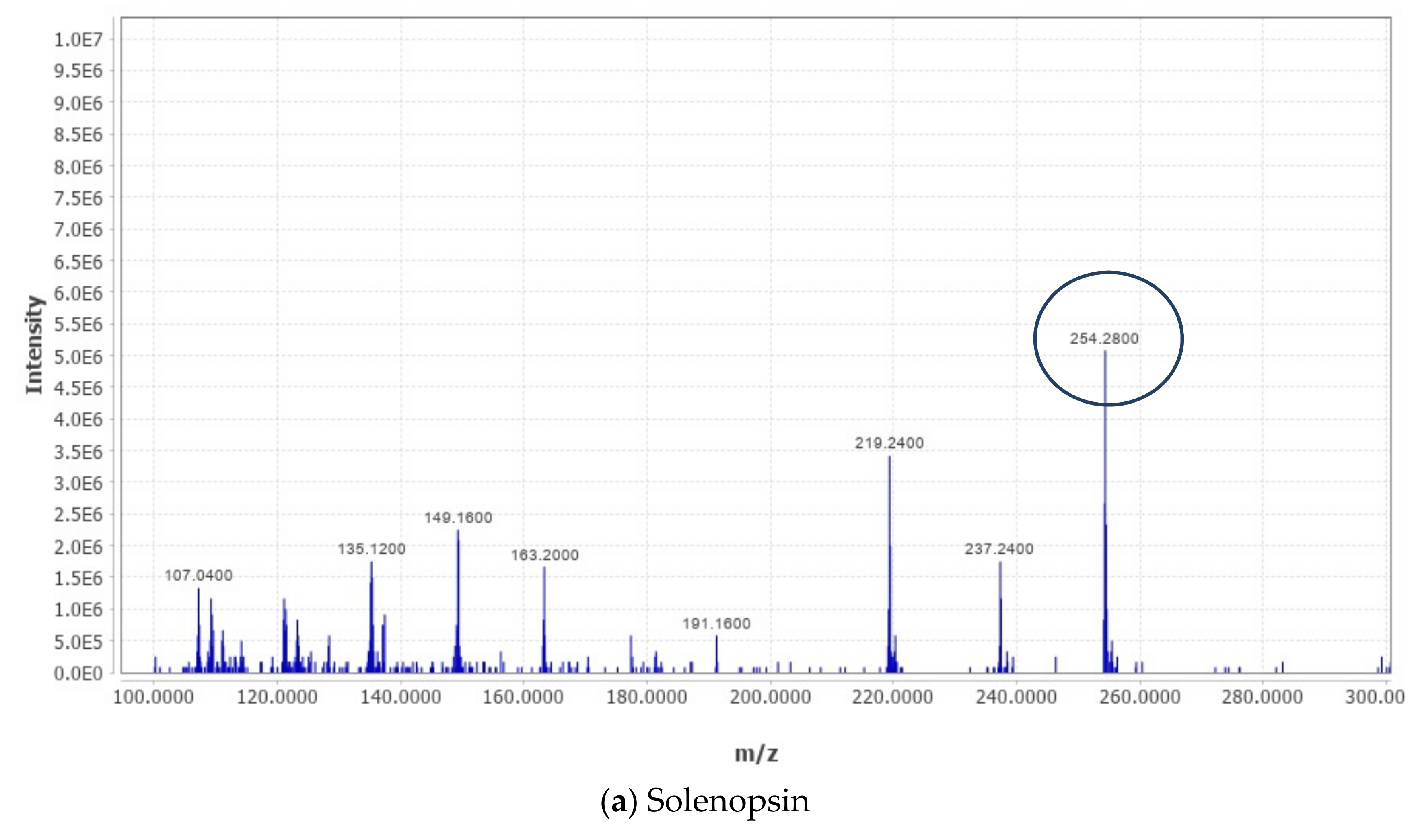
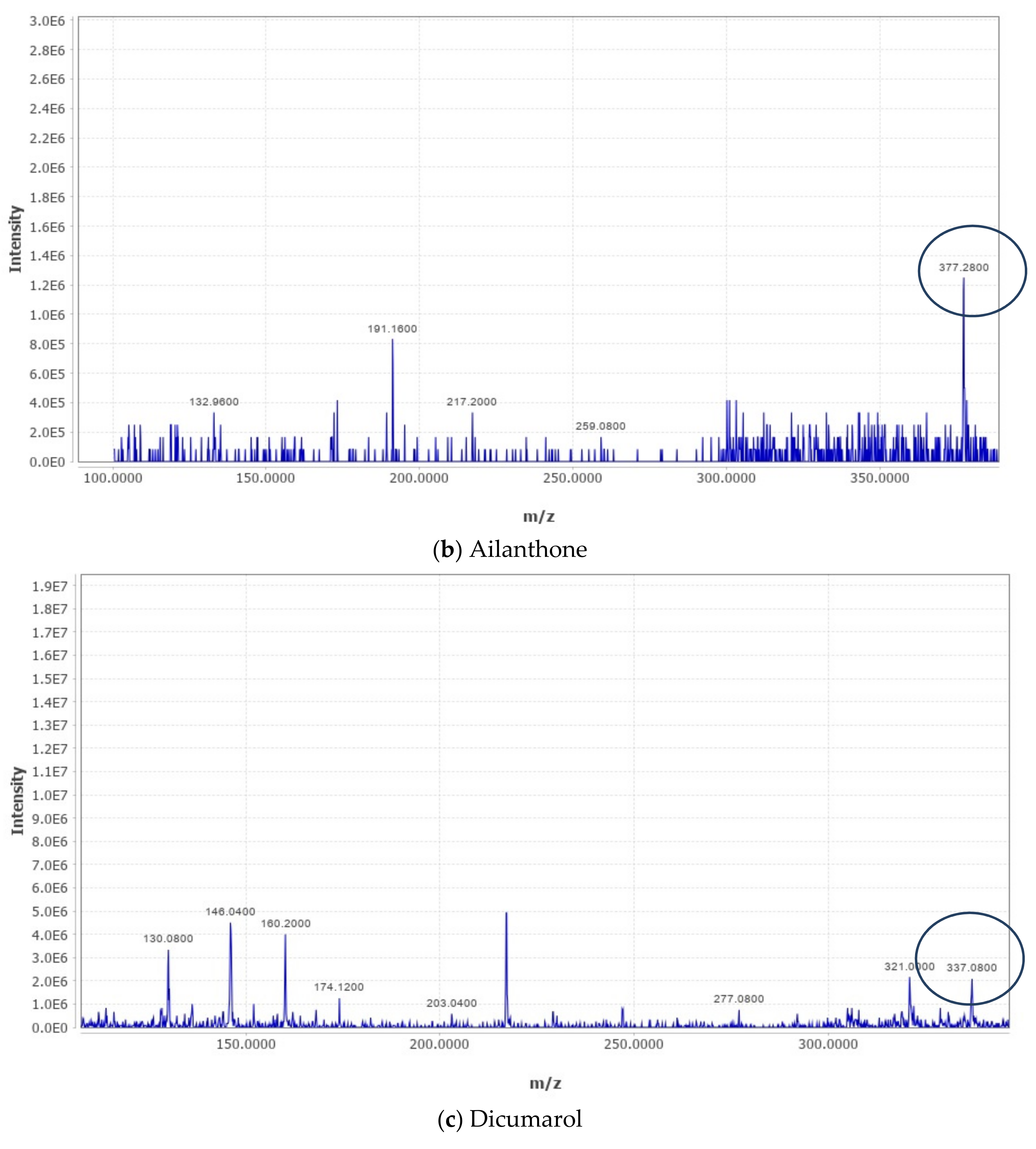
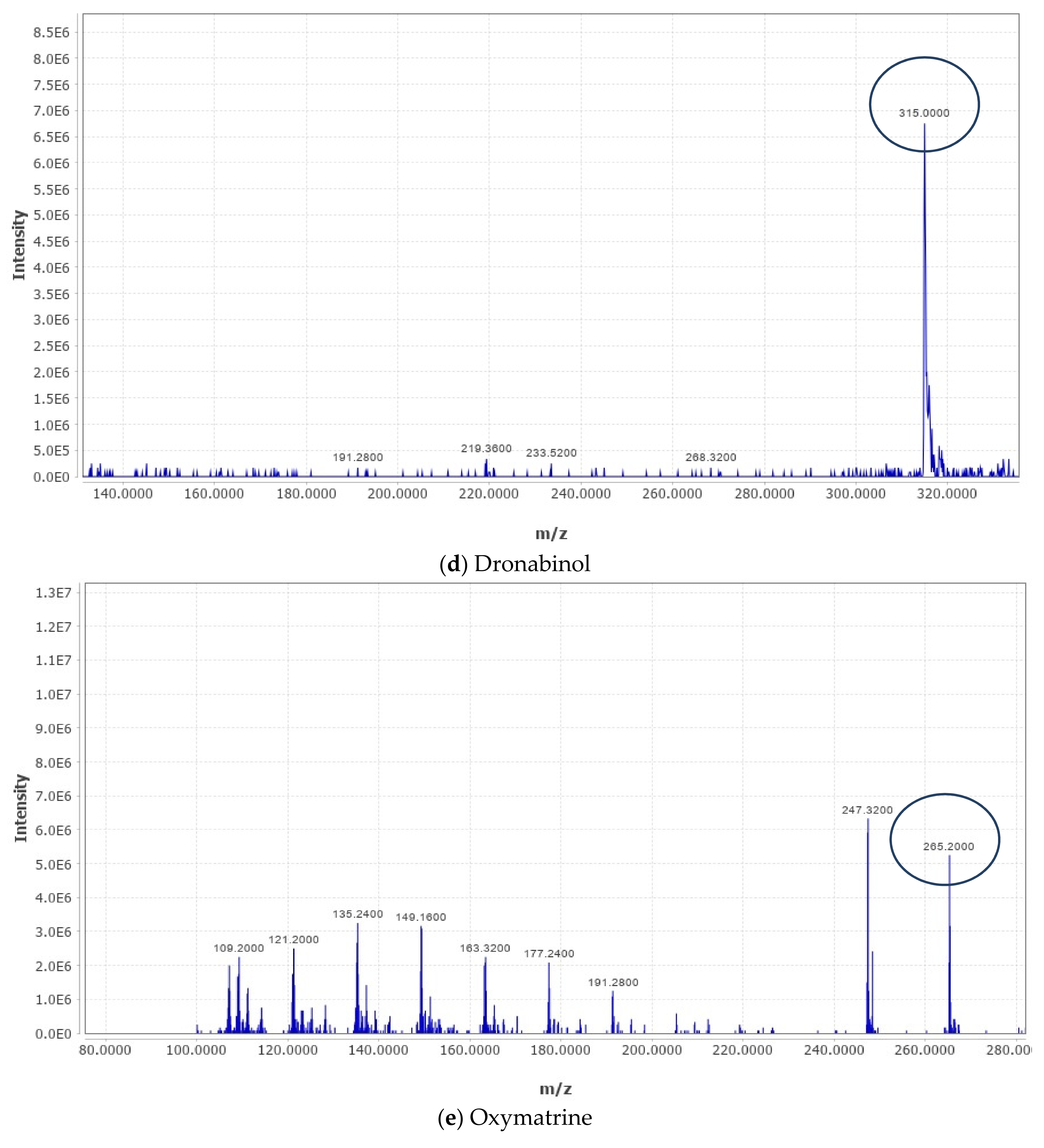
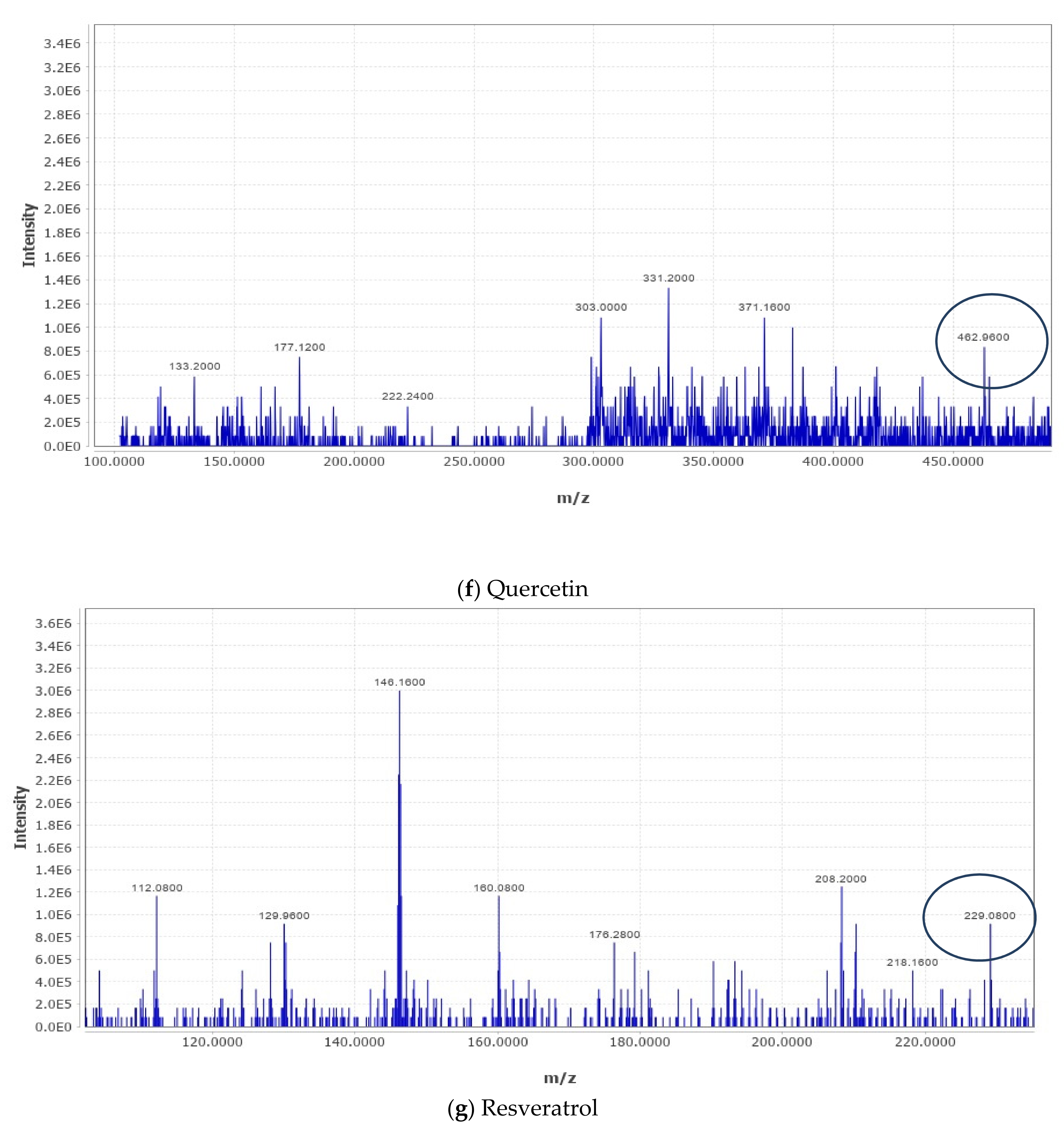
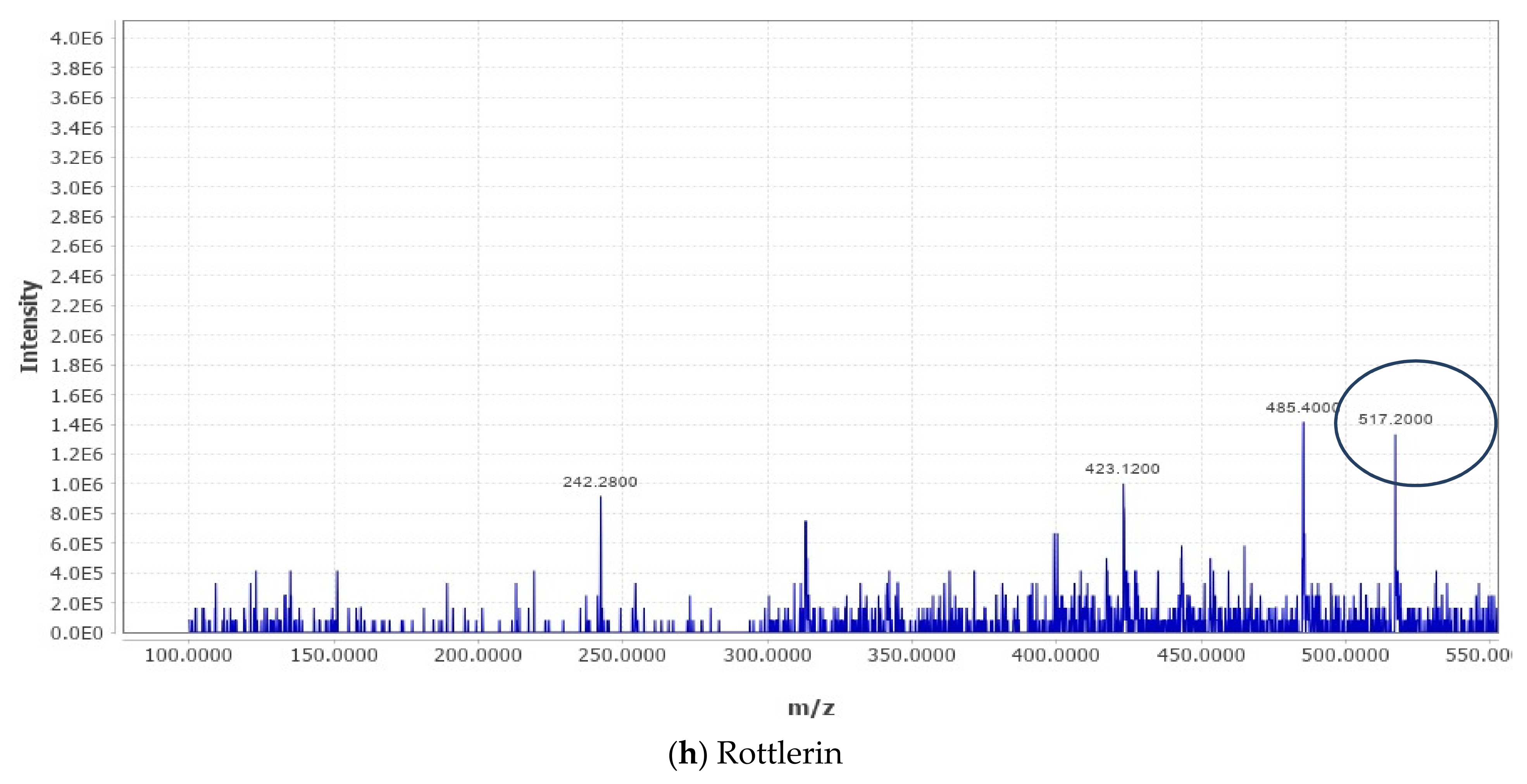
2.4. Liquid Chromatography–Mass Spectrometry (LC–MS) Metabolomic Analysis and Data Processing

The analysis was carried out as described in our recently published studies [18,20,21]. LC–MS metabolomic analysis was undertaken consisting of an ACQUITY UPLC I-Class System (Waters Technologies, Milford, MA, USA) coupled with a 6500Qtrap(AB Sciex, Concord, ON, Canada). Chromatographic separation was completed on a Zorbax XDB C18 column (2.1 × 150 mm. 3.5 µm) (Agilent, Santa Clara, CA, USA) maintained at 40 °C with a flow rate of 300 µL/min. The mobile phase consisted of A (0.1% formic acid in HPLC grade water) and B (0.1% formic acid in HPLC grade acetonitrile). The linear gradient elution was as follows: 2% B (from 0 to 2), 95% B (from 2 to 24), 95% B (held for 2 min), and then 4 min equilibration time. Electrospray ionization mass spectra (ESI-MS) were acquired in the positive mode (ES+), with an electrode voltage of 5500 V. The declustering potential was set to 90 V and the entrance potential was 10 V. Nitrogen was used as the curtain gas (30 psi) and nebulizer gas on the MS. Spectra were collected with a mass range of 100–900 m/z. Data files from the LC were converted to MZxml format using MS Convert (ProteoWizard 3.0.20270). Analysis of the data was conducted using MZ mine software (version 2.53). After importing the data into the MZ mine, a minimum intensity cutoff of 1000 was applied and the retention time was adjusted with a tolerance of 0.2 min. Adjusted peaks were then aligned to one mass list to facilitate identification and comparison. The KEGG Database was used to identify compounds of interest in the finalized list based on m/z with a tolerance of 30 ppm.

2.5. Experimental Animals

Male Wistar rats weighing 100–110 g were procured from KAUST, Saudi Arabia. All animals (Wistar rats) were acclimatized in the institute animal house for at least one week prior to experimentation with a 12 h dark and light cycle at room temperature. Animals were fed with a standard pellet diet and water ad libitum. This research was endorsed by the Ethics Committee of the College of Applied Medical Sciences, Qassim University (cams1-2019-1-14-s-3360).

2.6. CCl4-Induced Hepatotoxicity

The Wistar male rats acclimatized in the animal house were randomly grouped into 7 groups, with each group having 6 rats (42 Wistar rats) per experiment (n = 3, meaning 126 animals for the whole study). Animals were orally dosed with CCl4as mentioned previously [22,23], and other respective concentrations of TA extracts (30, 40, and 50 mg/kg b.w.) using gastric gavage without the administration of any anesthesia agent. Group A was designated as normal, without any chemical or extract being administered. Group B was designated as the CCl4-treated group and the animals were administered 40% CCl4mixed in olive oil orally for 3 alternate days a week for 8 weeks. Group C was designated as 40% CCl4mixed in olive oil and TA extract 30 mg/kg administered orally for 3 alternate days a week for 8 weeks. Group D was designated as 40% CCl4mixed in olive oil and TA extract 40 mg/kg administered orally for 3 alternate days a week for 8 weeks. Group E was designed as 40% CCl4mixed in olive oil and TA extract 50 mg/kg administered orally for 3 alternate days a week for 8 weeks. Group F was designated as 40% CCl4mixed in olive oil and TA extract 60 mg/kg administered orally for 3 alternate days a week for 8 weeks. Group G was designed as 40% CCl4mixed in olive oil and 100 mg/kg b.w. silymarin [24] administered orally for 3 alternate days a week for 8 weeks (Table 3).
To evaluate the effective dose of TA extract with the least toxicity to the animals, we intended to perform a preliminary experiment on animals. This was performed to optimize the dose of TA extract that could exhibit effective hepatoprotective activity with less deleterious effects on animals. After properly acclimatizing the animals, we categorized them randomly into seven groups, as shown in Table 3. The disease control (Group B) animals were orally administered 200 µL 40% CCl4dissolved in olive oil for three alternate days a week for 8 weeks, whereas the control group (Group A) animals were orally administered 200 µL of vehicle. The other groups (Group C, D, E, and F) were orally administered 200 µL of 40% CCl4dissolved in olive oil and 200 µL of TA extract with varying doses (30, 40, 50, and 60 mg/kg b.w., respectively) for three alternate days a week for 8 weeks. However, Group G animals were orally administered 200 µL of standard silymarin compound for three alternate days a week for 8 weeks; this was in addition to 200 µL of40% CCl4dissolved in olive oil. To evaluate the effective dose in terms of the hepatoprotective effect and safe toxicity in animals, we observed the hepatoprotective activity of TA extract against CCl4-mediated liver toxicity in a dose-dependent manner (Tables 4 and 5). Intriguingly, our results suggest that the ideal activity of TA extract was determined at 50 mg/kg b.w. (Group E), and the results were significantly compared with those of standard silymarin (Group G). When tested at 60 mg/kg b.w. (Group F), TA extract displayed toxicity-associated symptoms in animals, such as lethargy, diarrhea, muscle tremors, loss of appetite, etc., and they died before the completion of the study (Table 4 and Table 5). Collectively, the preliminary optimization results suggest that a 50 mg/kg b.w. oral dose of TA extract has the maximum hepatoprotective effect against CCl4-mediated liver toxicity.

2.7. Evaluation of Liver Function Test

Following the completion of the treatment schedules, the animals in each group were sacrificed following methods approved by the Ethics Committee of the College of Applied Medical Sciences, Qassim University (cams1-2019-1-14-s-3360). Once the study was completed, diethyl ether was used to anesthetize the animals, followed by cervical dislocation and operating procedures to extract internal organs for the current study. The blood (1.0 mL) was collected from the tail vein before sacrificing the animals. The collected blood was allowed to clot and was centrifuged at 3000 rpm and 4 °C for 10 min [25]. The supernatant (serum) was collected in sterile microfuge tubes and stored at −80 °C for the analysis of various biochemical parameters. The liver function enzyme levels of alkaline phosphatase (ALP), alanine transaminase (ALT), and aspartate aminotransferase (AST), and the bilirubin levels in serum were estimated as previously described [26]. However, the quantification of pro-inflammatory cytokine markers (TNF-α, IL-6, and TGF-β) and the anti-inflammatory cytokine marker adiponectin levels were investigated with the use of an enzyme-linked immunosorbent assay (ELISA) kit.

2.8. Histopathological Analysis

The liver tissue was extracted from the animals of each group (n = 6) and thoroughly cleaned in PBS at pH 7.4. A small chunk of liver tissue was excised and fixed in 10% formalin to preserve the morphology of the cells from putrification. After processing the samples for paraffin blocks, the thin paraffin sections were cut with ultra-microtome. The sections were subjected to hematoxylin–eosin for inflammation and Masson’s trichome staining to analyze the histopathological changes in the collagen fibers of the hepatic tissue using a microscope [27].

2.9. Detection of In Vivo Antioxidant Enzymes

Briefly, a small chunk of liver tissue (approximately 10%) was chopped into smaller pieces, followed by homogenization (Fisherband 150 efficient homogenizer, #15-340-167, Waltham, MA, USA) in a 2 mL sterile tube by using 1 mL of PBS (pH 7.4). The mixture obtained was subjected to centrifugation at 10,000 rpm for 10 min at 4 °C. The supernatant obtained was used to evaluate the antioxidant enzymes and pro-inflammatory markers. The enzymatic activity of catalase (CAT), superoxide dismutase (SOD), glutathione reductase (GSR), and malondialdehyde (MDA) per milligram of the protein concentration of hepatic tissue homogenate was estimated utilizing a spectrophotometric method (Bradford method) [28].

2.10. Determination of Reactive Oxygen Species (ROS)

After extracting the livers from the animals in each group (n = 6), part of the liver was excised, washed thoroughly with PBS followed by resuspension in culture media, and subjected to tissue dissociation with a tissue dissociator. After counting the hepatocytes using a hemocytometer (Neubauer chamber; #02-671-6, Waltham, MA, USA), 1 × 10⁶ hepatocytes obtained were exposed to DCFH-DA for 15 min at 37 °C in a 5% CO2incubator in the dark. The presence of free radicals or ROS converting DCFH-DA into dichlorofluorescein emits a green color; here, it was recorded with a fluorescence microplate reader at an excitation wavelength of 488 nm and emission wavelength of 525 nm [29].

2.11. Determination of Depolarization Mitochondrial Membrane Potential (MMP)

Briefly, 1 × 10⁶ hepatocytes obtained from the tissue dissociation process were exposed to JC-1 dye for 15 min at 37 °C in a 5% CO2incubator in the dark. Immediately, suspended stained cell mixtures were analyzed by flow cytometry to evaluate the depolarization of MMP with anexcitation wavelength of 488 nm and emission wavelength of 590 nm [29].

2.12. Statistical Analysis

All the results obtained represent the mean ± standard error of the mean (SEM), calculated and processed by one-way ANOVA. A p-value equal to or less than 0.05 was designated as significant.

3. Results

3.1. Phytochemical Analysis and Dose Optimization of TA Extract in Rats to Evaluate the Hepatoprotective Effect

The phytochemical analysis of the methanolic extract of TA by LC-MS analysis revealedthat more than 200 compounds were identified (Table 1). The major compounds are summarized in Table 2 and their respective chromatograms are presented in Figure 1. The key phytochemicals identified from the methanolic extract of TA by LC-MS display anticancer activities against various cellular models and are summarized in Table 2.
We have performed the dose optimization and treatment schedule of different groups of animals in Table 3.
We have performed the analysis of antioxidant enzymes of liver tissues of animals treated with different concentrations of TA extract in Table 4.
We have performed the biochemical parameters of liver function test in Table 5.

3.2. Effect of TA Extract on the Biochemistry of The Liver

Serum biochemistry analysis revealed that a substantial amount of hepatocellular damage was induced after the oral administration of 40% CCl4mixed in olive oil, which was evidenced by a significant (p < 0.01) elevation in the levels of liver function enzymes (ALT, ALP, and AST) and bilirubin (Table 5) when compared with the control group. However, for animals in the group dosed with 50 mg/kg b.w. TA extract, significant (p < 0.01) recovery was observed in the liver enzymes and bilirubin level when compared with CCl4mixed in olive oil and 100 mg/kg b.w. standard silymarin for 8 weeks. Collectively, these results suggest that a significant recovery of the serum liver and antioxidant enzymes to normal levels was observed in animals treated with a 50 mg/kg b.w oral dose after the induction of hepatotoxicity with CCl4.
Next, we aimed to evaluate the pro-inflammatory markers in the serum samples of different animal groups with the objective of analyzing the hepatocellular inflammation induced by CCl4and recovery after treatment with TA extract. We observed a sharp decline in the serum concentration of adiponectin, an anti-inflammatory marker cytokine protein derived from adipocytes, and an increase in the serum levels of pro-inflammatory cytokine markers TGF-β, IL-6, and TNF-α when the animals were administered CCl4compared with the control group animals (Figure 2). However, after treating the animals with 50 mg/kg b.w. of TA extract, we observed a significant increase in the serum level of anti-inflammatory cytokine adiponectin (p < 0.01) (Figure 2A). Furthermore, analysis revealed that a significant decrease in the serum levels of pro-inflammatory cytokine (TNF-α, p < 0.01; IL-6, p < 0.05; and TGF-β, p < 0.05) (Figure 2B–D) was observed when animals were dosed with 50 mg/kg b.w. of TA extract when compared with the CCl4-treated group and control group. Intriguingly, we observed that the results with a 50 mg/kg b.w. oral dose of TA were as good as those with a 100 mg/kg b.w. oral dose of standard silymarin. Together, these results suggest that TA extract firstly restored the level of anti-inflammatory adiponectin and secondly decreased the levels of the pro-inflammatory cytokine markers TNF-α, IL-6, and TGF-β. These were as good as those obtained for silymarin, a standard compound able to manage CCl4-mediated hepatocellular inflammation.

3.3. Effect of TA Extract on Histopathology of Hepatic Tissue

Histopathological analysis revealed that remarkable hepatocellular damage occurred in animals administered with CCl4 (Group B) when compared with the untreated group (A). Further microscopic analysis demonstrated that a significant necrotic population of hepatocytes with a deformed shape, large number of vacuolizations, infiltration of inflammatory cells, portal biliary damage, and hepatocyte blooming was observed in Group B animals when compared with control Group A. In the latter, hepatocytes with a definite polyhedral shape and a prominent nucleus and other cellular organelles were observed. However, in Group E, where animals were orally dosed with 40% CCl4mixed in olive oil or 50 mg/kg b.w. TA extract, they showed significant protection against CCl4-mediated hepatotoxicity with diminished necrosis of hepatocytes, infiltrated cells with less vacuolization and hepatocyte blooming, and hepatocytes were able to maintain their proper shape and architecture (Figure 3, Table 6). Further, Masson trichome staining revealed that the liver section of the CCl4-treated group showed significant fibrosis, which was characterized by the enlargement of collagenous tissue in and around the portal tract (Table 6). Interestingly, liver sections of the animals treated with 50 mg/kg b.w. TA extract (Group E) exhibited considerably little fibrotic tissue and low deposition of collagen in and around the portal tract compared with the groups treated with lower doses of TA extract (30 and 40 mg/kg b.w.; Groups C and D, respectively) (Figure 3). Collectively, these results suggest that TA extract demonstrates promising hepatoprotective activity against CCl4-mediated hepatotoxicity.

3.4. TA Extract Restores Antioxidant Enzymes against CCl4-Mediated Oxidative Stress and Prevents Lipid Peroxidation

Next, we conducted comparative analysis of antioxidant enzymes (SOD, CAT, and GSR) in the hepatic tissue lysates of animals in all groups. Our results confirmed that a significant decrease in the levels of antioxidant enzymes (SOD, p < 0.05; CAT, p < 0.05; and GSR, p < 0.05) occurred in the group orally administered CCl4 (Group B) when compared with the control group (Group A). However, hepatic tissue lysates obtained from the group treated with 50 mg/kg b.w. of TA extract (Group E) exhibited significantly restored antioxidant enzymatic levels when compared with those treated with lower doses of TA extract (30 and 40 mg/kg b.w.; Groups C and D, respectively) and the control group (Group A) (Figure 4A–C).
The tissue MDA level was verified in the animals of all groups, since it plays a critical role; therefore, the lipid peroxidation process during oxidative stress was induced by CCl4. Our results suggest that a significant increase in the MDA level occurred in animals treated with CCl4when compared with the control group after the induction of CCl4-mediated oxidative stress. However, the hepatic tissue lysates obtained from the TA extract (50 mg/kg b.w.)-treated group significantly decreased the level of MDA, which reflects the reduction in lipid peroxidation when compared with the control group and those treated with lower doses of TA extract (30 and 40 mg/kg b.w.).

3.5. TA Extracts Decrease ROS Generation and Offer Protection to Hepatocellular Mitochondria

Under normal physiological conditions, mitochondrial respiration generates some ROS while transferring electrons from adenosine triphosphate to the final acceptor, oxygen [38]. After dosing with CCl4orally and reaching the liver hepatocytes, the caused a disruption in the structure, function, and energy production of mitochondria, thereby dissolving lipids and impairing mitochondrial membrane potential. This impairment in the mitochondrial membrane potential led to a disruption in the electron transport chain function, subsequently causing additional ROS to be produced. This excessive ROS production disturbed the antioxidant balance within the hepatocytes and caused necrosis of cells. Our results showed that hepatocytes of the livers of animals initially dosed with CCl4hada significantly elevated level of ROS and disruption in the mitochondrial membrane potential when compared with hepatocytes obtained from the livers of animals in the control group. However, the hepatocytes collected from the group treated with 50 mg/kg b.w. of TA extract (Group E) exhibited a remarkable reduction in ROS generation and inhibited depolarization of mitochondrial membrane potential when compared with theCCl4-treated group and control group. Conversely, a good reduction in ROS production and the depolarization of mitochondrial membrane potential was also observed in the groups treated with lower doses of TA extract (30 and 40 mg/kg b.w.; Group C and D) when compared with theCCl4-treated group (Group B) and control group (Group C) (Figure 5A,B). Together, these results demonstrate that TA extract effectively neutralizes ROS generation, thus aiding in the protection of hepatocytes. It achieves this by reducing the depolarization of the mitochondrial membrane potential.

4. Discussion

In the previous study, we reported that TA extract has promising antiproliferative and antioxidant activities. Owing to the presence of high contents of flavonoid and polyphenolic compounds in TA extract [20], the present hypothesis supports the notion that TA extract might have hepatoprotective activity and anti-scavenging effect to restore antioxidant enzymes and neutralize ROS production induced by CCl4. Therefore, the current study aimed to evaluate the hepatoprotective activity of TA extract against CCl4-mediated hepatotoxicity in Wistar rats. Our results demonstrate that TA extract protects against CCl
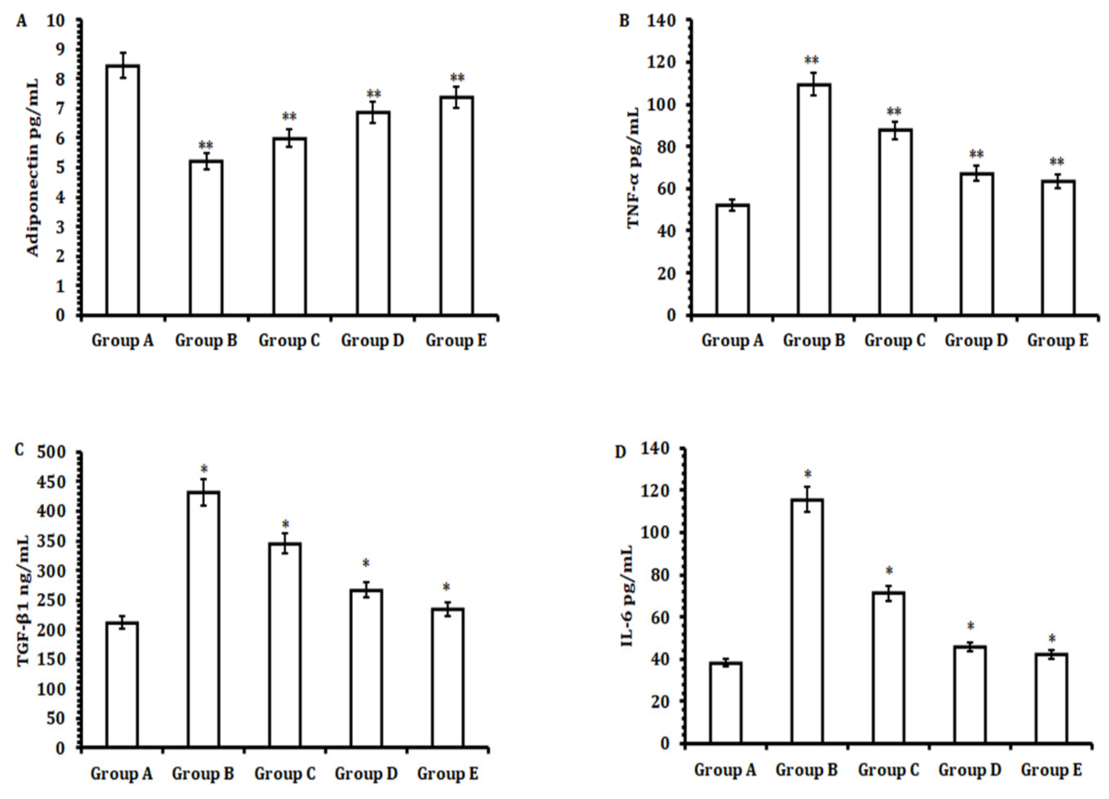
4-mediated hepatotoxicity by restoring serum liver biochemistry, thereby regulating the level of the pro-inflammatory cytokines TNF-α, IL-6, and TGF-β. This was checked by elevating the level of the anti-inflammatory cytokine adiponectin. It attenuates ROS production and maintains the integrity of mitochondrial membrane potential, which ultimately aids in reducing the necrotic population and fibrosis of hepatocytes.
Among the environmental toxicants, CCl4is considered one of the prominent toxicants used to study the hepatoprotective effect of various plant-based products for in vivo animal models [39]. CCl4, upon administration to the animal body, is converted into the more potent free radical CCl3OO+ with the help of a group of liver enzyme systems called cytochrome P-450 [40]. This conversion of a toxicant substance into a more toxicant substance by liver enzymes is known as biotransformation [41]. The free radical CCl3OO+ generated after the biotransformation process in the liver induces lipid peroxidation by interacting covalently with cellular macromolecules (membrane phospholipids) [42]. This interaction causes the disruption of membrane integrity and results in pore formation in the cell membrane, thereby releasing the hepatic enzymes AST, ALP, ALT, and bilirubin from hepatocytes [43]. Thus, CCl4administration causes injury to hepatocytes and thereby elevates the levels of liver enzymes in serum [29]. In the current study, we conducted preliminary experiments to evaluate the ideal dose of TA extract and the route of administration in terms of effective hepatoprotective activity with a safe toxicity profile in animals. Our preliminary results suggest that TA extract showed hepatoprotective activity against CCl4-mediated liver toxicity in a dose-dependent manner. The optimum activity of TA extract was observed at 50 mg/kg b.w. orally (Group E); the results were significant compared with those obtained with 100 mg/kg b.w. standard silymarin. Additionally, we observed that a dose higher than 60 mg/kg b.w.c aused the animals to show toxicity symptoms, such as lethargy, diarrhea, muscle tremors, loss of appetite, etc., and they died before the completion of the study. The administration of 50 mg/kg b.w. of TA extract (Group E) restored the levels of serum enzymes of the liver to normal by inhibiting the potency of the CCl3OO+ free radical, which in turn reduced lipid peroxidation, as evidenced by the reduced level of MDA upon the treatment of animals with TA extract compared with those administered CCl4 (Group B). The hepatoprotective activity of TA extract was further confirmed by the histopathology results of hepatic tissues. Remarkable hepatocellular damage was observed in animals administered CCl4 (Group B) when compared with untreated animals (Group A). Microscopic analysis demonstrated that a significant number of necrotic populations of hepatocytes with large vacuolization, infiltration of inflammatory cells, portal biliary damage, and hepatocyte blooming were observed in animals administeredCCl4orally (Group B). However, animals treated with an oral TA extract dose of 50 mg/kg b.w. (Group E) showed significant protection against CCl4-mediated hepatotoxicity, which was accompanied by a reduction in the necrosis of hepatocytes, infiltrated cells, less vacuolization, and hepatocyte blooming.
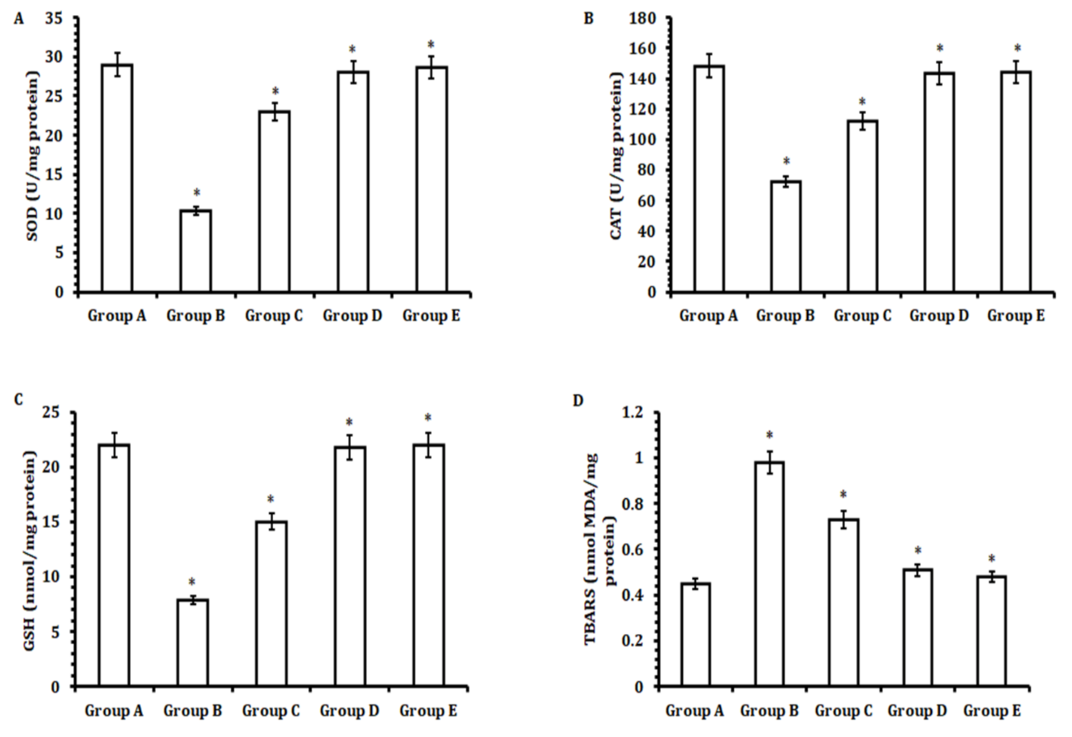
Another striking feature of hepatic tissue damage caused by CCl4is the induction of oxidative stress that affects the levels of antioxidant enzymes, such as SOD, CAT, and GSR [44]. These enzymes together create an anti-scavenging system by catalyzing biochemical reactions to neutralize any free radicals generated under normal physiological conditions into nontoxic compounds to nullify any detrimental effects of toxic compounds, thus helping in the regulation of cell homeostasis [45]. Upon the administration of CCl4to animals, the generation of free radicals increased many-fold [46]. This over-production of free radicals caused oxidative stress, which in turn affected the levels of antioxidant enzymes (SOD, CAT, and GSR). However, the animals treated with 50 mg/kg b.w. of TA extract (Group E) exhibited decreased CCl4-mediated oxidative stress, specifically by elevating the levels of antioxidant enzymes significantly when compared with the CCl4-treated group (Group B). This induction of antioxidant enzymes prevented oxidative stress by neutralizing the CCl4-mediated generation of free radicals.
The oxidative stress is induced by CCl4-stimulated liver fibrosis in animals dosed with CCl4 [47]. As evidenced by the high level of hydroxyproline, a major amino acid was found in collagen tissue by Masson’s trichome staining [48]. Another striking feature of fibrotic tissue was the elevated expression of pro-inflammatory cytokines, which play a crucial role in the stimulation of portal fibroblasts, thereby aiding in the synthesis of more fibrous tissue and associated extracellular matrix substances [49]. Herein, we observed, upon the administration of 50 mg/kg b.w. of TA extract (Group E), significantly decreased fibrosis markers, such as TNF-α, IL-6, and TGF-β, in serum samples when compared with CCL4-treated animals (Group B). However, after treating the animals with 50 mg/kg b.w. of TA extract (Group E), we observed a significant increase in the serum level of the anti-inflammatory cytokine adiponectin. Collectively, these results suggest that TA extract has two effects: firstly, it restores the level of anti-inflammatory adiponectin, and secondly, it decreases the level of the pro-inflammatory cytokine proteins TNF-α, IL-6, and TGF-β to control CCl4-mediated hepatocellular fibrosis and inflammation.
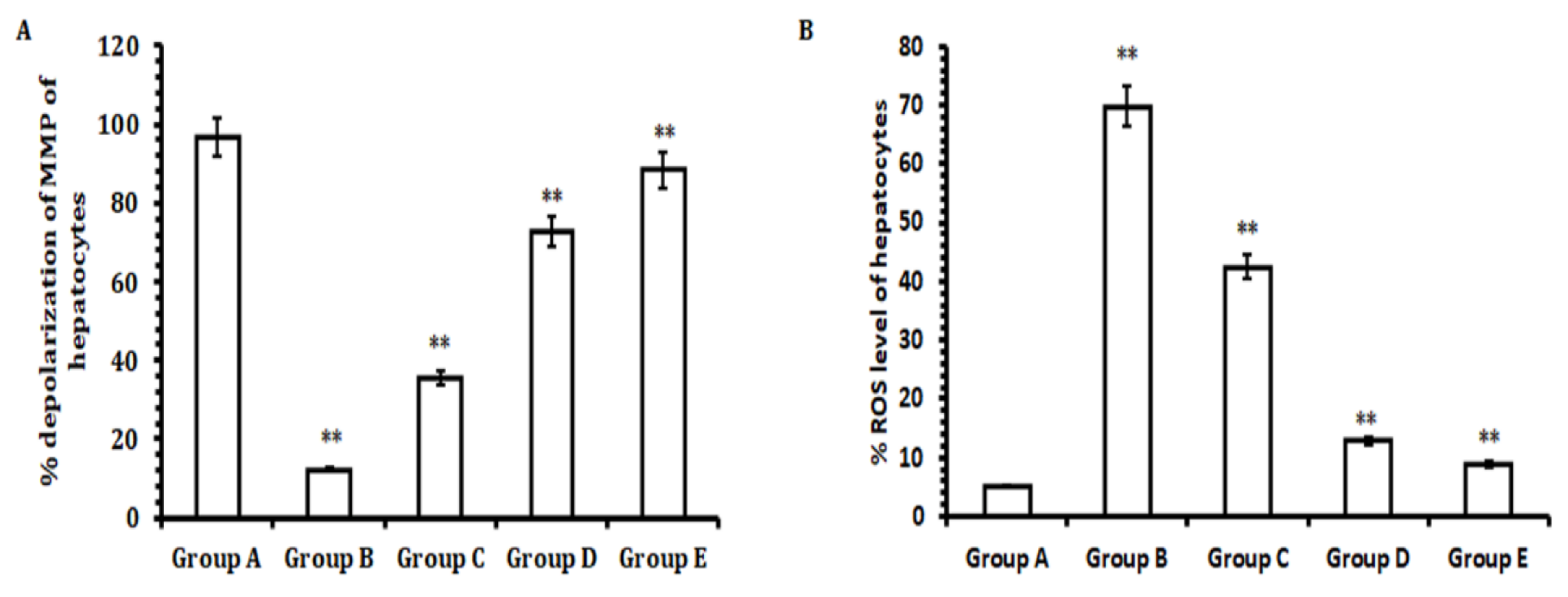
Besides other factors that cause the necrosis of hepatocytes, CCl4administration induces ROS production and mitochondrial membrane potential (MMP) depolarization that lead to hepatic tissue damage and induce the necrosis of hepatocytes [50]. In the current study, our results highlight that hepatocytes of rat livers initially dosed with CCl4had significantly elevated levels of ROS and mitochondrial membrane depolarization when compared with hepatocytes obtained from livers from rats in the control group. However, the hepatocytes collected from the animal group treated with TA extract showed a remarkable reduction in ROS generation and inhibited depolarization of mitochondrial membrane potential when compared with the CCl4-treated group and control group. Conversely, a significant reduction in ROS generation and the depolarization of mitochondrial membrane potential was observed for the groups treated with lower doses of TA extract (30 and 40 mg/kg b.w.; Groups C and D) when compared with the CCl4-treated group (Group B) and control group (Group A). Therefore, in effect, our results demonstrate that TA extract effectively normalizes ROS generation and helps to protect hepatocytes by reducing the depolarization of the mitochondrial membrane potential. Together, these hepatoprotective outcomes may be due to the presence of bioactive compounds in the TA extract. Although the TA extracts exhibited a promising hepatoprotective effect against CCl4-induced hepatotoxicity, the limitation of the study was the inability to evaluate the mechanism of action, pharmacokinetics, and bioavailability of its bioactive constituents.

5. Conclusions

Our study demonstrated that TA extract and its major bioactive (polyphenolic and flavonoid) compounds effectively alleviated CCl4-induced hepatotoxicity or injury in rat models. More importantly, the hepatoprotective effect of TA extract is mostly attributed to the reduction of ROS, which is associated with oxidative stress, restoration of serum liver biochemistry, inhibition of pro-inflammatory cytokines, and maintenance of mitochondrial membrane potential (Graphical Abstract). Additionally, these results provide a strong platform for the further evaluation of TA extract as a potential agent for treating and preventing liver ailments.

Author Contributions

A.M.A. and A.G.A. designed and conducted the experiments, and conducted visualization, data curation, acquisition, formal analysis, writing, and editing. M.A.A. and S.A.: review and editing. B.R.: conceptualization, design of experiments, writing, editing, and producing an original draft of the manuscript. All authors have read and agreed to the published version of the manuscript.

Funding

Researchers would like to thank the Deanship of Scientific Research, Qassim University, for funding the publication of this project.

Institutional Review Board Statement

Not applicable.

Informed Consent Statement

Not applicable.

Data Availability Statement

Data exists within the article.

Acknowledgments

The researchers would like to thank the Deanship of Scientific Research, Qassim University, for funding the publication of this project.

Conflicts of Interest

The authors declare that there are no conflicts of interest.

Abbreviations

TA, Tamarix articulata; CCl4, carbon tetrachloride; SOD, superoxide dismutase; CAT, catalase; GSR, glutathione reductase; TBARS, thiobarbituric reactive substances; TNF-α, tumor-necrosis factor-alpha; TGF-β, tumor-growth factor-beta; IL-6, interleukin-6; ROS, reactive oxygen species; CCl3+, trichloromethyl radical; CCl3OO+, trichloromethylperoxy; LFT, liver function test; MMP, mitochondrial membrane potential; LC–MS, liquid chromatography–mass spectrometry; ESI-MS, electrospray ionization mass spectra; KAUST, King Abdullah University of Science and Technology; ALP, alkaline phosphatase; ALT, alanine transaminase; AS, aspartate aminotransferase; ELISA, enzyme-linked immunosorbent assay; PBS, phosphate-buffered saline; MDA, malondialdehyde; DCFH-DA, 2′-7′dichlorofluorescin diacetate.

References

  1. Thibaut, R.; Gage, M.C.; Pineda-Torra, I.; Chabrier, G.; Venteclef, N.; Alzaid, F. Liver Macrophages and Inflammation in Physiology and Physiopathology of Non-alcoholic Fatty Liver Disease. FEBS J. 2022, 289, 3024–3057. [Google Scholar] [CrossRef] [PubMed]
  2. Jaeschke, H.; McGill, M.R.; Ramachandran, A. Oxidant Stress, Mitochondria, and Cell Death Mechanisms in Drug-Induced Liver Injury: Lessons Learned from Acetaminophen Hepatotoxicity. Drug Metab. Rev. 2012, 44, 88–106. [Google Scholar] [CrossRef] [PubMed]
  3. Cichoż-Lach, H.; Michalak, A. Oxidative Stress as a Crucial Factor in Liver Diseases. World J. Gastroenterol. 2014, 20, 8082. [Google Scholar] [CrossRef] [PubMed]
  4. Lima, G.P.P.; Vianello, F.; Corrêa, C.R.; Campos, R.A.d.S.; Borguini, M.G. Polyphenols in Fruits and Vegetables and Its Effect on Human Health. Food Nutr. Sci. 2014, 5, 1065–1082. [Google Scholar] [CrossRef]
  5. Zhang, A.; Sun, H.; Wang, X. Recent Advances in Natural Products from Plants for Treatment of Liver Diseases. Eur. J. Med. Chem. 2013, 63, 570–577. [Google Scholar] [CrossRef]
  6. Choudhury, S.; Ghosh, S.; Mukherjee, S.; Gupta, P.; Bhattacharya, S.; Adhikary, A.; Chattopadhyay, S. Pomegranate Protects against Arsenic-Induced P53-Dependent ROS-Mediated Inflammation and Apoptosis in Liver Cells. J. Nutr. Biochem. 2016, 38, 25–40. [Google Scholar] [CrossRef]
  7. Vun-Sang, S.; Rodrigues, K.F.; Dsouza, U.J.; Iqbal, M. Suppression of Oxidative Stress and Proinflammatory Cytokines Is a Potential Therapeutic Action of Ficus Lepicarpa B.(Moraceae) against Carbon Tetrachloride (CCl4)-Induced Hepatotoxicity in Rats. Molecules 2022, 27, 2593. [Google Scholar] [CrossRef]
  8. Maurya, H. Pharmacological Evaluation of Allium Cepa Extract as Hepatoprotective Potential in Albino Rat. Int. J. Health Clin. Res. 2018, 1, 7–13. [Google Scholar]
  9. Unsal, V.; Cicek, M.; Sabancilar, İ. Toxicity of Carbon Tetrachloride, Free Radicals and Role of Antioxidants. Rev. Environ. Health 2021, 36, 279–295. [Google Scholar] [CrossRef]
  10. Karapetsas, A.; Voulgaridou, G.-P.; Konialis, M.; Tsochantaridis, I.; Kynigopoulos, S.; Lambropoulou, M.; Stavropoulou, M.-I.; Stathopoulou, K.; Aligiannis, N.; Bozidis, P. Propolis Extracts Inhibit UV-Induced Photodamage in Human Experimental in Vitro Skin Models. Antioxidants 2019, 8, 125. [Google Scholar] [CrossRef]
  11. Neha, K.; Haider, M.R.; Pathak, A.; Yar, M.S. Medicinal Prospects of Antioxidants: A Review. Eur. J. Med. Chem. 2019, 178, 687–704. [Google Scholar] [CrossRef] [PubMed]
  12. Richter, K.; Kietzmann, T. Reactive Oxygen Species and Fibrosis: Further Evidence of a Significant Liaison. Cell Tissue Res. 2016, 365, 591–605. [Google Scholar] [CrossRef] [PubMed]
  13. Heeba, G.H.; Mahmoud, M.E. Therapeutic Potential of Morin against Liver Fibrosis in Rats: Modulation of Oxidative Stress, Cytokine Production and Nuclear Factor Kappa B. Environ. Toxicol. Pharmacol. 2014, 37, 662–671. [Google Scholar] [CrossRef] [PubMed]
  14. Sherif, I.O.; Al-Gayyar, M.M. Antioxidant, Anti-Inflammatory and Hepatoprotective Effects of Silymarin on Hepatic Dysfunction Induced by Sodium Nitrite. Eur. Cytokine Netw. 2013, 24, 114–121. [Google Scholar] [CrossRef]
  15. Bag, S.; Mondal, A.; Majumder, A.; Banik, A. Tea and Its Phytochemicals: Hidden Health Benefits & Modulation of Signaling Cascade by Phytochemicals. Food Chem. 2022, 371, 131098. [Google Scholar]
  16. Alnuqaydan, A.M.; Rah, B. Tamarix Articulata (T. Articulata)—An Important Halophytic Medicinal Plant with Potential Pharmacological Properties. Curr. Pharm. Biotechnol. 2019, 20, 285–292. [Google Scholar] [CrossRef]
  17. Said, S.; Noureddine, G.; Eddine, L.S.; Abdelmadjid, G.; Djamel, B.; Tliba, A. Phenolic Content, HPLC Analysis and Antioxidant Activity Extract from Tamarix Gallica and Tamarix Articulata Growing in Southeast of Algeria. Res. J. Pharm. Technol. 2018, 11, 3826–3832. [Google Scholar] [CrossRef]
  18. Rah, B.; Alnuqaydan, A.M.; Almutary, A.G.; Alshehri, O.Y.; Henidi, H.A.; Alajlan, A.M.; Tamim, A.A.; Alowaifeer, A.; Rather, M.Y. Evaluation of the Cytotoxic Activity of Tamarix Articulata and Its Anticancer Potential in Prostate Cancer Cells. J. Appl. Pharm. Sci. 2022, 12, 089–108. [Google Scholar] [CrossRef]
  19. Shaalan, Y.M.; Handoussa, H.; Youness, R.A.; Assal, R.A.; El-Khatib, A.H.; Linscheid, M.W.; El Tayebi, H.M.; Abdelaziz, A.I. Destabilizing the Interplay between MiR-1275 and IGF2BPs by Tamarix Articulata and Quercetin in Hepatocellular Carcinoma. Nat. Prod. Res. 2018, 32, 2217–2220. [Google Scholar] [CrossRef]
  20. Alnuqaydan, A.M.; Rah, B. Comparative Assessment of Biological Activities of Different Parts of Halophytic Plant Tamarix Articulata (T. Articulata) Growing in Saudi Arabia. Saudi J. Biol. Sci. 2020, 27, 2586–2592. [Google Scholar] [CrossRef]
  21. Alnuqaydan, A.M.; Rah, B. Tamarix Articulata Inhibits Cell Proliferation, Promotes Cell Death Mechanisms and Triggers G0/G1 Cell Cycle Arrest in Hepatocellular Carcinoma Cells. Food Technol. Biotechnol. 2021, 59, 162–173. [Google Scholar] [CrossRef] [PubMed]
  22. Nwaechefu, O.O.; Olaolu, T.D.; Akinwunmi, I.R.; Ojezele, O.O.; Olorunsogo, O.O. Cajanus Cajan Ameliorated CCl4-Induced Oxidative Stress in Wistar Rats via the Combined Mechanisms of Anti-Inflammation and Mitochondrial-Membrane Transition Pore Inhibition. J. Ethnopharmacol. 2022, 289, 114920. [Google Scholar] [CrossRef]
  23. Iqbal, M.O.; Manzoor, M.; Mumtaz, A.; Riaz, R.; Arshad, S.; Khan, I.A.; Javaid, U.; Manzoor, Z.; Munawar, S.H.; Andleeb, S. Evaluation of the Hepatoprotective Activity of Hydroalcoholic Extract of Alhagi Camelorum against Valproic Acid-Induced Hepatotoxicity in Rats. Biomed. Pharmacother. 2022, 150, 112953. [Google Scholar] [CrossRef]
  24. Mihailović, V.; Katanić, J.; Mišić, D.; Stanković, V.; Mihailović, M.; Uskoković, A.; Arambašić, J.; Solujić, S.; Mladenović, M.; Stanković, N. Hepatoprotective Effects of Secoiridoid-Rich Extracts from Gentiana Cruciata L. against Carbon Tetrachloride Induced Liver Damage in Rats. Food Funct. 2014, 5, 1795–1803. [Google Scholar] [CrossRef]
  25. Ammar, N.M.; Hassan, H.A.; Abdallah, H.M.; Afifi, S.M.; Elgamal, A.M.; Farrag, A.R.H.; El-Gendy, A.E.-N.G.; Farag, M.A.; Elshamy, A.I. Protective Effects of Naringenin from Citrus Sinensis (Var. Valencia) Peels against CCl4-Induced Hepatic and Renal Injuries in Rats Assessed by Metabolomics, Histological and Biochemical Analyses. Nutrients 2022, 14, 841. [Google Scholar] [CrossRef] [PubMed]
  26. Mazani, M.; Rezagholizadeh, L.; Shamsi, S.; Mahdavifard, S.; Ojarudi, M.; Salimnejad, R.; Salimi, A. Protection of CCl4-Induced Hepatic and Renal Damage by Linalool. Drug Chem. Toxicol. 2022, 45, 963–971. [Google Scholar] [CrossRef]
  27. Hamza, A.A.; Lashin, F.M.; Gamel, M.; Hassanin, S.O.; Abdalla, Y.; Amin, A. Hawthorn Herbal Preparation from Crataegus Oxyacantha Attenuates in Vivo Carbon Tetrachloride-Induced Hepatic Fibrosis via Modulating Oxidative Stress and Inflammation. Antioxidants 2020, 9, 1173. [Google Scholar] [CrossRef] [PubMed]
  28. Al-Megrin, W.A.; Alkhuriji, A.F.; Yousef, A.O.S.; Metwally, D.M.; Habotta, O.A.; Kassab, R.B.; Abdel Moneim, A.E.; El-Khadragy, M.F. Antagonistic Efficacy of Luteolin against Lead Acetate Exposure-Associated with Hepatotoxicity Is Mediated via Antioxidant, Anti-Inflammatory, and Anti-Apoptotic Activities. Antioxidants 2019, 9, 10. [Google Scholar] [CrossRef]
  29. Lin, S.-Y.; Xu, D.; Du, X.-X.; Ran, C.-L.; Xu, L.; Ren, S.-J.; Tang, Z.-T.; Yin, L.-Z.; He, C.-L.; Yuan, Z.-X. Protective Effects of Salidroside against Carbon Tetrachloride (CCl4)-Induced Liver Injury by Initiating Mitochondria to Resist Oxidative Stress in Mice. Int. J. Mol. Sci. 2019, 20, 3187. [Google Scholar] [CrossRef]
  30. Arbiser, J.L.; Kau, T.; Konar, M.; Narra, K.; Ramchandran, R.; Summers, S.A.; Vlahos, C.J.; Ye, K.; Perry, B.N.; Matter, W. Solenopsin, the Alkaloidal Component of the Fire Ant (Solenopsis Invicta), Is a Naturally Occurring Inhibitor of Phosphatidylinositol-3-Kinase Signaling and Angiogenesis. Blood 2007, 109, 560–565. [Google Scholar] [CrossRef]
  31. Hou, S.; Cheng, Z.; Wang, W.; Wang, X.; Wu, Y. Ailanthone Exerts an Antitumor Function on the Development of Human Lung Cancer by Upregulating MicroRNA-195. J. Cell. Biochem. 2018, 120, 10444–10451. [Google Scholar] [PubMed]
  32. Du, J.; Daniels, D.H.; Asbury, C.; Venkataraman, S.; Liu, J.; Spitz, D.R.; Oberley, L.W.; Cullen, J.J. Mitochondrial Production of Reactive Oxygen Species Mediate Dicumarol-Induced Cytotoxicity in Cancer Cells. J. Biol. Chem. 2006, 281, 37416–37426. [Google Scholar] [CrossRef] [PubMed] [Green Version]
  33. Kampa-Schittenhelm, K.M.; Salitzky, O.; Akmut, F.; Illing, B.; Kanz, L.; Salih, H.R.; Schittenhelm, M.M. Dronabinol Has Preferential Antileukemic Activity in Acute Lymphoblastic and Myeloid Leukemia with Lymphoid Differentiation Patterns. BMC Cancer 2016, 16, 1–12. [Google Scholar] [CrossRef]
  34. Dai, Z.; Wang, L.; Wang, X.; Zhao, B.; Zhao, W.; Bhardwaj, S.S.; Ye, J.; Yin, Z.; Zhang, J.; Zhao, S. Oxymatrine Induces Cell Cycle Arrest and Apoptosis and Suppresses the Invasion of Human Glioblastoma Cells through the EGFR/PI3K/Akt/MTOR Signaling Pathway and STAT3. Oncol. Rep. 2018, 40, 867–876. [Google Scholar] [CrossRef] [PubMed]
  35. Li, Y.; Yao, J.; Han, C.; Yang, J.; Chaudhry, M.T.; Wang, S.; Liu, H.; Yin, Y. Quercetin, Inflammation and Immunity. Nutrients 2016, 8, 167. [Google Scholar] [CrossRef]
  36. Wang, L.; Wang, C.; Jia, Y.; Liu, Z.; Shu, X.; Liu, K. Resveratrol Increases Anti-proliferative Activity of Bestatin through Downregulating P-glycoprotein Expression via Inhibiting PI3K/Akt/MTOR Pathway in K562/ADR Cells. J. Cell. Biochem. 2016, 117, 1233–1239. [Google Scholar] [CrossRef]
  37. Huang, T.-H.; Lin, C.-F.; Alalaiwe, A.; Yang, S.-C.; Fang, J.-Y. Apoptotic or Antiproliferative Activity of Natural Products against Keratinocytes for the Treatment of Psoriasis. Int. J. Mol. Sci. 2019, 20, 2558. [Google Scholar] [CrossRef]
  38. Li, X.; Fang, P.; Mai, J.; Choi, E.T.; Wang, H.; Yang, X. Targeting Mitochondrial Reactive Oxygen Species as Novel Therapy for Inflammatory Diseases and Cancers. J. Hematol. Oncol. 2013, 6, 1–19. [Google Scholar] [CrossRef]
  39. Suzek, H.; Celik, I.; Dogan, A.; Yildirim, S. Protective Effect and Antioxidant Role of Sweetgum (Liquidambar Orientalis) Oil against Carbon Tetrachloride-Induced Hepatotoxicity and Oxidative Stress in Rats. Pharm. Biol. 2016, 54, 451–457. [Google Scholar] [CrossRef]
  40. Dutta, S.; Chakraborty, A.K.; Dey, P.; Kar, P.; Guha, P.; Sen, S.; Kumar, A.; Sen, A.; Chaudhuri, T.K. Amelioration of CCl4 Induced Liver Injury in Swiss Albino Mice by Antioxidant Rich Leaf Extract of Croton Bonplandianus Baill. PLoS ONE 2018, 13, e0196411. [Google Scholar] [CrossRef]
  41. Kaur, S.; Sharma, D.; Singh, A.P.; Kaur, S. Amelioration of Hepatic Function, Oxidative Stress, and Histopathologic Damages by Cassia Fistula L. Fraction in Thioacetamide-Induced Liver Toxicity. Environ. Sci. Pollut. Res. 2019, 26, 29930–29945. [Google Scholar] [CrossRef] [PubMed]
  42. Bischoff, K.; Mukai, M.; Ramaiah, S.K. Liver Toxicity. In Veterinary Toxicology; Elsevier: Amsterdam, The Netherlands, 2018; pp. 239–257. [Google Scholar]
  43. Muthumani, M.; Miltonprabu, S. Ameliorative Efficacy of Tetrahydrocurcumin against Arsenic Induced Oxidative Damage, Dyslipidemia and Hepatic Mitochondrial Toxicity in Rats. Chem. Biol. Interact. 2015, 235, 95–105. [Google Scholar] [CrossRef] [PubMed]
  44. Li, S.; Tan, H.-Y.; Wang, N.; Zhang, Z.-J.; Lao, L.; Wong, C.-W.; Feng, Y. The Role of Oxidative Stress and Antioxidants in Liver Diseases. Int. J. Mol. Sci. 2015, 16, 26087–26124. [Google Scholar] [CrossRef] [PubMed]
  45. Salam, A.; Farwin, S. Biochemistry of Reactive Oxygen Species in Selective Cancer Cell Toxicity and Protection of Normal Cells; University of Cincinnati: Cincinnati, OH, USA, 2017. [Google Scholar]
  46. Zhao, Z.; Hou, Y.; Zhou, W.; Keerthiga, R.; Fu, A. Mitochondrial Transplantation Therapy Inhibit Carbon Tetrachloride-induced Liver Injury through Scavenging Free Radicals and Protecting Hepatocytes. Bioeng. Transl. Med. 2021, 6, e10209. [Google Scholar] [CrossRef]
  47. Tipoe, G.L.; Leung, T.M.; Liong, E.C.; Lau, T.Y.H.; Fung, M.L.; Nanji, A.A. Epigallocatechin-3-Gallate (EGCG) Reduces Liver Inflammation, Oxidative Stress and Fibrosis in Carbon Tetrachloride (CCl4)-Induced Liver Injury in Mice. Toxicology 2010, 273, 45–52. [Google Scholar] [CrossRef]
  48. Bahri, S.; Abdennabi, R.; Mlika, M.; Neji, G.; Jameleddine, S.; Ali, R.B. Effect of Phoenix Dactylifera L. Sap against Bleomycin-Induced Pulmonary Fibrosis and Oxidative Stress in Rats: Phytochemical and Therapeutic Assessment. Nutr. Cancer 2019, 71, 781–791. [Google Scholar] [CrossRef]
  49. Weiskirchen, R.; Weiskirchen, S.; Tacke, F. Organ and Tissue Fibrosis: Molecular Signals, Cellular Mechanisms and Translational Implications. Mol. Asp. Med. 2019, 65, 2–15. [Google Scholar] [CrossRef]
  50. Sengupta, D.; Chowdhury, K.D.; Sarkar, A.; Paul, S.; Sadhukhan, G.C. Berberine and S Allyl Cysteine Mediated Amelioration of DEN+CCl4 Induced Hepatocarcinoma. Biochim. Biophys. Acta 2014, 1840, 219–244. [Google Scholar] [CrossRef]
Figure 1. Chromatogram of key phytochemicals present in TA extract (this figure shows data from our recently published work [18,20,21]).
Antioxidants 11 01824 g001aAntioxidants 11 01824 g001bAntioxidants 11 01824 g001cAntioxidants 11 01824 g001dAntioxidants 11 01824 g001e
Figure 2. Effect of TA extract on anti-inflammatory (adiponectin) and pro-inflammatory cytokine markers (TNF-α, TGF-β, and IL-6) of liver tissues initially treated with CCl4to induce liver toxicity. (A) Adiponectin level, (B) TNF-α level, (C) TGF-β level, and (D) IL-6 level. The experiments were performed three times or more than three times, and the represented data are the mean values ± SEM. A p-value less than or equal to 0.05 was considered to be statistically significant; * p ≤ 0.05, ** p ≤ 0.01.
Antioxidants 11 01824 g002
Figure 3. Effect of TA extract on the microscopic analysis of hepatic tissue sections of animals initially treated with CCl4. Hematoxylin and eosin stain of hepatic tissues in control (Group A), CCl4-treated animals (Group B), CCl4+30 mg/kg b.w. TA extract-treated animals (Group C), CCl4+ 40 mg/kg b.w. TA extract-treated animals (Group D), and CCl4+ 50 mg/kg b.w. TA extract-treated animals (Group E). Masson’s trichrome staining in control (Group A), CCl4-treated animals (Group B), CCl4+ 30 mg/kg b.w. TA extract-treated animals (Group C), 40 mg/kg b.w. TA extract-treated animals (Group D), and CCl4+ 50 mg/kg b.w. TA extract-treated animals (Group E). Bar scale = 25 μm. Magnification × 100.
Antioxidants 11 01824 g003
Figure 4. Effect of TA extract on the antioxidant enzymes (SOD, CAT, GSR, and MDA) of liver tissues initially treated with CCl4to induce liver toxicity. (A) Determination of superoxide dismutase (SOD) activity, (B) determination of catalase (CAT) activity, (C) determination of glutathione reductase (GSR) activity, and (D) determination of malondialdehyde (MDA) activity. The experiments were performed three times or more than three times and data are represented as the mean value ± SEM. A p-value less than or equal to 0.05 was considered to be statistically significant, i.e., * p ≤ 0.05.
Antioxidants 11 01824 g004
Figure 5. Measurement of MMP and ROS production of liver hepatocytes extracted from the livers of rats initially treated with CCl4and the effect of TA extract on MMP and ROS production. (A) Measurement of the MMP of hepatocytes extracted from different animal groups initially dosed with CCl4, (B) ROS production of hepatocytes extracted from different animal groups initially dosed with CCl4. The experiments were performed three or more than three times and the data are represented as the mean ± SEM. A p-value less than or equal to 0.05 was considered statistically significant, i.e., ** p ≤ 0.01.
Antioxidants 11 01824 g005
Table 1. Phytochemicals detected from the methanolic extract of TA by LC-MS.
S. NoM/ZRTCompound
1103.086.4474813-,ethylbutanoic acid;2-methylbutyrate
2104.040.781022Pyruvate oxime
3105.1216.5714Choline
4107.0422.75023Aromatic aldehyde; D-glycerate
5108.9624.27732Bromoethane
6119.0411.48668Succinic acid
7126.1200031.72972046γ-Coniceine
8128.0424.264565-amino-4-imidazole carboxylate;1-methyl-4-nitroimidazole
9130.0800020.79923889L-pipecolate
10131.046.248767Itaconate;(E)-glutaconate
11133.0820.91498(R)-2-hydroxyisocaproate;6-hydroxyhexanoic acid
12134.046.839831L-aspartate
13135.1224.28927p-cymene
14136.088.6310822-phenylacetamide
15137.0415.86743Hypoxanthine; threonate
16139.086.4507364-hydroxyphenylethanol; Styrene-cis-2,3-dihydrodiol
17140.0399937.618814354-nitrophenol;2-nitrophenol;3-nitrophenol;3-hydroxypicolinic acid
18142.088.676849Hypoglycin; arecaidine
19145.0813.84979Trans-4-hydroxycyclohexanecarboxylate
20146.0399930.78391111α-ketoglutarate
21146.160.787356Spermidine
221475.374421Flupropanate
23147.1213.21838L-lysine
24149.0422.59839D-arabinono-1,4-lactone
25151.084.124735Tolylacetate
26160.0800020.81778333Indole-3-acetaldehyde
27161.03999317.29154442-oxoadipate
28162.1199956.89406976L-carnitine
29163.0824.28231Methyl cinnamate; safrole
30165.1224.24879Jasmone
31189.1211.11041Glycyl-leucine
32167.165.766924Robinobiose
33175.0822.49876N-formimino-L-glutamate
341779.8419883-Chloro-cis,cis-muconate
35178.0800029.756042864-hydroxy-4-methylglutamate
36177.1210.12313L-cladinose; metaldehyde
37181.088.720061D-glucose
38189.11999511.1007144Glycyl-leucine
39193.0800025.79493623Carpacin; myristicin
40197.1600048.60402733Linalyl acetate;alpha-terpinyl acetate
41199.087.810947L-mimosine
42205.1999979.61738333β-caryophyllene
4320726.1537176Chloroneb
44207.1226.10531,4-dimethylphenanthrene
45209.0399937.79105238Fraxetin
46209.1612.44344Ammodendrine
47211.08000210.4657552Sedoheptulose
48219.11999516.7337904N-acetylserotonin
49224.168.699876Tigloidine
50225.11999513.7993487Aspidinol
51228.11999520.8984778Ametryn
52229.0800021.01080798Resveratrol
53232.0800026.13259678N-acetyl-L-2-amino-6-oxopimelate
54233.16000410.3468355Alantolactone
55241.080.891821(1R,6R)-6-hydroxy-2-succinylcyclohexa-2,4-diene-1-carboxylate
56245.0399937.47793282L-fuculose 1-phosphate
57245.1622.48843Anagyrine
58248.0399938.71224279Pyridoxal phosphate
59249.123.2335286-hydroxymelatonin
60254.1621.66069Fenapanil
61254.27999921.6546583Solenopsin A
62256.07998723.6897444Apigeninidin
63257.16000423.6928028Chanoclavine-I
64265.224.29171Oxymatrine
65266.1624.28768Brevicolline
66270.1224.60784Acetochlor
67276.1199952.23423865(5-L-glutamyl)-L-glutamine
68277.0799870.93720015Biotin sulfone
69282.11999524.2929528O6-methyl-2’-deoxyguanosine
70299.16000411.7539109Ostruthin
71300.11999512.13215022,3,9,10-tetrahydroxyberbine
72300.9615.56441Tolclofos-methyl
73302.0415.565112-(3,5-dichlorophenylcarbamoyl)-1,2-dimethylcyclopropane-1-carboxylic acid
74307.0799879.30033418Leucocyanidin; gallocatechin
75307.089.293607Leucocyanidin; gallocatechin; epigallocatechin
76308.165.040135Tebuconazole
77311.049.821766Diflubenzuron; edifenphos
78311.1600049.16593779Nafenopin
79313.0818.69813Inosine-5’-carboxylate
80313.218.794392,4,6-triphenyl-1-hexene
81313.3218.65702Icosanoic acid
82314.0410.93399Isazofos
83315.1218.68262Dihydropteroate; pyraclonil
84315.2418.68325Dronabinol; cannabidiol; cannabichromene
85316.0818.69064Fenbendazole S-oxide
86316.218.68546Belladine
8731814.04215Phosmet
88319.0811.8284Lecanoric acid
89320.1618.29014Metconazole
90321.120.803094Mugineic acid
91322.0799872.18447318β-Citryl-L-glutamate
92323.0410.14008Digallate
93324.1199958.44852717Stylopine
94324.9611.66847Trichloroethanol glucuronide
95325.218.62262Affinine
96325.0813.86467Sterigmatocystin
97326.1620.01459Monocrotaline
9832727.18485Fenuron
99327.2427.2353512 α-Methylpregna-4,9(11)-diene-3,20-dione
100328.0818.00045Fluazifop
101328.29.572027Sethoxydim
102328.3215.33696N,N-dimethylsphing-4-enine
103329.0411.85282Fluorodifen
104329.1612.032591,2-Dehydroreticuline
105330.1212.42584Lithospermoside
106331.0813.817683,7-Di-O-methylquercetin; cirsiliol; tricin
107331.213.81691Carnosol
108336.11999517.2204175Trifluralin; befuraline
109337.085.901588Dicumarol
110337.2000125.67431458Catharanthine; tabersonine
111337.3200078.4919533(13Z,16Z)-docosadienoic acid
112338.0400092.47067165Azafenidin
113338.8810.747111,2,3,7,8-Pentachlorodibenzofuran
114339.127.357424Glyceollin I; glyceollin II
115341.0424.88731Diclofop methyl
11634224.85467Bifenox
117342.1220.51962Protodeoxyviolaceinic acid
118346.07998712.0925351Thiamine monophosphate
119349.20001211.5365785Gibberellin A53
120350.1600042.21731399Riddelline
121353.0411.55965Petunidin
122353.16000412.090492Palmatine
123355.226.82419Vincamine; yohimbine; stemmadenine;
124355.3227.4867622-Oxodocosanoate
125356.1627.42916S-Adenosylmethioninamine
126357.1224.57235Gentiopicrin
127357.2424.60749Arachidonyltrifluoromethane
128359.0412.91192Triflumuron
129360.129.024405Isopenicillin N
130360.9599912.27723168Chlorthiophos
131363.1212.82633Chelirubine; catalpol
132364.07998710.812962Flufenacet
133365.040.799356Xanthosine 5’-phosphate
134365.1600040.792625Gibberellin A8
135367.085.559117Salicin 6-phosphate
136367.2000125.6837843816-Methoxytabersonine; hirsuteine
137368.0400099.69718675Anilofos
137370.0814.05884S-(2-chloroethyl) glutathione
138371.1600046.76877846Bursehernin
139373.0810.125542-O-caffeoylglucarate
140373.2000127.36471102Biocytin
141375.11999523.1907687Secologanate
142375.1213.1768Portulacaxanthin II; swertiamarin; geniposidic acid; gardoside
143377.168.95533Ailanthone
144377.2819.447339-cis-10’-apo-β-carotenal
145379.0799870.822086117-Methylguanosine 5’-phosphate
146383.163.5401262-Methoxyestradiol-17β 3-sulfate
147385.3222.32738(8Z,11Z,14Z,17Z,20Z,23Z)-Hexacosahexaenoic acid
1483878.48086861Sulfentrazone
149387.1210.097971-O-Sinapoyl-β-D-glucose
150389.16000410.6243039Visnadin
151390.9599917.310337645-Phospho-α-D-ribose 1-diphosphate
152393.1199956.13023356Macarpine
153395.1615.13311Rotenone; deguelin
154395.39999417.3745676Ximenic acid
155397.3212.56728δ-Tocotrienol
156398.16000412.6067147Aureothin
157399.127.147485S-Adenosyl-4-methylthio-2-oxobutanoate
158401.287.685251Ophiobolin A
159403.29.7105Prednisolone acetate; citreoviridin
160407.1617.18432Asperlicin C
161409.29.246711Abyssinone V
162410.1600043.91140291Linustatin
16341111.26515Imibenconazole
164411.1214.16161Nicosulfuron; spirodiclofen
165411.2417.672941-Palmitoylglycerol almitoylglycerol 3-phosphate
166411.3618.696425-Dehydroavenasterol; avenastenone
167412.32000723.3697828Cyclopamine
168413.0422.43382Imazosulfuron; pyraflufen-ethyl
169413.04000922.4248369Pyraflufen-ethyl
170414.9617.5999Flusulfamide
171415.0819.1103(7R)-7-(5-Carboxy-5-oxopentanoyl) aminocephalosporinate
172415.32000725.4925863Yamogenin
173417.1211.14687Daidzin; frangulin A; puerarin
174418.20001213.5014327Casimiroedine
17542314.17587895-Fluorouridine diphosphate
176423.11999522.6974831Plicatic acid
177425.27999917.1062564α-Phocaecholic acid
178427.212.38413Abscisic acid glucose ester
179428.04000912.3795444Adenosine 3’,5’-bisphosphate
180428.1612.38573Vicianin
181429.1223.93726ε-Rhodomycinone; ouizalofop-P-tefuryl
182429.2423.93274Nummularine F
183430.32000721.7180979Imperialine
184437.51998917.0830779Hentriacontane
185438.239999.0921256Lunarine
186441.1210.95042Hallactone B
187441.3616.10699Soyasapogenol C; oleanolic aldehyde
189443.1600040.95589167Mallotochromene
190447.124.350199Cephamycin C; glycitin
191453.1226.06561Cinchonain 1a
192453.3625.52124Phylloquinol
193455.16000414.5180328ε-Viniferin
194458.1620.93682Amygdalin
195460.2000128.640175595-Methyltetrahydrofolate
196461.1600049.34915583Paeonolide
197461.27999921.0038789Sophoranone
198461.3999949.51068431Protopanaxadiol
199462.95999113.8378296Quercetin 3,3’-bissulfate; quercetin 3,4’-bissulfate
200463.07998714.0931398Luteolin 7-O-glucuronide
201463.20001216.7546761Plicamine
202467.16000422.6358392Agnuside
203467.27999920.3436707Cephaeline
204471.3621.25501Gypsogenin
205473.39999422.5188859α-Tocopherol acetate
206474.1216.33285CMP-N-trimethyl-2-aminoethylphosphonate
207485.2825.71086Stigmatellin
208486.965.8767785-Fluorodeoxyuridine triphosphate
209487.20001223.1899191Rutaevin; haplodimerine; nafenopin glucuronide
210489.3624.9554Asiatic acid
211489.129.603542CDP-choline
21249210.758352dATP
213495.11999523.74321485’-Methoxyhydnocarpin-D
214497.27999923.6242253Absinthin
215517.222.54293Rottlerin
216517.32000723.5029426Cucurbitacin D
217525.4799822.7749935Retinyl palmitate
218541.20001221.6297253Oleuropein
219553.4424.15608Zeinoxanthin; β-cryptoxanthin
220593.40002424.1761293Santiaguine
221625.20001213.1988915Verbascoside
222629.28002923.81974Resiniferatoxin
Table 2. Major compounds of Tamarix articulata exhibiting antitumor activity.
S. NoMajor CompoundsFunctionsReferences
1.SolenopsinExhibits antiproliferative activity and inhibits PI3K/Akt-driven angiogenesis[30]
2.AilanthonePromotes apoptosis and autophagy by increasing the expression of miR-195 in leukemia cells[31]
3.DicumarolExhibits promising antiproliferative effects and induces ROS-mediated mitochondria-dependent apoptosis in breast cancer cells[32]
4.DronabinolPromotes antiproliferative activity and induces apoptosis by cannabinoid receptor (CB) ½ in leukemia cells[33]
5.OxymatrineInduces apoptotic cell death and arrests the G0/G1 cell cycle phase by blocking EGFR/PI3K/Akt/mTOR signaling in glioma cells[34]
6.QuercetinImmuno-modulatory effect, anticancer, anti-inflammatory, and antiviral activities[35]
7.ResveratrolExhibits antioxidant, anti-inflammatory, and antiproliferative activities, interferes in many signaling pathways, and activates apoptosis in cancer cell models[36]
8.RottlerinAntihypertensive, antiallergic, and antifertility activities. Promotes antiproliferative activity by downregulating NF-kB and cyclin D1 expression[37]
Table 3. Dose optimization of TA extract. Forty-two Wistar male rats were divided into seven groups containing six animals each. Group A was designated as normal without any chemical or extract treatment. Group B was designated as the CCl4-treated group and the animals were administered 40% CCl4mixed in olive oil orally for 3 alternate days a week for 8 weeks. Groups C, D, E, and F were administered 40% CCl4mixed in olive oil and 30, 40, 50, and 60 mg/kg b.w. TA extract, respectively, and all treatments were administered for 3 alternate days a week for 8 weeks. Group G was designated as 40% CCl4mixed in olive oil and standard silymarin 100 mg/kg orally for 3 alternate days a week for 8 weeks.
GroupsTreatments/DosagesSchedules (200 µL)
AControl (without CCl4)Vehicle treatment (after 8 weeks animals were sacrificed)
BCCl4CCl4 (diluted 40%:60% in olive oil)/100 g rat, orally, thrice weekly, for 8 weeks; after 8 weeks, the animals were sacrificed
CCCl4 + TA extract (30 mg/kg b.w.)CCl4 (diluted 40%:60% in olive oil)/100 g rat, orally, thrice weekly. TA extract (30 mg/kg p.o.; 1 mL/100 g body weight) thrice weekly for 8 weeks; after 8 weeks, the animals were sacrificed
DCCl4 + TA extract (40 mg/kg b.w.)CCl4 (diluted 40%:60% in olive oil)/100 g rat, orally, thrice weekly. TA extract (40 mg/kg p.o.; 1 mL/100 g body weight) thrice weekly for 8 weeks; after 8 weeks the animals were sacrificed
ECCl4 + TA extract (50 mg/kg b.w.)CCl4 (diluted 40%:60% in olive oil)/100 g rat, orally, thrice weekly. TA extract (50 mg/kg p.o.; 1 mL/100 g body weight) thrice weekly for 8 weeks; after 8 weeks, the animals were sacrificed
FCCl4 + TA extract (60 mg/kg b.w.)CCl4 (diluted 40%:60% in olive oil)/100 g rat, orally, thrice weekly. TA extract (60 mg/kg p.o.; 1 mL/100 g body weight) thrice weekly for 8 weeks; after 8 weeks, the animals were sacrificed
GCCl4 + Silymarin (100 mg/kg b.w.)CCl4 (diluted 40%:60% in olive oil)/100 g rat, orally, thrice weekly. Silymarin (100 mg/kg p.o.; 1 mL/100 g body weight) thrice weekly for 8 weeks; after 8 weeks, the animals were sacrificed
Table 4. Analysis of the antioxidant enzymes (SOD, CAT, GSR, and MDA) of liver tissues initially treated with CCl4to induce liver toxicity and treatment with varying doses of TA extract (30, 40, 50, and 60 mg/kg b.w.) along with standard silymarin (100 mg/kg b.w.). The data represent the mean value ±SEM of three independent experiments. * p< 0.05, *** p< 0.001, and **** p< 0.0001.
GroupsTreatment/SOD (U/mg Protein)CAT (U/mg Protein)GSR (nmol/mg Protein)TBARS (nmol MDA/mg Protein)
Dosage
AControl (without CCl4)29.64 ± 0.16146.01 ± 2.8722.45 ± 0.350.457 ± 0.01
BCCl410.67 ± 0.14 ****79.76 ± 1.02 ****7.87 ± 0.16 ****1.18 ± 0.09 ****
CCCl4 + TA extract (30 mg/kg b.w.)13.59 ± 0.56 ****108.93 ± 1.91 ****12.09 ± 0.15 ****0.98 ± 0.03 ****
DCCl4 + TA extract (40 mg/kg b.w.)22.94 ± 0.53 ****119.98 ± 2.08 ****16.34 ± 0.42 ****0.73 ± 0.04 ****
ECCl4 + TA extract (50 mg/kg b.w.)27.78 ± 0.53 *145.97 ± 1.08 *21.91 ± 0.09 *0.49 ± 0.03 *
FCCl4 + TA extract (60 mg/kg b.w.)Animals died before the completion of the studyAnimals died before the completion of the studyAnimals died before the completion of the studyAnimals died before the completion of the study
GCCl4 + Silymarin (100 mg/kg b.w.)28.63 ± 0.43 ***149.02 ± 3.78 ****22.40 ± 0.36 ****0.39 ± 0.9 ****
Table 5. Analysis of the serum biochemical parameters in the liver function test after CCl4-induced liver toxicity and treatment with varying doses of TA extract (30, 40, 50, and 60 mg/kg b.w.) along with standard silymarin (100 mg/kg b.w.). The data represent the mean value ±SEM of three independent experiments. * p< 0.05, and **** p< 0.0001.
GroupsTreatmentAST (UL−1)ALT (UL−1)ALP (UL−1)Bil (µmol L−1)
AControl (without CCl4)32.31 ± 0.9866.01 ± 2.87498.98 ± 20.451.23 ± 0.11
BCCl42945 ± 82.54 ****388.66 ± 12.02 ****865 ± 17.76 ****3.98± 0.23 ****
CCCl4 + TA extract (30 mg/kg b.w.)576.12 ± 33.56 ****288.93 ± 16.91 ****509.19 ± 34.85 ****1.58 ± 0.19 ****
DCCl4 + TA extract (40 mg/kg b.w.)271.83 ± 19.53 ****159.98 ± 07.58 ****456.34 ± 16.42 ****1. 43 ± 0.04 ****
ECCl4 + TA extract (50 mg/kg b.w.)172.78 ± 12.73 *65.97 ± 1.08 *391.94 ± 23.02 *1. 19 ± 0.03 *
FCCl4 + TA extract (60 mg/kg b.w.)Animals died before the completion of the studyAnimals died before the completion of the studyAnimals died before the completion of the studyAnimals died before the completion of the study
GCCl4 + Silymarin (100 mg/kg b.w.)170.63 ± 10.03 ****69.02 ± 3.78 ****403.40 ± 13.56 ****1. 29 ± 0.43 ****
Table 6. Histopathological changes and grading of micro-sections of the liver.
GroupsHepatocyte BallooningHepatocyte NecrosisFatty Changes (Lipidosis)Inflammatory Cells InfiltrationPortal Fibrosis
A0+0+0
B++++++++++++++++++++
C+++++++++++
D+++++++
E+++++
Histological changes in micro-sections of liver were scored for hepatic injury through light microscopy, with 0 = visibly no cellular damage, + = focal hepatocyte damage (less than 10–20%), ++ = focal hepatocyte damage (20–40%), +++ = extensive focal hepatic damage, ++++ = extensive focal hepatic damage with lesions, and +++++ = global hepatocyte necrosis.
Publisher’s Note: MDPI stays neutral with regard to jurisdictional claims in published maps and institutional affiliations.

