
Progress in the Surface Functionalization of Selenium Nanoparticles and Their Potential Application in Cancer Therapy

Abstract
:1. Introduction
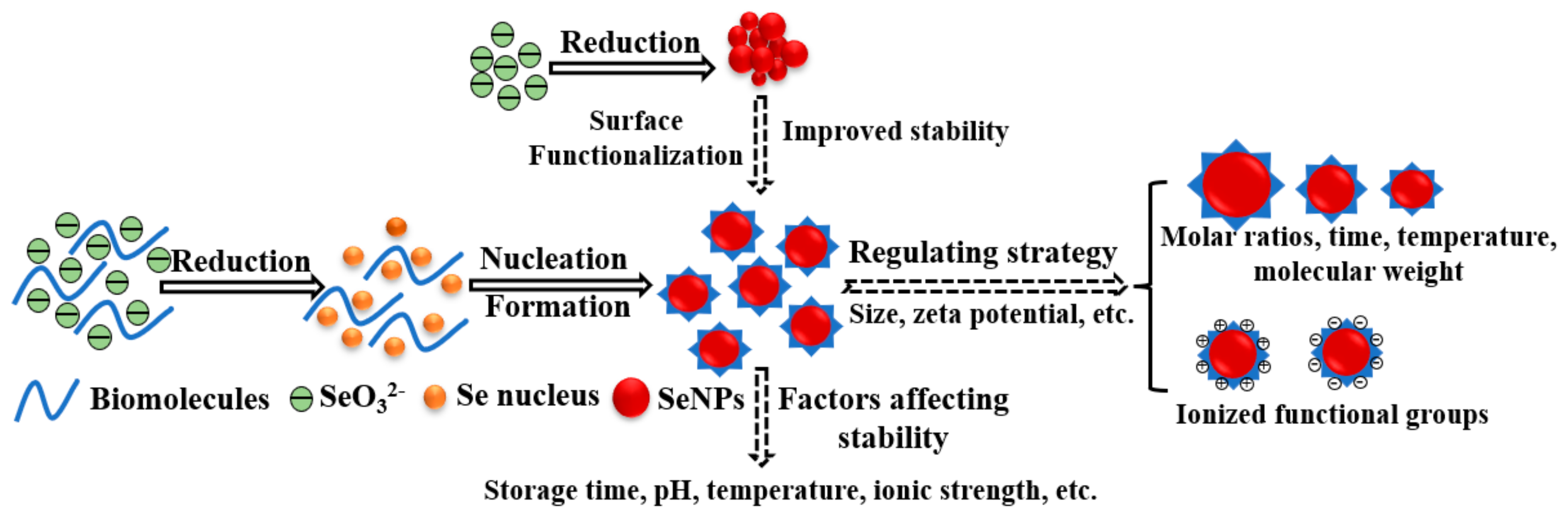
2. Synthesis, Surface Functionalization, and Stability of SeNPs
2.1. Synthesis of SeNPs
2.2. Surface Functionalization of SeNPs
2.2.1. Polysaccharides Functionalized SeNPs
2.2.2. Protein Functionalized SeNPs
2.2.3. Other Biomolecules Functionalized SeNPs

| Name | Biomolecules | Reaction Conditions | Size | Stability Conditions Tested | Stability | Ref. |
|---|---|---|---|---|---|---|
| Polysaccharides | ||||||
| T70-SeNPs | Dextran 70,000 | T70: 5 mg/mL, 20 mL; Na2SeO3: 200 mM, 250 μL; Vc: 200 mM, 1.25 mL; Temperature: 25℃; Time: 12 h | 110.3 ± 30.2 nm | Storage stability: 0, 1, 3, 6 months of storage at 4 °C | The freeze-dried powder of T70-SeNPs kept stable for 6 months at 4 °C, while the size of T70-SeNPs solution increased from 117.5 ± 30.4 to 178.5 ± 43 nm in the second month | [55] |
| PSP-SeNPs | Polygonatum sibiricum polysaccharide | PSP: 5 mg/mL; Na2SeO3: 50 mM, 1 mL; Vc: 200 mM, 1 mL; Temperature: 25 °C; Time: 30 min | 114 nm | Storage stability: 0, 1, 5, 10, 20, 30 days of storage at 4 °C; Thermal stability: 25 °C, 50 °C, 70 °C, 90 °C for 1 h; pH stability: pH 2–10 for 1 h; Ionic strength stability: 10, 50, 100 mM NaCl for 1 h | PSP-SeNPs were stable for 20 days of storage at 4 °C and stable at pH 3–10, but easily aggregated in 100 mM NaCl | [56] |
| ORPS-SeNPs | Oudemansiella raphanipies polysaccharide | ORPS: 1 mg/mL; Na2SeO3: 0.1 M, 0.3 mL; TW-80: 2 mg/mL; Vc: 0.1 M, 1.2 mL; Temperature: 30 °C; Time: 4 h; Dark condition | 60 nm | Storage stability: 0, 15, 30, 60, 90, 120 days of storage at 4 °C, 25 °C, 37 °C; pH stability: pH 2–10 | ORPS-SeNPs were stable at 4 °C for at least 90 days and stable at pH 4–10 | [57] |
| SPs-SeNPs | Spirulina platensis polysaccharide | SPs: 100 mg/mL; Na2SeO3: 2 mM; Vc: 6 mM; Time: 6 h | 73.42 nm | Storage stability: 0, 5, 15, 30, 45, 60, 75, 90 days of storage at 4 °C | SPs-SeNPs were stable for 75 days at 4 °C | [58] |
| GLP-SeNPs | Grateloupia Livida polysaccharide | GLP: 1 mg/mL, 5 mL; Na2SeO3: 0.01 M, 5 mL; Vc: 0.04 M, 5 mL; Temperature: 45 °C; Time: 3 h | 115.54 nm | Storage stability: 0, 5, 10, 15, 20, 25, 30 days of storage at 4 °C and 25 °C | GLP-SeNPs were stable at 4 °C after 30 days of storage and only kept stable for 15 days at 25 °C | [28] |
| AF1-Se nanocomposite | A highly-branched β-(1→3)-D-glucan | AF1: 1 mg/mL, 100 mL; Na2SeO3: 0.1 M, 1 mL; Vc: 0.2 M, 2 mL; Temperature: 25 °C; Time: 24 h | 92 nm | Storage stability: 1 day and 16 months of storage at room temperature | AF1-Se nanocomposite exhibited excellent stability during 16 months of storage | [59] |
| PUP-SeNPs | Polyporus umbellatus polysaccharide | PUP: 2.5 mg/mL, 0–8 mL; Na2SeO3: 50 mM, 1 mL; Vc: 20 mM, 10 mL; Temperature: 30 °C; Time: 12 h; Dark condition | 82.5 nm | Storage stability: Illumination (dark and 2500 ± 200 Lx), temperature (4 °C, 25°C, 37 °C), time (0–120 days); pH stability: 2–12 | PUP-SeNPs possessed good stability at 4 °C in dark conditions for 84 days and were stable at pH 4–12 | [60] |
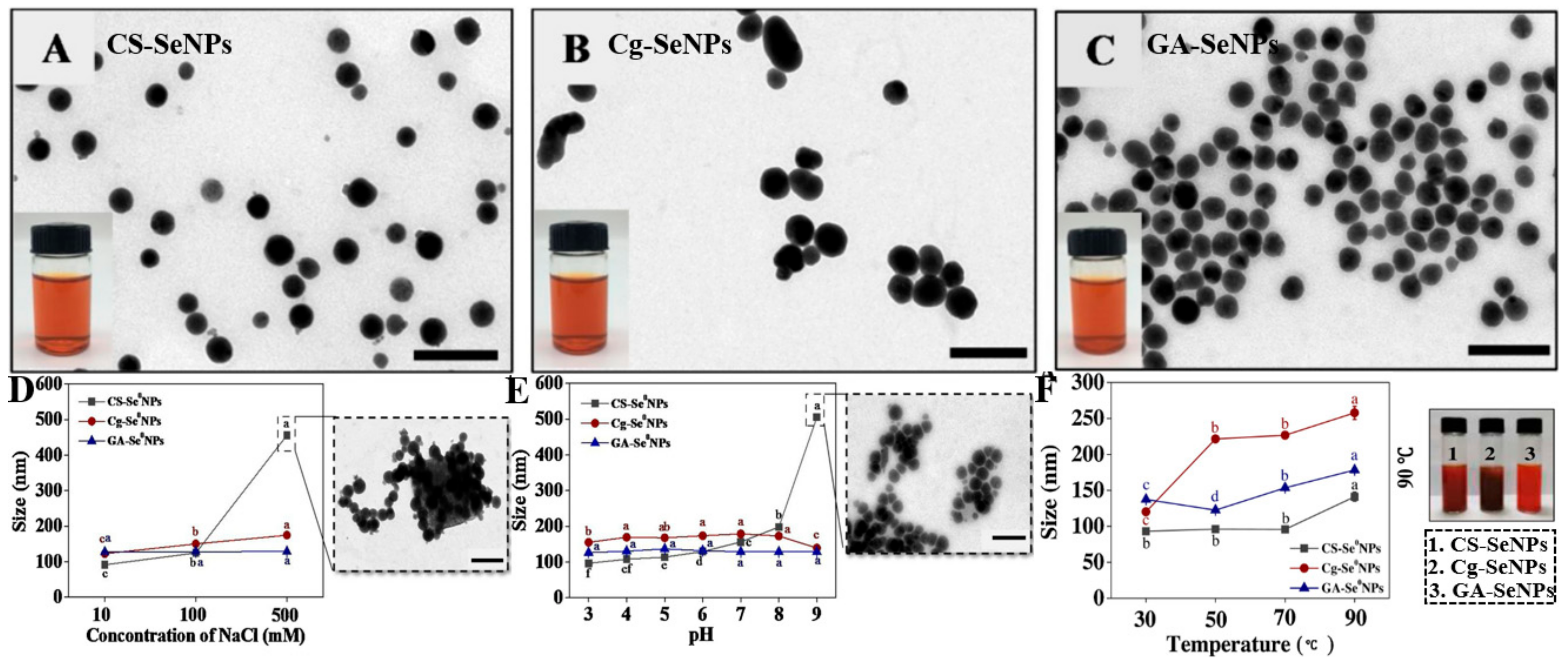
| CS-SeNPs | Chitosan | CS: 10 mg/mL, 1 mL; H2SeO3: 20 mM, 1 mL; Vc: 80 mM, 1 mL | 387.31 ± 8.13 nm | Storage stability: 0, 5, 10, 15, 20, 25, 30 days of storage at 4 °C; Thermal stability: 30 °C, 50 °C, 70°C, 90 °C for 1 h; pH stability: pH 3–9 for 1 h; Ionic strength stability: 10, 100, 500 mM NaCl for 1 h | CS-SeNPs exhibited good thermal stability and storage stability for 30 days, but easily aggregated in 500 mM NaCl or at pH > 8 | [25] |
| CPP-SeNPs | Fructose-enriched polysaccharide from Codonopsis pilosula | CPP: 2 mg/mL, 5 mL; Na2SeO3: 1.2 M; Vc: 4.8 M; The mass ratio of Na2SeO3/CPP: 1:20; Temperature: 25 °C; Dark condition | 75 nm | Storage stability: 0, 7, 14, 21, 28, 35 days of storage at 4 °C | CPP-SeNPs showed superior stability at 4 °C for at least 35 days | [61] |
| GLP-SeNPs | Polysaccharides of Gracilaria lemaneiformis | GLP: 2 mg/mL; Na2SeO3: 0.01 M; Vc: 0.04 M; Temperature: 40 °C; Time: 4 h; Dark condition | 92.5 nm | Storage stability: 0, 7, 14, 21, 28, 42 days of storage at 4 °C and 25 °C in dark conditions; pH stability: pH 3, 5, 7, 9 for 1 h; Ionic strength stability: 50, 100, 150, 200 mM for 1 h | GLPs-SeNPs were stable at 4 °C under dark conditions for 42 days and kept stable in 50–200 mM ion strengths and at a pH range from 3 to 10 | [62] |
| SFPS-SeNPs | Polysaccharides from Sargassum fusiforme | SFPS: 1 mg/mL; Na2SeO3: 0.01 M; Vc: 0.04 M; Temperature: 50 °C; Time: 4 h | 60 nm | Storage stability: 0, 7, 14, 21, 30, 40 days of storage at 4 °C | SFPS-SeNPs remained highly stable at 4 °C for 40 days | [63] |
| APS-SeNPs | Astragalus polysaccharide | APS: 2 mg/mL, 10 mL; Na2SeO3: 10 mM, 2.4 mL; Vc: 40 mM, 2.4 mL; Temperature: 25 °C; Time: 4 h | 62.3 nm | Storage stability: 0, 7, 14, 21, 28, 35 days of storage at 4 °C | APS-SeNPs exhibited good stability for 35 days at 4 °C | [64] |
| PEC-SeNPs | Pectin | PEC: 1 mg/mL, 200 mL; Na2SeO3: 0.1 M; Vc: 0.2 M, 1 mL; Temperature: 25 °C; Time: 24 h | 41 nm | Storage stability: 0, 5, 10, 15, 20, 25, 30 days of storage at 4 °C pH stability: pH 3, 4, and 5 during the storage time (0–30 days) | PEC-SeNPs were highly stable at pH > 4.0 for at least 1 month | [32] |
| SeNPs-C/C | Chitosan/citrate gel | CS: 10 mg/mL; Na2SeO3: 40 mg/mL; Vc: 40 mg/mL | 1–30 μm | Storage stability: 0, 5, 10 days of storage at 60 ± 1 °C, 80 ± 5% RH, and 5000 ± 500 Lx (Stress testing); 1, 2, 3, 6 months storage at 40 ± 2 °C, 75 ± 5% RH, dark (Accelerated testing) | SeNPs-C/C exhibited excellent stability after 6 months of storage in a simulated package environment (40 ± 2 °C, 75 ± 5% RH, dark) | [65] |
| Proteins | ||||||
| Blg-SeNPs | Beta-lactoglobulin | Blg: 10 mg/mL, 1 mL; Na2SeO3: 0.06 M, 1 mL; Vc: 0.3 M, 5 mL; Time: 30 min | 36.8 nm | Storage stability: 0, 30 days of storage at 4 °C or 25 ± 1 °C; pH stability: pH 2.5, 3.5, 4.5, 5.5, 6.5, 7.5, 8.5 | Blg-SeNPs were stable in acidic or neutral to basic solutions (pH 2.5–3.5 or 6.5–8.5) at 4 °C for 30 days | [47] |
| BSINPs | Bovine serum albumin | BSA: 20.63 mg/mL, 40 mL; Na2SeO3: 20 mM, 10 mL; Glutathione: 25 mM, 40 mL; Time: 1 h | 40 nm | Storage stability: 0, 1, 2, 3, 4 weeks of storage | BSINPs remained around the initial particle size without aggregation or precipitation over 4 weeks | [66] |
| Other biomolecules | ||||||
| EWP-SeNPs | Egg white polypeptide | EWP: 35 mg/mL; Na2SeO3: 1.038 mg/mL; Temperature: 82 °C; Time: 3.5 h | 30–50 nm | Storage stability: 0, 8, 30 days of storage at 4 °C; pH stability: pH 2, 4, 6, 8, 10 for 8 and 30 days | EWP-SeNPs showed excellent stability in an alkaline environment (pH = 10) for 30 days at 4 °C | [67] |
| TP-SeNPs | Tilapia polypeptide | TP: 20 mg/mL; Na2SeO3: 0.692 mg/mL; Temperature: 50 °C; Time: 21 h | 200 nm | Storage stability: 0, 8 days of storage at 4 °C; pH stability: pH 2, 4, 6, 8, 10 for 8 days at 4 °C | TP-SeNPs were relatively stable in an alkaline environment (pH = 8) after 8 days of storage at 4 °C | [49] |
| Ppm-SeNPs | Peanut meal peptides mixture | Ppm: 3–4 mg/mL; Na2SeO3: 1 mM; Vc: 4 mM; Temperature: 55℃; Time: 6 h | 140 nm | Storage stability: Two months of storage at 4 °C and 25 ± 1 °C; Thermal stability: 90 °C for 1, 3, and 6 h; pH stability: pH 2, 6, 10 for 2 weeks at 4 °C | Ppm-SeNPs exhibited good thermal stability and alkali resistance, and were stable for 60 days at 4 °C | [48] |
| PSP-SeNPs | Polysaccharide-protein complex | PSP: 0.08%; Na2SeO3: 1 mM; Vc: 4 mM; Temperature: 25 °C | 63.33 nm | Storage stability: 0, 4, 8, 12 months of storage at 4 °C | PSP-SeNPs remained stable for 12 months at 4 °C | [68] |
| Bc@SeNPs | Betacyanins | Bc: 14 mg/mL, 5 mL; Na2SeO3: 7 mg/mL, 5 mL; Vc: 30 mg/mL, 5 mL; Temperature: 4 °C; Time: 24 h | 133 nm | Storage stability: 0, 5, 10, 15, 20, 25, 30 days of storage at 4 °C | Bc@SeNPs maintained good stability in an aqueous solution at 4 °C for 30 days | [69] |
2.3. The Stability of Functionalized SeNPs
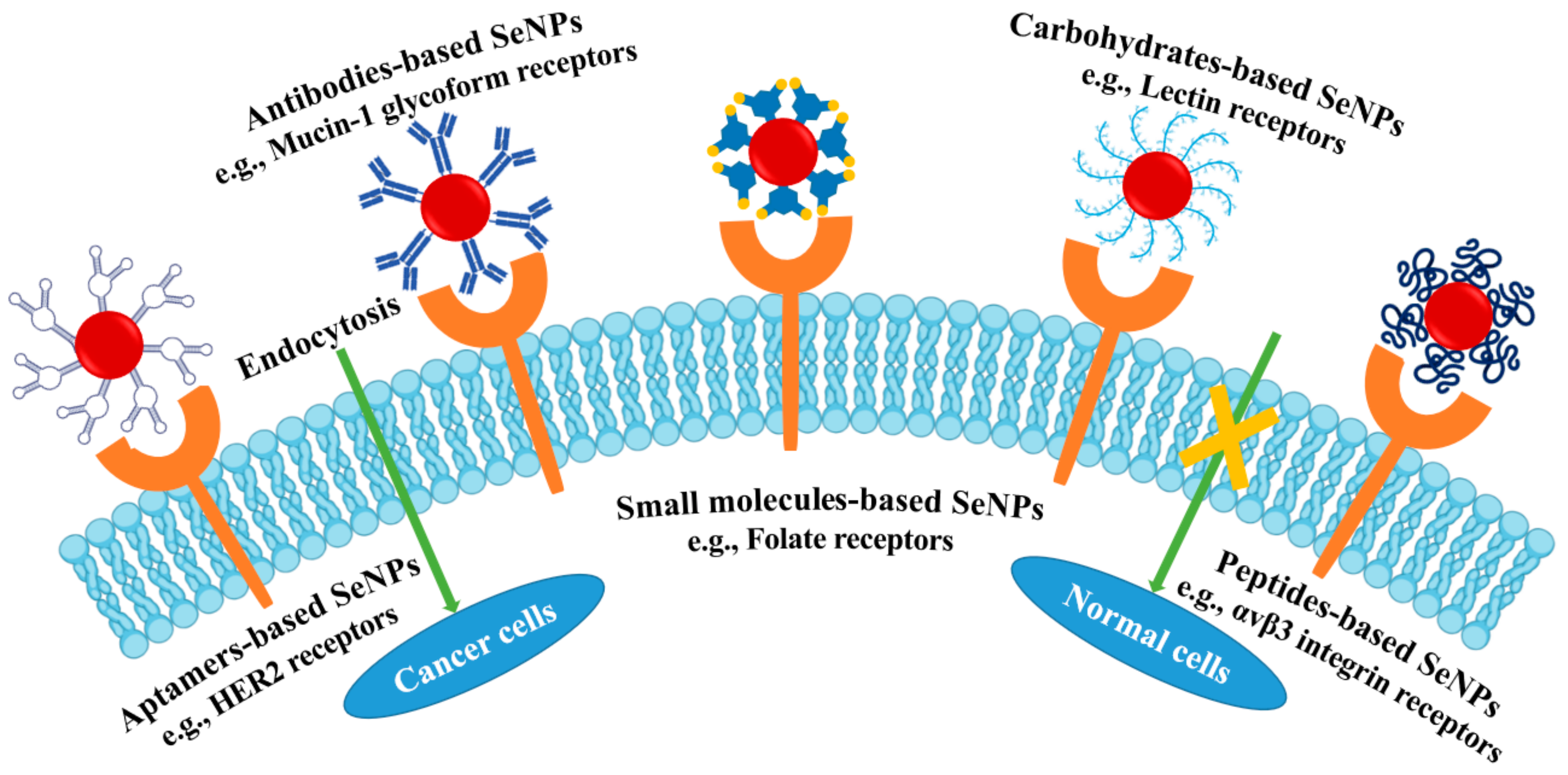
3. Targeting Strategies for SeNPs
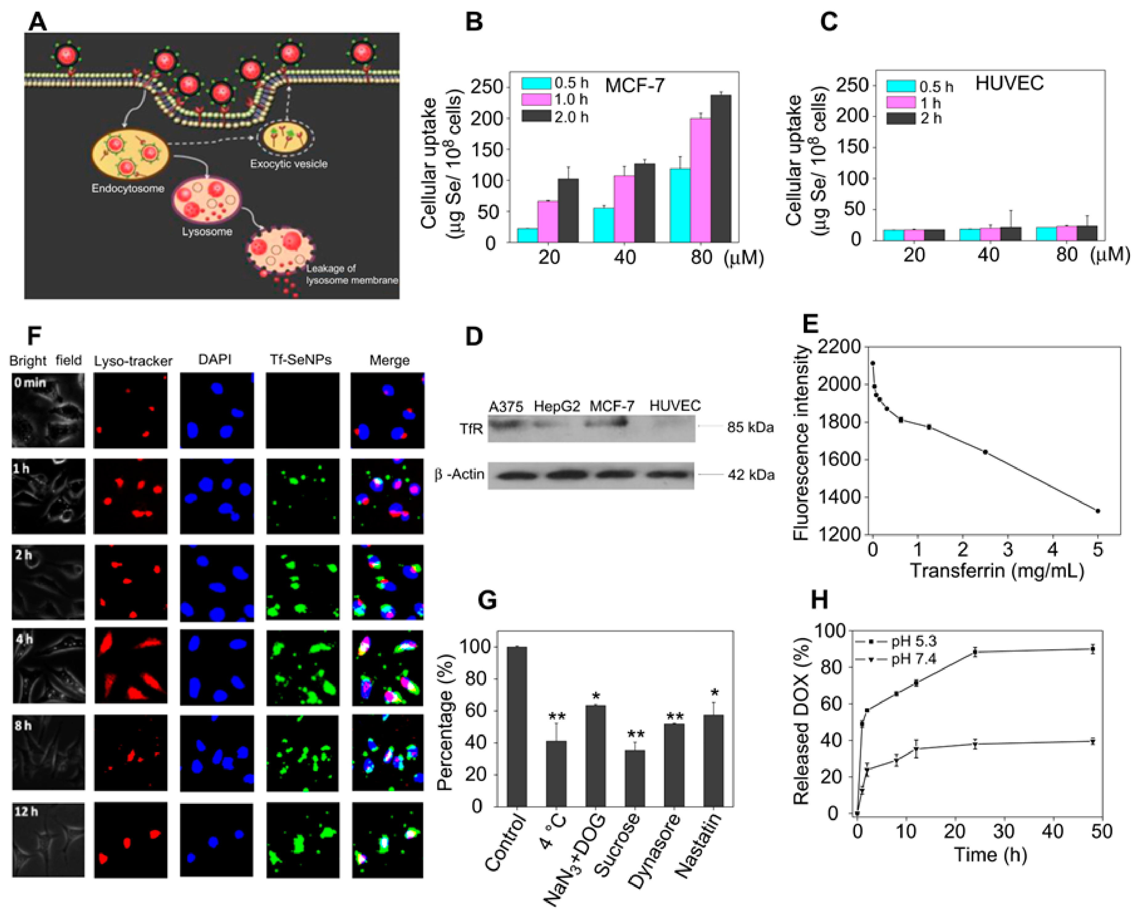
3.1. Achieving Targeting by Avoiding Reticuloendothelial System (RES)
3.2. Tumor-Activated Targeting
3.2.1. Small Molecules-Based Targeting SeNPs
3.2.2. Carbohydrate-Based Targeting SeNPs
3.2.3. Peptides-Based Targeting SeNPs
3.2.4. Antibodies-Based Targeting SeNPs
3.2.5. Aptamers-Based Targeting SeNPs
| Name | Ligand | Receptors | Endocytosis Mechanism | Biochemical Mechanism | Ref. |
|---|---|---|---|---|---|
| Small molecules-based | |||||
| FA-SeNPs | Folic acid | Folate receptors | Clathrin and caveolin-mediated endocytosis | ROS overproduction and mitochondrial depolarization | [92] |
| SeNPs@TMC-FA | Folic acid | Folate receptors | Folate receptor-mediated endocytosis | Regulation of caspase-3 and PARP | [96] |
| LA-PLL-SeNPs | Lactobionic acid | ASGPR | ASGPR-mediated endocytosis | N/A | [101] |
| Carbohydrate-based | |||||
| GA-SeNPs | Galactose | Lectin receptors | Clathrin-mediated endocytosis | Activating caspase signaling and Bcl-2 family proteins | [102] |
| GLP-SeNPs | Gracilaria lemaneiformis polysaccharide | αvβ3 integrin | αvβ3 integrin-mediated endocytosis | Downregulation of intracellular reactive oxygen species and activation of p53, MAPKs, and AKT pathways | [110] |
| LNT-SeNPs | lentinan | TLR4/TNF receptor | Caveolae-mediated endocytosis | Regulation of mitochondrial membrane fusion pathway, mediated by factor 3 (TRAF3)/mitofusin-1 (MFN1) protein complex | [111] |
| Peptides-based | |||||
| RGD-NPs | RGD peptide | αvβ3 integrin receptor | αvβ3 integrin-mediated endocytosis | Induction of apoptosis and cell cycle arrest in HUVECs via suppression of VEGF-VEGFR2-ERK/AKT signaling axis | [113] |
| GE11-Se NPs | GE11 peptide | EGFR | Lipid raft-mediated and clathrin-mediated endocytic pathway | Induction of reactive oxygen species production, activation of the mitochondria-dependent pathway, and inhibition of EGFR-mediated PI3K/AKT, and Ras/Raf/MEK/ERK pathways | [117] |
| Antibodies-based | |||||
| HER2@NP | HER2 antibody | HER2 receptors | Receptor-mediated endocytosis | Triggered DNA damage-mediated p53 signaling pathways | [123] |
| OX26-PEG-Se NPs | Monoclonal antibody (OX26) | Anti-transferrin receptor | Transferrin receptor-mediated endocytosis | Regulation of cellular metabolic state (TSC1/TSC2, p-mTOR, mTORC1), oxidative defense system (FoxO1, β-catenin/Wnt, Yap1), inflammatory reactions (jak2/stat3, Adamts-1), autophagy and apoptotic cell death (Mst1, ULK1, Bax, caspase-3 and Bcl-2) | [124] |
| Aptamers-based | |||||
| PEI-PEG-5TR1 aptamer coated SeNPs | 5TR1 aptamer | Mucin-1-glycoform | MUC1 receptor-mediated endocytosis. | N/A | [128] |
4. SeNPs as Delivery Vehicles
4.1. Single Delivery
4.2. Co-Delivery
5. Conclusions and Perspective
Author Contributions
Funding
Institutional Review Board Statement
Informed Consent Statement
Data Availability Statement
Conflicts of Interest
References
- Miyoshi, N.; Haraguchi, N.; Mizushima, T.; Ishii, H.; Yamamoto, H.; Mori, M. Targeting cancer stem cells in refractory cancer. Regen. Ther. 2021, 17, 13–19. [Google Scholar] [CrossRef]
- Wei, Q.Y.; Xu, Y.M.; Lau, A.T.Y. Recent progress of nanocarrier-based therapy for solid malignancies. Cancers 2020, 12, 2783. [Google Scholar] [CrossRef]
- Patra, A.R.; Hajra, S.; Baral, R.; Bhattacharya, S. Use of selenium as micronutrients and for future anticancer drug: A review. Nucl. India 2020, 63, 107–118. [Google Scholar] [CrossRef]
- Li, J.; Shen, B.; Nie, S.; Duan, Z.; Chen, K. A combination of selenium and polysaccharides: Promising therapeutic potential. Carbohydr. Polym. 2019, 206, 163–173. [Google Scholar] [CrossRef]
- Kursvietiene, L.; Mongirdiene, A.; Bernatoniene, J.; Sulinskiene, J.; Staneviciene, I. Selenium anticancer properties and impact on cellular redox status. Antioxidants 2020, 9, 80. [Google Scholar] [CrossRef]
- Zhu, Y.; Zhao, T.J.; Liu, M.; Wang, S.Y.; Liu, S.L.; Yang, Y.R.; Yang, Y.Q.; Nan, Y.Y.; Huang, Q.; Ai, K.L. Rheumatoid arthritis microenvironment insights into treatment effect of nanomaterials. Nano Today 2022, 42, 101358. [Google Scholar] [CrossRef]
- Garza-Garcia, J.J.O.; Hernandez-Diaz, J.A.; Zamudio-Ojeda, A.; Leon-Morales, J.M.; Guerrero-Guzman, A.; Sanchez-Chipres, D.R.; Lopez-Velazquez, J.C.; Garcia-Morales, S. The role of selenium nanoparticles in agriculture and food technology. Biol. Trace Elem. Res. 2021, 200, 2528–2548. [Google Scholar] [CrossRef]
- Ikram, M.; Javed, B.; Raja, N.I.; Mashwani, Z.-u.-R. Biomedical potential of plant-based selenium nanoparticles: A comprehensive review on therapeutic and mechanistic aspects. Int. J. Nano Med. 2021, 16, 249–268. [Google Scholar] [CrossRef]
- Liu, Q.; Duan, B.; Xu, X.; Zhang, L. Progress in rigid polysaccharide-based nanocomposites with therapeutic functions. J. Mater. Chem. B 2017, 5, 5690–5713. [Google Scholar] [CrossRef]
- Cheng, L.Z.; Wang, Y.F.; He, X.X.; Wei, X.L. Preparation, structural characterization and bioactivities of Se-containing polysaccharide: A review. Int. J. Biol. Macromol. 2018, 120, 82–92. [Google Scholar] [CrossRef]
- Ferro, C.; Florindo, H.F.; Santos, H.A. Selenium nanoparticles for biomedical applications: From development and characterization to therapeutics. Adv. Healthc. Mater. 2021, 10, 2100598. [Google Scholar] [CrossRef] [PubMed]
- Singh, A.; Guleria, A.; Kunwar, A.; Neogy, S.; Rath, M.C. Highly facile and rapid one-pot synthetic protocol for the formation of Se nanoparticles at ambient conditions with controlled phase and morphology: Role of starch and cytotoxic studies. Mater. Res. Express. 2019, 6, 015029. [Google Scholar] [CrossRef]
- Rehman, A.; John, P.; Bhatti, A. Biogenic selenium nanoparticles: Potential solution to oxidative stress mediated inflammation in rheumatoid arthritis and associated complications. Nanomaterials 2021, 11, 2005. [Google Scholar] [CrossRef] [PubMed]
- Mellinas, C.; Jimenez, A.; Garrigos, M.d. Microwave-assisted green synthesis and antioxidant activity of selenium nanoparticles using theobroma cacao L. bean shell extract. Molecules 2019, 24, 4048. [Google Scholar] [CrossRef]
- Varlamova, E.G.; Goltyaev, M.V.; Mal’tseva, V.N.; Turovsky, E.A.; Sarimov, R.M.; Simakin, A.V.; Gudkov, S.V. Mechanisms of the cytotoxic effect of selenium nanoparticles in different human cancer cell lines. Int. J. Mol. Sci. 2021, 22, 7798. [Google Scholar] [CrossRef]
- Ren, L.; Zhang, H.Z.; Tan, P.H.; Chen, Y.F.; Zhang, Z.S.; Chang, Y.Q.; Xu, J.; Yang, F.H.; Yu, D.P. Hexagonal selenium nanowires synthesized via vapor-phase growth. J. Phys. Chem. B 2004, 108, 4627–4630. [Google Scholar] [CrossRef]
- Triantis, T.; Troupis, A.; Gkika, E.; Alexakos, G.; Boukos, N.; Papaconstantinou, E.; Hiskia, A. Photocatalytic synthesis of Se nanoparticles using polyoxometalates. Catal. Today 2009, 144, 2–6. [Google Scholar] [CrossRef]
- Chaudhary, S.; Umar, A.; Mehta, S.K. Selenium nanomaterials: An overview of recent developments in synthesis, properties and potential applications. Prog. Mater. Sci. 2016, 83, 270–329. [Google Scholar] [CrossRef]
- Quintana, M.; Haro-Poniatowski, E.; Morales, J.; Batina, N. Synthesis of selenium nanoparticles by pulsed laser ablation. Appl. Surf. Sci. 2002, 195, 175–186. [Google Scholar] [CrossRef]
- Alagesan, V.; Venugopal, S. Green synthesis of selenium nanoparticle using leaves extract of withania somnifera and its biological applications and photocatalytic activities. Bionanoscience 2019, 9, 105–116. [Google Scholar] [CrossRef] [Green Version]
- Sowndarya, P.; Ramkumar, G.; Shivakumar, M.S. Green synthesis of selenium nanoparticles conjugated Clausena dentata plant leaf extract and their insecticidal potential against mosquito vectors. Artif. Cell Nanomed. B 2017, 45, 1490–1495. [Google Scholar] [CrossRef]
- Zhai, X.N.; Zhang, C.Y.; Zhao, G.H.; Stoll, S.; Ren, F.Z.; Leng, X.J. Antioxidant capacities of the selenium nanoparticles stabilized by chitosan. J. Nanobiotechnol. 2017, 15, 4. [Google Scholar] [CrossRef]
- Maiyo, F.; Singh, M. Selenium nanoparticles: Potential in cancer gene and drug delivery. Nanomedicine 2017, 12, 1075–1089. [Google Scholar] [CrossRef]
- Su, L.; Feng, Y.; Wei, K.; Xu, X.; Liu, R.; Chen, G. Carbohydrate-based macromolecular biomaterials. Chem. Rev. 2021, 121, 10950–11029. [Google Scholar] [CrossRef]
- Song, X.X.; Chen, Y.Y.; Sun, H.B.; Liu, X.N.; Leng, X.J. Physicochemical stability and functional properties of selenium nanoparticles stabilized by chitosan, carrageenan, and gum Arabic. Carbohydr. Polym. 2021, 255, 117379. [Google Scholar] [CrossRef]
- Nie, T.; Wu, H.; Wong, K.H.; Chen, T. Facile synthesis of highly uniform selenium nanoparticles using glucose as the reductant and surface decorator to induce cancer cell apoptosis. J. Mater. Chem. B 2016, 4, 2351–2358. [Google Scholar] [CrossRef]
- Liu, L.; Xiao, Z.; Niu, S.; He, Y.; Wang, G.; Pei, X.; Tao, W.; Wang, M. Preparation, characteristics and feeble induced-apoptosis performance of non-dialysis requiring selenium nanoparticles@chitosan. Mater. Des. 2019, 182, 108024. [Google Scholar] [CrossRef]
- Cao, B.; Zhang, Q.; Guo, J.; Guo, R.; Fan, X.; Bi, Y. Synthesis and evaluation of Grateloupia Livida polysaccharides-functionalized selenium nanoparticles. Int. J. Biol. Macromol. 2021, 191, 832–839. [Google Scholar] [CrossRef]
- Chen, W.W.; Yue, L.; Jiang, Q.X.; Liu, X.L.; Xia, W.S. Synthesis of varisized chitosan-selenium nanocomposites through heating treatment and evaluation of their antioxidant properties. Int. J. Biol. Macromol. 2018, 114, 751–758. [Google Scholar] [CrossRef]
- Ye, X.G.; Chen, Z.Z.; Zhang, Y.Y.; Mu, J.J.; Chen, L.Y.; Li, B.; Lin, X.R. Construction, characterization, and bioactive evaluation of nano-selenium stabilized by green tea nano-aggregates. LWT-Food Sci. Technol. 2020, 129, 109475. [Google Scholar] [CrossRef]
- Xiao, Y.D.; Huang, Q.L.; Zheng, Z.M.; Guan, H.; Liu, S.Y. Construction of a Cordyceps sinensis exopolysaccharide-conjugated selenium nanoparticles and enhancement of their antioxidant activities. Int. J. Biol. Macromol. 2017, 99, 483–491. [Google Scholar] [CrossRef]
- Qiu, W.Y.; Wang, Y.Y.; Wang, M.; Yan, J.K. Construction, stability, and enhanced antioxidant activity of pectin-decorated selenium nanoparticles. Colloid. Surface B 2018, 170, 692–700. [Google Scholar] [CrossRef]
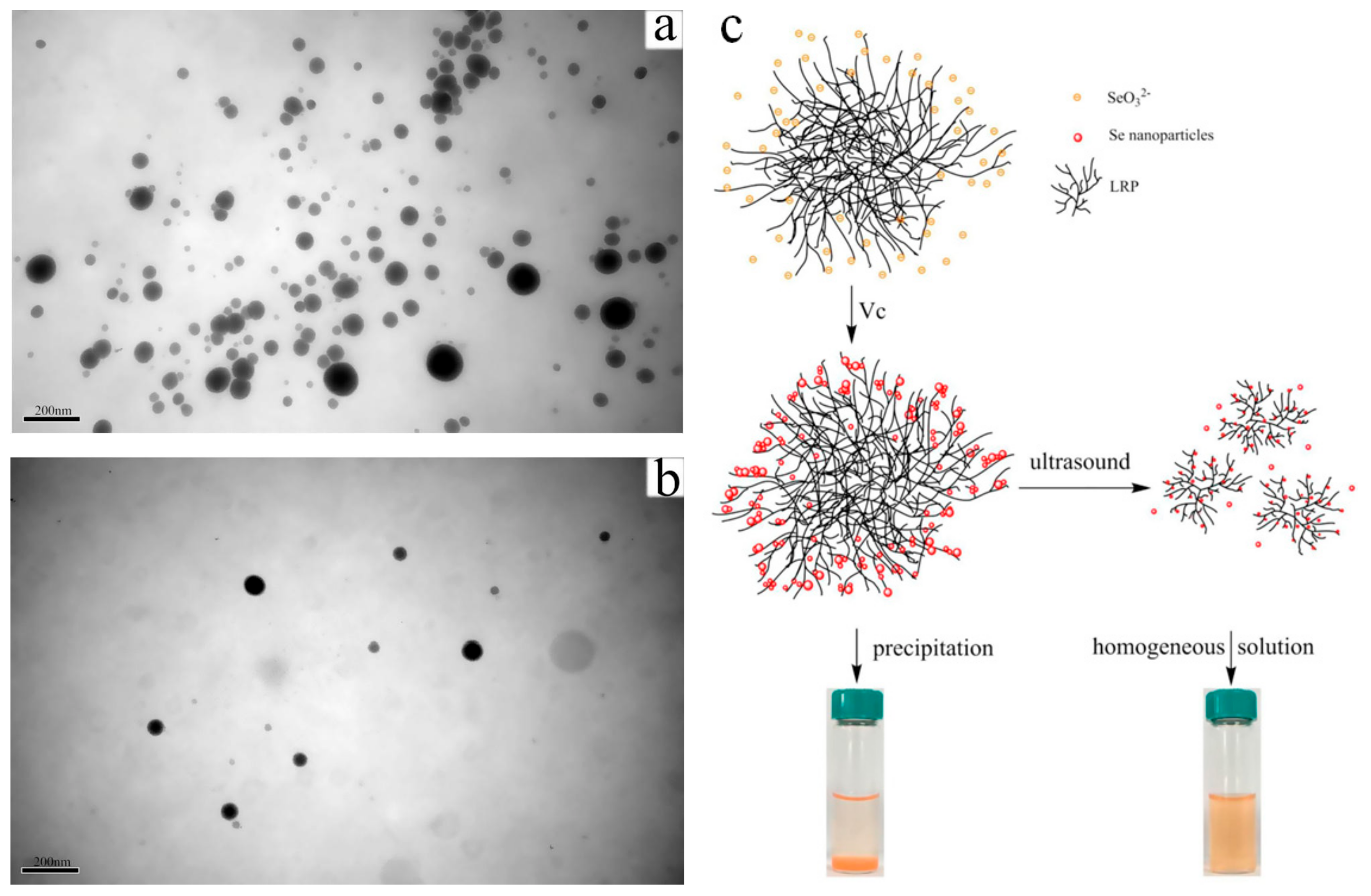
- Cai, W.F.; Hu, T.; Bakry, A.M.; Zheng, Z.M.; Xiao, Y.D.; Huang, Q.L. Effect of ultrasound on size, morphology, stability and antioxidant activity of selenium nanoparticles dispersed by a hyperbranched polysaccharide from Lignosus rhinocerotis. Ultrason Sonochem. 2018, 42, 823–831. [Google Scholar] [CrossRef]
- Liu, Y.; Huang, W.; Han, W.; Li, C.; Zhang, Z.; Hu, B.; Chen, S.; Cui, P.; Luo, S.; Tang, Z.; et al. Structure characterization of Oudemansiella radicata polysaccharide and preparation of selenium nanoparticles to enhance the antioxidant activities. LWT-Food Sci. Technol. 2021, 146, 111469. [Google Scholar] [CrossRef]
- Hien, N.Q.; Tuan, P.D.; Phu, D.V.; Quoc, L.A.; Lan, N.T.K.; Duy, N.N.; Hoa, T.T. Gamma Co-60 ray irradiation synthesis of dextran stabilized selenium nanoparticles and their antioxidant activity. Mater. Chem. Phys. 2018, 205, 29–34. [Google Scholar] [CrossRef]
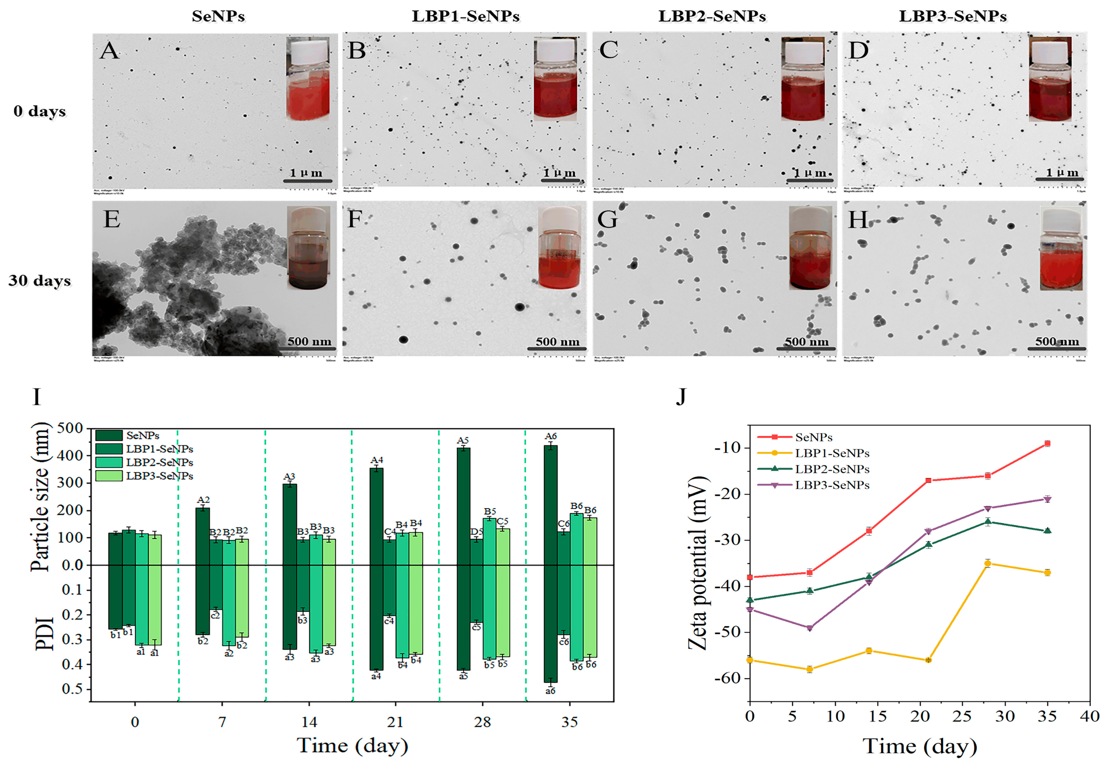
- Liu, G.Y.; Yang, X.; Zhang, J.X.; Liang, L.; Miao, F.; Ji, T.; Ye, Z.Q.; Chu, M.; Ren, J.Y.; Xu, X. Synthesis stability and anti-fatigue activity of selenium nanoparticles stabilized by Lycium barbarum polysaccharides. Int. J. Biol. Macromol. 2021, 179, 418–428. [Google Scholar]
- Chen, W.W.; Yue, L.; Jiang, Q.X.; Xia, W.S. Effect of chitosan with different molecular weight on the stability, antioxidant and anticancer activities of well-dispersed selenium nanoparticles. IET Nanobiotechnol. 2019, 13, 30–35. [Google Scholar] [CrossRef]
- Zhang, C.Y.; Zhai, X.N.; Zhao, G.H.; Ren, F.Z.; Leng, X.J. Synthesis, characterization, and controlled release of selenium nanoparticles stabilized by chitosan of different molecular weights. Carbohydr. Polym. 2015, 134, 158–166. [Google Scholar] [CrossRef]
- Yan, J.K.; Qiu, W.Y.; Wang, Y.Y.; Wang, W.H.; Yang, Y.; Zhang, H.N. Fabrication and stabilization of biocompatible selenium nanoparticles by carboxylic curdlans with various molecular properties. Carbohydr. Polym. 2018, 179, 19–27. [Google Scholar] [CrossRef]
- Lin, J.T.; Chang, Y.Y.; Chen, Y.C.; Shen, B.Y.; Yang, D.J. Molecular mechanisms of the effects of the ethanolic extract of Muntingia calabura Linn. fruit on lipopolysaccharide-induced pro-inflammatory mediators in macrophages. Food Funct. 2017, 8, 1245–1253. [Google Scholar]
- Hu, S.Q.; Hu, W.C.; Li, Y.R.; Li, S.J.; Tian, H.F.; Lu, A.; Wang, J.G. Construction and structure-activity mechanism of polysaccharide nano-selenium carrier. Carbohydr. Polym. 2020, 236, 116052. [Google Scholar] [CrossRef] [PubMed]
- Chen, W.W.; Li, Y.F.; Yang, S.; Yue, L.; Jiang, Q.X.; Xia, W.S. Synthesis and antioxidant properties of chitosan and carboxymethyl chitosan-stabilized selenium nanoparticles. Carbohydr. Polym. 2015, 132, 574–581. [Google Scholar] [CrossRef] [PubMed]
- Teng, Z.; Xu, R.; Wang, Q. Beta-lactoglobulin-based encapsulating systems as emerging bioavailability enhancers for nutraceuticals: A review. Rsc. Adv. 2015, 5, 35138–35154. [Google Scholar] [CrossRef]
- Valueva, S.V.; Borovikova, L.N.; Koreneva, V.V.; Nazarkina, Y.I.; Kipper, A.I.; Kopeikin, V.V. Structural-morphological and biological properties of selenium nanoparticles stabilized by bovine serum albumin. Russ. J. Phys. Chem. A 2007, 81, 1170–1173. [Google Scholar] [CrossRef]
- Wang, H.; Zhang, J.; Yu, H. Elemental selenium at nano size possesses lower toxicity without compromising the fundamental effect on selenoenzymes: Comparison with selenomethionine in mice. Free Radical Bio. Med. 2007, 42, 1524–1533. [Google Scholar] [CrossRef]
- Cao, H.; Xiao, J.; Liu, H. Enhanced oxidase-like activity of selenium nanoparticles stabilized by chitosan and application in a facile colorimetric assay for mercury (II). Biochem. Eng. J. 2019, 152, 107384. [Google Scholar] [CrossRef]
- Zhang, J.L.; Teng, Z.; Yuan, Y.; Zeng, Q.Z.; Lou, Z.Y.; Lee, S.H.; Wang, Q. Development, physicochemical characterization and cytotoxicity of selenium nanoparticles stabilized by beta-lactoglobulin. Int. J. Biol. Macromol. 2018, 107, 1406–1413. [Google Scholar] [CrossRef]
- Ye, M.J.; Xu, Q.L.; Tang, H.Y.; Jiang, W.Y.; Su, D.X.; He, S.; Zeng, Q.Z.; Yuan, Y. Development and stability of novel selenium colloidal particles complex with peanut meal peptides. LWT-Food Sci. Technol. 2020, 126, 109280. [Google Scholar] [CrossRef]
- Tang, H.Y.; Huang, Q.; Wang, Y.L.; Yang, X.Q.; Su, D.-X.; He, S.; Tan, J.C.; Zeng, Q.Z.; Yuan, Y. Development, structure characterization and stability of food grade selenium nanoparticles stabilized by tilapia polypeptides. J. Food Eng. 2020, 275, 109878. [Google Scholar] [CrossRef]
- Liao, W.; Zhang, R.; Dong, C.; Yu, Z.; Ren, J. Novel walnut peptide-selenium hybrids with enhanced anticancer synergism: Facile synthesis and mechanistic investigation of anticancer activity. Int. J. Nanomed. 2016, 11, 1305–1321. [Google Scholar]
- Feng, Y.X.; Su, J.Y.; Zhao, Z.N.; Zheng, W.J.; Wu, H.L.; Zhang, Y.B.; Chen, T.F. Differential effects of amino acid surface decoration on the anticancer efficacy of selenium nanoparticles. Dalton T. 2014, 43, 1854–1861. [Google Scholar] [CrossRef]
- Guo, Y.; Sun, Q.; Wu, F.G.; Dai, Y.; Chen, X. Polyphenol-containing nanoparticles: Synthesis, properties, and therapeutic delivery. Adv. Mater. 2021, 33, 2007356. [Google Scholar] [CrossRef]
- Chen, L.; Deng, Z.; Zhong, C.; Zhou, Y.; Bai, Y. Modulation of calcium oxalate crystallization by colloidal selenium nanoparticles-polyphenol complex. Cryst. Growth Des. 2016, 16, 2581–2589. [Google Scholar] [CrossRef]
- Wu, S.S.; Sun, K.; Wang, X.; Wang, D.X.; Wan, X.C.; Zhang, J.S. Protonation of epigallocatechin-3-gallate (EGCG) results in massive aggregation and reduced oral bioavailability of EGCG-dispersed selenium nanoparticles. J. Agric. Food Chem. 2013, 61, 7268–7275. [Google Scholar] [CrossRef]
- Wang, Z.Z.; Ji, L.Y.; Ren, Y.M.; Liu, M.H.; Ai, X.Y.; Yang, C. Preparation and anti-tumor study of dextran 70,000-selenium nanoparticles and poloxamer 188-selenium nanoparticles. Aaps Pharmscitech. 2022, 23, 2329. [Google Scholar] [CrossRef]
- Chen, W.W.; Cheng, H.; Xia, W.S. Construction of Polygonatum sibiricum polysaccharide functionalized selenium nanoparticles for the enhancement of stability and antioxidant activity. Antioxidants 2022, 11, 240. [Google Scholar] [CrossRef]
- Jiang, H.T.; Wang, R.L.; Zhou, F.; Wu, Y.L.; Li, S.J.; Huo, G.M.; Ye, J.C.; Hua, C.; Wang, Z.J. Preparation, physicochemical characterization, and cytotoxicity of selenium nanoparticles stabilized by Oudemansiella raphanipies polysaccharide. Int. J. Biol. Macromol. 2022, 211, 35–46. [Google Scholar] [CrossRef]
- Zhang, X.; Yan, H.; Ma, L.; Zhang, H.; Ren, D.F. Preparation and characterization of selenium nanoparticles decorated bySpirulina platensispolysaccharide. J. Food Biochem. 2020, 44, e13363. [Google Scholar] [CrossRef]
- Ping, Z.; Liu, T.; Xu, H.; Meng, Y.; Li, W.; Xu, X.; Zhang, L. Construction of highly stable selenium nanoparticles embedded in hollow nanofibers of polysaccharide and their antitumor activities. Nano Res. 2017, 10, 3775–3789. [Google Scholar] [CrossRef]
- Gao, X.; Li, X.; Mu, J.; Ho, C.T.; Su, J.; Zhang, Y.; Lin, X.; Chen, Z.; Li, B.; Xie, Y. Preparation, physicochemical characterization, and anti-proliferation of selenium nanoparticles stabilized by Polyporus umbellatus polysaccharide. Int. J. Biol. Macromol. 2020, 152, 605–615. [Google Scholar] [CrossRef]
- Yu, J.; Dong, X.D.; Jiao, J.S.; Yu, S.S.; Ji, H.Y.; Liu, A.J.; Chen, Y. The inhibitory effects of selenium nanoparticles modified by fructose-enriched polysaccharide from Codonopsis pilosula on HepG2 cells. Ind. Crops Prod. 2022, 176, 114335. [Google Scholar] [CrossRef]
- Tang, L.; Luo, X.M.; Wang, M.Y.; Wang, Z.; Guo, J.; Kong, F.S.; Bi, Y.G. Synthesis characterization, in vitro antioxidant and hypoglycemic activities of selenium nanoparticles decorated with polysaccharides of Gracilaria lemaneiformis. Int. J. Biol. Macromol. 2021, 193, 923–932. [Google Scholar] [CrossRef]
- Wang, T.; Zhao, H.Y.; Bi, Y.G.; Fan, X.D. Preparation and antioxidant activity of selenium nanoparticles decorated by polysaccharides from Sargassum fusiforme. J. Food Sci. 2021, 86, 977–986. [Google Scholar] [CrossRef]
- Jiao, J.S.; Yu, J.; Ji, H.Y.; Liu, A.J. Synthesis of macromolecular Astragalus polysaccharide-nano selenium complex and the inhibitory effects on HepG2 cells. Int. J. Biol. Macromol. 2022, 211, 481–489. [Google Scholar] [CrossRef]
- Bai, K.K.; Hong, B.H.; Hong, Z.; Sun, J.P.; Wang, C.S. Selenium nanoparticles-loaded chitosan/citrate complex and its protection against oxidative stress in D-galactose-induced aging mice. J. Nanobiotechnol. 2017, 15, 92. [Google Scholar] [CrossRef]
- Deng, G.J.; Zhu, T.; Zhou, L.H.; Zhang, J.N.; Li, S.P.; Sun, Z.H.; Lai, J.Z.; Meng, X.Q.; Li, W.J.; Zhang, P.F.; et al. Bovine serum albumin-loaded nano-selenium/ICG nanoparticles for highly effective chemo-photothermal combination therapy. Rsc. Adv. 2017, 7, 30717–30724. [Google Scholar] [CrossRef]
- Tan, J.C.; Tang, H.Y.; Xu, Q.L.; Zheng, Y.M.; Su, D.X.; He, S.; Zeng, Q.Z.; Yuan, Y. The formation of egg white polypeptide-selenium complex particles: Mechanism, stability and functional properties. Int. J. Food Sci. Tech. 2022, 57, 6155–6164. [Google Scholar] [CrossRef]
- Ren, L.R.; Wu, Z.C.; Ma, Y.; Jian, W.J.; Xiong, H.J.; Zhou, L.N. Preparation and growth-promoting effect of selenium nanoparticles capped by polysaccharide-protein complexes on tilapia. J. Sci. Food Agr. 2021, 101, 476–485. [Google Scholar] [CrossRef]
- Tang, X.Y.; Yu, S.J.; Guo, X.M.; Li, H.; Chen, M.S.; Zhang, T.; Lei, C.Y.; Zhao, Z.G.; Meng, H.C. Betacyanins functionalized selenium nanoparticles inhibit HepG2 cells growth via mitochondria-mediated pathway. J. Funct. Foods 2021, 78, 104359. [Google Scholar] [CrossRef]
- Lei, W.; Chao, L.; Qiang, H.; Xiong, F. Biofunctionalization of selenium nanoparticles with a polysaccharide from Rosa roxburghii fruit and their protective effect against H2O2-induced apoptosis in INS-1 cells. Food Funct. 2019, 10, 539–553. [Google Scholar]
- Shi, X.D.; Tian, Y.Q.; Wu, J.L.; Wang, S.Y. Synthesis, characterization, and biological activity of selenium nanoparticles conjugated with polysaccharides. Crit. Rev. Food Sci. Nutr. 2021, 61, 2225–2236. [Google Scholar] [PubMed]
- Zhu, C.H.; Zhang, S.M.; Song, C.W.; Zhang, Y.B.; Ling, Q.J.; Hoffmann, P.R.; Li, J.; Chen, T.F.; Zheng, W.J.; Huang, Z. Selenium nanoparticles decorated with Ulva lactuca polysaccharide potentially attenuate colitis by inhibiting NF-kappa B mediated hyper inflammation. J. Nanobiotechnol. 2017, 15, 20. [Google Scholar] [CrossRef] [PubMed]
- Meng, D.; Guo, L.; Shi, D.D.; Sun, X.; Shang, M.M.; Zhou, X.Y.; Li, J. Charge-conversion and ultrasound-responsive O-carboxymethyl chitosan nanodroplets for controlled drug delivery. Nanomedicine 2019, 14, 2549–2565. [Google Scholar] [CrossRef] [PubMed]
- Xiaoxiao, S.; Yuying, C.; Hongbo, S.; Xinnan, L.; Xiaojing, L. Physicochemical and functional properties of chitosan-stabilized selenium nanoparticles under different processing treatments. Food Chem. 2020, 331, 127378. [Google Scholar]
- Cavalu, S.; Prokisch, J.; Laslo, V.; Vicas, S. Preparation, structural characterisation and release study of novel hybrid microspheres entrapping nanoselenium, produced by green synthesis. Iet. Nanobiotechnol. 2017, 11, 426–432. [Google Scholar] [CrossRef]
- Nguyen, D.N.; Dang, P.V.; Le, Q.A.; Nguyen, L.T.K.; Nguyen, H.Q.; Tran, N.T.T.; Tran, H.L.B.; Phan, T.D.; Bui, H.M. Preparation and effect of selenium nanoparticles/oligochitosan on the white blood cell recovery of mice exposed to gamma-ray radiation. J. Chem. 2021, 2021, 6635022. [Google Scholar] [CrossRef]
- Dung, N.T.; Trong, T.D.; Vu, N.T.; Binh, N.T.; Minh, T.T.L.; Luan, L.Q. Radiation synthesis of selenium nanoparticles capped with beta-glucan and its immunostimulant activity in cytoxan-induced immunosuppressed mice. Nanomaterials 2021, 11, 2439. [Google Scholar] [CrossRef]
- He, J.; Li, C.; Ding, L.; Huang, Y.; Yin, X.; Zhang, J.; Zhang, J.; Yao, C.; Liang, M.; Pirraco, R.P.; et al. Tumor targeting strategies of smart fluorescent nanoparticles and their applications in cancer diagnosis and treatment. Adv. Mater. 2019, 31, 1902409. [Google Scholar] [CrossRef]
- Duan, L.; Yang, L.; Jin, J.; Yang, F.; Liu, D.; Hu, K.; Wang, Q.; Yue, Y.; Gu, N. Micro/nano-bubble-assisted ultrasound to enhance the EPR effect and potential theranostic applications. Theranostics 2020, 10, 462–483. [Google Scholar] [CrossRef]
- Veiseh, O.; Gunn, J.W.; Zhang, M. Design and fabrication of magnetic nanoparticles for targeted drug delivery and imaging. Adv. Drug Deliv. Rev. 2010, 62, 284–304. [Google Scholar] [CrossRef]
- Wang, R.; Zhang, Z.; Liu, B.; Xue, J.; Liu, F.; Tang, T.; Liu, W.; Feng, F.; Qu, W. Strategies for the design of nanoparticles: Starting with long-circulating nanoparticles, from lab to clinic. Biomater. Sci. 2021, 9, 3621–3637. [Google Scholar] [CrossRef]
- Kipper, A.I.; Titova, A.V.; Borovikova, L.N.; Pisarev, O.A. Morphological characteristics of selenium-polyethylene glycol nanocomposites. Russ. J. Phys. Chem. A 2015, 89, 1625–1627. [Google Scholar] [CrossRef]
- Valueva, S.V.; Kipper, A.I. Morphology and thermodynamics of selenium-containing nanosystems: The effect of polymer stabilizers. Russ. J. Phys. Chem. A 2017, 91, 609–612. [Google Scholar] [CrossRef]
- Zheng, S.; Li, X.; Zhang, Y.; Xie, Q.; Wong, Y.-S.; Zheng, W.; Chen, T. PEG-nanolized ultrasmall selenium nanoparticles overcome drug resistance in hepatocellular carcinoma HepG2 cells through induction of mitochondria dysfunction. Int. J. Nanomed. 2012, 7, 3939–3949. [Google Scholar]
- Tan, S.W.; Wu, T.T.; Zhang, D.; Zhang, Z.P. Cell or cell membrane-based drug delivery systems. Theranostics 2015, 5, 863–881. [Google Scholar] [CrossRef]
- Zhang, E.D.; Phan, P.; Algarni, H.A.; Zhao, Z.M. Red blood cell inspired strategies for drug delivery: Emerging concepts and new advances. Pharm. Res. 2022. [CrossRef]
- Lin, A.G.; Liu, Y.A.; Zhu, X.F.; Chen, X.; Liu, J.W.; Zhou, Y.H.; Qin, X.Y.; Liu, J. Bacteria-responsive biomimetic selenium nanosystem for multidrug-resistant bacterial infection detection and inhibition. Acs Nano 2019, 13, 13965–13984. [Google Scholar] [CrossRef]
- Liu, T.; Shi, C.Z.; Duan, L.Q.; Zhang, Z.H.; Luo, L.P.; Goel, S.; Cai, W.B.; Chen, T.F. A highly hemocompatible erythrocyte membrane-coated ultrasmall selenium nanosystem for simultaneous cancer radiosensitization and precise antiangiogenesis. J. Mater. Chem. B 2018, 6, 4756–4764. [Google Scholar] [CrossRef]
- Yu, M.K.; Park, J.; Jon, S. Targeting strategies for multifunctional nanoparticles in cancer imaging and therapy. Theranostics 2012, 2, 3–44. [Google Scholar] [CrossRef]
- Narmani, A.; Rezvani, M.; Farhood, B.; Darkhor, P.; Mohammadnejad, J.; Amini, B.; Refahi, S.; Goushbolagh, N.A. Folic acid functionalized nanoparticles as pharmaceutical carriers in drug delivery systems. Drug Develop. Res. 2019, 80, 404–424. [Google Scholar] [CrossRef]
- Pi, J.; Jin, H.; Liu, R.Y.; Song, B.; Wu, Q.; Liu, L.; Jiang, J.; Yang, F.; Cai, H.H.; Cai, J.Y. Pathway of cytotoxicity induced by folic acid modified selenium nanoparticles in MCF-7 cells. Appl. Microbiol. Biot. 2013, 97, 1051–1062. [Google Scholar] [CrossRef]
- Bidkar, A.P.; Sanpui, P.; Ghosh, S.S. Combination therapy with MAPK-pathway-specific inhibitor and folic-acid-receptor-targeted selenium nanoparticles induces synergistic antiproliferative response in BRAF mutant cancer cells. Acs. Biomater. Sci. Eng. 2019, 5, 2222–2234. [Google Scholar] [CrossRef]
- Maiyo, F.; Singh, M. Folate-targeted mRNA delivery using chitosan-functionalized selenium nanoparticles: Potential in cancer immunotherapy. Pharmaceuticals 2019, 12, 164. [Google Scholar] [CrossRef]
- Maiyo, F.; Singh, M. Polymerized selenium nanoparticles for folate-receptor-targeted delivery of anti-luc-siRNA: Potential for gene silencing. Biomedicines 2020, 8, 76. [Google Scholar] [CrossRef]
- Luesakul, U.I.F.; Komenek, S.; Puthong, S.; Muangsin, N. Shape-controlled synthesis of cubic-like selenium nanoparticles via the self-assembly method. Carbohydr. Polym. 2016, 153, 435–444. [Google Scholar] [CrossRef]
- Luesakul, U.; Puthong, S.; Neamati, N.; Muangsin, N. pH-responsive selenium nanoparticles stabilized by folate-chitosan delivering doxorubicin for overcoming drug-resistant cancer cells. Carbohydr. Polym. 2018, 181, 841–850. [Google Scholar] [CrossRef]
- Xia, Y.; Zhao, M.; Chen, Y.; Hua, L.; Xu, T.; Wang, C.; Li, Y.; Zhu, B. Folate-targeted selenium nanoparticles deliver therapeutic siRNA to improve hepatocellular carcinoma therapy. Rsc. Adv. 2018, 8, 25932–25940. [Google Scholar] [CrossRef]
- Jain, A.; Jain, A.; Parajuli, P.; Mishra, V.; Ghoshal, G.; Singh, B.; Shivhare, U.S.; Katare, O.P.; Kesharwani, P. Recent advances in galactose-engineered nanocarriers for the site-specific delivery of siRNA and anticancer drugs. Drug Discov. Today 2018, 23, 960–973. [Google Scholar] [CrossRef]
- Liu, M.; Huang, Q.; Zhu, Y.; Chen, L.; Li, Y.M.; Gong, Z.C.; Ai, K.L. Harnessing reactive oxygen/nitrogen species and inflammation: Nanodrugs for liver injury. Mater. Today Bio. 2022, 13, 100215. [Google Scholar] [CrossRef]
- Singh, D.; Singh, M. Hepatocellular-targeted mRNA delivery using functionalized selenium nanoparticles in vitro. Pharmaceutics 2021, 13, 298. [Google Scholar] [CrossRef]
- Naidoo, S.; Daniels, A.; Habib, S.; Singh, M. Poly-L-lysine-lactobionic acid-capped selenium nanoparticles for liver-targeted gene delivery. Int. J. Mol. Sci. 2022, 23, 1492. [Google Scholar] [CrossRef] [PubMed]
- Xia, Y.; Zhong, J.; Zhao, M.; Tang, Y.; Han, N.; Hua, L.; Xu, T.; Wang, C.; Zhu, B. Galactose-modified selenium nanoparticles for targeted delivery of doxorubicin to hepatocellular carcinoma. Drug Deliv. 2019, 26, 1–11. [Google Scholar] [CrossRef] [PubMed]
- Chen, Q.H.; Li, N.S.; Wang, X.Y.; Yang, Y.Q.; Xiang, Y.T.; Long, X.Y.; Zhang, J.P.; Huang, J.; Chen, L.; Huang, Q. Mitochondria-targeting chemodynamic therapy nanodrugs for cancer treatment. Front. Pharmacol. 2022, 13, 847048. [Google Scholar] [CrossRef] [PubMed]
- Bock, F.J.; Tait, S.W.G. Mitochondria as multifaceted regulators of cell death. Nat. Rev. Mol. Cell Bio. 2020, 21, 85–100. [Google Scholar] [CrossRef] [PubMed]
- Zhuang, Y.; Li, L.J.; Feng, L.D.; Wang, S.S.; Su, H.M.; Liu, H.J.; Liu, H.M.; Wu, Y.Z. Mitochondrion-targeted selenium nanoparticles enhance reactive oxygen species-mediated cell death. Nanoscale 2020, 12, 1389–1396. [Google Scholar] [CrossRef] [PubMed]
- Marin, M.J.; Schofield, C.L.; Field, R.A.; Russell, D.A. Glyconanoparticles for colorimetric bioassays. Analyst 2015, 140, 59–70. [Google Scholar] [CrossRef] [Green Version]
- Chen, F.; Huang, G.; Huang, H. Sugar ligand-mediated drug delivery. Future Med. Chem. 2020, 12, 161–171. [Google Scholar] [CrossRef]
- Ren, Y.; Zhao, T.; Mao, G.; Zhang, M.; Li, F.; Zou, Y.; Yang, L.; Wu, X. Antitumor activity of hyaluronic acid-selenium nanoparticles in Heps tumor mice models. Int. J. Biol. Macromol. 2013, 57, 57–62. [Google Scholar] [CrossRef]
- Xia, Y.; Xiao, M.; Zhao, M.; Xu, T.; Guo, M.; Wang, C.; Li, Y.; Zhu, B.; Liu, H. Doxorubicin-loaded functionalized selenium nanoparticles for enhanced antitumor efficacy in cervical carcinoma therapy. Mat. Sci. Eng. C-Mater. 2020, 106, 110100. [Google Scholar] [CrossRef]
- Jiang, W.T.; Fu, Y.T.; Yang, F.; Yang, Y.F.; Liu, T.; Zheng, W.J.; Zeng, L.L.; Chen, T.F. Gracilaria iemaneiformis polysaccharide as integrin-targeting surface decorator of selenium nanoparticles to achieve enhanced anticancer efficacy. Acs. Appl. Mater. Inter. 2014, 6, 13738–13748. [Google Scholar] [CrossRef]
- Liu, H.J.; Qin, Y.; Zhao, Z.H.; Zhang, Y.; Yang, J.H.; Zhai, D.H.; Cui, F.; Luo, C.; Lu, M.X.; Liu, P.P.; et al. Lentinan-functionalized selenium nanoparticles target tumor cell mitochondria via TLR4/TRAF3/MFN1 pathway. Theranostics 2020, 10, 9083–9099. [Google Scholar] [CrossRef]
- Tesauro, D.; Accardo, A.; Diaferia, C.; Milano, V.; Guillon, J.; Ronga, L.; Rossi, F. Peptide-based drug-delivery systems in biotechnological applications: Recent advances and perspectives. Molecules 2019, 24, 351. [Google Scholar] [CrossRef]
- Fu, X.; Yang, Y.; Li, X.; Lai, H.; Huang, Y.; He, L.; Zheng, W.; Chen, T. RGD peptide-conjugated selenium nanoparticles: Antiangiogenesis by suppressing VEGF-VEGFR2-ERK/AKT pathway. Nanomed. Nanotechnol. 2016, 12, 1627–1639. [Google Scholar] [CrossRef]
- Xia, Y.; Lin, Z.; Li, Y.; Zhao, M.; Wang, C.; Guo, M.; Zhang, B.; Zhu, B. Targeted delivery of siRNA using RGDfC-conjugated functionalized selenium nanoparticles for anticancer therapy. J. Mater. Chem. B 2017, 5, 6941–6952. [Google Scholar] [CrossRef]
- Xia, Y.; Tang, G.; Wang, C.; Zhong, J.; Chen, Y.; Hua, L.; Li, Y.; Liu, H.; Zhu, B. Functionalized selenium nanoparticles for targeted siRNA delivery silence Derlin1 and promote antitumor efficacy against cervical cancer. Drug Deliv. 2020, 27, 15–25. [Google Scholar] [CrossRef]
- Xia, Y.; Xu, T.; Wang, C.; Li, Y.; Lin, Z.; Zhao, M.; Zhu, B. Novel functionalized nanoparticles for tumor-targeting co-delivery of doxorubicin and siRNA to enhance cancer therapy. Int. J. Nanomed. 2018, 13, 143–159. [Google Scholar] [CrossRef] [Green Version]
- Pi, J.; Jiang, J.; Cai, H.; Yang, F.; Jin, H.; Yang, P.; Cai, J.; Chen, Z.W. GE11 peptide conjugated selenium nanoparticles for EGFR targeted oridonin delivery to achieve enhanced anticancer efficacy by inhibiting EGFR-mediated PI3K/AKT and Ras/Raf/MEK/ERK pathways. Drug Deliv. 2017, 24, 1549–1564. [Google Scholar] [CrossRef]
- Yang, L.; Sun, J.; Xie, W.; Liu, Y.; Liu, J. Dual-functional selenium nanoparticles bind to and inhibit amyloid beta fiber formation in Alzheimer’s disease. J. Mater Chem. B 2017, 5, 5954–5967. [Google Scholar] [CrossRef]
- Huang, J.; Liu, Y.; Liu, T.; Chang, Y.; Chen, T.; Li, X. Dual-targeting nanotherapeutics antagonize hyperinsulinemia-promoted tumor growth via activating cell autophagy. J. Mater Chem. B 2019, 7, 6751–6758. [Google Scholar] [CrossRef]
- Wang, J.; Masehi-Lano, J.J.; Chung, E.J. Peptide and antibody ligands for renal targeting: Nanomedicine strategies for kidney disease. Biomater. Sci. 2017, 5, 1450–1459. [Google Scholar] [CrossRef]
- Wang, R.E.; Zhang, Y.; Tian, L.; Cai, W.; Cai, J. Antibody-based imaging of HER-2: Moving into the clinic. Curr. Mol. Med. 2013, 13, 1523–1537. [Google Scholar] [CrossRef]
- Liu, T.; Lai, L.; Song, Z.; Chen, T. A sequentially triggered nanosystem for precise drug delivery and simultaneous inhibition of cancer growth, migration, and invasion. Adv. Funct. Mater. 2016, 26, 7775–7790. [Google Scholar] [CrossRef]
- Song, Z.; Liu, T.; Chen, T. Overcoming blood-brain barrier by HER2-targeted nanosystem to suppress glioblastoma cell migration, invasion and tumor growth. J. Mater. Chem. B 2018, 6, 568–579. [Google Scholar] [CrossRef] [PubMed]
- Amani, H.; Habibey, R.; Shokri, F.; Hajmiresmail, S.J.; Akhavans, O.; Mashaghi, A.; Pazoki-Toroudi, H. Selenium nanoparticles for targeted stroke therapy through modulation of inflammatory and metabolic signaling. Sci. Rep. 2019, 9, 6044. [Google Scholar] [CrossRef]
- Huang, Y.Y.; He, L.Z.; Liu, W.; Fan, C.D.; Zheng, W.J.; Wong, Y.S.; Chen, T.F. Selective cellular uptake and induction of apoptosis of cancer-targeted selenium nanoparticles. Biomaterials 2013, 34, 7106–7116. [Google Scholar] [CrossRef] [PubMed]
- Citartan, M.; Kaur, H.; Presela, R.; Tang, T.-H. Aptamers as the chaperones (Aptachaperones) of drugs-from siRNAs to DNA nanorobots. Int. J. Pharmaceut. 2019, 567, 118483. [Google Scholar] [CrossRef] [PubMed]
- Liu, M.L.; Zou, H.Y.; Li, C.M.; Li, R.S.; Huang, C.Z. Aptamer-modified selenium nanoparticles for dark-field microscopy imaging of nucleolin. Chem. Commun. 2017, 53, 13047–13050. [Google Scholar] [CrossRef] [PubMed]
- Jalalian, S.H.; Ramezani, M.; Abnous, K.; Taghdisi, S.M. Targeted co-delivery of epirubicin and NAS-24 aptamer to cancer cells using selenium nanoparticles for enhancing tumor response in vitro and in vivo. Cancer Lett. 2018, 416, 87–93. [Google Scholar] [CrossRef]
- Huang, S.; Yang, W.; Huang, G. Preparation and activities of selenium polysaccharide from plant such as Grifola frondosa. Carbohydr. Polym. 2020, 242, 116409. [Google Scholar] [CrossRef]
- Menon, S.; Devi, S.K.S.; Santhiya, R.; Rajeshkumar, S.; Kumar, V.S. Selenium nanoparticles: A potent chemotherapeutic agent and an elucidation of its mechanism. Colloid. Surface B 2018, 170, 280–292. [Google Scholar] [CrossRef]
- Pucci, C.; Martinelli, C.; Ciofani, G. Innovative approaches for cancer treatment: Current perspectives and new challenges. Ecancermedicalscience 2019, 13, 961. [Google Scholar] [CrossRef]
- Zou, J.; Su, S.; Chen, Z.; Liang, F.; Zeng, Y.; Cen, W.; Zhang, X.; Xia, Y.; Huang, D. Hyaluronic acid-modified selenium nanoparticles for enhancing the therapeutic efficacy of paclitaxei in lung cancer therapy. Artif. Cells Nanomed. B 2019, 47, 3456–3464. [Google Scholar] [CrossRef]
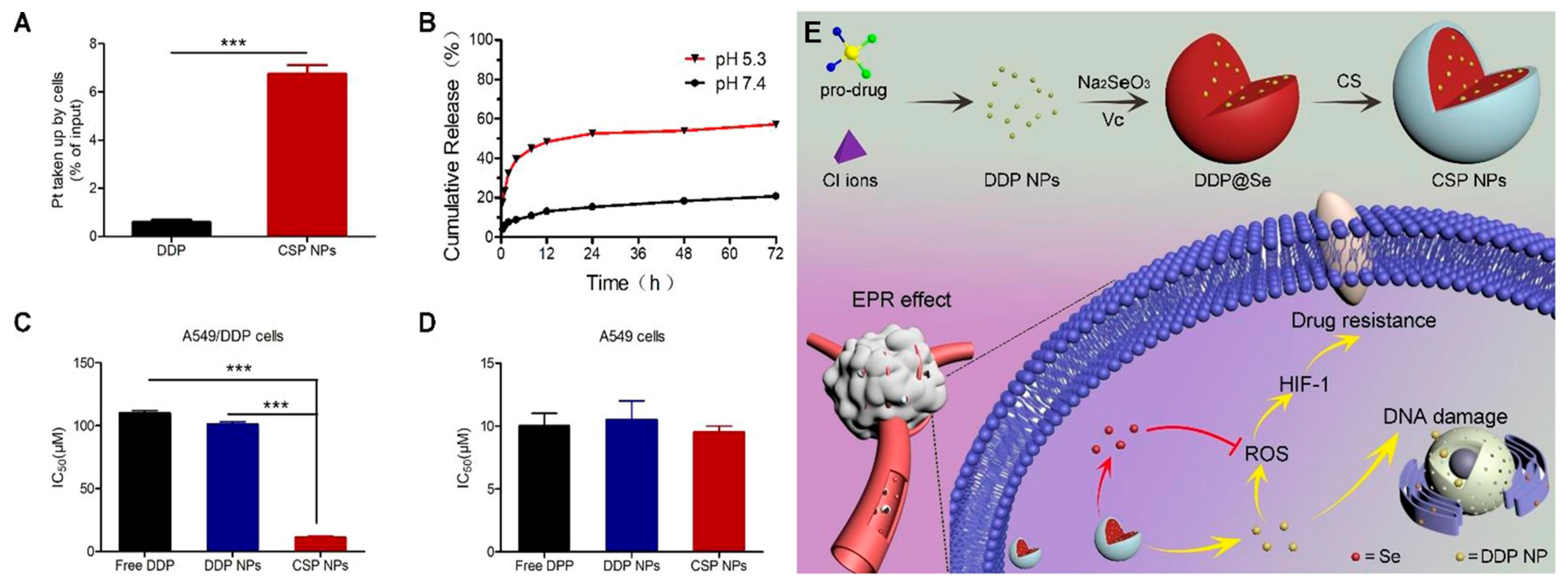
- Zhang, X.; He, C.; Yan, R.; Chen, Y.; Zhao, P.; Li, M.; Fan, T.; Yang, T.; Lu, Y.; Luo, J.; et al. HIF-1 dependent reversal of cisplatin resistance via anti-oxidative nano selenium for effective cancer therapy. Chem. Eng. J. 2020, 380, 122540. [Google Scholar] [CrossRef]
- Wu, Y.; Liu, H.; Li, Z.; Huang, D.; Nong, L.; Ning, Z.; Hu, Z.; Xu, C.; Yan, J.-K. Pectin-decorated selenium nanoparticles as a nanocarrier of curcumin to achieve enhanced physicochemical and biological properties. Iet. Nanobiotechnol. 2019, 13, 880–886. [Google Scholar] [CrossRef]
- Liu, F.; Liu, H.; Liu, R.; Xiao, C.; Duan, X.; McClements, D.J.; Liu, X. Delivery of sesamol using polyethylene-glycol-functionalized selenium nanoparticles in human liver cells in culture. J. Agri. Food Chem. 2019, 67, 2991–2998. [Google Scholar] [CrossRef]
- Sharifiaghdam, M.; Shaabani, E.; Sharifiaghdam, Z.; de Keersmaecker, H.; de Rycke, R.; de Smedt, S.; Faridi-Majidi, R.; Braeckmans, K.; Fraire, J.C. Enhanced siRNA delivery and selective apoptosis induction in H1299 cancer cells by layer-by-layer-assembled Se nanocomplexes: Toward more efficient cancer therapy. Front. Mol. Biosci. 2021, 8, 639184. [Google Scholar] [CrossRef]
- Zheng, W.; Cao, C.; Liu, Y.; Yu, Q.; Zheng, C.; Sun, D.; Ren, X.; Liu, J. Multifunctional polyamidoamine-modified selenium nanoparticles dual-delivering siRNA and cisplatin to A549/DDP cells for reversal multidrug resistance. Acta Biomater. 2015, 11, 368–380. [Google Scholar] [CrossRef]
- Fang, X.; Li, C.e.; Zheng, L.; Yang, F.; Chen, T. Dual-targeted selenium nanoparticles for synergistic photothermal therapy and chemotherapy of tumors. Chem-Asian J. 2018, 13, 996–1004. [Google Scholar] [CrossRef]
- Guan, B.; Yan, R.; Li, R.; Zhang, X. Selenium as a pleiotropic agent for medical discovery and drug delivery. Int. J. Nanomed. 2018, 13, 7473–7490. [Google Scholar] [CrossRef]
- Liu, W.; Li, X.L.; Wong, Y.S.; Zheng, W.J.; Zhang, Y.B.; Cao, W.Q.; Chen, T.F. Selenium nanoparticles as a carrier of 5-fluorouracil to achieve anticancer synergism. Acs Nano 2012, 6, 6578–6591. [Google Scholar] [CrossRef]
- Yu, S.X.; Wang, Y.R.; Zhang, W.T.; Zhang, Y.H.; Zhu, W.X.; Liu, Y.N.; Zhang, D.H.; Wang, J.L. pH-Assisted surface functionalization of selenium nanoparticles with curcumin to achieve enhanced cancer chemopreventive activity. Rsc. Adv. 2016, 6, 72213–72223. [Google Scholar] [CrossRef]
- Guo, M.; Li, Y.; Lin, Z.; Zhao, M.; Xiao, M.; Wang, C.; Xu, T.; Xia, Y.; Zhu, B. Surface decoration of selenium nanoparticles with curcumin induced HepG2 cell apoptosis through ROS mediated p53 and AKT signaling pathways. Rsc. Adv. 2017, 7, 52456–52464. [Google Scholar] [CrossRef]
- Mazarei, M.; Arvejeh, P.M.; Mozafari, M.R.; Khosravian, P.; Ghasemi, S. Anticancer potential of temozolomide-loaded Eudragit-chitosan coated selenium nanoparticles: In vitro evaluation of cytotoxicity, apoptosis and gene regulation. Nanomaterials 2021, 11, 1704. [Google Scholar] [CrossRef]
- Hosnedlova, B.; Kepinska, M.; Skalickova, S.; Fernandez, C.; Ruttkay-Nedecky, B.; Peng, Q.; Baron, M.; Melcova, M.; Opatrilova, R.; Zidkova, J.; et al. Nano-selenium and its nanomedicine applications: A critical review. Int. J. Nanomed. 2018, 13, 2107–2128. [Google Scholar] [CrossRef]
- Liu, T.; Zeng, L.; Jiang, W.; Fu, Y.; Zheng, W.; Chen, T. Rational design of cancer-targeted selenium nanoparticles to antagonize multidrug resistance in cancer cells. Nanomed. Nanotechnol. 2015, 11, 947–958. [Google Scholar] [CrossRef]
- Pillay, N.S.; Daniels, A.; Singh, M. Folate-targeted transgenic activity of dendrimer functionalized selenium nanoparticles in vitro. Int. J. Mol. Sci. 2020, 21, 7177. [Google Scholar] [CrossRef]
- Sharifiaghdam, M.; Shaabani, E.; Sharifiaghdam, Z.; de Keersmaecker, H.; Lucas, B.; Lammens, J.; Ghanbari, H.; Teimoori-Toolabi, L.; Vervaet, C.; de Beer, T.; et al. Macrophage reprogramming into a pro-healing phenotype by siRNA delivered with LBL assembled nanocomplexes for wound healing applications. Nanoscale 2021, 13, 15445–15463. [Google Scholar] [CrossRef]
- Wang, Z.; Zhang, P.; Ding, X.; Wang, J.; Sun, Y.; Yin, C.; Wang, W.; Fan, C.; Sun, D. Co-delivery of ampicillin and fl-lactamase inhibitor by selenium nanocomposite to achieve synergistic anti-infective efficiency through overcoming multidrug resistance. Chem. Eng. J. 2021, 414, 128908. [Google Scholar] [CrossRef]


| Drug/Gene Loaded | Nanocarrier | Release Properties | Effects | Ref. |
|---|---|---|---|---|
| Doxorubicin | Folic acid-N-trimethyl chitosan stabilized SeNPs | The release rate was 12.3% and 54.1% at pH 7.4 and 5.3 within 2 h | Induce cell death through the apoptosis pathway by the involvement of caspase-3 and PARP proteins. | [96] |
| Paclitaxel | Hyaluronic acid-modified SeNPs | The release rates were 45.7% and 59.4% in pH 7.4 and 6.8 | Activate the caspase-3-related apoptosis pathway | [132] |
| Cisplatin | Chitosan-coated SeNPs | The release rates reached 50% at 12 h in pH 5.3 while less than 15% was released at pH 7.4 | Reduce ROS levels to prevent HIF-1 activation | [133] |
| Curcumin | Pectin-decorated SeNPs | The cumulative release was 60% at pH 3.0 and 17% at pH 7.0 within 8 h | Inhibit the growth of HepG2 cells | [134] |
| Seamol | Polyethylene-glycol-functionalized SeNPs | The release rate reached 65.4% at 24 h and 84.7% at 48 h (pH 5.4) | Induce apoptosis by down-regulating of Bcl-2 and procaspase-3, up-regulating Bax and PARP | [135] |
| siRNA | Layer-by-layer Se-based nanocomplexes | The release rate was only 35% after 7 days | Induce around 32% apoptosis in H1299 cancer cells | [136] |
| siRNA/cisplatin | Amine-terminated generation 5 polyamidoamine dendrimers-modified SeNPs | The accumulated siRNA released rate reached 80% | Induce apoptosis involving the AKT and ERK signaling pathways | [137] |
| Doxorubicin/ indocyanine green | RC-12 and PG-6 peptides functionalized SeNPs | The release ratio was 82.5% for pH 5.3 and 36% for pH 7.4 with NIR laser irradiation | Induce apoptosis by triggering ROS overproduction | [138] |
Publisher’s Note: MDPI stays neutral with regard to jurisdictional claims in published maps and institutional affiliations. |
© 2022 by the authors. Licensee MDPI, Basel, Switzerland. This article is an open access article distributed under the terms and conditions of the Creative Commons Attribution (CC BY) license (https://creativecommons.org/licenses/by/4.0/).
Share and Cite
Chen, W.; Cheng, H.; Xia, W. Progress in the Surface Functionalization of Selenium Nanoparticles and Their Potential Application in Cancer Therapy. Antioxidants 2022, 11, 1965. https://doi.org/10.3390/antiox11101965
Chen W, Cheng H, Xia W. Progress in the Surface Functionalization of Selenium Nanoparticles and Their Potential Application in Cancer Therapy. Antioxidants. 2022; 11(10):1965. https://doi.org/10.3390/antiox11101965
Chicago/Turabian StyleChen, Wanwen, Hao Cheng, and Wenshui Xia. 2022. "Progress in the Surface Functionalization of Selenium Nanoparticles and Their Potential Application in Cancer Therapy" Antioxidants 11, no. 10: 1965. https://doi.org/10.3390/antiox11101965