
Neoadjuvant Chemotherapy with Laser Interstitial Thermal Therapy in Central Nervous System Neuroblastoma: Illustrative Case and Literature Review

Abstract
:1. Introduction
2. Methods
3. Results
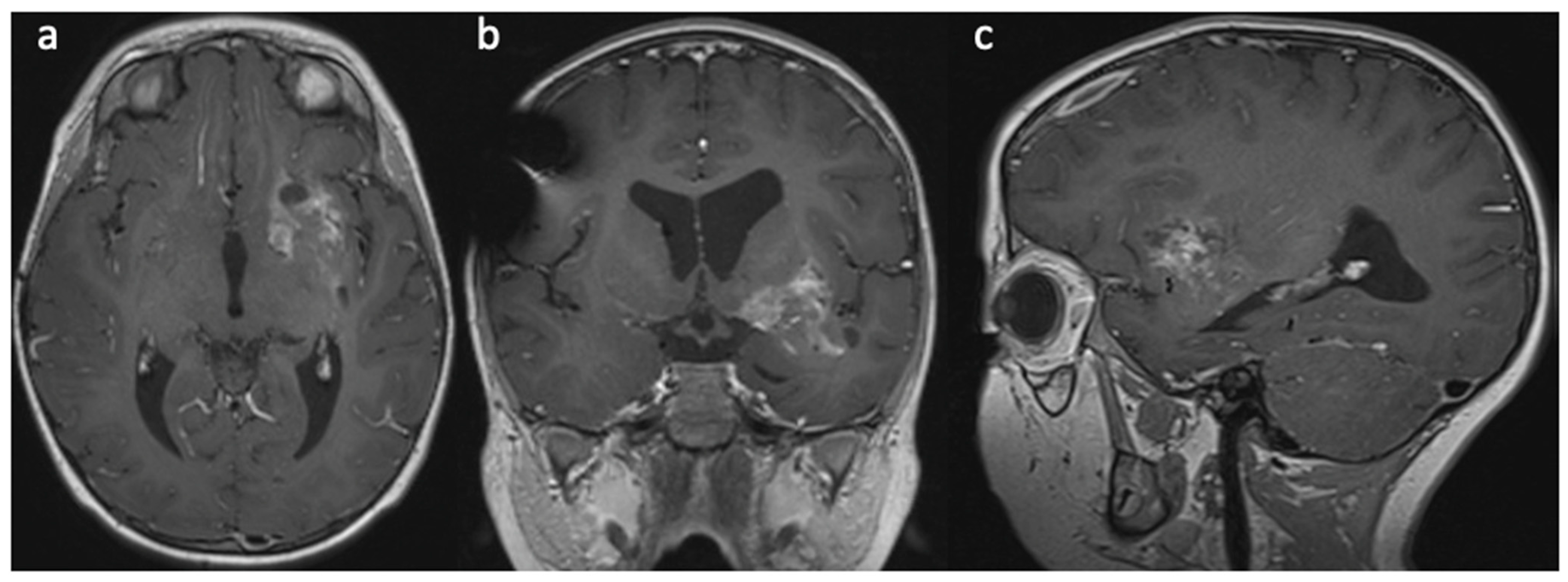
Case Report
4. Discussion
5. Conclusions
Author Contributions
Funding
Institutional Review Board Statement
Informed Consent Statement
Data Availability Statement
Conflicts of Interest
References
- Sturm, D.; Orr, B.A.; Toprak, U.H.; Hovestadt, V.; Jones, D.T.W.; Capper, D.; Sill, M.; Buchhalter, I.; Northcott, P.A.; Leis, I.; et al. New Brain Tumor Entities Emerge from Molecular Classification of CNS-PNETs. Cell 2016, 164, 1060–1072. [Google Scholar] [CrossRef] [PubMed]
- Holsten, T.; Lubieniecki, F.; Spohn, M.; Mynarek, M.; Bison, B.; Löbel, U.; Rutkowski, S.; Schüller, U. Detailed Clinical and Histopathological Description of 8 Cases of Molecularly Defined CNS Neuroblastomas. J. Neuropathol. Exp. Neurol. 2021, 80, 52–59. [Google Scholar] [CrossRef] [PubMed]
- Furuta, T.; Moritsubo, M.; Muta, H.; Koga, M.; Komaki, S.; Nakamura, H.; Morioka, M.; Ohshima, K.; Sugita, Y. Central nervous system neuroblastic tumor with FOXR2 activation presenting both neuronal and glial differentiation: A case report. Brain Tumor Pathol. 2020, 37, 100–104. [Google Scholar] [CrossRef] [PubMed]
- Von Hoff, K.; Haberler, C.; Schmitt-Hoffner, F.; Schepke, E.; De Rojas, T.; Jacobs, S.; Zapotocky, M.; Sumerauer, D.; Perek-Polnik, M.; Dufour, C.; et al. Therapeutic implications of improved molecular diagnostics for rare CNS embryonal tumor entities: Results of an international, retrospective study. Neuro-Oncology 2021, 23, 1597–1611. [Google Scholar] [CrossRef] [PubMed]
- Korshunov, A.; Okonechnikov, K.; Schmitt-Hoffner, F.; Ryzhova, M.; Sahm, F.; Stichel, D.; Schrimpf, D.; Reuss, D.E.; Sievers, P.; Suwala, A.K.; et al. Molecular analysis of pediatric CNS-PNET revealed nosologic heterogeneity and potent diagnostic markers for CNS neuroblastoma with FOXR2-activation. Acta Neuropathol. Commun. 2021, 9, 20. [Google Scholar] [CrossRef] [PubMed]
- Schepke, E.; Löfgren, M.; Pietsch, T.; Kling, T.; Nordborg, C.; Olsson Bontell, T.; Holm, S.; Öberg, A.; Nyman, P.; Eliasson-Hofvander, M.; et al. Supratentorial CNS-PNETs in children; a Swedish population-based study with molecular re-evaluation and long-term follow-up. Clin. Epigenetics 2023, 15, 40. [Google Scholar] [CrossRef] [PubMed]
- Chen, C.; Lee, I.; Tatsui, C.; Elder, T.; Sloan, A.E. Laser interstitial thermotherapy (LITT) for the treatment of tumors of the brain and spine: A brief review. J. Neurooncol. 2021, 151, 429–442. [Google Scholar] [CrossRef] [PubMed]
- Kanner, A.M.; Irving, L.T.; Cajigas, I.; Saporta, A.; Cordeiro, J.G.; Ribot, R.; Velez-Ruiz, N.; Detyniecki, K.; Melo-Bicchi, M.; Rey, G.; et al. Long-term seizure and psychiatric outcomes following laser ablation of mesial temporal structures. Epilepsia 2022, 63, 812–823. [Google Scholar] [CrossRef] [PubMed]
- Iwama, J.; Ogiwara, H.; Kiyotani, C.; Terashima, K.; Matsuoka, K.; Iwafuchi, H.; Morota, N. Neoadjuvant chemotherapy for brain tumors in infants and young children. J. Neurosurg. Pediatr. 2015, 15, 488–492. [Google Scholar] [CrossRef] [PubMed]
- Giantini-Larsen, A.M.; Chakravarthy, V.B.; Barzilai, O.; Newman, W.C.; Wexler, L.; Bilsky, M.H. The role of neoadjuvant denosumab in the treatment of aneurysmal bone cysts: A case series and review of the literature. J. Neurosurg. Pediatr. 2022, 30, 547–554. [Google Scholar] [CrossRef] [PubMed]
- Louis, D.N.; Perry, A.; Wesseling, P.; Brat, D.J.; Cree, I.A.; Figarella-Branger, D.; Hawkins, C.; Ng, H.K.; Pfister, S.M.; Reifenberger, G.; et al. The 2021 WHO Classification of Tumors of the Central Nervous System: A summary. Neuro-Oncology 2021, 23, 1231–1251. [Google Scholar] [CrossRef] [PubMed]
- Shimazaki, K.; Kurokawa, R.; Franson, A.; Kurokawa, M.; Baba, A.; Bou-Maroun, L.; Kim, J.; Moritani, T. Neuroimaging features of FOXR2-activated CNS neuroblastoma: A case series and systematic review. J. Neuroimaging 2023, 33, 359–367. [Google Scholar] [CrossRef] [PubMed]
- Tietze, A.; Mankad, K.; Lequin, M.H.; Ivarsson, L.; Mirsky, D.; Jaju, A.; Kool, M.; Hoff, K.V.; Bison, B.; Löbel, U. Imaging Characteristics of CNS Neuroblastoma- FOXR2: A Retrospective and Multi-Institutional Description of 25 Cases. Am. J. Neuroradiol. 2022, 43, 1476–1480. [Google Scholar] [CrossRef] [PubMed]
- Łastowska, M.; Trubicka, J.; Sobocińska, A.; Wojtas, B.; Niemira, M.; Szałkowska, A.; Krętowski, A.; Karkucińska-Więckowska, A.; Kaleta, M.; Ejmont, M.; et al. Molecular identification of CNS NB-FOXR2, CNS EFT-CIC, CNS HGNET-MN1 and CNS HGNET-BCOR pediatric brain tumors using tumor-specific signature genes. Acta Neuropathol. Commun. 2020, 8, 105. [Google Scholar] [CrossRef] [PubMed]
- Patel, N.V.; Jethwa, P.R.; Shetty, A.; Danish, S.F. Does the real-time thermal damage estimate allow for estimation of tumor control after MRI-guided laser-induced thermal therapy? Initial experience with recurrent intracranial ependymomas. J. Neurosurg. Pediatr. 2015, 15, 363–371. [Google Scholar] [CrossRef] [PubMed]
- Tovar-Spinoza, Z.; Choi, H. Magnetic resonance–guided laser interstitial thermal therapy: Report of a series of pediatric brain tumors. J. Neurosurg. Pediatr. 2016, 17, 723–733. [Google Scholar] [CrossRef]
- Dadey, D.Y.A.; Kamath, A.A.; Leuthardt, E.C.; Smyth, M.D. Laser interstitial thermal therapy for subependymal giant cell astrocytoma: Technical case report. Neurosurg. Focus 2016, 41, E9. [Google Scholar] [CrossRef]
- De Groot, J.F.; Kim, A.H.; Prabhu, S.; Rao, G.; Laxton, A.W.; Fecci, P.E.; O’Brien, B.J.; Sloan, A.; Chiang, V.; Tatter, S.B.; et al. Efficacy of laser interstitial thermal therapy (LITT) for newly diagnosed and recurrent IDH wild-type glioblastoma. Neuro-Oncol. Adv. 2022, 4, vdac040. [Google Scholar] [CrossRef]


Disclaimer/Publisher’s Note: The statements, opinions and data contained in all publications are solely those of the individual author(s) and contributor(s) and not of MDPI and/or the editor(s). MDPI and/or the editor(s) disclaim responsibility for any injury to people or property resulting from any ideas, methods, instructions or products referred to in the content. |
© 2023 by the authors. Licensee MDPI, Basel, Switzerland. This article is an open access article distributed under the terms and conditions of the Creative Commons Attribution (CC BY) license (https://creativecommons.org/licenses/by/4.0/).
Share and Cite
Chung, J.E.; Iqbal, O.; Krishnan, C.; Harrod, V.; Tyler-Kabara, E.; Lu, R.O.; Ho, W.S. Neoadjuvant Chemotherapy with Laser Interstitial Thermal Therapy in Central Nervous System Neuroblastoma: Illustrative Case and Literature Review. Brain Sci. 2023, 13, 1515. https://doi.org/10.3390/brainsci13111515
Chung JE, Iqbal O, Krishnan C, Harrod V, Tyler-Kabara E, Lu RO, Ho WS. Neoadjuvant Chemotherapy with Laser Interstitial Thermal Therapy in Central Nervous System Neuroblastoma: Illustrative Case and Literature Review. Brain Sciences. 2023; 13(11):1515. https://doi.org/10.3390/brainsci13111515
Chicago/Turabian StyleChung, Jason E., Omar Iqbal, Chandra Krishnan, Virginia Harrod, Elizabeth Tyler-Kabara, Rongze O. Lu, and Winson S. Ho. 2023. "Neoadjuvant Chemotherapy with Laser Interstitial Thermal Therapy in Central Nervous System Neuroblastoma: Illustrative Case and Literature Review" Brain Sciences 13, no. 11: 1515. https://doi.org/10.3390/brainsci13111515