Article

Amorphous Calcium Magnesium Fluoride Phosphate—Novel Material for Mineralization in Preventive Dentistry

1
Psilox AB, Vallvägen 4A, 756 51 Uppsala, Sweden
2
Department of Materials Science and Engineering, Division of Applied Materials Science, Uppsala University, 751 21 Uppsala, Sweden
*
Author to whom correspondence should be addressed.
Appl. Sci. 2023, 13(10), 6298; https://doi.org/10.3390/app13106298
Submission received: 4 May 2023 / Revised: 19 May 2023 / Accepted: 20 May 2023 / Published: 22 May 2023
(This article belongs to the Special Issue Dental Materials: Latest Advances and Prospects - Volume II)

Abstract

:

Featured Application

The article presents a novel fluoride particle technology for rapid remineralization, applicable for caries prevention and treatment of dentin hypersensitivity.

Abstract

This paper describes novel and innovative amorphous calcium magnesium fluoride phosphate (ACMFP) core-shell microparticles that may be applied in preventive dentistry for the prevention of caries and the treatment of dentin hypersensitivity. The particles can be synthesized with varied fluoride content, up to approximately 6 wt%, without any observable differences in morphology or crystallinity. Fluoride release from the particles is correlated to the fluoride content, and the particles are readily converted to fluoride-substituted hydroxyapatite or fluorapatite in a simulated saliva solution. The remineralization and dentin tubule occlusion potential of the particles was evaluated in vitro on acid-etched dentin specimens, and treatment with the ACMFP particles resulted in complete tubule occlusion and the formation of a dense mineralization layer. The acid resistance of the mineralization layer was improved compared to treatment with analogous particles without fluoride inclusion. A cross-sectional evaluation of dentin specimens after treatment revealed the formation of high aspect ratio fluorapatite crystals and poorly crystalline hydroxyapatite, respectively. The particles of the current study provide a single source vehicle of readily available calcium, phosphate, and fluoride ions for the potential remineralization of carious lesions as well as exposed dentin tubules for the reduction of hypersensitivity.

1. Introduction

Caries (tooth decay or dental cavities) is a major global healthcare issue, and according to the 2019 Global Burden of Disease Study, it is estimated that 2.03 billion people have caries in their permanent teeth and that 520 million children have caries in their primary teeth, making it the most prevalent non-communicable disease in the world [1,2]. Caries is a result of plaque formation on the tooth surface, where bacteria excrete acids while metabolizing fermentable carbohydrates. This causes demineralization of the enamel and dentin and, if allowed to progress, will cause the formation of cavities. In order to prevent caries, it is important to reduce the daily intake of dietary sugars and to exercise regular dental hygiene care, such as tooth brushing and flossing. Tooth brushing should be performed with a fluoridated toothpaste, which can prevent and arrest the progression of caries by forming fluorapatite, i.e., inhibiting demineralization [3,4]. Demineralization of dentin and enamel may be countered by remineralization, in which calcium and phosphate ions present in the saliva can deposit to form new mineral. This requires a local supersaturation of ions to form hydroxyapatite. If fluoride is present, the newly formed mineral may be fluorapatite rather than hydroxyapatite, making it less soluble and more resistant to acid erosion [5,6]. Repeated acid attacks or poor salivary flow may, however, tip the scale towards continued demineralization, leading to a net mineral loss. This has prompted the need for additional calcium phosphate sources and the development of specialized oral care products [7]. Different calcium phosphate technologies and particles have thus been introduced in the past 20 years to promote the remineralization of enamel as well as the occlusion of dentin tubules for the reduction of dentin hypersensitivity [8,9]. Dentin hypersensitivity is generally triggered by thermal, mechanical, or evaporative stimuli of the tooth and is highly prevalent among adults, reported at 34% in a recent study [10]. It is a clinical condition that can cause significant oral discomfort and pain, and the underlying cause is that the dentin tubules have become exposed due to gingival recession or loss of enamel, which is in turn caused by, e.g., abrasion or acid erosion, excessive tooth brushing or flossing, pocket reduction surgery, or as a secondary reaction to periodontal disease [10,11]. According to the generally accepted hydrodynamic theory, occluding the dentin tubules will hinder fluid movement within the tubules, which otherwise excites nerve endings in the pulp, causing the sensation of pain [12]. For a long-lasting and minimally invasive treatment of dentin hypersensitivity, the tubules should therefore ideally be deeply occluded with an acid-resistant mineralization layer resistant to both abrasion and erosion, and it should preferably be easy to apply at home or as a topical desensitizer product at the dental office. Successful treatment and prevention of both caries and dentin hypersensitivity thus rely on effective remineralization, requiring adequate access to calcium, phosphate, and fluoride ions.
There are a number of products and technologies on the market today that deliver various forms of calcium phosphate to the tooth surface, the most prevalent being hydroxyapatite (as nano- or microparticles, available in many kinds of toothpaste), different bioglasses (e.g., NovaMin®), and casein phosphopeptide–amorphous calcium phosphate (CPP–ACP, marketed as RECALDENT™). All of these technologies have successful clinical track records and have been demonstrated to be effective as remineralization agents in multiple studies [9,13,14]. They are regularly combined with fluoride in oral care products for daily caries protection. Each of these technologies has drawbacks or limitations, however, ranging from poor solubility (hydroxyapatite) to coarse particles (bioglass) [15] and milk protein allergy (CPP–ACP). A recent comparative study showcased the efficacy in dentin mineralization and tubule occlusion of an alternative technology based on stabilized microparticles of amorphous calcium magnesium phosphate (ACMP, marketed as CAPOSAL®) [16]. In the study, CAPOSAL® was the only technology resulting in complete occlusion and deep mineralization of the dentin tubules. The mineralization layer was also able to withstand an acid challenge, increasing the chances for long-term sensitivity relief. The mode of action of these particles, as well as the characteristics of intra-tubular mineralization layers, have been demonstrated in separate studies [17,18]. Combined, these studies demonstrate that the application of ACMP particles is one of the most effective approaches for promoting dentin remineralization and tubule occlusion, caused by the high aqueous solubility that supplies a high local concentration of calcium and phosphate ions. The metastable nature of ACMP particles also means that they are easily transformed into more stable calcium phosphate phases in situ, such as hydroxyapatite. In fact, amorphous calcium phosphate has long been considered a precursor to natural apatite in teeth and bone and therefore plays an important role in endogenous mineralization [19,20], and its unique properties have rendered it a highly interesting material for dental and other biomedical applications [21,22]. With the innovative CAPOSAL® technology, the previously elusive nature of amorphous calcium phosphate particles as a functional ingredient in oral care products has been made commercially available. Until now, however, the ACMP particles have relied on separate application and external supply of fluoride ions to form the more acid- and caries-resistant fluorapatite mineralization layer [18]. In the current study, we present a development of the ACMP particles to also contain a fluoride inclusion, making the particles essentially self-sustaining in the process of remineralizing dentin for the proposed reduction of dentin hypersensitivity and the prevention of caries.

2. Materials and Methods

2.1. Particle Synthesis

The amorphous calcium magnesium fluoride phosphate (ACMFP) particles described in this study were produced based on the development of a previously described method for the synthesis of ion-substituted calcium phosphate particles [17,23,24,25]. The amorphous nature of the particles is obtained by substituting Mg2+ for Ca2+, which inhibits the nucleation and growth of hydroxyapatite during synthesis. The particles may be synthesized by heating a phosphate buffer containing calcium and substitution ions or by a continuous flow mixing process of pre-heated calcium/magnesium and phosphate/fluoride salt solutions. Both methods result in the precipitation of core-shell particles with a hollow interior, with the latter method generating finer particles and being better suited for large-scale production as it allows for higher throughput. The fluoride content in the current particles was adjusted by changing the relative fluoride concentration in the appropriate starting solution. The formed particles were collected by filtration and washed with deionized water to remove any salt residues. To obtain long-term stability, the resulting particle slurry was mixed with glycerol, and residual water was evaporated by drying the mixture in a forced convection oven. The dried product was then homogenized to obtain a viscous paste consisting of stabilized and well-dispersed ACMFP particles in glycerol, suitable for use in the formulation of oral care products.

2.2. Characterization

The appearance and elemental composition of the synthesized particles were evaluated using the in-lens secondary electron detector of a field emission scanning electron microscope (SEM, Zeiss LEO 1530) equipped with an energy-dispersive X-ray spectroscopy system (EDS, Oxford AZtec). In preparation for analysis, the particles were dispersed in alcohol by ultrasound, pipetted onto sample stubs, and sputter coated with a thin Au/Pd conductive layer. Imaging was conducted using an acceleration voltage of 2 keV and a working distance of approximately 3 mm, whereas EDS data was collected using an acceleration voltage of 6 keV and a working distance of 8 mm. Crystallinity of the particles was evaluated on powder samples by X-ray diffraction (XRD, Bruker D8 ADVANCE), using CuKα-radiation and scanning 2θ from 10 to 60° with a step size of 0.0125°. Particle size distribution was evaluated using dynamic light scattering (DLS, Malvern Zetasizer Nano) after dispersing a fine amount of particles by ultrasound in ethanol. The Brunauer–Emmett–Teller (BET) particle-specific surface area was measured using N2 adsorption (Micromeritics TriStar II). In addition to the EDS measurements, the content of Ca, Mg, and P in the particles was quantified using inductively coupled plasma optical emission spectroscopy (ICP-OES, Perkin Elmer Avio 200) after dissolution in 5% HNO3. The instrument was calibrated with stock standard solutions (Perkin Elmer) and corresponding blanks. Fluoride concentration was additionally determined using a calibrated fluoride ion selective electrode coupled to a benchtop meter (Hanna Instruments HI 4110, HI 5522-02), measuring in total ionic strength adjustment buffer (TISAB II) after dissolving the particles in diluted HCl. Fluoride release from the particles was analyzed by adding 50 mg of particles to 100 mL of TISAB II under stirring and measuring the fluoride concentration in solution after 1, 5, 10, and 30 min.

2.3. In Vitro Mineralization and Dentin Tubule Occlusion
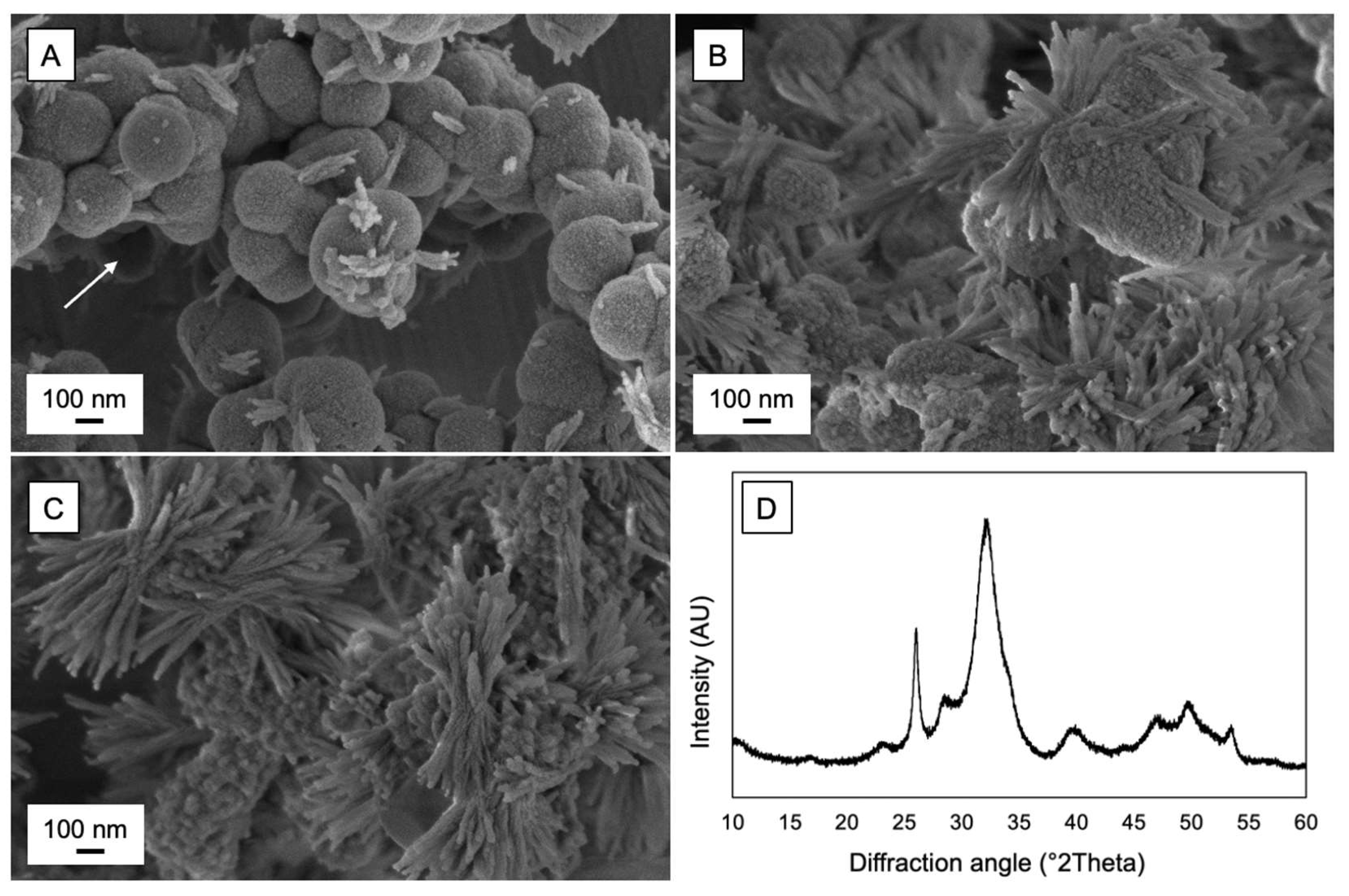
Bioactivity of the ACMFP particles, in terms of their propensity to transform/crystallize into hydroxyapatite or fluorapatite in biological conditions, was evaluated by submerging the particles in a simulated saliva solution composed as described in Table 1 (pH 7). Particles were left static in the solution for 18 h at 37 °C, then filtered, dried, and characterized in SEM and by XRD. Phase composition analysis of the crystallized material was performed using Rietveld refinement (Profex v4.3.5 software, an open source software, available from www.profex-xrd.org, accessed on 3 May 2023), quantifying hydroxyapatite and fluorapatite.
In vitro mineralization and dentin tubule occlusion testing were performed based on a previously described protocol [16]. Dentin specimens were sectioned from extracted human molars without any caries or anatomical defects using a water-cooled low-speed saw (Buehler IsoMet 2000). The sectioning was performed in the buccolingual plane, yielding specimens with approximate dimensions of 1 × 8 × 8 mm. Utilization of human molars in the study was performed in compliance with guidelines from the Swedish Ethical Review Authority (2016/039). After sectioning, the dentin specimens were etched in 30% phosphoric acid for 15 s to remove the smear layer and expose the tubules, followed by a thorough rinsing in deionized water. The dentin specimens were then subjected to a mineralization treatment with formulas containing glycerol and either 5% ACMFP particles or 5% ACMP particles (without fluoride) for comparison. A blank treatment formula without any mineralizing particles was also included for reference. Each treatment formula was applied to two dentin specimens twice daily for seven days, using a soft-bristled toothbrush and manual brushing with light hand pressure for approximately 30 s at each application. In between applications, the dentin specimens were stored in a simulated saliva solution (Table 1) at 37 °C, which was exchanged daily. Upon completion of the treatment, one specimen of each treatment formula was subjected to an acid challenge by swirling in a 2% citric acid solution (pH 2) for 30 s. The specimens were then rinsed, vacuum dried, mounted on sample stubs, and sputter coated with a thin conductive Au/Pd layer to allow SEM evaluation. One dentin specimen for each treatment cycle was evaluated, for a total of six specimens. Cross-sectional images of the dentin tubules were taken after manually breaking the specimens.

3. Results

The EDS elemental composition of ACMFP particles synthesized with various fluoride contents is listed in Table 2. The fluoride concentration ranged from essentially 0 wt% in ACMP particles synthesized without fluoride to nearly 6 wt% in the particles denoted ACMFP-4. Results from ICP-OES analysis were in general agreement with EDS data for Ca, P, and Mg content, whereas fluoride ion selective electrode measurements indicated a lower F content than those quantified with EDS (Table 3).
Morphology of synthesized particles is shown in Figure 1, demonstrating a generally spherical shape and a mixture of singular and fused particles with diameters ranging from 100 to 300 nm. The DLS z-average particle size of approximately 360 nm (Table 3) reflects the aggregation of particles. There were no apparent differences in the appearance between ACMP particles (Figure 1A) and ACMFP particles (Figure 1B,C), demonstrating that the fluoride inclusion did not affect the particle morphology.
XRD analysis of the same particles demonstrated that they were amorphous, with identical spectra regardless of fluoride inclusion (Figure 2A). Fluoride release, however, as shown in Figure 2B, demonstrates a significant difference between particles synthesized with and without fluoride. The relative fluoride release from particles was reflective of the concentrations listed in Table 2. The particles can be stabilized after synthesis by suspension in glycerol, with no observed changes in particle morphology or crystallinity after six months of storage at 40 °C.
The gradual transformation of ACMFP particles from amorphous core-shell particles to crystalline bundles of fluoride-substituted hydroxyapatite in simulated saliva solution is shown in Figure 3A–C. In Figure 3A, protrusions of high aspect ratio crystals are observed stemming directly from the ACMFP particle surfaces, with the image capturing an early phase of the transformation process. In Figure 3B, some features of the origin of ACMFP particles are still visible, but sharp crystals dominate the appearance. In Figure 3C, the particles are completely crystallized, demonstrating the final phase of transformation. All three SEM images in Figure 3 were captured from the same sample after static incubation for 18 h, and the global crystallinity of the sample is demonstrated in the XRD spectrum in Figure 3D. Phase identification of the crystalline part using Rietveld refinement yielded a share of approximately 58% fluorapatite and 42% hydroxyapatite.
The results of the in vitro mineralization and dentin tubule occlusion tests are shown in Figure 4. The top panel (Figure 4A–C) displays a reference dentin surface and a tubule in cross-section after treatment with a blank formula without any mineralizing particles. The tubules were fully exposed both before and after the acid challenge, demonstrating that the simulated saliva solution used for storage was not sufficiently mineralizing to have any notable effect. The middle panel (Figure 4D–F) shows the dentin surface and the mineralization product within a tubule after treatment with ACMP particles, i.e., without any fluoride inclusion. The tubules were completely occluded with a mineralized layer before the acid challenge (Figure 4D), but the orifices became partly visible again after the acid challenge (Figure 4E). The mineralized material both on the surface and within the tubules was characteristic of poorly crystalline nano-hydroxyapatite, as observed in previous studies [17,18]. Treatment with the ACMFP particles also resulted in complete tubule occlusion (Figure 4G) but with the added feature of resisting dissolution during the acid challenge (Figure 4H). The mineralization product within the tubules was dense and characterized by high aspect ratio crystals extending from the peritubular wall (Figure 4I), similar in appearance to the crystallized particles shown in Figure 3C as well as to fluoride-substituted hydroxyapatite structures reported in previous studies [16,18]. The deep (>10 µm) mineralization within the dentin tubules was unaffected by the acid challenge.

4. Discussion

Fluoride treatment is the gold standard when it comes to caries prevention, and brushing with fluoridated toothpaste is at the core of a sound oral care routine. Much of the effectiveness of fluoride in preventing caries and helping maintain good oral health depends on its capacity to enhance the remineralization of dentin and enamel and subsequently inhibit demineralization due to increased acid resistance [5]. For remineralization to be effective, there should be a net mineral gain in the tooth structure, i.e., the degree of apatite deposition should outweigh the dissolution caused by plaque bacteria. Facilitating the right conditions to remineralize or repair early enamel defects such as sub-surface lesions (“white spots”) may be complex, as it generally depends on ion diffusion through a more intact surface layer. A key aspect of reversing sub-surface demineralization is thus to clear the acid-producing plaque and allow saliva to contact the surface, which can neutralize the area and provide calcium and phosphate ions. If the saliva is supersaturated with calcium and phosphate, ions may then diffuse back into the lesion and remineralize partially demineralized crystals in a process that is accelerated in the presence of fluoride [26,27]. To create conditions that favor remineralization, especially in areas subjected to repeated acid challenges or in cases of poor salivary flow, supplementing the saliva and the lesion surface with readily available calcium, phosphate, and fluoride ions is therefore crucial. Application of a successful remineralization therapy may often be sufficient to stop caries progression and repair early lesions, reducing the need for costly and irreversible restorations [26]. As such, there is evidently a need for the development of calcium phosphate technologies that can contribute to the remineralization process in an effective way.
The ACMFP particles of the current study present an opportunity to deliver a single source vehicle of readily available calcium, phosphate, and fluoride ions for remineralization of carious lesions as well as exposed dentin tubules for the reduction of hypersensitivity. The particles may also be produced in a simple and cost-effective way. The unique spherical shape and sub-micron size of the particles make them especially suitable for dentin tubule occlusion, as they enable deep penetration and mineralization that can resist both abrasion and acid erosion. The prospect of having all key remineralization ions from a single source that is readily converted into fluoride-substituted hydroxyapatite could make products for preventive dentistry more effective and potentially safer, as excessive use of fluoride during tooth development may lead to fluorosis. The potential risk of fluorosis should not deter from the proper use of fluoride, however, as the benefits clearly outweigh the risks [3,4,5]. Nevertheless, the current findings demonstrate that the degree of fluoride inclusion in the particles and subsequent fluoride release can be adjusted without altering their characteristic features in terms of morphology and crystallinity (Figure 1 and Figure 2), which effectively allows tailoring of the particles to suit a wide range of applications. The fluoride inclusion may be high to maximize the fluoride release and remineralization potential of the particles, or it can be optimized at a lower level to reduce overall fluoride exposure. As the direct crystallization and remineralization triggered by the particles are independent of long-range fluoride ion diffusion from other sources, the ACMFP particles can offer local remineralization with a lower overall fluoride concentration.
The rapid in vitro conversion from amorphous core-shell particles into crystalline fluorapatite-like mineral depicted in Figure 3 illustrates the bioactive and remineralizing properties of the ACMFP particles. Several mechanisms of the conversion process from ACP to apatite in aqueous media have been proposed in the literature, including dissolution–reprecipitation, reorganization of Posner’s clusters, and surface-mediated transformation triggered by phosphate hydrolysis, leading to apatite nucleation and growth supported by surface ion migration [22]. It is plausible that all these processes occur to a varying degree at different stages of conversion of the particles, i.e., once apatite is nucleated at the particle surface, the rate of dissolution–reprecipitation increases. It is further established that this conversion process is accelerated in the presence of fluoride [22,27]. The corresponding conversion was demonstrated in an in vitro mineralization and tubule occlusion test, showing that the ACMFP particles generated a more acid-resistant mineralization layer than the fluoride-free ACMP particles (Figure 4). It should be noted that similar mineralization and tubule occlusion results have been observed in previous studies [16,18], but only when the ACMP particles have been applied in combination with an external fluoride source, e.g., successive application of a toothpaste containing sodium fluoride. Although the current findings are based on in vitro data from a limited set of dentin specimens, the results indicate that a similarly effective remineralization and dentin tubule occlusion effect can be obtained by the sole use of the ACMFP particles, eliminating the need for external or additional fluoride sources.

5. Conclusions

This study briefly describes the development, characteristic features, and potential application of ACMFP core-shell particles for use as a remineralization agent in preventive dentistry. It is demonstrated that the particles may be synthesized with a varied fluoride content and thereby allow fine-tuning and optimization for various needs and indications related to caries prevention. The amorphous particles are rapidly converted into fluorapatite structures in simulated saliva, and in vitro analysis demonstrates the formation of an acid-resistant mineralization layer both on the dentin surface and deep within dentin tubules. Based on the hydrodynamic theory, the complete tubule occlusion observed after the application of the ACMFP particles makes them a viable option for the effective treatment of dentin hypersensitivity.

Author Contributions

Conceptualization, E.U., W.X. and H.E.; methodology, E.U., D.F. and W.X.; investigation, E.U. and D.F.; resources, E.U. and H.E.; writing—original draft preparation, E.U.; writing—review and editing, E.U., D.F., W.X. and H.E.; visualization, E.U.; supervision, E.U. and W.X. All authors have read and agreed to the published version of the manuscript.

Funding

This research received no external funding.

Institutional Review Board Statement

The use of extracted human molars was approved according to the guidelines from the Regional Ethics Review Board in Uppsala (Sweden) (2016/039).

Informed Consent Statement

The study did not involve any interventions or handling of personal data that are covered in the Swedish Ethics Review Act (2006:615). The use of would-be discarded extracted molars for scientific purposes was therefore waived on the condition of anonymity, and provisions regarding personal consent did not apply to the use of human samples in the study.

Data Availability Statement

Data presented in the study are available upon request from the authors.

Conflicts of Interest

E.U., W.X. and H.E. are connected to Psilox AB, which has developed the material family (CAPOSAL®) evaluated in the study. E.U. is employed by the company, and W.X. and H.E. are shareholders. D.F. has no conflict of interest.

References

  1. Qin, X.; Zi, H.; Zeng, X. Changes in the global burden of untreated dental caries from 1990 to 2019: A systematic analysis for the Global Burden of Disease study. Heliyon 2022, 8, e10714. [Google Scholar] [CrossRef]
  2. Pitts, N.B.; Twetman, S.; Fisher, J.; Marsh, P.D. Understanding dental caries as a non-communicable disease. Brit. Dent. J. 2021, 231, 749–753. [Google Scholar] [CrossRef]
  3. Tenuta, L.M.A.; Cury, J.A. Fluoride: Its role in dentistry. Braz. Oral Res. 2010, 24, 9–17. [Google Scholar] [CrossRef]
  4. Cate, J.M.T. Contemporary perspective on the use of fluoride products in caries prevention. Brit. Dent. J. 2013, 214, 161–167. [Google Scholar] [CrossRef]
  5. Featherstone, J.D.B. Prevention and reversal of dental caries: Role of low level fluoride. Community Dent. Oral 1999, 27, 31–40. [Google Scholar] [CrossRef]
  6. Pajor, K.; Pajchel, L.; Kolmas, J. Hydroxyapatite and Fluorapatite in Conservative Dentistry and Oral Implantology—A Review. Materials 2019, 12, 2683. [Google Scholar] [CrossRef]
  7. Neel, E.A.A.; Aljabo, A.; Strange, A.; Ibrahim, S.; Coathup, M.; Young, A.M.; Bozec, L.; Mudera, V. Demineralization–remineralization dynamics in teeth and bone. Int. J. Nanomed. 2016, 11, 4743–4763. [Google Scholar] [CrossRef]
  8. Philip, N. State of the Art Enamel Remineralization Systems: The Next Frontier in Caries Management. Caries Res. 2019, 53, 284–295. [Google Scholar] [CrossRef]
  9. Meyer, F.; Amaechi, B.T.; Fabritius, H.-O.; Enax, J. Overview of Calcium Phosphates used in Biomimetic Oral Care. Open. Dent. J. 2018, 12, 406–423. [Google Scholar] [CrossRef]
  10. Alcântara, P.M.; Barroso, N.F.F.; Botelho, A.M.; Douglas-de-Oliveira, D.W.; Gonçalves, P.F.; Flecha, O.D. Associated factors to cervical dentin hypersensitivity in adults: A transversal study. BMC Oral. Health 2018, 18, 155. [Google Scholar] [CrossRef]
  11. Davari, A.; Ataei, E.; Assarzadeh, H. Dentin hypersensitivity: Etiology; diagnosis; treatment; a literature review. J. Dent. Shiraz Iran. 2013, 14, 136–145. [Google Scholar]
  12. Bartold, P. Dentinal hypersensitivity: A review. Aust. Dent. J. 2006, 51, 212–218. [Google Scholar] [CrossRef]
  13. Chen, L.; Al-Bayatee, S.; Khurshid, Z.; Shavandi, A.; Brunton, P.; Ratnayake, J. Hydroxyapatite in Oral Care Products—A Review. Materials 2021, 14, 4865. [Google Scholar] [CrossRef]
  14. Li, J.; Xie, X.; Wang, Y.; Yin, W.; Antoun, J.S.; Farella, M.; Mei, L. Long-term remineralizing effect of casein phosphopeptide-amorphous calcium phosphate (CPP-ACP) on early caries lesions in vivo: A systematic review. J. Dent. 2014, 42, 769–777. [Google Scholar] [CrossRef]
  15. LaTorre, G.; Greenspan, D.C. The role of ionic release from NovaMin (calcium sodium phosphosilicate) in tubule occlusion: An exploratory in vitro study using radio-labeled isotopes. J. Clin. Dent. 2010, 21, 72–76. [Google Scholar]
  16. Berg, C.; Unosson, E.; Engqvist, H.; Xia, W. Comparative Study of Technologies for Tubule Occlusion and Treatment of Dentin Hypersensitivity. J. Funct. Biomater. 2021, 12, 27. [Google Scholar] [CrossRef]
  17. Berg, C.; Unosson, E.; Engqvist, H.; Xia, W. Amorphous Calcium Magnesium Phosphate Particles for Treatment of Dentin Hypersensitivity: A Mode of Action Study. Acs Biomater. Sci. Eng. 2020, 6, 3599–3607. [Google Scholar] [CrossRef]
  18. Berg, C.; Unosson, E.; Riekehr, L.; Xia, W.; Engqvist, H. Electron microscopy evaluation of mineralization on peritubular dentin with amorphous calcium magnesium phosphate microspheres. Ceram. Int. 2020, 46, 19469–19475. [Google Scholar] [CrossRef]
  19. Posner, A.S.; Betts, F. Synthetic amorphous calcium phosphate and its relation to bone mineral structure. Acc. Chem. Res. 1975, 8, 273–281. [Google Scholar] [CrossRef]
  20. Boskey, A.L. Amorphous Calcium Phosphate: The Contention of Bone. J. Dent. Res. 1997, 76, 1433–1436. [Google Scholar] [CrossRef]
  21. Zhao, J.; Liu, Y.; Sun, W.; Zhang, H. Amorphous calcium phosphate and its application in dentistry. Chem. Cent. J. 2011, 5, 40. [Google Scholar] [CrossRef]
  22. Combes, C.; Rey, C. Amorphous calcium phosphates: Synthesis, properties and uses in biomaterials. Acta Biomater. 2010, 6, 3362–3378. [Google Scholar] [CrossRef] [PubMed]
  23. Xia, W.; Grandfield, K.; Schwenke, A.; Engqvist, H. Synthesis and release of trace elements from hollow and porous hydroxyapatite spheres. Nanotechnology 2011, 22, 305610. [Google Scholar] [CrossRef] [PubMed]
  24. Xia, W.; Qin, T.; Suska, F.; Engqvist, H. Bioactive Spheres: The Way of Treating Dentin Hypersensitivity. ACS Biomater. Sci. Eng. 2016, 2, 734–740. [Google Scholar] [CrossRef] [PubMed]
  25. Mellgren, T.; Qin, T.; Öhman-Mägi, C.; Zhang, Y.; Wu, B.; Xia, W.; Engqvist, H. Calcium Phosphate Microspheres as a Delivery Vehicle for Tooth-Bleaching Agents. J. Dent. Res. 2018, 97, 283–288. [Google Scholar] [CrossRef]
  26. Featherstone, J. Dental caries: A dynamic disease process. Aust. Dent. J. 2008, 53, 286–291. [Google Scholar] [CrossRef]
  27. Cate, J.M.T.; Featherstone, J.D.B. Mechanistic Aspects of the Interactions between Fluoride and Dental Enamel. Crit. Rev. Oral Biol. Med. 1991, 2, 283–296. [Google Scholar] [CrossRef]
Figure 1. SEM images of particles: (A) ACMP synthesized without fluoride; (B) ACMFP-2 particles; (C) ACMFP-4 particles.
Applsci 13 06298 g001
Figure 2. (A) XRD spectra of ACMP, ACMFP-2, and ACMFP-4 particles; (B) Fluoride release curves from ACMP and ACMFP particles with varied fluoride content.
Applsci 13 06298 g002
Figure 3. (AC) SEM images of ACMFP particles at different stages of crystallization in simulated saliva. Arrow in (A) displays a hollow particle core; (D) XRD spectrum of crystallized particles.
Applsci 13 06298 g003
Figure 4. SEM images of dentin surfaces and dentin tubules in cross-section: Top panel (AC) shows appearance after treatment with blank formula (no particles); Middle panel (DF) shows appearance after treatment with the ACMP formula; Bottom panel (GI) shows appearance after treatment with the ACMFP formula. The left-hand panel (A,D,G) shows surfaces before the acid challenge, and the center panel (B,E,H) after the acid challenge. Cross-section images of dentin tubules beneath the treated surface are shown in the right-hand panel (C,F,I).
Applsci 13 06298 g004
Table 1. Composition of the simulated saliva solution.
SaltConc. (mM)
NaCl30
KCl3
CaCl2∙2H2O1.5
MgCl2∙6H2O0.5
Na2HPO4∙2H2O2.9
KH2PO42.1
Table 2. Elemental composition of particles according to EDS analysis.
SampleO (wt%)Ca (wt%)P (wt%)Mg (wt%)F (wt%)
ACMP54.3 ± 0.823.2 ± 1.014.6 ± 0.17.9 ± 1.80.1 ± 0.1
ACMFP-148.4 ± 2.025.6 ± 0.619.0 ± 1.05.9 ± 0.11.2 ± 0.2
ACMFP-250.2 ± 1.523.0 ± 1.517.9 ± 2.36.4 ± 0.42.6 ± 0.3
ACMFP-350.3 ± 3.324.7 ± 0.815.5 ± 3.36.3 ± 0.03.2 ± 0.7
ACMFP-448.6 ± 1.825.7 ± 1.512.8 ± 0.67.1 ± 0.85.8 ± 0.2
Table 3. Characteristics of ACMFP-2 particles. Elemental composition according to ICP-OES (Ca, P, and Mg) and fluoride ion selective electrode (F). Z-average particle size according to DLS and specific surface area according to N2 BET analysis.
SampleCa (wt%)P (wt%)Mg (wt%)F (wt%)Z-Avg. Particle Size (nm)BET Surface Area (g/m2)
ACMFP-221.5 ± 0.320.0 ± 0.56.9 ± 0.11.51 ± 0.05362 ± 3824.5 ± 1.1
Disclaimer/Publisher’s Note: The statements, opinions and data contained in all publications are solely those of the individual author(s) and contributor(s) and not of MDPI and/or the editor(s). MDPI and/or the editor(s) disclaim responsibility for any injury to people or property resulting from any ideas, methods, instructions or products referred to in the content.

