Article

Antimicrobial Resistant Staphylococcus Species Colonization in Dogs, Their Owners, and Veterinary Staff of the Veterinary Teaching Hospital of Naples, Italy

by
Francesca Paola Nocera
1,*,
Francesca Pizzano
1,
Angelo Masullo
1,
Laura Cortese
1 and
Luisa De Martino
1,2
1
Department of Veterinary Medicine and Animal Production, University of Naples ‘Federico II’, Via F. Delpino 1, 80137 Naples, Italy
2
Task Force on Microbiome Studies, University of Naples ‘Federico II’, 80137 Naples, Italy
*
Author to whom correspondence should be addressed.
Pathogens 2023, 12(8), 1016; https://doi.org/10.3390/pathogens12081016
Submission received: 26 June 2023 / Revised: 2 August 2023 / Accepted: 4 August 2023 / Published: 5 August 2023

Abstract

:
This study aimed to identify Staphylococcus species isolated from nasal swabs of both healthy and diseased dogs, and those of human origin, obtained from nasal swabs of both owners and veterinary staff. Firstly, pet owners were requested to complete a questionnaire relating to the care and relationship with their pets, whose results mainly showed a statistically significant higher frequency of hand washing in diseased dogs’ owners than in healthy dogs’ owners. Canine nasal swabs were obtained from 43 diseased dogs and 28 healthy dogs, while human nasal swabs were collected from the respective dogs’ owners (71 samples) and veterinary staff (34 samples). The isolation and identification of Staphylococcus spp. were followed by disk diffusion method to define the antimicrobial resistance profiles against 18 different molecules. Staphylococcus pseudintermedius was the most frequent isolated strain in both diseased (33.3%) and healthy (46.1%) dogs. Staphylococcus epidermidis was the most frequent isolated bacterium in diseased dogs’ owners (66.6%), while in nasal samples of healthy dogs’ owners, the same frequency of isolation (38.4%) was observed for both Staphylococcus epidermidis and Staphylococcus aureus. All the isolated strains showed good susceptibility levels to the tested antimicrobials; however, the carriage of oxacillin-resistant strains was significantly higher in diseased dogs than in healthy ones (71% and 7.7%, respectively). Only in three cases the presence of the same bacterial species with similar antimicrobial resistance profiles in dogs and their owners was detected, suggesting the potential bacterial transmission. In conclusion, this study suggests potential transmission risk of staphylococci from dogs to humans or vice versa, and highlights that the clinical relevance of Staphylococcus pseudintermedius transmission from dog to human should not be underestimated, as well as the role of Staphylococcus aureus from human to dog transmission.

1. Introduction

Staphylococci are normal inhabitants of skin and mucus membranes of both humans and animals [1]. In dogs, nasal carriage of both coagulase-positive and coagulase-negative staphylococci can be influenced not only by diseases but also by some environmental factors, such as the close contact with humans or vice versa [2]. Although it is still not clear how the transmission of staphylococci between dogs and humans occurs, previous studies suggest that close contact might cause a higher colonization of Staphylococcus aureus in dogs and Staphylococcus pseudintermedius in humans [2,3,4].
Staphylococcus aureus (S. aureus) is widely disseminated as a colonizer and as an opportunistic pathogen among humans and animals [3]. In the human community, it colonizes the anterior nares in 20% to 80% of healthy humans [5], and these people can be defined as “carriers”, as in normal physiological conditions their immunity system is able to fight off this opportunistic pathogen. However, in specific conditions (e.g., injuries, skin disorders, metabolic diseases), this microorganism, finding propitious conditions for its reproduction, can give rise to both mild and life-threatening infections. Currently, the spread of methicillin-resistant S. aureus (MRSA), showing also multidrug-resistant profile, has increased worldwide and, in particular, it has emerged not only as a nosocomial pathogen as healthcare-associated MRSA, but also as community-associated MRSA and livestock-acquired MRSA [6,7].
Rates of methicillin resistance among clinical S. aureus isolates vary greatly among countries, from rates as low as 9% in Scandinavian countries [8] to over 50% in countries such as the United States and China [9,10]. Although nosocomial MRSA infections are declining in the United States, Europe, China, and many other countries, possibly due to increased surveillance and hygiene measures, they are still increasing in less-developed countries [11].
Human-to-human transmission of MRSA occurs through direct contact with carriers, infected people, or with contaminated medical devices and equipment. Therefore, MRSA is of greatest concern in hospital settings, where patients with weakened immune systems are more prone to infection than the healthy population. Furthermore, animals, mainly food-producing ones, have been described as MRSA reservoirs [12]. The close contact with an MRSA-positive animal can favor colonization by this pathogen in humans, but they rarely become infected [13]. Therefore, farmers and livestock breeders, but also veterinary staff, are exposed to a higher risk of being colonized by MRSA. Indeed, Neradova et al. [14] reported that the prevalence of MRSA nasal carriage was 6.72% in veterinary professionals attending a conference for mixed animal practice, which took place in Czech Republic, confirming the isolation of both animal and nosocomial strains. In this scenario, it appears clear that skin contact with colonized individuals, fomites, or animals may result in either asymptomatic colonization or subsequent clinically significant MRSA disease. Furthermore, globalization, including the increasing of international travel, has facilitated the transmission of multidrug-resistant bacteria such as MRSA across continents, potentially favoring the replacement of existing endemic MRSA with fitter and more transmissible strains [15].
Staphylococcus pseudintermedius (S. pseudintermedius) is a member of the Staphylococcus intermedius group (SIG), together with Staphylococcus intermedius, Staphylococcus delphini, Staphylococcus ursi, and Staphylococcus cornubiensis [16]. This coagulase-positive Staphylococcus is a normal skin inhabitant and colonizer of healthy dogs [17]. Precisely, S. pseudintermedius can be isolated from the nares, oral mucosa, pharynx, forehead, groin, and anus of healthy dogs [18]. Being an opportunistic pathogen, it represents one of the main causes of pyoderma and otitis, along with postoperative wound infections [19,20,21]. In recent years, methicillin-resistant S. pseudintermedius strains (MRSP) have also been isolated and identified, and often these MRSP strains showed multidrug-resistant profiles, being also resistant to most of the antimicrobial agents approved for veterinary applications [21,22].
Although S. pseudintermedius is not a normal human commensal bacterium, for its zoonotic potential it can be responsible for serious opportunistic infections in humans, particularly affecting pet owners and veterinary personnel [23,24,25,26,27].
According to the International Organization for the Protection of Animals [28] report, in Italy, 13,209,745 dogs and 956,308 cats were registered in the regional registers of pets through 4 February 2022. This number, gradually increased over the years, shows that pet dogs and cats are present in Italian households. In particular, household pet number has risen sharply during the years of the pandemic. The context of the COVID-19 pandemic has provided an opportunity to understand how animals can offer social support to their owners and alleviate heightened symptoms of stress, anxiety, and depression, and contribute to happiness during this major global crisis. In fact, pet dogs and cats may have provided people with a stronger sense of social support, which in turn may have helped reduce some of the negative psychological impacts caused by the COVID-19 pandemic [29]. Contact and interactions between owners and their pets can have benefits [30] but can also facilitate the transmission of zoonotic agents. Indeed, microorganisms transmitted from animals to humans represent about 60% of pathogens, and close contact is essential for transmission [27,31]. However, to estimate the risk of these contacts, more information is needed, such as frequency and intensity.
People can be exposed to zoonoses by direct contact through biting, scratching, sneezing, coughing, licking, or by handling pets and their droppings; or by indirect contact through bedding, food, and water [32]. Furthermore, the possibility of intraspecific transmission should also be considered, for example, during interactions between dogs during playful activities.
The purpose of this study was to identify Staphylococcus species isolated from nasal swabs of both healthy and diseased dogs, and those of human origin, obtained from nasal swabs of both owners and veterinary staff. In addition, in order to determine the frequency and intensity of owners’ interactions with their dogs, a survey was conducted among dog-owning households, with the collection of 71 pet-care questionnaires. The antimicrobial resistance profiles of collected strains were also investigated.

2. Materials and Methods

2.1. Ethical Statement

This study was approved by the Veterinary Service Center of the University of Naples Federico II, Italy (certificate number PG/2021/0009256), in compliance with the Italian Legislative Decree 26/2014, Article 2, implementing Directive 2010/63/EU.

2.2. Informed Consent

No animals were used in this study. Samples, represented by only noninvasive nasal swabs, were obtained from dogs being investigated for clinical reasons and for their benefit. Furthermore, nasal swabs were collected from the dogs’ owners and veterinary staff, who agreed to take part in this project by signing an informed consent and performing the nasal swabs by themselves, following the given sampling instructions. After sampling, swabs were held at 4 °C during transport to the bacteriology laboratory.

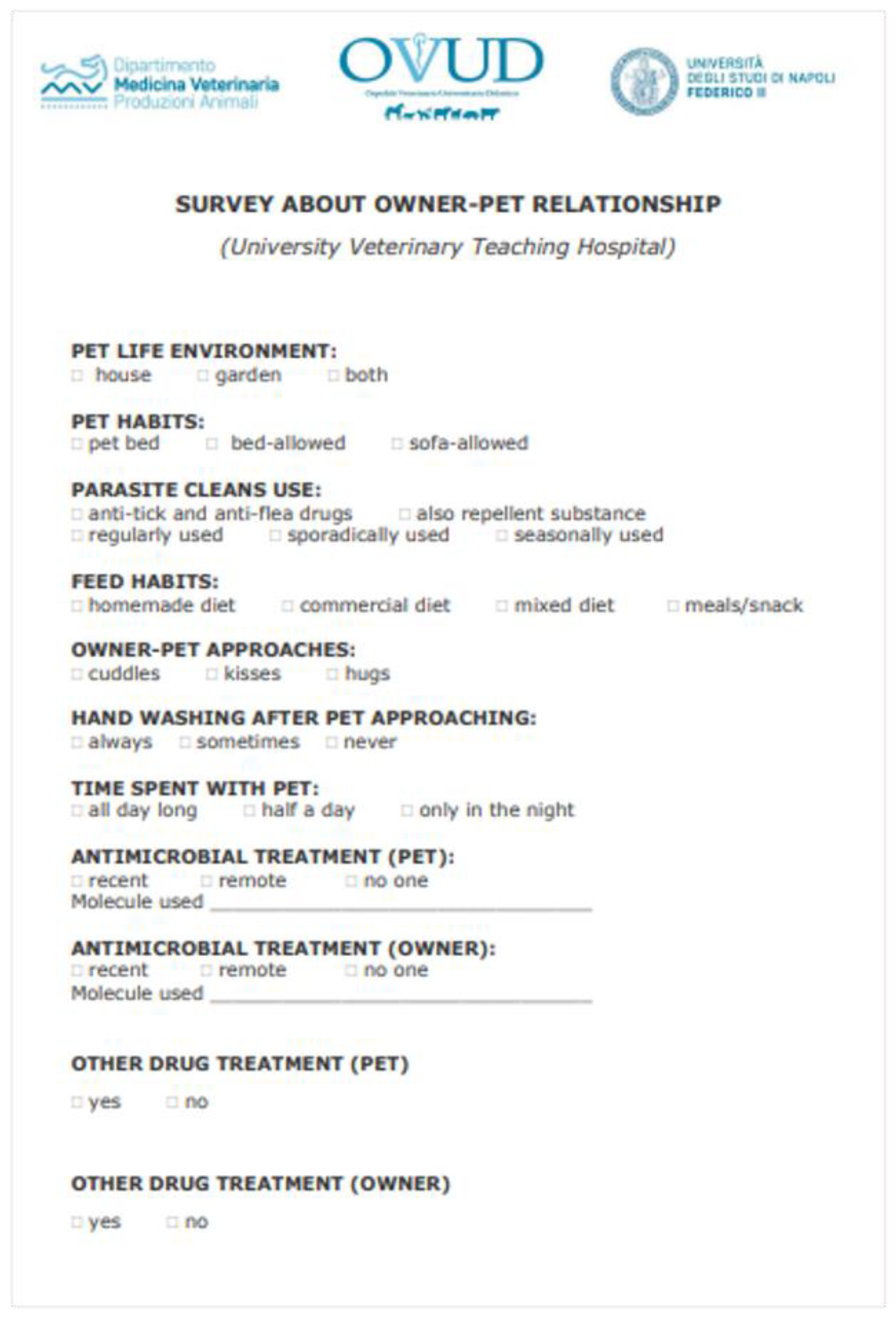
2.3. Participant Groups and Small Pet-Care Questionnaire Survey

In this study, performed in 2022, five different groups of participants were enrolled: I group_ healthy dogs, II group _ healthy dogs’ owners, III group _ diseased dogs, IV group_ diseased dogs’ owners, and V group _ veterinary staff.
Pet owners were asked to complete a questionnaire relating to the care and relationship with their pets (Figure 1). In particular, the questionnaire had the aim to assess some aspects of the dog–owner relationship and to understand if constant and prolonged contact with dogs might represent a potential risk for the transmission of pathogens and their antimicrobial resistance determinants, leading to the selection and sharing of strains with zoonotic potential. The survey consisted of a series of 11 questions having a 2–4 multiple-choice format, and the possibility to provide additional information. The questions focused on the dog–owner relationship, the pet’s lifestyle, and the canine medical history such as recent or previous antimicrobial treatments. This last question was also foreseen for owners. Most patients and people enrolled in this study had not received an antimicrobial treatment for at least two months, classified in the survey as remote antimicrobial therapy (in the last two months/two years), while only few patients received a recent antimicrobial treatment (in the last ten days/one month).

2.4. Sampling

Nasal samples were collected from dogs and their owners attending the University Veterinary Teaching Hospital of the Department of Veterinary Medicine and Animal Production (University of Naples “Federico II”, Italy). In particular, the sampling was performed on dogs not affected by skin disorders, referred to the hospital for routine checks or for annual vaccination booster, thereafter, defined healthy, and their owners; but also on dogs clinically suffering from otitis or pyoderma, here indicated as diseased, and their owners. Further nasal swabs were obtained from veterinary staff involved in the daily clinical activity at the University Veterinary Teaching Hospital of Naples.
For each patient, a single swab was inserted about 2/3 cm deep and rotated 360° a couple of times in both nostrils and then placed into Stuart W/O CH (Aptaca Spa, Asti, Italy) transport medium. Within a maximum of 24 h after sampling, specimens, kept at 4 °C, were transferred in an icebox to the bacteriology laboratory of the abovementioned department for bacteriological investigation.

2.5. Staphylococcus spp. Isolation and Identification

Once arrived at the laboratory, nasal swabs were streaked on Mannitol salt agar (MSA) plates purchased from Liofilchem Srl (Teramo, Italy). MSA is a selective medium used for isolation, enumeration, and differentiation of Staphylococcus spp. After an overnight incubation at 37 °C, the recovered colonies were firstly evaluated for their morphologic and fermentative features (colony morphology, mannitol fermentation) and subjected to rapid standard screening techniques such as Gram’s staining, catalase (Biomérieux, Marcy-lÉtoile, France), and oxidase (Liofilchem, Teramo, Italy) reactions. In addition, staphylocoagulase reaction was performed (Oxoid, Ltd., Hampshire, UK).
Thereafter, colonies, grown on MSA, were subcultivated on Columbia CNA agar plates (Liofilchem, Teramo, Italy) and incubated aerobically at 37° for 24 h. Subsequently, the identification of the isolated strains was carried out by using matrix-assisted laser desorption/ionization–time of flight mass spectrometry (MALDI-TOF MS) (Bruker Daltonics Inc., Bremen, Germany), according to manufacturer’s guidelines. Specifically, according to Bruker biotyper’s guidelines, a score of ≥2.0 indicated highly probable species-level identification, a score of 1.70 to 1.99 indicated a secure identification to the genus level, and a score of <1.7 was interpreted as no identification.
Staphylococcus aureus ATCC® 33591TM and Staphylococcus pseudintermedius ATCC® 49444TM were used as quality control strains.

2.6. Antimicrobial Susceptibility Testing of Isolated Staphylococci

The susceptibility profiles of the isolated strains were evaluated for 18 antimicrobials, belonging to 11 different classes, by the disk diffusion method on Mueller Hinton agar plates. The tested antimicrobials comprised amoxicillin–clavulanate (AUG, 20/10 µg), ampicillin (AMP, 30 µg), cephalothin (KF, 30 µg), cefoxitin (FOX, 30 µg), ciprofloxacin (CIP, 5 µg), clindamycin (CD, 2 µg), doxycycline (DO, 30 µg), enrofloxacin (ENR, 5 µg), erythromycin (E, 15 µg), gentamicin (CN, 10 µg), levofloxacin (LVX, 5 µg), linezolid (LNZ, 30 µg), nitrofurantoin (F, 300 µg), oxacillin (OX, 1 µg), penicillin (P, 10 IU), sulfamethoxazole–trimethoprim (SXT, 23.75/1.25 µg), tetracycline (TE, 30 µg), and vancomycin (VA, 30 µg), all of which were supplied by Liofilchem Srl (Teramo, Italy). For the classification of the isolated strains as susceptible, intermediate susceptibility, or resistant, the Clinical and Laboratory Standards Institute guidelines [33] were taken into consideration for all tested antimicrobials except for AMP, LVX, and LNZ, for which the European Committee on Antimicrobial Susceptibility Testing guidelines [34] were used.

2.7. Statistical Analysis

All diagnostic results generated by the Microbiological Diagnostic Laboratory were recorded and entered into a Microsoft 365 Excel™ spreadsheet for successive analysis. Descriptive statistical analysis was employed to evaluate the prevalence of isolated Staphylococcus species and the frequencies of antibiotic resistance among recovered isolates. All graphics were made by using Excel software. The statistical significance level between variable groups was investigated using two-tailored Fisher’s exact test (Social Science Statistics https://www.socscistatistics.com, accessed on 20 May 2023) and p-values ≤ 0.05 were considered statistically significant at 95% confidence interval.

3. Results

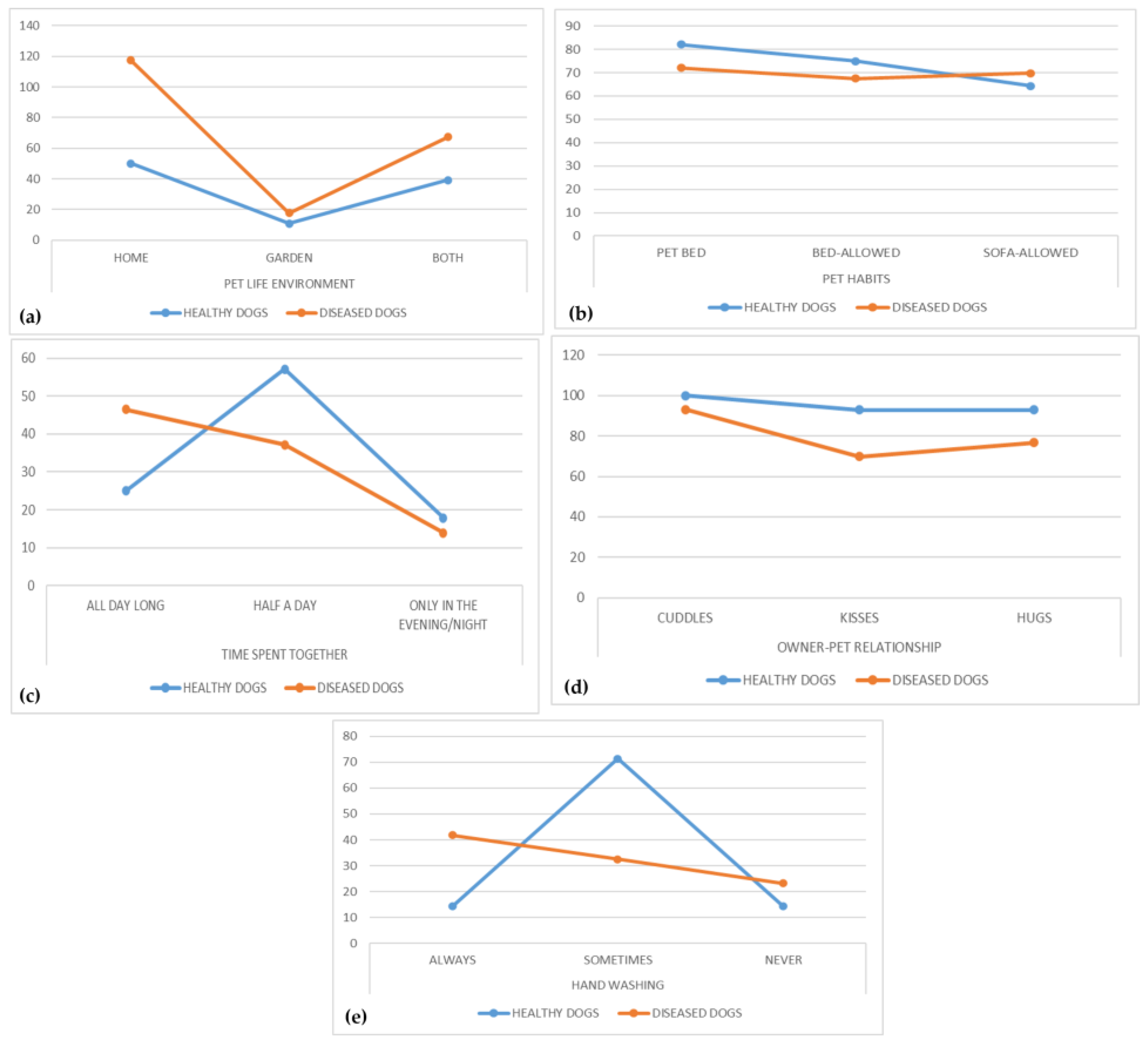
3.1. Pet-Care Questionnaire Survey

During this study, 71 questionnaire surveys were collected. In particular, 28 and 43 were filled by healthy dogs’ and diseased dogs’ owners, respectively. The survey consisted of a series of 11 questions having a 2–4 multiple-choice format, with the possibility to provide additional information, above all for the question concerning the use of antimicrobials. In this case, the administered antimicrobial agent was always indicated; for diseased dogs, the reason for presentation in the University Veterinary Teaching Hospital was also requested. In most cases, the diseased dogs were referred to the hospital because of skin disorders, such as otitis externa, pyoderma, hair loss, presence of scales and/or dandruff, or presence of skin wounds.
The answers are summarized in Table 1.
Comparing the answers obtained, it should be noted that, despite the different numbers in the two sampled owners groups, the behaviors and habits of the pet owners are quite similar (Table 1, Figure 2), especially concerning the intimate pet–owner relationship and the intensity of the contact with high percentages of both healthy and diseased dogs’ owners giving cuddles (100% and 93%, respectively), kisses (93% and 70%, respectively), and hugs (93% and 77%, respectively) to their dogs (Figure 2c,d). Furthermore, 75% of healthy dogs’ owners and 67% of diseased dogs’ owners allowed their pets the access to bed (Figure 2b). In addition, reduced attention given to hand washing was observed among owners, with 14% and 23% of healthy and diseased owners, respectively, never washing their hands after a contact with their dog (Table 1, Figure 2e). However, a significantly higher number (p < 0.05) of diseased dogs’ owners (18/43; 42%) indicated always washing their hands after interaction with their pet compared to healthy dogs’ owners (4/28; 14%).

3.2. Identification of Staphylococcus spp. from Nasal Swabs of Pet Dogs and Their Owners

During the investigation period, a higher number of samples were processed than reported in this study, as in dogs affected by skin disorders, other specimens, besides nasal swabs, were also collected (e.g., auricular and skin swabs, hair, etc.) to perform a proper diagnosis. Therefore, this study focused only on Staphylococcus spp. strains isolated from the nasal swabs of both healthy and diseased dogs and those of their respective owners, in order to evaluate and highlight a possible dog–owner correspondence.
Only 52% dog–owner couples (37/71 dog–owner couples) had positive results for Staphylococcus spp. strains during the bacteriological investigation. Staphylococci recovered from canine and human nasal swabs were identified by MALDI-TOF MS with a log(score) ≥ 2.2, indicating a highly probable species-level identification.
In the group of the 13 healthy dogs positive to bacteriological examination, S. pseudintermedius was the most frequently isolated strain (46.1%, 6/13 strains), followed by S. aureus and S. epiderdimis, showing both an isolation frequency of 23% (3/13 strains). A strain of S. xylosus was also identified (7.6%).
S. aureus and S. epidermidis were both the predominant species (38.4%; 5/13 strains) among the staphylococcal strains isolated from the nasal swabs in the group_ healthy dogs’ owners. S. haemolyticus was also recovered with an isolation frequency of 23% (3/13 strains).
A comparison of the staphylococcal identifications among the 13 owner–healthy dog couples highlighted a higher occurrence for the following bacterial pairings (Table 2): S. epidermidisS. pseudintermedius (23%; 3/13), S. aureusS. epidermidis (15.4%; 2/13), S. haemolyticusS. aureus (15.4%; 2/13), S. aureusS. pseudintermedius (15.4%; 2/13), S. aureusS. aureus (7.7%; 1/13), S. haemolyticusS. pseudintermedius (7.7%; 1/13), S. epidermidisS. xylosus (7.7%; 1/13), and S. epidermidisS. epidermidis (7.7%; 1/13).
As for healthy dogs, also in the group composed of diseased dogs, S. pseudintermedius was the most commonly identified staphylococcal species (75%; 18/24 strains) from nasal swabs, followed by S. lugdnensis (8.3%; 2/24), and S. hyicus, S. lentus, S. sciuri, S. simulans, all four showing an isolation rate of 4.2% (1/24 strains).
Interestingly, S. epidermidis was the most predominant microorganism (66.7%; 16/24) in the diseased dogs’ owner group. S. aureus and S. pseudintermedius were also recovered, but with lower frequencies: 29.2% (7/24 strains) and 4.2% (1/24 strains), respectively.
The comparison of staphylococcal identifications among the 24 owner–diseased dog couples highlighted a higher occurrence for the following bacterial pairings (Table 3): S. epidermidisS. pseudintermedius (45.8%; 11/24), S. aureusS. pseudintermedius (25%; 6/24), S. epidermidisS. lugdunensis (8.3%; 2/24), S. epidermidisS. sciuri (4.2%; 1/24), S. epidermidisS. lentus (4.2%; 1/24), S. epidermidisS. hyicus (4.2%; 1/24), S. pseudintermediusS. pseudintermedius (4.2%; 1/24), S. aureusS. simulans (4.2%; 1/24).
The analysis of obtained identification highlighted isolation of 12 S. aureus from 37 owners (32.4%) and 3 from 37 dogs (8.1%), whereas S. pseudintermedius isolation frequencies were 64.8% (24/37) and 2.7% (1/37) for dogs and owners, respectively.

3.3. Identification of Staphylococcus spp. from Nasal Swabs of Veterinary Staff

The possible dog–human transmission of Staphylococcus spp. was also investigated among the veterinary staff (V group) at the University Veterinary Teaching Hospital of Naples, considering their daily contact with small animal pets referred to the hospital. In particular, 34 nasal swabs were collected, of which 19/34 (55.9%) swabs yielded Staphylococcus spp. isolates, while 15/34 (44.1%) nasal swabs did not yield any Staphylococcus spp. growth. In contrast to the results obtained for diseased dogs’ owners where S. epidermidis was predominant, S. aureus (52.6%; 10/19 strains), followed by S. pseudintermedius (26.3%; 5/19 strains), S. epidermidis (15.8%; 3/19 strains), and S. haemolyticus (5.3%; 1/19 strains) were detected in nasal specimens of veterinary staff.

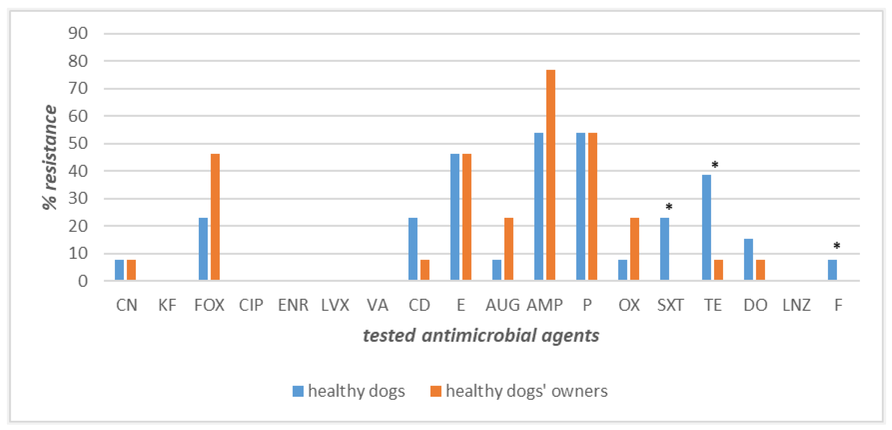
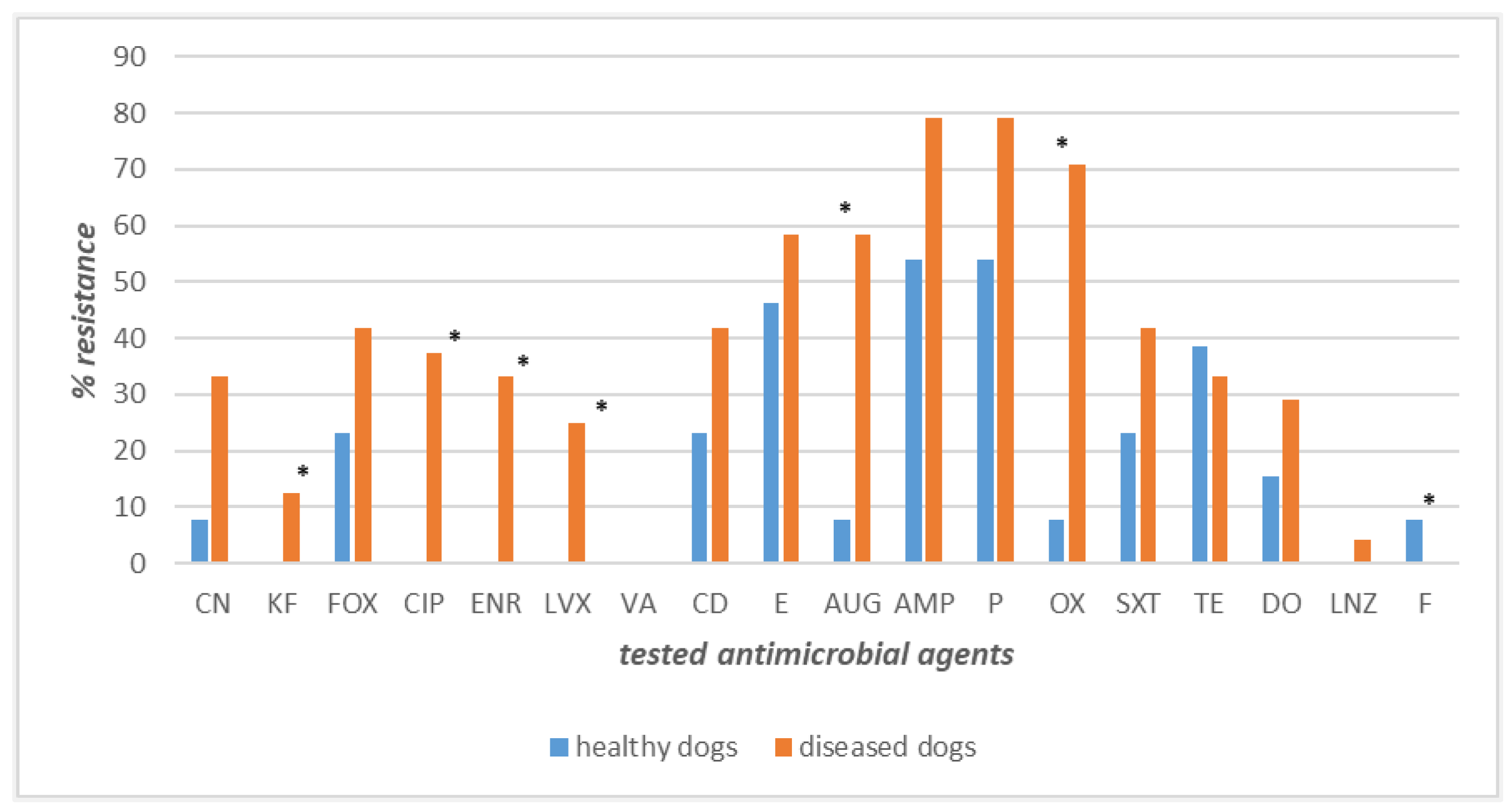
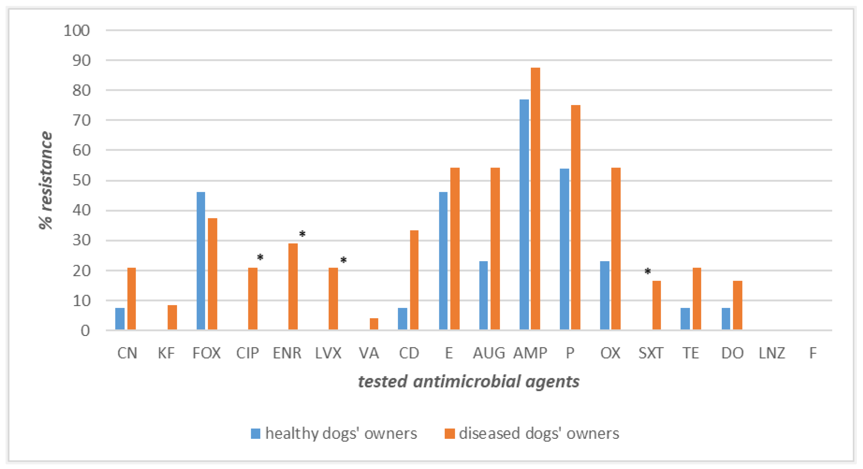
3.4. Antimicrobial Resistance Profiles of Staphylococcus spp. Strains Recovered from Dogs’ and Owners’ Nasal Swabs

In the current study, the antimicrobial resistance profiles of Staphylococcus spp. strains isolated from both healthy and diseased dogs and their respective owners were evaluated and compared.
The strains recovered from the 13 owner–healthy dog couples displayed high levels of susceptibility for most of the antimicrobial agents tested. Precisely, 100% of the canine and human isolates were found to be susceptible to cephalothin, ciprofloxacin, enrofloxacin, levofloxacin, vancomycin, and linezolid (Figure 3). Conversely, the highest levels of resistance were observed for ampicillin (77% owners and 54% dogs), penicillin (54% in both), and erythromycin (43% in both). Different percentages of resistance, with higher values among healthy dogs’ strains than human ones, were observed for tetracycline (39% vs. 8%), clindamycin (23% vs. 7.7%), and doxycycline (15.4% vs. 7.7%), whilst an inverted trend, with higher values among human isolates, was obtained for cefoxitin (42.2% vs. 23%), oxacillin (23% vs. 7.7%), and amoxicillin–clavulanate (23% vs. 7.7%) (Figure 3). Gentamicin was the only antimicrobial to which a lower and identical value of resistance was recorded (7.7%) (Figure 3). The differences in resistance frequencies between canine and human strains were significantly higher (p < 0.05) in healthy dogs than their owners for tetracycline, nitrofurantoin, and sulfamethoxazole–trimethoprim (Figure 3).
Intriguingly, higher resistance levels were recorded for the strains isolated from the 24 diseased dog–owner couples (Figure 4). Specifically, the highest values of resistance were exhibited by ampicillin (87% in owners and 79% in dogs), penicillin (79% in dogs and 75% in owners), oxacillin (71% in dogs, 54% in owners), amoxicillin–clavulanate, and erythromycin, displaying the same level both in diseased dogs and their owners (58% and 54%, respectively). Important differences in resistance frequencies were found for sulfamethoxazole–trimethoprim, ciprofloxacin, and linezolid against which canine strains showed higher percentages of resistance than owner strains: 42%, 38%, and 4%, respectively (Figure 4). The differences in resistance frequencies between canine and human strains were significantly higher (p < 0.05) in dogs suffering from bacterial skin disorders than their owners only for sulfamethoxazole–trimethoprim (Figure 4).
A comparison of the antimicrobial resistance profiles of staphylococci isolated from healthy and diseased dogs underlined higher resistance values among strains isolated from diseased dogs than those recovered from healthy dogs, except for only tetracycline, where, instead, lower resistance levels were recorded than in healthy dogs (Figure 5). It is worth noting, however, that the highest resistance levels in diseased dogs were shown especially by ampicillin (79%), penicillin (79%), and oxacillin (71%), indicative of a greater spread of methicillin-resistant strains. Furthermore, the staphylococcal strains isolated from diseased dogs were also resistant to ciprofloxacin (38%), enrofloxacin (33%), levofloxacin (25%), cephalothin (13%), and linezolid (4.16%).
The differences in resistance frequencies among canine strains were significantly higher (p < 0.05) in diseased dogs than healthy dogs for the following antimicrobials: cephalothin, ciprofloxacin, enrofloxacin, levofloxacin, amoxicillin–clavulanate, oxacillin, and nitrofurantoin (Figure 5).
In Figure 6, the antimicrobial resistance frequencies recorded for the staphylococci isolated from healthy dogs’ and diseased dogs’ owners are compared. In this context, outstanding resistances were exhibited by ampicillin (88% for diseased dogs’ owners vs. 78% for healthy dogs’ owners), penicillin (75% for diseased dogs’ owners vs. 54% for healthy dogs’ owners), and oxacillin and amoxicillin–clavulanate (54% for diseased dogs’ owners vs. 23% for healthy dogs’ owners, same rates for both molecules). Resistance to enrofloxacin (30%), ciprofloxacin and levofloxacin (21%), vancomycin (4%), and cephalothin (8%) were observed only for the strains isolated from diseased dogs’ owners.
The differences in resistance frequencies between human strains were significantly higher (p < 0.05) in diseased dogs’ owners than healthy dogs’ owners for the following antimicrobials: ciprofloxacin, enrofloxacin, levofloxacin, and sulfamethoxazole–trimethoprim (Figure 6).
In addition, the presence of the same bacterial species (S. aureus, S. pseudintermedius, S. epidermidis) with similar antimicrobial resistance profiles in dogs and their owners was detected in only three cases, suggesting potential bacterial transmission. However, further molecular profiling studies are needed to confirm this hypothesis and to possibly demonstrate at least the origin from a same ancestry.

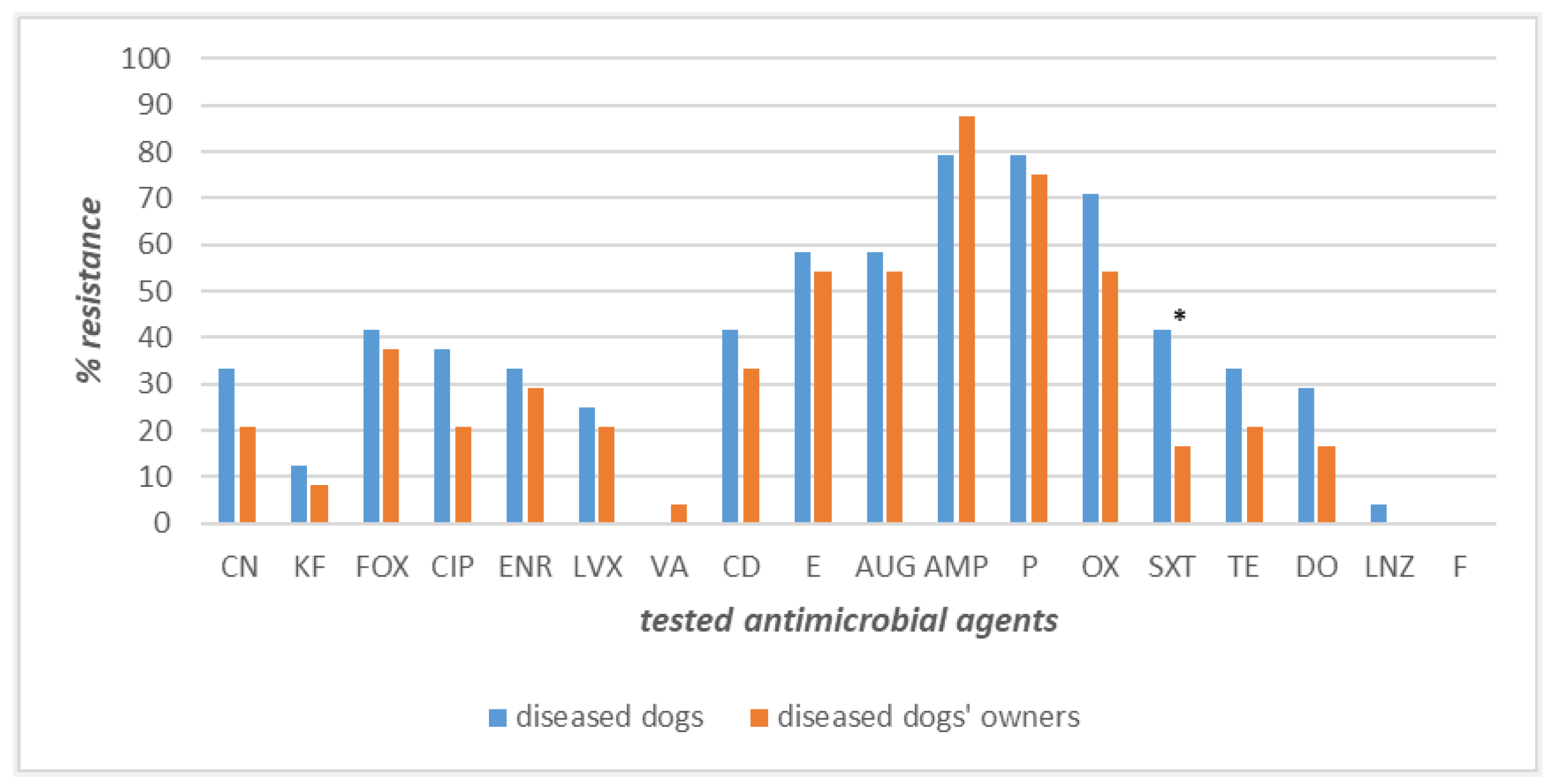
3.5. Antimicrobial Resistance Profiles of Staphylococcus spp. Strains Recovered from Nasal Swabs of Veterinary Staff

All the strains collected from the veterinary staff showed 100% of susceptibility to cephalothin, gentamicin, levofloxacin, nitrofurantoin, vancomycin, and linezolid. Differently, high values of resistance were observed against beta-lactams with a resistance rate of 89.4% for ampicillin and penicillin, followed by amoxicillin–clavulanate (78.9%), oxacillin (57.8%), and cefoxitin (52.6%). Among non-beta-lactam antibiotics, high-level resistance to erythromycin (73.6%) and clindamycin (57.8%) was observed. Furthermore, additional resistances to antibiotics belonging to the following classes were observed: (i) fluoroquinolones, with enrofloxacin and ciprofloxacin exhibiting a resistance of 47.3% and 36.8%, respectively; (ii) tetracyclines, with the same resistance to both doxycycline and tetracycline (42.1%); (iii) sulfonamides, with 36.8% of resistance to sulfamethoxazole–trimethoprim. In addition, the lowest-level resistance was displayed by the three strains identified as S. epidermidis.

4. Discussion

In recent years, the consideration of dogs as potential reservoirs of opportunistic and antimicrobial-resistant pathogens has increased, even though it is not completely clear how the extent and the timing of the transmission of opportunistic pathogens as Staphylococcus spp. between household dog and owner occurs [27,35]. During the COVID-19 pandemic, the number of pet dogs has notably increased. In fact, in the European Union there are more than 72 million households that own dogs [36], of which 25% is in Italy [37]. Furthermore, it is worth noting that pet owners often have intensive and close contact with their pet animals, and this is the reason why the European Medicines Agency has already addressed the minimal knowledge about risk factors and transmission routes for transfer of antimicrobial-resistant pathogens with zoonotic potential between pet animals and humans, also pointing out the risk of antimicrobial resistance transfer from pet dogs and cats [38].
In this study, the results of the questionnaire completed by the pet owners underlined a very close relationship with their dogs. Indeed, in most cases (61%), dogs lived at home, and 70% of household dogs had free access to bed, sleeping with owners, and 68% to the sofa. These results agree with those from other studies. In particular, Stull et al. [39] described that in Canada, 26% of pet dogs slept in the bed with the owning family children, and this habit was also reported in Netherlands [40,41] and in UK [42], with dogs sleeping under the blankets at rates of 18% and 14%, respectively. Since sleeping in the same bed represents a very intimate direct contact, it really may constitute a potential risk favoring the transmission of zoonotic and opportunistic pathogens (i.e., Staphylococcus spp., Enterobacterales, etc.) to their owners through the fur or paw pads, and this risk is higher above all when dogs are affected by skin bacterial disorders. Therefore, it would be desirable for the owners to be adequately informed and educated about this potential risk, to responsibly interact with their dogs.
In this study, 81.7% of dog-owning households described a very tender relationship, characterized by hugs and kisses with their dogs, demonstrating a strong dog–owner interaction. Our results are comparable in extent and frequency to those described in another study [30] in which 85.3% of dogs licked the owner’s hands and 49.3% of owners reported being licked in the face (intense contacts). Moreover, in situations where saliva can be transferred, for example, when a dog licks its owner’s face or hand, microorganisms can be easily transferred from the dog to the owner or vice versa. In this study, 30.9% of participants claimed to always wash their hands after petting or touching their dog, a higher rate compared to that found in another report, where only 15% of respondents washed their hands [40]; 19.7% of the pet owners involved in our study stated that they never washed their hands. Specifically, our pet-care questionnaire highlighted a significantly greater (p < 0.05) attention given to hand washing by diseased dogs’ owners, with 42% of them always washing their hands, than healthy dogs’ owners, with only 14% declaring to always wash hands. This result is probably linked to the presence of skin disorders in their dogs at the time of the interview. Surprisingly, despite the attention given to hand washing; diseased dogs’ owners referred to occasional use of anti-tick and anti-flea drugs and repellents.
The oral cavity and the skin of dogs host a varied microbial flora, including many species of opportunistic pathogens, which can be responsible for even serious health problems in high-risk individuals (young, elderly, pregnant women, and immunocompromised people). Transmission generally occurs through the bite, contact with saliva, mucous membranes, or on an open wound; this can cause serious infections in high-risk patients [43,44]. Indeed, it is known that hand washing is a critical point, a moment to pay attention to, as it is one of the best hygiene measures to remove germs, avoid getting sick, and prevent the spread and the transmission of opportunistic germs to others.
In this study, from a total of 172 nasal samples of both canine and human origin, 60.5% were positive for Staphylococcus spp. Among identified canine nasal staphylococcal strains, S. pseudintermedius was the most predominant identified bacterium (46%). This high S. pseudintermedius occurrence is not surprising, as other published studies described S. pseudintermedius as a normal constituent of the microbial flora in the nasal cavities of dogs [45], but also as the main opportunistic canine pathogen associated with canine skin disorders, such as otitis externa and pyoderma [21,46,47,48,49]. Moreover, this opportunistic microorganism has been reported as responsible for diseases in humans, associated with contact with dogs, suggesting zoonotic transmission [16,50,51]. Transmission can be hypothesized for the dog–owner interaction in our study, where both the dog suffering from pyoderma and its owner were positive for S. pseudintermedius strains, showing a comparable antimicrobial resistance profile. Even though humans are known not to be its natural host, they can be transiently colonized by S. pseudintermedius, including methicillin-resistant S. pseudintermedius (MRSP). In this regard, Guardabassi et al. [52] demonstrated that owners of dogs suffering from pyoderma were more likely to be positive for S. pseudintermedius compared to people not having daily contact with dogs, but there was no evidence of colonization at the time of a second sampling 2 months later, suggesting that S. pseudintermedius long-term colonization is uncommon in humans. This may be one of the reasons why currently the prevalence of human colonization is still unknown; the other one could be represented by the misidentification, above all in past years, of this pathogen as S. aureus, due to the lack of knowledge of this bacterial species in human medicine [53,54].
In two other cases, it was possible to recover the same bacterial strain both in the dog and its owner. Precisely, in one case, a strain of S. epidermidis was isolated from the nasal swab of the dog and its owner; in the other case, the isolated strain was represented by S. aureus. In these two cases, the strains of different origins showed overlapping phenotypes and similar antimicrobial resistance profiles, and led us to hypothesize a possible transmission from the owner to dog, as these microorganisms are more commonly isolated in humans [55,56]; however, this hypothesis also needs support from molecular-typing studies.
Concerning the species belonging to the genus Staphylococcus isolated from the nasal cavities of dogs’ owners, S. aureus was the most commonly isolated species, as well as from the nasal cavities of veterinary staff. These results agree with those of other studies, since S. aureus is a normal constituent of the microbial flora of both skin and nasal mucous membranes of humans, found in up to 68% of examined cases [57,58]. However, it has also been shown how the skin microbiome is primarily dependent on the physiology of the skin site [59] and can be greatly influenced by a series of factors such as age, environment rather than the habits of the person (smokers and nonsmokers), and the onset of pathologies [60].
In this study, another Staphylococcus species often isolated from human nasal specimens was S. epidermidis, a coagulase-negative Staphylococcus (CoNS) described as the species that competes with S. aureus for stable colonization in human nasal mucosa. Furthermore, it is also able to produce a serine protease (Esp), which inhibits S. aureus biofilm formation [61], an activity documented in many studies carried out in hospitalized infants, adolescents, and adults [61,62].
Staphylococcus aureus (29.4%) followed by S. pseudintermedius (14.7%) were the most frequently isolated strains from the culture of nasal swabs sampled among the staff working at the University Veterinary Teaching Hospital. These results partially agree with those obtained for pet owners’ nasal samples, with S. aureus and S. epidermidis as the most prevalent ones. This variation and replacement of S. epidermidis with S. pseudintermedius could be related to the continuous contact that the veterinary staff has daily with diseased dogs and cats, especially those affected by skin infections, such as pyoderma and otitis. Although traditionally S. pseudintermedius is not considered a risk for humans, an increase in its zoonotic transmission has been reported above all for MRSP in recent years [24,25,26]. In this context, the veterinary environments (hospital, clinics) seem to play an important role in the dissemination of MRSP between animals and people, especially people who have constant contact with pets (veterinary personnel and pet owners) [20,63]. Paul et al. [63] pointed out that 3.9% of small animal dermatologists attending a national veterinary conference in Italy carried MRSP in their nasal cavities. This is an important rate of MRSP carriage, considering the relative rare occurrence in humans and, consequently, it might be considered an occupational risk [63].
Referring to the antimicrobial resistance profiles of isolated staphylococci, high levels of resistance to penicillins (oxacillin and penicillin) were recorded. In particular, the highest level of resistance, statistically significant, against oxacillin was observed for the strains isolated from diseased dogs (71%) compared to the healthy dogs (7.7%), representing an alarming result, since MRSP growing spread has become a relevant issue, causing great concern in veterinary medicine [64,65,66]. The emergence of MRSP has for some time highlighted the need for careful long-term surveillance of this strain. However, the high rate of resistance (46% for healthy dog–owner couples and >50% for diseased dog–owner couples) to erythromycin, a macrolide widely used for the treatment of human and animal infections but not generally used in canine infections, was here exhibited by staphylococci isolates of both human and canine origin. The increased spread of bacteria showing worrying antimicrobial resistance profiles underlines the crucial role of the One Health Approach in helping to better control the antimicrobial resistance issue and, consequently, the importance of prudent use and proper administration of antimicrobials in both pet animals and humans [67,68].
In conclusion, the results of this study suggested the potential risk of transmission of S. aureus from dog owners to their dogs and of S. pseudintermedius from dogs to their owners and to veterinary staff. Continuous monitoring is desirable to define the circulation of antimicrobial-resistant staphylococci and to further explore the possibility of animal–human–animal transmission through epidemiological studies concerning interspecies transmission. Particular attention is required for strains such as S. pseudintermedius and S. aureus, which are important candidates for “One Health” initiatives.

5. Conclusions

Dog–human relationships, which have become closer and more continuous over time, can bring benefits but can also allow the possible transmission of opportunistic pathogens. The high prevalence of S. pseudintermedius among dogs, S. aureus and S. epidermidis among owners, and S. aureus and S. pseudintermedius among veterinary staff suggests the possible transmission of these staphylococci between people and dogs or vice versa. In any case, this hypothesis should be confirmed by molecular analysis techniques such as whole genome sequencing. In addition, monitoring of antimicrobial resistance profiles is desirable to evaluate the emergence of new resistances among the circulating strains, along with their transmissibility and pathogenicity. Thus, adequate prevention and control measures for domestic infections control could be better applied.

Author Contributions

Conceptualization, L.D.M. and L.C.; swab collection, L.C., software, F.P.N. and F.P. investigation, F.P.N., F.P. and A.M.; writing—original draft preparation, F.P.N., L.C. and L.D.M.; writing—review and editing, F.P.N., L.C. and L.D.M. All authors have read and agreed to the published version of the manuscript.

Funding

This study was supported by a project of the Regional Center of “Farmacovigilanza Veterinaria Campania” (Naples, Italy), approved by Decree No. 126 of 22 September 2017 for the in-depth program concerning the study of multidrug-resistant bacteria in the Veterinary Teaching Hospital of Naples, Italy (OVUD). Francesca Paola Nocera is supported by a research fellowship (fixed-term researcher type A) financed by the Ministry of Education, University and Scientific Research (MIUR), Italy (PON Ricerca e Innovazione 2014-2020-Azione IV.4- Contratti di Ricerca su tematiche dell’Innovazione, Dipartimento di Medicina Veterinaria e Produzioni Animali, Università degli Studi di Napoli “Federico II”, code n. PON_INN_RTDA_2021_62). The funder of Francesca Paola Nocera has no role in study design, data collection and analysis, decision to publish, or preparation of the manuscript.

Institutional Review Board Statement

This study was approved by the Veterinary Service Center of the University of Naples Federico II, Italy (certificate number PG/2021/0009256), in compliance with the Italian Legislative Decree 26/2014, Article 2, implementing Directive 2010/63/EU.

Informed Consent Statement

Informed consent was obtained from dogs’ owners and veterinary staff prior the sampling of their nasal cavities by use of swabs.

Data Availability Statement

Not applicable.

Acknowledgments

Authors would like to thank all the dog owners and veterinary staff of the Veterinary Teaching Hospital of Naples who decided to take part to this study.

Conflicts of Interest

The authors declare no conflict of interest.

References

  1. Misic, A.M.; Davis, M.F.; Tyldsley, A.S.; Hodkinson, B.P.; Tolomeo, P.; Hu, B.; Nachamkin, I.; Lautenbach, E.; Morris, D.O.; Grice, E.A. The shared microbiota of humans and companion animals as evaluated from Staphylococcus carriage sites. Microbiome 2015, 3, 2. [Google Scholar] [CrossRef] [Green Version]
  2. Weese, J.S. The canine and feline skin microbiome in health and disease. Vet. Dermatol. 2013, 24, 137–145.e31. [Google Scholar] [CrossRef]
  3. Haag, A.F.; Fitzgerald, J.R.; Penadés, J.R. Staphylococcus aureus in animals. Microbiol Spectr. 2019, 7, 1–19. [Google Scholar] [CrossRef] [PubMed]
  4. Cuny, C.; Layer-Nicolaou, F.; Weber, R.; Köck, R.; Witte, W. Colonization of dogs and their owners with Staphylococcus aureus and Staphylococcus pseudintermedius in households, veterinary practices, and healthcare facilities. Microorganisms 2022, 10, 677. [Google Scholar] [CrossRef] [PubMed]
  5. Sakr, A.; Brégeon, F.; Mège, J.L.; Rolain, J.M.; Blin, O. Staphylococcus aureus nasal colonization: An update on mechanisms, epidemiology, risk factors, and subsequent infections. Front. Microbiol. 2018, 9, 2419. [Google Scholar] [CrossRef] [PubMed]
  6. Grema, H.A.; Geidam, Y.A.; Gadzama, G.B.; Ameh, J.A.; Suleiman, A. Methicillin resistant Staphylococcus aureus (MRSA): A review. Adv. Anim. Vet. 2015, 3, 79–98. [Google Scholar] [CrossRef] [Green Version]
  7. Garoy, E.Y.; Gebreab, Y.B.; Achila, O.O.; Tekeste, D.G.; Kesete, R.; Ghirmay, R.; Kiflay, R.; Tesfu, T. Methicillin-resistant Staphylococcus aureus (MRSA): Prevalence and antimicrobial sensitivity pattern among patients-a multicenter study in Asmara, Eritrea. Can. J. Infect. Dis. Med. Microbiol. 2019, 2019, 8321834. [Google Scholar] [CrossRef] [PubMed] [Green Version]
  8. Petersen, A.; Larssen, K.W.; Gran, F.W.; Enger, H.; Hæggman, S.; Mäkitalo, B.; Haraldsson, G.; Lindholm, L.; Vuopio, J.; Henius, A.E.; et al. Increasing incidences and clonal diversity of methicillin-resistant Staphylococcus aureus in the Nordic countries - results from the Nordic MRSA surveillance. Front. Microbiol. 2021, 12, 668900. [Google Scholar] [CrossRef]
  9. Kourtis, A.P.; Hatfield, K.; Baggs, J.; Mu, Y.; See, I.; Epson, E.; Nadle, J.; Kainer, M.A.; Dumyati, G.; Petit, S.; et al. Vital Signs: Epidemiology and recent trends in methicillin-resistant and in methicillin-susceptible Staphylococcus aureus bloodstream infections—United States. MMWR. Morb. Mortal. Wkly. Rep. 2019, 68, 214–219. [Google Scholar] [CrossRef] [Green Version]
  10. Wu, M.; Tong, X.; Liu, S.; Wang, D.; Wang, L.; Fan, H. Prevalence of methicillin-resistant Staphylococcus aureus in healthy Chinese population: A system review and meta-analysis. PLoS ONE 2019, 14, e0223599. [Google Scholar] [CrossRef] [Green Version]
  11. Cheung, G.Y.C.; Bae, J.S.; Otto, M. Pathogenicity and virulence of Staphylococcus aureus. Virulence 2021, 12, 547–569. [Google Scholar] [CrossRef] [PubMed]
  12. Verkade, E.; Kluytmans, J. Livestock-associated Staphylococcus aureus CC398: Animal reservoirs and human infections. Infect. Genet. Evol. 2014, 21, 523–530. [Google Scholar] [CrossRef] [PubMed]
  13. Cuny, C.; Wieler, L.H.; Witte, W. Livestock-associated MRSA: The impact on humans. Antibiotics 2015, 4, 521–543. [Google Scholar] [CrossRef] [Green Version]
  14. Neradova, K.; Jakubu, V.; Pomorska, K.; Zemlickova, H. Methicillin-resistant Staphylococcus aureus in veterinary professionals in 2017 in the Czech Republic. BMC Vet. Res. 2020, 16, 4. [Google Scholar] [CrossRef] [Green Version]
  15. Zhou, Y.P.; Wilder-Smith, A.; Hsu, L.Y. The role of international travel in the spread of methicillin-resistant Staphylococcus aureus. J. Travel Med. 2014, 21, 272–281. [Google Scholar] [CrossRef] [PubMed] [Green Version]
  16. Carroll, K.C.; Burnham, C.-A.D.; Westblade, L.F. From canines to humans: Clinical importance of Staphylococcus pseudintermedius. PLoS Pathog. 2021, 17, e1009961. [Google Scholar] [CrossRef]
  17. Bannoehr, J.; Guardabassi, L. Staphylococcus pseudintermedius in the dog: Taxonomy, diagnostics, ecology, epidemiology and pathogenicity. Vet. Dermatol. 2012, 23, 253–266.e51–e52. [Google Scholar] [CrossRef] [PubMed]
  18. Garbacz, K.; Zarnowska, S.; Piechowicz, L.; Haras, K. Pathogenicity potential of Staphylococcus pseudintermedius strains isolated from canine carriers and from dogs with infection signs. Virulence 2013, 4, 255–259. [Google Scholar] [CrossRef] [Green Version]
  19. Rubin, R.H. Surgical wound infection: Epidemiology, pathogenesis, diagnosis and management. BMC Infect. Dis. 2006, 6, 171. [Google Scholar] [CrossRef] [Green Version]
  20. van Duijkeren, E.; Kamphuis, M.; van der Mije, I.C.; Laarhoven, L.M.; Duim, B.; Wagenaar, J.A.; Houwers, D.J. Transmission of methicillin-resistant Staphylococcus pseudintermedius between infected dogs and cats and contact pets, humans and the environment in households and veterinary clinics. Vet. Microbiol. 2011, 150, 338–343. [Google Scholar] [CrossRef]
  21. Nocera, F.P.; Meroni, G.; Fiorito, F.; De Martino, L.; Martino, P.A. Occurrence and antimicrobial susceptibility patterns of canine Staphylococcus pseudintermedius strains isolated from two different Italian university veterinary hospitals. Vet. Ital. 2020, 56, 263–269. [Google Scholar] [PubMed]
  22. Kadlec, K.; Schwarz, S. Antimicrobial resistance of Staphylococcus pseudintermedius. Vet. Dermatol. 2012, 23, 276–282.e55. [Google Scholar] [CrossRef] [PubMed]
  23. Stegmann, R.; Burnens, A.; Maranta, C.A.; Perreten, V. Human infection associated with methicillin-resistant Staphylococcus pseudintermedius ST71. J. Antimicrob. Chemother. 2010, 65, 2047–2048. [Google Scholar] [CrossRef] [PubMed]
  24. Somayaji, R.; Priyantha, M.A.; Rubin, J.E.; Church, D. Human infections due to Staphylococcus pseudintermedius, an emerging zoonosis of canine origin: Report of 24 cases. Diagn. Microbiol. Infect. Dis. 2016, 85, 471–476. [Google Scholar] [CrossRef]
  25. Lozano, C.; Rezusta, A.; Ferrer, I.; Pérez-Laguna, V.; Zarazaga, M.; Ruiz-Ripa, L.; Revillo, M.J.; Torres, C. Staphylococcus pseudintermedius human infection cases in Spain: Dog-to-Human Transmission. Vector. Borne Zoonotic. Dis. 2017, 17, 268–270. [Google Scholar] [CrossRef]
  26. Robb, A.R.; Wright, E.D.; Foster, A.M.E.; Walker, R.; Malone, C. Skin infection caused by a novel strain of Staphylococcus pseudintermedius in a Siberian husky dog owner. JMM Case Rep. 2017, 4, jmmcr005087. [Google Scholar] [CrossRef]
  27. Wegener, A.; Duim, B.; van der Graaf-van Bloois, L.; Zomer, A.L.; Visser, C.E.; Spaninks, M.; Timmerman, A.J.; Wagenaar, J.A.; Broens, E.M. Within-Household Transmission and Bacterial Diversity of Staphylococcus pseudintermedius. Pathogens 2022, 11, 850. [Google Scholar] [CrossRef]
  28. International Organization for Protection of Animals (OIPA). Available online: https://www.oipa.org/italia/14-milioni-gli-animali-microchippati-in-italia-loipa-diffonde-i-numeri-della-banca-dati-del-ministero-della-salute/ (accessed on 8 August 2022).
  29. Martin, F.; Bachert, K.E.; Snow, L.; Tu, H.W.; Belahbib, J.; Lyn, S.A. Depression, anxiety, and happiness in dog owners and potential dog owners during the COVID-19 pandemic in the United States. PLoS ONE 2021, 16, e0260676. [Google Scholar] [CrossRef]
  30. Joosten, P.; Ceccarelli, D.; Odent, E.; Sarrazin, S.; Graveland, H.; Van Gompel, L.; Battisti, A.; Caprioli, A.; Franco, A.; Wagenaar, J.A.; et al. Antimicrobial usage and resistance in companion animals: A cross-sectional study in three European countries. Antibiotics 2020, 9, 87. [Google Scholar] [CrossRef] [Green Version]
  31. Orsini, M.; Petrin, S.; Corrò, M.; Baggio, G.; Spagnolo, E.; Losasso, C. Anthroponotic-Based Transfer of Staphylococcus to Dog: A Case Study. Pathogens 2022, 11, 802. [Google Scholar] [CrossRef]
  32. Robinson, R.A.; Pugh, R.N. Dogs, zoonoses and immunosuppression. J. R. Soc. Promot. Health 2002, 122, 95–98. [Google Scholar] [CrossRef]
  33. CLSI, VET01S; Performance Standards for Antimicrobial Disk and Dilution Susceptibility Test for Bacteria Isolate from Animals, 5th ed. Clinical and Laboratory Standards Institute: Philadelphia, PA, USA, 2020.
  34. EUCAST. The European Committee on Antimicrobial Susceptibility Testing. Breakpoint Tables for Interpretation of MICs and zone Diameters. Version 11.0. 2021. Available online: http://www.eucast.org (accessed on 12 January 2021).
  35. Thomson, P.; García, P.; Miles, J.; Isla, D.; Yáñez, C.; Santibáñez, R.; Núñez, A.; Flores-Yáñez, C.; del Río, C.; Cuadra, F. Isolation and identification of Staphylococcus species obtained from healthy companion animals and humans. Vet. Sci. 2022, 9, 79. [Google Scholar] [CrossRef] [PubMed]
  36. Number of Pet Animals in European Union in 2021, by Animal Type. Available online: https://www.statista.com/statistics/515010/pet-population-european-union-eu-by-animal/ (accessed on 20 May 2023).
  37. Share of Households Owning at Least One Dog in the European Union in 2021, by Country. Available online: https://www.statista.com/statistics/515475/dog-ownership-european-union-eu-by-country/ (accessed on 20 May 2023).
  38. European Medicines Agency Committee for Medicinal Products for Veterinary Use (CVMP) Reflection Paper on the Risk of Antimicrobial Resistance Transfer from Companion Animals. 2015. Available online: https://www.ema.europa.eu/en/documents/scientific-guideline/reflection-paper-risk-antimicrobial-resistance-transfer-companion-animals_en.pdf (accessed on 5 May 2022).
  39. Stull, J.W.; Peregrine, A.S.; Sargeant, J.M.; Weese, J.S. Pet husbandry and infection control practices related to zoonotic disease risks in Ontario, Canada. BMC Public Health 2013, 13, 520. [Google Scholar] [CrossRef] [PubMed] [Green Version]
  40. Overgaauw, P.A.; van Zutphen, L.; Hoek, D.; Yaya, F.O.; Roelfsema, J.; Pinelli, E.; van Knapen, F.; Kortbeek, L.M. Zoonotic parasites in fecal samples and fur from dogs and cats in The Netherlands. Vet. Parasitol. 2009, 163, 115–122. [Google Scholar] [CrossRef] [PubMed]
  41. Zanen, L.A.; Kusters, J.G.; Overgaauw, P.A.M. Zoonotic Risks of Sleeping with Pets. Pathogens 2022, 11, 1149. [Google Scholar] [CrossRef]
  42. Westgarth, C.; Pinchbeck, G.L.; Bradshaw, J.W.; Dawson, S.; Gaskell, R.M.; Christley, R.M. Dog-human and dog-dog interactions of 260 dog-owning households in a community in Cheshire. Vet. Rec. 2008, 162, 436–442. [Google Scholar] [CrossRef]
  43. Morelli, M.K.; Veve, M.P.; Shorman, M.A. Maternal bacteremia caused by Staphylococcus aureus with a focus on infective endocarditis. Open Forum Infect. Dis. 2020, 7, ofaa239. [Google Scholar] [CrossRef] [PubMed]
  44. Khan, S.; Siddiqui, S. Community-associated methicillin-resistant Staphylococcus aureus: Case report of acute sinusitis with orbital extension in a pregnant lady. Cureus 2020, 12, e12054. [Google Scholar] [CrossRef]
  45. Ruiz-Ripa, L.; Simón, C.; Ceballos, S.; Ortega, C.; Zarazaga, M.; Torres, C.; Gómez-Sanz, E.S. Pseudintermedius and S. aureus lineages with transmission ability circulate as causative agents of infections in pets for years. BMC Vet. Res. 2021, 17, 42. [Google Scholar] [CrossRef]
  46. De Martino, L.; Nocera, F.P.; Mallardo, K.; Nizza, S.; Masturzo, E.; Fiorito, F.; Iovane, G.; Catalanotti, P. An update on microbiological causes of canine otitis externa in Campania Region, Italy. Asian Pac. J. Trop. Biomed. 2016, 6, 384–389. [Google Scholar] [CrossRef] [Green Version]
  47. Stefanetti, V.; Bietta, A.; Pascucci, L.; Marenzoni, M.L.; Coletti, M.; Franciosini, M.P.; Passamonti, F.; Casagrande Proietti, P. Investigation of the antibiotic resistance and biofilm formation of Staphylococcus pseudintermedius strains isolated from canine pyoderma. Vet. Ital. 2017, 53, 289–296. [Google Scholar] [PubMed]
  48. Nocera, F.P.; Ambrosio, M.; Fiorito, F.; Cortese, L.; De Martino, L. On Gram-positive- and Gram-negative-bacteria-associated canine and feline skin infections: A 4-year retrospective study of the University Veterinary Microbiology Diagnostic Laboratory of Naples, Italy. Animals 2021, 11, 1603. [Google Scholar] [CrossRef] [PubMed]
  49. Lynch, S.A.; Helbig, K.J. The complex diseases of Staphylococcus pseudintermedius in canines: Where to next? Vet. Sci. 2021, 8, 11. [Google Scholar] [CrossRef] [PubMed]
  50. Pires Dos Santos, T.; Damborg, P.; Moodley, A.; Guardabassi, L. Systematic review on global epidemiology of methicillin-resistant Staphylococcus pseudintermedius: Inference of population structure from multilocus sequence typing data. Front. Microbiol. 2016, 7, 1599. [Google Scholar] [CrossRef] [PubMed] [Green Version]
  51. Moses, I.B.; Santos, F.F.; Gales, A.C. Human colonization and infection by Staphylococcus pseudintermedius: An emerging and underestimated zoonotic pathogen. Microorganisms 2023, 11, 581. [Google Scholar] [CrossRef]
  52. Guardabassi, L.; Loeber, M.E.; Jacobson, A. Transmission of multiple antimicrobial-resistant Staphylococcus intermedius between dogs affected by deep pyoderma and their owners. Vet. Microbiol. 2004, 98, 23–27. [Google Scholar] [CrossRef]
  53. Börjesson, S.; Gómez-Sanz, E.; Ekström, K.; Torres, C.; Grönlund, U. Staphylococcus pseudintermedius can be misdiagnosed as Staphylococcus aureus in humans with dog bite wounds. Eur. J. Clin. Microbiol. Infect. Dis. 2015, 34, 839–844. [Google Scholar] [CrossRef]
  54. Yarbrough, M.L.; Lainhart, W.; Burnham, C.A. Epidemiology, clinical characteristics, and antimicrobial susceptibility profiles of human clinical isolates of Staphylococcus intermedius group. J. Clin. Microbiol. 2018, 56, e01788-17. [Google Scholar] [CrossRef] [Green Version]
  55. Krismer, B.; Weidenmaier, C.; Zipperer, A.; Peschel, A. The commensal lifestyle of Staphylococcus aureus and its interactions with the nasal microbiota. Nat. Rev. Microbiol. 2017, 15, 675–687. [Google Scholar] [CrossRef]
  56. Brown, M.M.; Horswill, A.R. Staphylococcus epidermidis-skin friend or foe? PLoS Pathog. 2020, 16, e1009026. [Google Scholar] [CrossRef]
  57. Human Microbiome Project Consortium. Structure, function and diversity of the healthy human microbiome. Nature 2012, 486, 207–214. [Google Scholar] [CrossRef] [Green Version]
  58. Ramakrishnan, V.R.; Feazel, L.M.; Gitomer, S.A.; Ir, D.; Robertson, C.E.; Frank, D.N. The microbiome of the middle meatus in healthy adults. PLoS ONE 2013, 3, e85507. [Google Scholar] [CrossRef] [Green Version]
  59. Byrd, A.L.; Belkaid, Y.; Segre, J.A. The human skin microbiome. Nat. Rev. Microbiol. 2018, 16, 143–155. [Google Scholar] [CrossRef]
  60. Lloyd-Price, J.; Abu-Ali, G.; Huttenhower, C. The healthy human microbiome. Genome Med. 2016, 8, 51. [Google Scholar] [CrossRef] [PubMed] [Green Version]
  61. Lee, D.C.; Kananurak, A.; Tran, M.T.; Connolly, P.A.; Polage, C.R.; Iwase, T.; Bevins, C.L.; Underwood, M.A. Bacterial colonization of the hospitalized newborn: Competition between Staphylococcus aureus and Staphylococcus epidermidis. Pediatr. Infect. Dis. J. 2019, 38, 682–686. [Google Scholar] [CrossRef]
  62. Fredheim, E.G.A.; Flægstad, T.; Askarian, F.; Klingenberg, C. Colonisation and interaction between S. epidermidis and S. aureus in the nose and throat of healthy adolescents. Eur. J. Clin. Microbiol. Infect. Dis. 2015, 34, 123–129. [Google Scholar] [CrossRef] [PubMed]
  63. Paul, N.C.; Moodley, A.; Ghibaudo, G.; Guardabassi, L. Carriage of methicillin-resistant Staphylococcus pseudintermedius in small animal veterinarians: Indirect evidence of zoonotic transmission. Zoonoses Public Health 2011, 58, 533–539. [Google Scholar] [CrossRef] [PubMed]
  64. McCarthy, A.J.; Harrison, E.M.; Stanczak-Mrozek, K.; Leggett, B.; Waller, A.; Holmes, M.A.; Lloyd, D.H.; Lindsay, J.A.; Loeffler, A. Genomic insights into the rapid emergence and evolution of MDR in Staphylococcus pseudintermedius. J. Antimicrob. Chemother. 2015, 70, 997–1007. [Google Scholar] [CrossRef] [Green Version]
  65. Nisa, S.; Bercker, C.; Midwinter, A.C.; Bruce, I.; Graham, C.F.; Venter, P.; Bell, A.; French, N.P.; Benschop, J.; Bailey, K.M.; et al. Combining MALDI-TOF and genomics in the study of methicillin resistant and multidrug resistant Staphylococcus pseudintermedius in New Zealand. Sci. Rep. 2019, 9, 1271. [Google Scholar] [CrossRef] [Green Version]
  66. Nocera, F.P.; Parisi, A.; Corrente, M.; De Martino, L. Evidence of new sequence types of methicillin-resistant Staphylococcus pseudintermedius in Italy. Pol. J. Vet. Sci. 2020, 23, 465–468. [Google Scholar]
  67. Collignon, P.J.; McEwen, S.A. One Health-its importance in helping to better control antimicrobial resistance. Trop. Med. Infect. Dis. 2019, 4, 22. [Google Scholar] [CrossRef] [PubMed] [Green Version]
  68. Chirollo, C.; Nocera, F.P.; Piantedosi, D.; Fatone, G.; Della Valle, G.; De Martino, L.; Cortese, L. Data on before and after the traceability system of veterinary antimicrobial prescriptions in small animals at the University Veterinary Teaching Hospital of Naples. Animals 2021, 11, 913. [Google Scholar] [CrossRef] [PubMed]
Figure 1. Pet-care questionnaire survey.
Pathogens 12 01016 g001
Figure 2. Comparison of answers between healthy and diseased dogs’ owners: (a) pet life environment; (b) pet habits; (c) time spent together; (d) owner–pet relationship; (e) hand washing.
Pathogens 12 01016 g002
Figure 3. Antimicrobial resistance frequencies of Staphylococcus spp. isolated from healthy dogs and their owners. Legend_ Tested antimicrobials: amoxicillin–clavulanate (AUG), ampicillin (AMP), cephalothin (KF), cefoxitin (FOX), ciprofloxacin (CIP), clindamycin (CD), doxycycline (DO), enrofloxacin (ENR), erythromycin (E), gentamicin (CN), levofloxacin (LVX), linezolid (LNZ), nitrofurantoin (F), oxacillin (OX), penicillin (P), sulfamethoxazole–trimethoprim (SXT), tetracycline (TE), and vancomycin (VA). * p < 0.05 for SXT, TE, F.
Pathogens 12 01016 g003
Figure 4. Antimicrobial resistance frequencies of Staphylococcus spp. isolated from diseased dogs and their owners. Legend_ Tested antimicrobials: amoxicillin–clavulanate (AUG), ampicillin (AMP), cephalothin (KF), cefoxitin (FOX), ciprofloxacin (CIP), clindamycin (CD), doxycycline (DO), enrofloxacin (ENR), erythromycin (E), gentamicin (CN), levofloxacin (LVX), linezolid (LNZ), nitrofurantoin (F), oxacillin (OX), penicillin (P), sulfamethoxazole–trimethoprim (SXT), tetracycline (TE), and vancomycin (VA). * p < 0.05 for SXT.
Pathogens 12 01016 g004
Figure 5. Antimicrobial resistance frequencies of Staphylococcus spp. isolated from diseased and healthy dogs. Legend_ Tested antimicrobials: amoxicillin–clavulanate (AUG), ampicillin (AMP), cephalothin (KF), cefoxitin (FOX), ciprofloxacin (CIP), clindamycin (CD), doxycycline (DO), enrofloxacin (ENR), erythromycin (E), gentamicin (CN), levofloxacin (LVX), linezolid (LNZ), nitrofurantoin (F), oxacillin (OX), penicillin (P), sulfamethoxazole–trimethoprim (SXT), tetracycline (TE), and vancomycin (VA). * p < 0.05 for KF, CIP, ENR, LVX, AUG, OX, F.
Pathogens 12 01016 g005
Figure 6. Antimicrobial resistance frequencies of Staphylococcus spp. isolated from diseased dogs’ and healthy dogs’ owners. Legend_Tested antimicrobials: amoxicillin–clavulanate (AUG), ampicillin (AMP), cephalothin (KF), cefoxitin (FOX), ciprofloxacin (CIP), clindamycin (CD), doxycycline (DO), enrofloxacin (ENR), erythromycin (E), gentamicin (CN), levofloxacin (LVX), linezolid (LNZ), nitrofurantoin (F), oxacillin (OX), penicillin (P), sulfamethoxazole–trimethoprim (SXT), tetracycline (TE), and vancomycin (VA). * p < 0.05 for CIP, ENR, LVX, SXT.
Pathogens 12 01016 g006
Table 1. Pet-owner interview questions and answers.
Healthy
(N.28)
Diseased
(N.43)
Pet life environmentHome14 (50%)29 (67%)
Garden3 (11%)3 (7%)
Both11 (39%)11 (26%)
Pet habitsPet bed23 (82%)31 (72%)
Bed-allowed21 (75%)29 (67%)
Sofa-allowed18 (64%)30 (70%)
Use of parasite cleanersAnti-tick and -flea drugs26 (93%)36 (84%)
Repellent substances10 (36%)7 (16%)
Regularly used15 (54%)15 (35%)
Sporadically used5 (18%)5 (12%)
Once a month9 (32%)6 (14%)
Seasonally used 7 (25%)13 (30%)
Feed habitsHomemade diet2 (7%)4 (9%)
Commercial diet14 (50%)30 (70%)
Mixed diet12 (43%)9 (21%)
Snacks18 (64%)6 (14%)
Owner–pet
relationship
Cuddles28 (100%)40 (93%)
Kisses26 (93%)30 (70%)
Hugs26 (93%)33 (77%)
Hand washingAlways 4 (14%)18 (42%)
Sometimes20 (71%)15 (35%)
Never4 (14%)10 (23%)
Time spent togetherAll day long 7 (25%)20 (47%)
Half a day16 (57%)16 (37%)
Only in the evening/night5 (18%)7 (16%)
Drug treatment
(dog)
Recent4 (14%)6 (14%)
Remote6 (21%)20 (47%)
No one18 (64%)17 (40%)
Antibiotic
administration
(dog)
Yes6 (21%)6 (14%)
No22 (79%)37 (86%)
Drug treatment
(owner)
Recent00
Remote19 (68%)10 (23%)
No one 9 (32%)33 (77%)
Antibiotic
administration
(owner)
Yes5 (18%)6 (14%)
No23 (82%)37 (86%)
Table 2. Occurrence of Staphylococcus spp. identified in owner–healthy dogs couples.
Strains from OwnersStrains from Healthy Dogs Occurrence (%)
S. epidermidisS. pseudintermedius23%
S. aureusS. epidermidis15.4%
S. haemolyticusS. aureus15.4%
S. aureusS. pseudintermedius15.4%
S. aureusS. aureus7.7%
S. haemolyticusS. pseudintermedius7.7%
S. epidermidisS. xylosus7.7%
S. epidermidisS. epidermidis7.7%
Table 3. Occurrence of Staphylococcus spp. identified in owner–diseased dogs couples.
Strains from OwnersStrains from Diseased Dogs Occurrence (%)
S. epidermidisS. pseudintermedius45.8%
S. aureusS. pseudintermedius25%
S. epidermidisS. lugdunensis8.3%
S. epidermidisS. sciuri4.2%
S. epidermidisS. lentus4.2%
S. epidermidisS. hyicus4.2%
S. pseudintermediusS. pseudintermedius4.2%
S. aureusS. simulans4.2%
Disclaimer/Publisher’s Note: The statements, opinions and data contained in all publications are solely those of the individual author(s) and contributor(s) and not of MDPI and/or the editor(s). MDPI and/or the editor(s) disclaim responsibility for any injury to people or property resulting from any ideas, methods, instructions or products referred to in the content.

