Next Issue
Volume 12, September
Previous Issue
Volume 12, July


Pathogens, Volume 12, Issue 8 (August 2023) – 97 articles

Cover Story (view full-size image): 
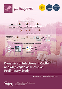
Tick-borne pathogens (TBPs) pose a significant problem globally, particularly in tropical and subtropical regions, impacting both livestock industries and animal health. In Cuba, key pathogens affecting cattle include Babesia bovis, Babesia bigemina, and Anaplasma marginale. These pathogens are primarily transmitted by the Rhipicephalus microplus tick. Diagnosing these infections is challenging, due to low parasitemia levels during persistent infections and cross-reactivity. Molecular techniques such as PCR variants are essential for diagnosis. A drawback of PCR-based methods is their inability to simultaneously detect pathogens. However, high-throughput microfluidic methods enable for the concurrent detection of various pathogens. This study aims to investigate TBPs in cattle and ticks at four sampling points using real-time microfluidic PCR. View this paper
  • Issues are regarded as officially published after their release is announced to the table of contents alert mailing list.
  • You may sign up for e-mail alerts to receive table of contents of newly released issues.
  • PDF is the official format for papers published in both, html and pdf forms. To view the papers in pdf format, click on the "PDF Full-text" link, and use the free Adobe Reader to open them.
Order results
Result details
Section
Select all
Export citation of selected articles as:
14 pages, 2117 KiB  
Article
Factors Affecting the Spread, Diagnosis, and Control of African Swine Fever in the Philippines
by Chia-Hui Hsu, Rachel Schambow, Maximino Montenegro, Ruth Miclat-Sonaco and Andres Perez
Pathogens 2023, 12(8), 1068; https://doi.org/10.3390/pathogens12081068 - 21 Aug 2023
Cited by 2 | Viewed by 5100
Abstract
African Swine Fever (ASF) is a highly contagious disease that threatens the swine industry globally. Since its introduction into the Philippines in 2019, ASF has spread extensively in both commercial and backyard farms. Here, using a mix of qualitative and quantitative methods, including [...] Read more.
African Swine Fever (ASF) is a highly contagious disease that threatens the swine industry globally. Since its introduction into the Philippines in 2019, ASF has spread extensively in both commercial and backyard farms. Here, using a mix of qualitative and quantitative methods, including conjoint and SWOT analyses, world café discussions, and multivariable regression models, the most important factors that influence the spread, diagnosis, and control of ASF in the Philippines were identified. Research findings suggest that swill or contaminated feed, inadequate biosecurity protocols, and movement of personnel were the top risk factors favoring ASF spread among farms in general. For commercial farms, contaminated vehicles and personnel were also important, whereas for backyard farms, the introduction of new pigs, environmental contamination, and poor feeding quality were relevant risk factors. Notable clinical signs of ASF in pigs include reduced feed intake, huddled behavior, and reluctance to stand. This study highlights the need for timely reporting, trust-building initiatives, and enhanced biosecurity measures to effectively manage ASF outbreaks in the country. Results here contribute to the knowledge of factors affecting ASF spread in the Philippines and can help design prevention and control measures in ASF-infected countries while enhancing preparedness in countries free from the disease. Full article
(This article belongs to the Special Issue Emergence and Control of African Swine Fever)
Show Figures

Figure 1

21 pages, 7202 KiB  
Systematic Review
Comparative Analysis of the Prevalence of Bovine Viral Diarrhea Virus in Cattle Populations Based on Detection Methods: A Systematic Review and Meta-Analysis
by Gebremeskel Mamu Werid, Farhid Hemmatzadeh, Darren Miller, Michael P. Reichel, Yohannes E. Messele and Kiro Petrovski
Pathogens 2023, 12(8), 1067; https://doi.org/10.3390/pathogens12081067 - 21 Aug 2023
Cited by 1 | Viewed by 1363
Abstract
Infectious diseases of cattle, including bovine viral diarrhea (BVD), pose a significant health threat to the global livestock industry. This study aimed to investigate the prevalence and risk factors associated with bovine viral diarrhea virus (BVDV) infections in cattle populations through a systematic [...] Read more.
Infectious diseases of cattle, including bovine viral diarrhea (BVD), pose a significant health threat to the global livestock industry. This study aimed to investigate the prevalence and risk factors associated with bovine viral diarrhea virus (BVDV) infections in cattle populations through a systematic review and meta-analysis. PubMed, Web of Science, and Scopus were systematically searched for relevant articles reporting the prevalence of and associated risk factors in studies published between 1 January 2000 and 3 February 2023. From a total of 5111 studies screened, 318 studies were included in the final analysis. BVDV prevalence in cattle populations was estimated using various detection methods. The analysis detected heterogeneity in prevalence, attributed to detection techniques and associated risk factors. Antibody detection methods exhibited a higher prevalence of 0.43, reflecting the cumulative effect of detecting both active and past infections. Antigen detection methods showed a prevalence of 0.05, which was lower than antibody methods. A prevalence of 0.08 was observed using nucleic acid detection methods. The health status of the examined cattle significantly influenced the prevalence of BVDV. Cattle with bovine respiratory disease complex (BRDC) exhibited higher antibody (prevalence of 0.67) and antigen (prevalence 0.23) levels compared to cattle with reproductive problems (prevalence 0.13) or diarrhea (prevalence 0.01). Nucleic acid detection methods demonstrated consistent rates across different health conditions. Age of cattle influenced prevalence, with higher rates in adults compared to calves. Risk factors related to breeding and reproduction, such as natural or extensive breeding and a history of abortion, were associated with increased prevalence. Coinfections with pathogens like bovine herpesvirus-1 or Neospora caninum were linked to higher BVDV prevalence. Management practices, such as commingling, introducing new cattle, and direct contact with neighboring farms, also influenced prevalence. Herd attributes, including larger herd size, and the presence of persistently infected cattle, were associated with higher prevalence. These findings indicated the importance of detection methods and risk factors in BVDV epidemiological studies. Full article
Show Figures

Figure 1

5 pages, 194 KiB  
Communication
Frequency of Positive Polymerase Chain Reaction (PCR) Testing for Borrelia burgdorferi on Whole Blood Samples That Tested Positive for Babesia microti by PCR from an Endemic Area for Both Infections in New York State
by Guiqing Wang, Jian Zhuge and Gary P. Wormser
Pathogens 2023, 12(8), 1066; https://doi.org/10.3390/pathogens12081066 - 21 Aug 2023
Viewed by 1729
Abstract
Because both Babesia microti and Borrelia burgdorferi can be transmitted by the bite of a single coinfected Ixodes scapularis tick, an attempt was made to determine the frequency with which whole blood samples that tested positive for B. microti infection by polymerase chain [...] Read more.
Because both Babesia microti and Borrelia burgdorferi can be transmitted by the bite of a single coinfected Ixodes scapularis tick, an attempt was made to determine the frequency with which whole blood samples that tested positive for B. microti infection by polymerase chain reaction (PCR) would also test positive by PCR for B. burgdorferi infection. Over a 7-year period from 2013 to 2019, 119 different patients tested positive for B. microti infection by PCR on at least one blood sample. Among the 118 patients with a positive B. microti PCR blood sample that could also be tested by a qualitative PCR for B. burgdorferi, only one patient tested positive (0.85%, 95% CI 0.02 to 4.6%). Routine PCR testing of every B. microti PCR-positive blood specimen to detect B. burgdorferi coinfection appears to have a low yield, even in a highly endemic geographic area for both of these infections. Full article
18 pages, 16402 KiB  
Article
Genomic and Phylogenetic Analysis of Bacillus cereus Biovar anthracis Isolated from Archival Bone Samples Reveals Earlier Natural History of the Pathogen
by Michael H. Norris, Diansy Zincke, David J. Daegling, John Krigbaum, W. Scott McGraw, Alexander Kirpich, Ted L. Hadfield and Jason K. Blackburn
Pathogens 2023, 12(8), 1065; https://doi.org/10.3390/pathogens12081065 - 20 Aug 2023
Viewed by 1379
Abstract
(1) Background: Bacillus cereus biovar anthracis (Bcbva) was the causative agent of an anthrax-like fatal disease among wild chimpanzees in 2001 in Côte d’Ivoire. Before this, there had not been any description of an anthrax-like disease caused by typically avirulent Bacillus cereus. Genetic [...] Read more.
(1) Background: Bacillus cereus biovar anthracis (Bcbva) was the causative agent of an anthrax-like fatal disease among wild chimpanzees in 2001 in Côte d’Ivoire. Before this, there had not been any description of an anthrax-like disease caused by typically avirulent Bacillus cereus. Genetic analysis found that B. cereus had acquired two anthrax-like plasmids, one a pXO1-like toxin producing plasmid and the other a pXO2-like plasmid encoding capsule. Bcbva caused animal fatalities in Cameroon, Democratic Republic of Congo, and the Central African Republic between 2004 and 2012. (2) Methods: The pathogen had acquired plasmids in the wild and that was discovered as the cause of widespread animal fatalities in the early 2000s. Primate bones had been shipped out of the endemic zone for anthropological studies prior to the realized danger of contamination with Bcbva. Spores were isolated from the bone fragments and positively identified as Bcbva. Strains were characterized by classical microbiological methods and qPCR. Four new Bcbva isolates were whole-genome sequenced. Chromosomal and plasmid phylogenomic analysis was performed to provide temporal and spatial context to these new strains and previously sequenced Bcbva. Tau and principal component analyses were utilized to identify genetic and spatial case patterns in the Taï National Park anthrax zone. (3) Results: Preliminary studies positively identified Bcbva presence in several archival bone fragments. The animals in question died between 1994 and 2010. Previously, the earliest archival strains of Bcbva were identified in 1996. Though the pathogen has a homogeneous genome, spatial analyses of a subset of mappable isolates from Taï National Park revealed strains found closer together were generally more similar, with strains from chimpanzees and duikers having the widest distribution. Ancestral strains were located mostly in the west of the park and had lower spatial clustering compared to more recent isolates, indicating a local increase in genetic diversity of Bcbva in the park over space and time. Global clustering analysis indicates patterns of genetic diversity and distance are shared between the ancestral and more recently isolated type strains. (4) Conclusions: Our strains have the potential to unveil historical genomic information not available elsewhere. This information sheds light on the evolution and emergence of a dangerous anthrax-causing pathogen. Full article
(This article belongs to the Special Issue Anthrax—a Threat beyond Bacillus anthracis)
Show Figures

Figure 1

12 pages, 1831 KiB  
Article
Host Soluble Factors Cause Changes in Staphylococcus epidermidis Antibiotic Susceptibility and Biofilm Formation Ability
by Fernando Oliveira, Vânia Gaio, Susana Brás, Sofia Oliveira and Angela França
Pathogens 2023, 12(8), 1064; https://doi.org/10.3390/pathogens12081064 - 19 Aug 2023
Viewed by 834
Abstract
Staphylococcus epidermidis is a major nosocomial pathogen with a remarkable ability to adhere to the surfaces of indwelling medical devices and form biofilms. Unlike other nosocomial pathogens, the interaction of S. epidermidis with host factors has not been the focus of substantial research. [...] Read more.
Staphylococcus epidermidis is a major nosocomial pathogen with a remarkable ability to adhere to the surfaces of indwelling medical devices and form biofilms. Unlike other nosocomial pathogens, the interaction of S. epidermidis with host factors has not been the focus of substantial research. This study aimed to assess the alterations in the antibiotic susceptibility and biofilm formation ability of S. epidermidis in the presence of host serum factors. S. epidermidis strain RP62A was cultured in a laboratory culture medium with or without human serum/plasma, and changes in antibiotic susceptibility, biofilm formation, and gene expression were evaluated. The data obtained revealed that exposure to host serum factors increased the susceptibility of S. epidermidis to glycopeptide antibiotics and was also detrimental to biofilm formation. Gene expression analysis revealed downregulation of both dltA and fmtC genes shortly after human serum/plasma exposure. The importance of transferrin-mediated iron sequestration as a host anti-biofilm strategy against S. epidermidis was also emphasized. We have demonstrated that serum factors play a pivotal role as part of the host’s anti-infective strategy against S. epidermidis infections, highlighting the importance of incorporating such factors during in vitro studies with this pathogen. Full article
(This article belongs to the Section Bacterial Pathogens)
Show Figures

Figure 1

18 pages, 3967 KiB  
Article
The Therapeutic Potential of Angeli’s Salt in Mitigating Acute Trypanosoma cruzi Infection in Mice
by Vera Lúcia Hideko Tatakihara, Aparecida Donizette Malvezi, Rito Santo Pereira, Bruno Fernando Cruz Lucchetti, Lucas Felipe Dos Santos, Rubens Cecchini, Lucy Megumi Yamauchi, Sueli Fumie Yamada-Ogatta, Katrina M. Miranda, Waldiceu A. Verri, Marli Cardoso Martins-Pinge and Phileno Pinge-Filho
Pathogens 2023, 12(8), 1063; https://doi.org/10.3390/pathogens12081063 - 19 Aug 2023
Viewed by 956
Abstract
Chagas disease (CD), caused by Trypanosoma cruzi, is a neglected tropical disease prevalent in Latin America. Infected patients are treated to eliminate the parasite, reduce the cardiomyopathy risk, and interrupt the disease transmission cycle. The World Health Organization recognizes benznidazole (BZ) and [...] Read more.
Chagas disease (CD), caused by Trypanosoma cruzi, is a neglected tropical disease prevalent in Latin America. Infected patients are treated to eliminate the parasite, reduce the cardiomyopathy risk, and interrupt the disease transmission cycle. The World Health Organization recognizes benznidazole (BZ) and nifurtimox as effective drugs for CD treatment. In the chronic phase, both drugs have low cure rates and serious side effects. T. cruzi infection causes intense tissue inflammation that controls parasite proliferation and CD evolution. Compounds that liberate nitric oxide (NO) (NO donors) have been used as anti-T. cruzi therapeutics. Currently, there is no evidence that nitroxyl (HNO) affects T. cruzi infection outcomes. This study investigated the effects of the HNO donor Angeli’s salt (AS) on C57BL/6 mice infected with T. cruzi (Y strain, 5 × 10³ trypomastigotes, intraperitoneally). AS reduced the number of parasites in the bloodstream and heart nests and increased the protective antioxidant capacity of erythrocytes in infected animals, reducing disease severity. Furthermore, in vitro experiments showed that AS treatment reduced parasite uptake and trypomastigote release by macrophages. Taken together, these findings from the murine model and in vitro testing suggest that AS could be a promising therapy for CD. Full article
(This article belongs to the Section Parasitic Pathogens)
Show Figures

Figure 1

24 pages, 3766 KiB  
Article
Genomic Characterization of Listeria innocua Isolates Recovered from Cattle Farms, Beef Abattoirs, and Retail Outlets in Gauteng Province, South Africa
by James Gana, Nomakorinte Gcebe, Rian Ewald Pierneef, Yi Chen, Rebone Moerane and Abiodun Adewale Adesiyun
Pathogens 2023, 12(8), 1062; https://doi.org/10.3390/pathogens12081062 - 18 Aug 2023
Cited by 2 | Viewed by 962
Abstract
Whole-genome sequencing (WGS) was used for the genomic characterization of one hundred and ten strains of Listeria innocua (L. innocua) isolated from twenty-three cattle farms, eight beef abattoirs, and forty-eight retail outlets in Gauteng province, South Africa. In silico multilocus sequence [...] Read more.
Whole-genome sequencing (WGS) was used for the genomic characterization of one hundred and ten strains of Listeria innocua (L. innocua) isolated from twenty-three cattle farms, eight beef abattoirs, and forty-eight retail outlets in Gauteng province, South Africa. In silico multilocus sequence typing (MLST) was used to identify the isolates’ sequence types (STs). BLAST-based analyses were used to identify antimicrobial and virulence genes. The study also linked the detection of the genes to the origin (industries and types of samples) of the L. innocua isolates. The study detected 14 STs, 13 resistance genes, and 23 virulence genes. Of the 14 STs detected, ST637 (26.4%), ST448 (20%), 537 (13.6%), and 1085 (12.7%) were predominant, and the frequency varied significantly (p < 0.05). All 110 isolates of L. innocua were carriers of one or more antimicrobial resistance genes, with resistance genes lin (100%), fosX (100%), and tet(M) (30%) being the most frequently detected (p < 0.05). Of the 23 virulence genes recognized, 13 (clpC, clpE, clpP, hbp1, svpA, hbp2, iap/cwhA, lap, lpeA, lplA1, lspA, oatA, pdgA, and prsA2) were found in all 110 isolates of L. innocua. Overall, diversity and significant differences were detected in the frequencies of STs, resistance, and virulence genes according to the origins (source and sample type) of the L. innocua isolates. This, being the first genomic characterization of L. innocua recovered from the three levels/industries (farm, abattoir, and retail) of the beef production system in South Africa, provides data on the organism’s distribution and potential food safety implications. Full article
Show Figures

Figure 1

19 pages, 2303 KiB  
Review
Malaria Genomics, Vaccine Development, and Microbiome
by Xinzhuan Su, Rachel V. Stadler, Fangzheng Xu and Jian Wu
Pathogens 2023, 12(8), 1061; https://doi.org/10.3390/pathogens12081061 - 18 Aug 2023
Viewed by 1969
Abstract
Recent advances in malaria genetics and genomics have transformed many aspects of malaria research in areas of molecular evolution, epidemiology, transmission, host–parasite interaction, drug resistance, pathogenicity, and vaccine development. Here, in addition to introducing some background information on malaria parasite biology, parasite genetics/genomics, [...] Read more.
Recent advances in malaria genetics and genomics have transformed many aspects of malaria research in areas of molecular evolution, epidemiology, transmission, host–parasite interaction, drug resistance, pathogenicity, and vaccine development. Here, in addition to introducing some background information on malaria parasite biology, parasite genetics/genomics, and genotyping methods, we discuss some applications of genetic and genomic approaches in vaccine development and in studying interactions with microbiota. Genetic and genomic data can be used to search for novel vaccine targets, design an effective vaccine strategy, identify protective antigens in a whole-organism vaccine, and evaluate the efficacy of a vaccine. Microbiota has been shown to influence disease outcomes and vaccine efficacy; studying the effects of microbiota in pathogenicity and immunity may provide information for disease control. Malaria genetics and genomics will continue to contribute greatly to many fields of malaria research. Full article
(This article belongs to the Special Issue Models in Parasite and Pathogen Evolution)
Show Figures

Figure 1

13 pages, 3296 KiB  
Article
The Program for the Control of Visceral Leishmaniasis in Brazil: The Effect of the Systematic Euthanasia of Seropositive Dogs as a Single Control Action in Porteirinha, a Brazilian City with an Intense Transmission of Visceral Leishmaniasis
by João Carlos França-Silva, Rodolfo Cordeiro Giunchetti, Reysla Maria da Silveira Mariano, George Luiz Lins Machado-Coelho, Luciana de Almeida Silva Teixeira, Ricardo Andrade Barata, Érika Monteiro Michalsky, Marília Fonseca Rocha, Consuelo Latorre Fortes-Dias and Edelberto Santos Dias
Pathogens 2023, 12(8), 1060; https://doi.org/10.3390/pathogens12081060 - 18 Aug 2023
Cited by 1 | Viewed by 1060
Abstract
Background: Porteirinha is endemic for visceral leishmaniasis (VL), with intense disease transmission of the disease. We evaluated the impact of canine euthanasia as a single control measure on the incidence of VL in humans and canines. Methods: A prospective observational cohort study was [...] Read more.
Background: Porteirinha is endemic for visceral leishmaniasis (VL), with intense disease transmission of the disease. We evaluated the impact of canine euthanasia as a single control measure on the incidence of VL in humans and canines. Methods: A prospective observational cohort study was carried out over four years (1998–2002) in 8 of the 12 neighborhoods of the city. The dynamics of canine visceral leishmaniasis (CVL) transmission were evaluated for 2 years, before beginning the screening–culling intervention. The comparative morbidity index (CMI) was used to stratify areas with the greatest risk of CVL, and the spatial distribution of human and canine VL cases was compared using univariate and bivariate K-functions. Results: Human cases conglomerated in three neighborhoods. Spatial clusters were detected for CVL in 1998, 2000, and 2001, but not in 1999, when greater spatial dispersion occurred. The screening and culling intervention reduced the number of human VL cases and decreased the incidence of CVL, mainly in neighborhoods with a high CMI. Conclusions: The systematic euthanasia of seropositive dogs was shown to be an effective control action of the Program for Control of Visceral Leishmaniasis (PCLV) in Brazil. The fundamental role of domestic dogs in the epidemiological chain of VL was reaffirmed. Full article
(This article belongs to the Special Issue New Insights on Canine Visceral Leishmaniasis)
Show Figures

Figure 1

9 pages, 4049 KiB  
Case Report
Bovine Papillomavirus Type 1 Infection in an Equine Congenital Papilloma
by Raffaella Maggi, Livia De Paolis, Daria De Santis, Valerio Gaetano Vellone, Chiara Grazia De Ciucis, Floriana Fruscione, Katia Mazzocco, Alessandro Ghelardi, Giuseppe Marruchella and Elisabetta Razzuoli
Pathogens 2023, 12(8), 1059; https://doi.org/10.3390/pathogens12081059 - 18 Aug 2023
Viewed by 862
Abstract
Papillomas are benign epithelial lesions protruding on the epithelial surfaces as finger-like or warty projections. These lesions are often caused by papillomavirus (PV) infection. Congenital papillomas have been reported in foals. However, to date, no evidence of PV infection has been provided. In [...] Read more.
Papillomas are benign epithelial lesions protruding on the epithelial surfaces as finger-like or warty projections. These lesions are often caused by papillomavirus (PV) infection. Congenital papillomas have been reported in foals. However, to date, no evidence of PV infection has been provided. In the present paper, we describe the main clinical–pathological features of a congenital papilloma observed in a foal. In addition, biomolecular tests demonstrated BPV1 infection in the case under study. Such data stimulate further investigations, even on archived samples, aiming to clarifying the etiology of equine congenital papilloma and the clinical relevance, if any, of BPV1 vertical transmission in horses. Full article
Show Figures

Figure 1

13 pages, 4020 KiB  
Article
Generation of Stable Cell Lines Expressing Akabane Virus N Protein and Insight into Its Function in Viral Replication
by Jingjing Wang, Dongjie Chen, Fang Wei, Junhua Deng, Jia Su, Xiangmei Lin and Shaoqiang Wu
Pathogens 2023, 12(8), 1058; https://doi.org/10.3390/pathogens12081058 - 18 Aug 2023
Viewed by 1007
Abstract
Akabane virus (AKAV) is a world wide epidemic arbovirus belonging to the Bunyavirales order that predominantly infects livestock and causes severe congenital malformations. The nucleocapsid (N) protein of AKAV possesses multiple important functions in the virus life cycle, and it is an ideal [...] Read more.
Akabane virus (AKAV) is a world wide epidemic arbovirus belonging to the Bunyavirales order that predominantly infects livestock and causes severe congenital malformations. The nucleocapsid (N) protein of AKAV possesses multiple important functions in the virus life cycle, and it is an ideal choice for AKAV detection. In this study, we successfully constructed two stable BHK-21 cell lines (C8H2 and F7E5) that constitutively express the AKAV N protein using a lentivirus system combined with puromycin selection. RT-PCR analysis confirmed that the AKAV N gene was integrated into the BHK-21 cell genome and consistently transcribed. Indirect immunofluorescence (IFA) and Western blot (WB) assays proved that both C8H2 and F7E5 cells could react with the AKAV N protein mAb specifically, indicating potential applications in AKAV detection. Furthermore, we analyzed the growth kinetics of AKAV in the C8H2 and F7E5 cell lines and observed temporary inhibition of viral replication at 12, 24 and 36 h postinfection (hpi) compared to BHK-21 cells. Subsequent investigations suggested that the reduced viral replication was linked to the down-regulation of the viral mRNAs (Gc and RdRp). In summary, we have established materials for detecting AKAV and gained new insights into the function of the AKAV N protein. Full article
Show Figures

Figure 1

13 pages, 2135 KiB  
Article
Additive Effects of Cyclic Peptide [R4W4] When Added Alongside Azithromycin and Rifampicin against Mycobacterium avium Infection
by Melissa Kelley, Kayvan Sasaninia, Arbi Abnousian, Ali Badaoui, James Owens, Abrianna Beever, Nala Kachour, Rakesh Kumar Tiwari and Vishwanath Venketaraman
Pathogens 2023, 12(8), 1057; https://doi.org/10.3390/pathogens12081057 - 18 Aug 2023
Viewed by 993
Abstract
Mycobacterium avium (M. avium), a type of nontuberculous mycobacteria (NTM), poses a risk for pulmonary infections and disseminated infections in immunocompromised individuals. Conventional treatment consists of a 12-month regimen of the first-line antibiotics rifampicin and azithromycin. However, the treatment duration and [...] Read more.
Mycobacterium avium (M. avium), a type of nontuberculous mycobacteria (NTM), poses a risk for pulmonary infections and disseminated infections in immunocompromised individuals. Conventional treatment consists of a 12-month regimen of the first-line antibiotics rifampicin and azithromycin. However, the treatment duration and low antibiotic tolerability present challenges in the treatment of M. avium infection. Furthermore, the emergence of multidrug-resistant mycobacterium strains prompts a need for novel treatments against M. avium infection. This study aims to test the efficacy of a novel antimicrobial peptide, cyclic [R4W4], alongside the first-line antibiotics azithromycin and rifampicin in reducing M. avium survival. Colony-forming unit (CFU) counts were assessed after treating M. avium cultures with varying concentrations of cyclic [R4W4] alone or in conjunction with azithromycin or rifampicin 3 h and 4 days post-treatment. M. avium growth was significantly reduced 4 days after cyclic [R4W4] single treatment. Additionally, cyclic [R4W4]–azithromycin and cyclic [R4W4]–rifampicin combination treatments at specific concentrations significantly reduced M. avium survival 3 h and 4 days post-treatment compared with single antibiotic treatment alone. These findings demonstrate cyclic [R4W4] as a potent treatment method against M. avium and provide insight into novel therapeutic approaches against mycobacterium infections. Full article
Show Figures

Figure 1

15 pages, 3479 KiB  
Article
Lyme Disease Agent Reservoirs Peromyscus leucopus and P. maniculatus Have Natively Inactivated Genes for the High-Affinity Immunoglobulin Gamma Fc Receptor I (CD64)
by Alan G. Barbour, Jonathan V. Duong and Anthony D. Long
Pathogens 2023, 12(8), 1056; https://doi.org/10.3390/pathogens12081056 - 18 Aug 2023
Cited by 2 | Viewed by 1100
Abstract
The abundant and widely distributed deermice Peromyscus leucopus and P. maniculatus are important reservoirs for several different zoonotic agents in North America. For the pathogens they persistently harbor, these species are also examples of the phenomenon of infection tolerance. In the present study [...] Read more.
The abundant and widely distributed deermice Peromyscus leucopus and P. maniculatus are important reservoirs for several different zoonotic agents in North America. For the pathogens they persistently harbor, these species are also examples of the phenomenon of infection tolerance. In the present study a prior observation of absent expression of the high-affinity Fc immunoglobulin gamma receptor I (FcγRI), or CD64, in P. leucopus was confirmed in an experimental infection with Borreliella burgdorferi, a Lyme disease agent. We demonstrate that the null phenotype is attributable to a long-standing inactivation of the Fcgr1 gene in both species by a deletion of the promoter and coding sequence for the signal peptide for FcγRI. The Fcgr1 pseudogene was also documented in the related species P. polionotus. Six other Peromyscus species, including P. californicus, have coding sequences for a full-length FcγRI, including a consensus signal peptide. An inference from reported phenotypes for null Fcgr1 mutations engineered in Mus musculus is that one consequence of pseudogenization of Fcgr1 is comparatively less inflammation during infection than in animals, including humans, with undisrupted, fully active genes. Full article
Show Figures

Figure 1

9 pages, 1157 KiB  
Article
Optimization and Validation of Reverse Transcription Recombinase-Aided Amplification (RT-RAA) for Sorghum Mosaic Virus Detection in Sugarcane
by Fenglin Wang, Qinmin Liang, Rongman Lv, Shakeel Ahmad, Mishal Bano, Guangzhen Weng and Ronghui Wen
Pathogens 2023, 12(8), 1055; https://doi.org/10.3390/pathogens12081055 - 18 Aug 2023
Cited by 2 | Viewed by 833
Abstract
Sorghum mosaic virus (SrMV) causes sugarcane mosaic disease and has significant adverse economic impacts on the cultivation of sugarcane. This study aimed to develop a rapid isotherm nucleic acid amplification method for detecting SrMV. Specific primers were designed to target the conserved region [...] Read more.
Sorghum mosaic virus (SrMV) causes sugarcane mosaic disease and has significant adverse economic impacts on the cultivation of sugarcane. This study aimed to develop a rapid isotherm nucleic acid amplification method for detecting SrMV. Specific primers were designed to target the conserved region of the P3 gene of SrMV. The reverse transcription recombinase-aided amplification (RT-RAA) method was developed by screening primers and optimizing reaction conditions. Comparative analyses with RT-PCR demonstrated that the RT-RAA method exhibited superior specificity, sensitivity, and reliability for SrMV detection. Notably, using a standard plasmid diluted 10-fold continuously as a template, the sensitivity of RT-RAA was 100-fold higher than that of RT-PCR. Moreover, the RT-RAA reaction displayed flexibility in a temperature range of 24–49 °C, eliminating the need for expensive and complex temperature control equipment. Thus, this method could be utilized at ambient or even human body temperature. Within a short duration of 10 min at 39 °C, the target sequence of SrMV could be effectively amplified. Specificity analysis revealed no cross-reactivity between SrMV and other common sugarcane viruses detected via the RT-RAA. With its high sensitivity, rapid reaction time, and minimal equipment requirements, this method presents a promising diagnostic tool for the reliable and expedited detection of SrMV. Furthermore, it indicates broad applicability for successfully detecting other sugarcane viruses. Full article
(This article belongs to the Special Issue Advances in Plant Viruses)
Show Figures

Figure 1

11 pages, 6498 KiB  
Case Report
Granulomatous Secondary Syphilis: A Case Report with a Brief Overview of the Diagnostic Role of Immunohistochemistry
by Francesca Ambrogio, Gerardo Cazzato, Caterina Foti, Mauro Grandolfo, Gisella Biagina Mennuni, Gino Antonio Vena, Nicoletta Cassano, Teresa Lettini, Cosimo Castronovi, Vito Ingordo, Paolo Romita and Raffaele Filotico
Pathogens 2023, 12(8), 1054; https://doi.org/10.3390/pathogens12081054 - 18 Aug 2023
Viewed by 3844
Abstract
The diagnosis of syphilis can be challenging for dermatologists and dermatopathologists. In particular, secondary syphilis can have different clinical and histopathological presentations. A granulomatous tissue response is an unusual finding in secondary syphilis. We report the case of a 77-year-old man who presented [...] Read more.
The diagnosis of syphilis can be challenging for dermatologists and dermatopathologists. In particular, secondary syphilis can have different clinical and histopathological presentations. A granulomatous tissue response is an unusual finding in secondary syphilis. We report the case of a 77-year-old man who presented with a 4-week history of non-pruritic generalised macules, papules, nodules and plaques. Histopathologically, there was a dense perivascular and periadnexal lympho-histiocytic dermal infiltrate with non-palisading and non-caseifying epithelioid granulomas and abundant plasma cells. The diagnosis of syphilis was confirmed by serology and immunohistochemical detection of Treponema pallidum in the biopsy specimen. A brief overview of the diagnostic role of immunohistochemistry is also provided, with particular emphasis on reported cases of granulomatous secondary syphilis. Full article
(This article belongs to the Special Issue Diagnosis and Prevention of Sexually Transmitted Infections (STIs))
Show Figures

Figure 1

14 pages, 1303 KiB  
Article
Biologically Relevant Murine Models of Chronic Pseudomonas aeruginosa Respiratory Infection
by Aoife M. Rodgers, Jaime Lindsay, Avril Monahan, Alice V. Dubois, Aduragbemi A. Faniyi, Barry J. Plant, Marcus A. Mall, Miquel B. Ekkelenkamp, Stuart Elborn and Rebecca J. Ingram
Pathogens 2023, 12(8), 1053; https://doi.org/10.3390/pathogens12081053 - 17 Aug 2023
Cited by 1 | Viewed by 1450
Abstract
Pseudomonas aeruginosa (P. aeruginosa) is an opportunistic pathogen and the leading cause of infection in patients with cystic fibrosis (CF). The ability of P. aeruginosa to evade host responses and develop into chronic infection causes significant morbidity and mortality. Several mouse models have [...] Read more.
Pseudomonas aeruginosa (P. aeruginosa) is an opportunistic pathogen and the leading cause of infection in patients with cystic fibrosis (CF). The ability of P. aeruginosa to evade host responses and develop into chronic infection causes significant morbidity and mortality. Several mouse models have been developed to study chronic respiratory infections induced by P. aeruginosa, with the bead agar model being the most widely used. However, this model has several limitations, including the requirement for surgical procedures and high mortality rates. Herein, we describe novel and adapted biologically relevant models of chronic lung infection caused by P. aeruginosa. Three methods are described: a clinical isolate infection model, utilising isolates obtained from patients with CF; an incomplete antibiotic clearance model, leading to bacterial bounce-back; and the establishment of chronic infection; and an adapted water bottle chronic infection model. These models circumvent the requirement for a surgical procedure and, importantly, can be induced with clinical isolates of P. aeruginosa and in wild-type mice. We also demonstrate successful induction of chronic infection in the transgenic βENaC murine model of CF. We envisage that the models described will facilitate the investigations of host and microbial factors, and the efficacy of novel antimicrobials, during chronic P. aeruginosa respiratory infections. Full article
(This article belongs to the Special Issue Animal Models of Infectious Disease-2nd Volume)
Show Figures

Figure 1

18 pages, 11379 KiB  
Article
Using Antimicrobial Photodynamic Therapy with Ultrasound Devices and Bioactive Glasses as a Combined Approach for Treating Dentin Caries Lesions
by João Felipe Besegato, Priscila Borges Gobbo de Melo, Adilson César Abreu Bernardi, Marina Trevelin Souza, Edgar Dutra Zanotto, Vanderlei Salvador Bagnato and Alessandra Nara de Souza Rastelli
Pathogens 2023, 12(8), 1052; https://doi.org/10.3390/pathogens12081052 - 17 Aug 2023
Cited by 1 | Viewed by 1055
Abstract
Novel approaches for caries lesion removal and treatment have been proposed. This study evaluates the combined use of an experimental ultrasound, aPDT (antimicrobial photodynamic therapy) and bioactive glasses on the removal, decontamination and remineralization of dentin caries lesions. A biological model created with [...] Read more.
Novel approaches for caries lesion removal and treatment have been proposed. This study evaluates the combined use of an experimental ultrasound, aPDT (antimicrobial photodynamic therapy) and bioactive glasses on the removal, decontamination and remineralization of dentin caries lesions. A biological model created with a duo species biofilm (Streptococcus mutans and Lactobacillus acidophilus) was used for the development of a caries-like lesion over the dentin for 7 days. Bovine dentin specimens (4 × 4 × 2 mm) were randomized according to the following caries removal techniques: bur (BUR) or ultrasound (ULT), decontamination (with or without aPDT) and remineralization materials (45S5 or F18 bioactive glasses). The following different groups were investigated: caries lesion (control); sound dentin (control); BUR; BUR + aPDT; ULT; ULT + aPDT; BUR + 45S5, BUR + F18; ULT + 45S5; ULT + F18; BUR + aPDT + 45S5; BUR + aPDT + F18; ULT + aPDT + 45S5; and ULT + aPDT + F18. Transverse microradiography (TMR), cross-sectional microhardness (CSH), FT-Raman spectroscopy and confocal microscopy (CLSM) were performed. A two-way ANOVA and Tukey’s test were used (α = 0.05). (3) Results: The TMR revealed a lesion depth of 213.9 ± 49.5 μm and a mineral loss of 4929.3% vol.μm. The CSH increases as a function of depth, regardless of the group (p < 0.05). Removal with BUR (24.40–63.03 KHN) has a greater CSH than ULT (20.01–47.53 KHN; p < 0.05). aPDT did not affect the CSH (p > 0.05). No difference was observed between 45S5 or F18 (p > 0.05), but a change was observed for ULT (p > 0.05). The FT-Raman shows no differences for the phosphate (p > 0.05), but a difference is observed for the carbonate and C-H bonds. The CLSM images show that aPDT effectively inactivates residual bacteria. A combination of ULT, aPDT and bioactive glasses can be a promising minimally invasive treatment. Full article
(This article belongs to the Special Issue Oral Bacteria: Friends and Foes?)
Show Figures

Graphical abstract

16 pages, 1416 KiB  
Article
Naked Oat and Fusarium culmorum (W.G.Sm.) Sacc. Responses to Growth Regulator Effects
by Sulukhan K. Temirbekova, Oksana B. Polivanova, Irina I. Sardarova, Sholpan O. Bastaubaeva, Elena A. Kalashnikova, Marat Sh. Begeulov, Mukhtar Zh. Ashirbekov, Yuliya V. Afanasyeva, Natalya S. Zhemchuzhina, Natalya E. Ionova, Natalia V. Statsyuk, Rima N. Kirakosyan and Abdulrahman Saleh
Pathogens 2023, 12(8), 1051; https://doi.org/10.3390/pathogens12081051 - 17 Aug 2023
Viewed by 978
Abstract
The antioxidant defense system can be stimulated by growth regulators in plants when they are under stress, such as exposure to pathogens. There are a lot of natural growth regulators on the market, but no research has been carried out yet to determine [...] Read more.
The antioxidant defense system can be stimulated by growth regulators in plants when they are under stress, such as exposure to pathogens. There are a lot of natural growth regulators on the market, but no research has been carried out yet to determine how effective they are. This field and laboratory study examines the impact of two commonly used Russian growth regulators, Crezacin and Zircon, along with artificial infection with Fusarium culmorum on the antioxidant system of naked oat. The results show that, compared to the control, Crezacin-treated plants had higher contents of low molecular weight fructose and nonenzymatic antioxidants like proline, phenolic compounds, and flavonoids. Zircon-treated plants had a lower content of proline, carbohydrates, and lower total antioxidant activity than the control plants. The obtained data show that Crezacin treatment mainly affected nonenzymatic systems of the antioxidant defense. This treatment was more successful than the Zircon application, which did not show any appreciable effectiveness and was typically associated with an improvement in oat productivity. The treatment with growth regulators and a fungal suspension performed at the flowering phase provided the best effect on the biochemical parameters and productivity of naked oats. Moreover, oat treatment with the pathogen promoted the reproductive capabilities of the plants, while growth regulators helped in avoiding infectious stress. Full article
(This article belongs to the Special Issue Host Response to Fungal Infections)
Show Figures

Figure 1

20 pages, 727 KiB  
Review
Epidemiology of Blastocystis Infection: A Review of Data from Poland in Relation to Other Reports
by Monika Rudzińska and Katarzyna Sikorska
Pathogens 2023, 12(8), 1050; https://doi.org/10.3390/pathogens12081050 - 16 Aug 2023
Cited by 7 | Viewed by 1454
Abstract
Blastocystis is a common gut protist of humans and various animals worldwide, with a high level of genetic diversity. Neither its zoonotic potential and transmission routes nor its pathogenicity are fully known. This fact, and the fact that Blastocystis is the most abundant [...] Read more.
Blastocystis is a common gut protist of humans and various animals worldwide, with a high level of genetic diversity. Neither its zoonotic potential and transmission routes nor its pathogenicity are fully known. This fact, and the fact that Blastocystis is the most abundant eukaryote in human faeces, raises the question of its relevance to public health. Here, we summarise (in relation to other reports) the results of studies on the prevalence and genotypic variation of Blastocystis, which were carried out in animals, humans, and in water environments in Poland. In humans, the prevalence ranged between 0.14 and 23.6%, in some animals reached 58.97%, and in water environments was 5.1%. Seven subtypes were identified in humans (ST1-ST4, ST6, ST7, and ST9), of which ST3 was the most common. Among animals (wild, livestock, and pet animals), eleven STs were identified, with differential host specificity. Humans and animals shared ST1, ST2, ST3, ST6, and ST7, while ST1 and ST3 were present in humans, animals, and water sources. These observations indicate the possibility of Blastocystis transmission between animals and humans. Further studies should be continued in search of the sources and transmission routes of Blastocystis in order to prevent the spread of infections among humans and animals. Full article
Show Figures

Figure 1

14 pages, 2530 KiB  
Article
Taurine, a Component of the Tear Film, Exacerbates the Pathogenic Mechanisms of Acanthamoeba castellanii in the Ex Vivo Amoebic Keratitis Model
by Lizbeth Salazar-Villatoro, Bibiana Chávez-Munguía, Celia Esther Guevara-Estrada, Anel Lagunes-Guillén, Dolores Hernández-Martínez, Ismael Castelan-Ramírez and Maritza Omaña-Molina
Pathogens 2023, 12(8), 1049; https://doi.org/10.3390/pathogens12081049 - 16 Aug 2023
Cited by 1 | Viewed by 858
Abstract
Acanthamoeba spp. is the etiological agent of amoebic keratitis. In this study, the effect of taurine in physiological concentrations in tears (195 μM) on trophozoites of Acanthamoeba castellanii through the ex vivo amoebic keratitis model was evaluated. Trophozoites were coincubated with the Syrian [...] Read more.
Acanthamoeba spp. is the etiological agent of amoebic keratitis. In this study, the effect of taurine in physiological concentrations in tears (195 μM) on trophozoites of Acanthamoeba castellanii through the ex vivo amoebic keratitis model was evaluated. Trophozoites were coincubated with the Syrian golden hamster cornea (Mesocricetus auratus) for 3 and 6 h. Group 1: Control (−). Corneas coincubated with amoebic culture medium and taurine. Group 2: Control (+). Corneas coincubated with trophozoites without taurine. Group 3: Corneas coincubated with taurine 15 min before adding trophozoites. Group 4: Trophozoites coincubated 15 min with taurine before placing them on the cornea. Group 5: Corneas coincubated for 15 min with trophozoites; subsequently, taurine was added. Results are similar for both times, as evaluated by scanning electron microscopy. As expected, in the corneas of Group 1, no alterations were observed in the corneal epithelium. In the corneas of Group 2, few adhered trophozoites were observed on the corneal surface initiating migrations through cell junctions as previously described; however, in corneas of Groups 3, 4 and 5, abundant trophozoites were observed, penetrating through different corneal cell areas, emitting food cups and destabilizing corneal surface in areas far from cell junctions. Significant differences were confirmed in trophozoites adherence coincubated with taurine (p < 0.05). Taurine does not prevent the adhesion and invasion of the amoebae, nor does it favor its detachment once these have adhered to the cornea, suggesting that taurine in the physiological concentrations found in tears stimulates pathogenic mechanisms of A. castellanii. Full article
(This article belongs to the Special Issue Free-Living Amoebae Infections)
Show Figures

Graphical abstract

11 pages, 934 KiB  
Article
Occurrence of Leishmania infantum in Wild Mammals Admitted to Recovery Centers in Spain
by Iris Azami-Conesa, Paula Pérez-Moreno, Pablo Matas Méndez, Jose Sansano-Maestre, Fernando González, Marta Mateo Barrientos and María Teresa Gómez-Muñoz
Pathogens 2023, 12(8), 1048; https://doi.org/10.3390/pathogens12081048 - 16 Aug 2023
Viewed by 1125
Abstract
Zoonotic leishmaniasis caused by Leishmania infantum is distributed worldwide and affects humans and domestic and wild mammals. In Europe, specifically in the Mediterranean basin, leishmaniasis is endemic due to the concurrence of the phlebotomine vectors and reservoir mammals, including carnivorous wildlife species and [...] Read more.
Zoonotic leishmaniasis caused by Leishmania infantum is distributed worldwide and affects humans and domestic and wild mammals. In Europe, specifically in the Mediterranean basin, leishmaniasis is endemic due to the concurrence of the phlebotomine vectors and reservoir mammals, including carnivorous wildlife species and other less studied wild species. In this article, spleen, skin, and eye or oral swabs taken from 134 wild mammals admitted to five wildlife recovery centers in Spain were used. PCR employing fragments of the Repeat region, ITS1, and SSUrRNA were used for detection, and positive samples were processed for sequencing. L. infantum was detected in three out of the nine species analyzed, including European hedgehog, European badger, and red squirrel, with percentages ranging from 11.53 to 35.71%, depending on the species. Most of the species showed higher percentages of positivity in spleen samples than in skin samples. A small number of animals from the remaining six species tested negative, including Algerian hedgehog, stone marten, least weasel, garden dormouse, western polecat, and Egyptian mongoose. Hedgehogs and badgers are good candidates for consideration as epidemiological sentinels and pose a higher risk as potential reservoirs of leishmaniasis based on their percentage of infection and wide distribution. Full article
Show Figures

Figure 1

17 pages, 3102 KiB  
Article
Timeline of SARS-CoV-2 Transmission in Sabah, Malaysia: Tracking the Molecular Evolution
by Krishnan Nair Balakrishnan, Chee Wei Yew, Eric Tzyy Jiann Chong, Sylvia Daim, Nurul Elyani Mohamad, Kenneth Rodrigues and Ping-Chin Lee
Pathogens 2023, 12(8), 1047; https://doi.org/10.3390/pathogens12081047 - 15 Aug 2023
Cited by 2 | Viewed by 1161
Abstract
Background: The COVID-19 pandemic poses an unprecedented public health challenge in Malaysia. The impact of COVID-19 varies between countries, including geographically divided states within a country. The deadly transmission of COVID-19 has taken a heavy toll in Sabah, Malaysia’s third most populous state, [...] Read more.
Background: The COVID-19 pandemic poses an unprecedented public health challenge in Malaysia. The impact of COVID-19 varies between countries, including geographically divided states within a country. The deadly transmission of COVID-19 has taken a heavy toll in Sabah, Malaysia’s third most populous state, contributing nearly 10% to the recorded national death toll as of 31 December 2022. Although several SARS-CoV-2 genome sequences have been analysed in Malaysia, molecular epidemiology data from Sabah focusing on the diversity and evolution of SARS-CoV-2 variants are still lacking. This study examines the major SARS-CoV-2 variants and emerging mutations from Sabah, the Malaysian Borneo, which is geographically divided from West Malaysia by the South China Sea. Methods: A total of 583 COVID-19 samples were subjected to whole genome sequencing and analysed with an additional 1123 Sabah COVID-19 sequences retrieved from the GISAID EpiCoV consortium. Nextclade and Pangolin were used to classify these sequences according to the clades and lineages. To determine the molecular evolutionary characteristics, Bayesian time-scaled phylogenetic analysis employing the maximum likelihood algorithm was performed on selected SARS-CoV-2 genome sequences, using the Wuhan-Hu-1 sequence as a reference. Results: Sabah was affected starting from the second COVID-19 wave in Malaysia, and the early sequences were classified under the O clade. The clade was gradually replaced during subsequent waves by G, GH, GK and GRA, with the latter being dominant as of December 2022. Phylogenetically, the Delta isolates in this study belong to the three main subclades 21A, 21J and 21I, while Omicron isolates belong to 21M, 21L and 22B. The time-scaled phylogeny suggested that SARS-CoV-2 introduced into Sabah originated from Peninsular Malaysia in early March 2020, and phylodynamic analysis indicated that increased viral spread was observed in early March and declined in late April, followed by an evolutionary stationary phase in June 2020. Conclusion: Continuous molecular epidemiology of SARS-CoV-2 in Sabah will provide a deeper understanding of the emergence and dominance of each variant in the locality, thus facilitating public health intervention measures. Full article
Show Figures

Figure 1

11 pages, 1675 KiB  
Article
First Molecular Evidence of Babesia vogeli, Babesia vulpes, and Theileria ovis in Dogs from Kyrgyzstan
by Kursat Altay, Ufuk Erol, Omer Faruk Sahin, Mehmet Fatih Aydin, Ayperi Aytmirzakizi and Nazir Dumanli
Pathogens 2023, 12(8), 1046; https://doi.org/10.3390/pathogens12081046 - 15 Aug 2023
Cited by 4 | Viewed by 1071
Abstract
Tick-borne parasitic diseases cause mild to severe infections among vertebrate hosts, including dogs. Species in the genus Babesia are important tick-borne pathogens and have worldwide distributions. Although there are data on the prevalence and distribution of Babesia species among dogs around the world, [...] Read more.
Tick-borne parasitic diseases cause mild to severe infections among vertebrate hosts, including dogs. Species in the genus Babesia are important tick-borne pathogens and have worldwide distributions. Although there are data on the prevalence and distribution of Babesia species among dogs around the world, there is no information available in Kyrgyzstan, according to a literature review. In this study, 337 dogs were screened by nested PCR for the presence of the 18S small subunit ribosomal RNA (18S SSU rRNA) gene of piroplasm species. Overall prevalence was 6.23% (21/337) for Babesia/Theileria spp. DNA sequencing of positively tested samples revealed that eighteen samples were infected with Babesia vogeli (B. vogeli) (5.34%), two samples with B. vulpes (0.59%), and one sample with Theileria ovis (T. ovis) (0.29%). The phylogenetic analyses and nucleotide sequences in contrast with those present in GenBank revealed that two nucleotide substitutions (594th and 627th) were found between B. vogeli isolates, including ours, indicating that the mutation is relatively rare. The sequences of other pathogens obtained in this study confirmed 100% nucleotide identity with B. vulpes and T. ovis sequences in GenBank. To the best of our knowledge, B. vogeli, B. vulpes, and T. ovis were detected for the first time in dogs from Kyrgyzstan, and it is thought that results will contribute to the understanding of the epidemiology of canine tick-borne pathogens in the country. Full article
(This article belongs to the Special Issue Babesia and Babesiosis in Animals)
Show Figures

Figure 1

13 pages, 1829 KiB  
Article
Molecular Detection and Identification of Piroplasm in Cattle from Kathmandu Valley, Nepal
by Medhavi Dhakal, Tulsi Ram Gompo, Prakash Devkota, Sharmila Chapagain Kafle, Janak Raj Subedi, Haiyan Gong, Hiroaki Arima, Richard Culleton, Masahito Asada and Kishor Pandey
Pathogens 2023, 12(8), 1045; https://doi.org/10.3390/pathogens12081045 - 15 Aug 2023
Cited by 1 | Viewed by 2093
Abstract
Background: Tick-borne protozoan parasites (TBPPs) cause significant problems for domestic animals’ health in Nepal. TBPPs are routinely diagnosed by labor-intensive blood smear microscopy. In Nepal, there are some reports of Babesia and Theileria in cattle, although species identification is rarely performed. Therefore, we [...] Read more.
Background: Tick-borne protozoan parasites (TBPPs) cause significant problems for domestic animals’ health in Nepal. TBPPs are routinely diagnosed by labor-intensive blood smear microscopy. In Nepal, there are some reports of Babesia and Theileria in cattle, although species identification is rarely performed. Therefore, we performed conventional nested PCR (nPCR) followed by sequence analysis to identify TBPP species infecting cattle in Nepal. Methods: One hundred and six blood samples were collected from cattle in the Kathmandu Valley. Thin blood smears were prepared for microscopic examination. Parasite DNA was extracted from the blood, and nPCR and sequencing were performed to identify the TBPPs present. Results: Among the 106 samples, 45 (42.5%) were positive for piroplasm (Babesia spp. and Theileria spp.) via microscope observation and 56 (52.8%) samples were positive via nPCR. The obtained PCR products were used for direct sequencing, and we identified the species as B. bigemina, B. bovis, T. annulate and T. orientalis. Phylogenetic analyses showed that the B. bovis, B. bigemina and T. orientalis sequences from this study belonged to each species clade. On the other hand, T. annulate was divided into two clades in the analysis, and our T. annulate sequences were also divided in these two clades. The piroplasm-positive cattle showed lower hemoglobin and red blood cells than healthy cattle. Conclusions: To the best of our knowledge, this study is the first to apply molecular detection and species determination of TBPPs in cattle in Nepal. The results of this study may be used as a starting point for the development of successful TBPP surveillance and prevention programs in Nepal. Full article
(This article belongs to the Section Parasitic Pathogens)
Show Figures

Figure 1

13 pages, 383 KiB  
Article
Patterns, Cost, and Immunological Response of MDR vs. Non MDR-Bacteremia: A Prospective Cohort Study
by Georgios Schinas, Katerina Skintzi, Anne-Lise De Lastic, Maria Rodi, Charalambos Gogos, Athanasia Mouzaki and Karolina Akinosoglou
Pathogens 2023, 12(8), 1044; https://doi.org/10.3390/pathogens12081044 - 15 Aug 2023
Viewed by 842
Abstract
Background: Antimicrobial resistance (AMR) is a significant global health concern, posing a critical challenge for the effective management of infectious diseases. This study aimed to compare the immunological response, clinical outcomes, and associated costs in patients with bacteremia due to antibiotic-resistant vs. susceptible [...] Read more.
Background: Antimicrobial resistance (AMR) is a significant global health concern, posing a critical challenge for the effective management of infectious diseases. This study aimed to compare the immunological response, clinical outcomes, and associated costs in patients with bacteremia due to antibiotic-resistant vs. susceptible bacterial microorganisms. Methods: This study was a single-center, prospective cohort study conducted from May 2017 to November 2019. The study population consisted of patients admitted with a confirmed diagnosis of bacteremia. Results: A total of 116 patients were included, with 53 (45.7%) harboring non-multidrug-resistant (non-MDR) bacterial isolates and 63 (54.3%) harboring multidrug-resistant (MDR) bacterial isolates. Patients with MDR bacteremia had more severe clinical presentations, as indicated by higher SOFA and APACHE II scores. Results revealed higher all-cause mortality rates (39.7% vs. 17%) and median healthcare costs (€4791 vs. €2843.5) in the MDR bacteremia group. Moreover, MDR bacteremia was linked to higher levels of TNF-a, indicating a differential immune response. Furthermore, MDR bacteremia was found to be an independent predictor of mortality (OR = 3.216, 95% CI: 1.338–7.730, p = 0.009) and increased healthcare costs (effect size of approximately 27.4%). Conclusion: These findings underscore the significant impact of antimicrobial resistance in healthcare settings, highlighting the urgency of addressing the challenges posed by MDR microorganisms. Full article
(This article belongs to the Special Issue Recent Research on Hospital-Acquired Bloodstream Infections)
Show Figures

Figure 1

13 pages, 710 KiB  
Article
Presence of Equine and Bovine Coronaviruses, Endoparasites, and Bacteria in Fecal Samples of Horses with Colic
by Moritz Stummer, Vicky Frisch, Frauke Glitz, Barbara Hinney, Joachim Spergser, Jürgen Krücken, Irina Diekmann, Katharina Dimmel, Christiane Riedel, Jessika-Maximiliane V. Cavalleri, Till Rümenapf, Anja Joachim, Manolis Lyrakis and Angelika Auer
Pathogens 2023, 12(8), 1043; https://doi.org/10.3390/pathogens12081043 - 15 Aug 2023
Viewed by 1323
Abstract
Acute abdominal pain (colic) is one of the major equine health threats worldwide and often necessitates intensive veterinary medical care and surgical intervention. Equine coronavirus (ECoV) infections can cause colic in horses but are rarely considered as a differential diagnosis. To determine the [...] Read more.
Acute abdominal pain (colic) is one of the major equine health threats worldwide and often necessitates intensive veterinary medical care and surgical intervention. Equine coronavirus (ECoV) infections can cause colic in horses but are rarely considered as a differential diagnosis. To determine the frequency of otherwise undetected ECoV infections in horses with acute colic, fresh fecal samples of 105 horses with acute colic and 36 healthy control horses were screened for viruses belonging to the Betacoronavirus 1 species by RT-PCR as well as for gastrointestinal helminths and bacteria commonly associated with colic. Horses with colic excreted significantly fewer strongyle eggs than horses without colic. The prevalence of anaerobic, spore-forming, gram-positive bacteria (Clostridium perfringens and Clostridioides difficile) was significantly higher in the feces of horses with colic. Six horses with colic (5.7%) and one horse from the control group (2.8%) tested positive for Betacoronaviruses. Coronavirus-positive samples were sequenced to classify the virus by molecular phylogeny (N gene). Interestingly, in three out of six coronavirus-positive horses with colic, sequences closely related to bovine coronaviruses (BCoV) were found. The pathogenic potential of BCoV in horses remains unclear and warrants further investigation. Full article
(This article belongs to the Special Issue Coronaviruses: Virology and Zoonotic Potential)
Show Figures

Graphical abstract

2 pages, 191 KiB  
Editorial
African Swine Fever: Epidemiology, the Design of New Diagnostic Methods, and Vaccine Development
by Marta Martínez-Avilés
Pathogens 2023, 12(8), 1042; https://doi.org/10.3390/pathogens12081042 - 15 Aug 2023
Viewed by 872
Abstract
African swine fever (ASF) is a pandemic viral disease that poses a major threat to the health of wild and domestic pigs, national economies, and subsistence livelihoods around the world [...] Full article
15 pages, 2351 KiB  
Article
18FDG-PET/CT-Scans and Biomarker Levels Predicting Clinical Outcome in Patients with Alveolar Echinococcosis—A Single-Center Cohort Study with 179 Patients
by Lynn Peters, Wanjie Jiang, Nina Eberhardt, Jürgen Benjamin Hagemann, Beate Grüner and Dennis Tappe
Pathogens 2023, 12(8), 1041; https://doi.org/10.3390/pathogens12081041 - 14 Aug 2023
Cited by 1 | Viewed by 839
Abstract
Background: Alveolar echinococcosis (AE) is a severe larval tapeworm infection with a variable clinical course of the disease. Reliable imaging techniques and biomarkers are needed to predict the course of the disease. Methods: 179 AE patients that received PET/CT scans between 2008 and [...] Read more.
Background: Alveolar echinococcosis (AE) is a severe larval tapeworm infection with a variable clinical course of the disease. Reliable imaging techniques and biomarkers are needed to predict the course of the disease. Methods: 179 AE patients that received PET/CT scans between 2008 and 2012 were retrospectively included. From stored blood samples taken on the day of the scan, levels of IgE, parasite-specific serology, amyloid A, C-reactive protein, soluble interleukin 2 receptor, cytokeratin fragments, eosinophilic cell count, and eosinophil cationic protein were measured. Additionally, the current clinical outcome (cured, stable, or progressive disease) after a median duration of 8 years after baseline examination was assessed. Ultimately, an ordinal logistic regression was conducted to evaluate which imaging parameters and biomarkers independently influence the clinical outcome. Results: In general, patients in need of medical treatment or with progressive disease, advanced PNM stages, and positive PET/CT scans exhibited higher levels of the respective biomarkers. However, only the parasite-specific serological markers and total IgE levels differed significantly between clinical groups, WHO PNM stages, and the results of the PET/CT scan. In the multivariate analysis, PET/CT results were a strong predictor of the clinical outcome (OR 8.908, 95%CI 3.019–26.285; p < 0.001), and age at baseline was a moderate predictor (OR 1.031, 95%CI 1.003–1.060; p = 0.029). Conclusions: The PET/CT scan is, preferably in combination with parasite-specific serology and IgE levels, a valuable tool in the clinical management of AE and is able to predict the course of the disease. Full article
(This article belongs to the Special Issue Alveolar Echinococcosis: Joining Hands to Tackle a Lethal Parasitosis)
Show Figures

Figure 1

14 pages, 1857 KiB  
Article
Multiplex Real-Time RT-PCR Assays for Detection and Differentiation of Porcine Enteric Coronaviruses
by Christina M. Lazov, Alice Papetti, Graham J. Belsham, Anette Bøtner, Thomas Bruun Rasmussen and Maria Beatrice Boniotti
Pathogens 2023, 12(8), 1040; https://doi.org/10.3390/pathogens12081040 - 14 Aug 2023
Cited by 1 | Viewed by 1017
Abstract
It is important to be able to detect and differentiate between distinct porcine enteric coronaviruses that can cause similar diseases. However, the existence of naturally occurring recombinant coronaviruses such as swine enteric coronavirus (SeCoV) can give misleading results with currently used diagnostic methods. [...] Read more.
It is important to be able to detect and differentiate between distinct porcine enteric coronaviruses that can cause similar diseases. However, the existence of naturally occurring recombinant coronaviruses such as swine enteric coronavirus (SeCoV) can give misleading results with currently used diagnostic methods. Therefore, we have developed and validated three duplex real-time quantitative RT-PCR assays for the simultaneous detection of, and differentiation between, porcine epidemic diarrhea virus (PEDV) and SeCoV. Transmissible gastroenteritis virus (TGEV) is also detected by two out of these three assays. In addition, a novel triplex assay was set up that was able to detect and differentiate between these alphacoronaviruses and the porcine deltacoronavirus (PDCoV). The validated assays have low limits of detection, close to 100% efficiency, and were able to correctly identify the presence of PEDV and SeCoV in 55 field samples, whereas 20 samples of other pathogens did not give a positive result. Implementing one or more of these multiplex assays into the routine diagnostic surveillance for PEDV will ensure that the presence of SeCoV, TGEV, and PDCoV will not go unnoticed. Full article
(This article belongs to the Special Issue Molecular Detection and Characterisation of Viral Pathogens)
Show Figures

Figure 1

13 pages, 651 KiB  
Article
Macroelement and Microelement Levels in the Urine in Experimental Acanthamoebiasis
by Natalia Łanocha-Arendarczyk, Karolina Kot, Irena Baranowska-Bosiacka, Patrycja Kupnicka, Dagmara Przydalska, Aleksandra Łanocha, Dariusz Chlubek, Iwona Wojciechowska-Koszko and Danuta Izabela Kosik-Bogacka
Pathogens 2023, 12(8), 1039; https://doi.org/10.3390/pathogens12081039 - 14 Aug 2023
Viewed by 881
Abstract
Free-living amoebas can impact the excretion of macroelements and microelements in urine. The aim of the present study was to examine the concentrations of macroelements, including calcium (Ca), phosphorus (P), sodium (Na), potassium (K), and magnesium (Mg), as well as microelements such as [...] Read more.
Free-living amoebas can impact the excretion of macroelements and microelements in urine. The aim of the present study was to examine the concentrations of macroelements, including calcium (Ca), phosphorus (P), sodium (Na), potassium (K), and magnesium (Mg), as well as microelements such as manganese (Mn), zinc (Zn), copper (Cu), iron (Fe), and chromium (Cr), in the urine during acanthamoebiasis while considering the host’s immunological status. This is the first study to show an increase in urinary excretion of Ca, Mn, Cu, Fe, Na, and Cr, along with a decreased excretion of K, in immunocompetent mice 16 days post Acanthamoeba sp. infection. In the final phase of infection (24 dpi), there was a further decrease in urinary K excretion and a lower level of P in Acanthamoeba sp. infected immunocompetent hosts. During acanthamoebiasis in immunosuppressed hosts, increased excretion of Zn, Fe, and Cr was observed at the beginning of the infection, and increased Na excretion only at 16 days post Acanthamoeba sp. infection. Additionally, host immunosuppression affected the concentration of Fe, Cr, Zn, Cu, Mn, and Ca in urine. Full article
Show Figures

Figure 1

Previous Issue
Back to TopTop