
Canine Leptospirosis in a Northwestern Region of Colombia: Serological, Molecular and Epidemiological Factors


Abstract
:1. Introduction
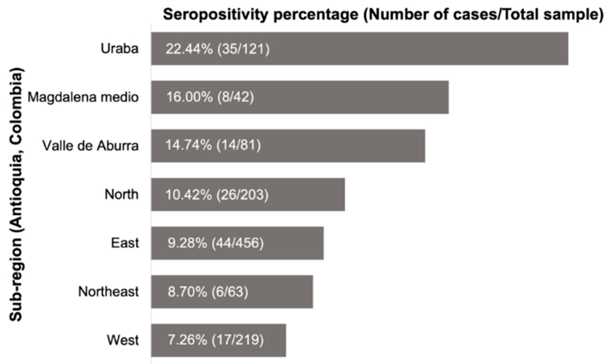
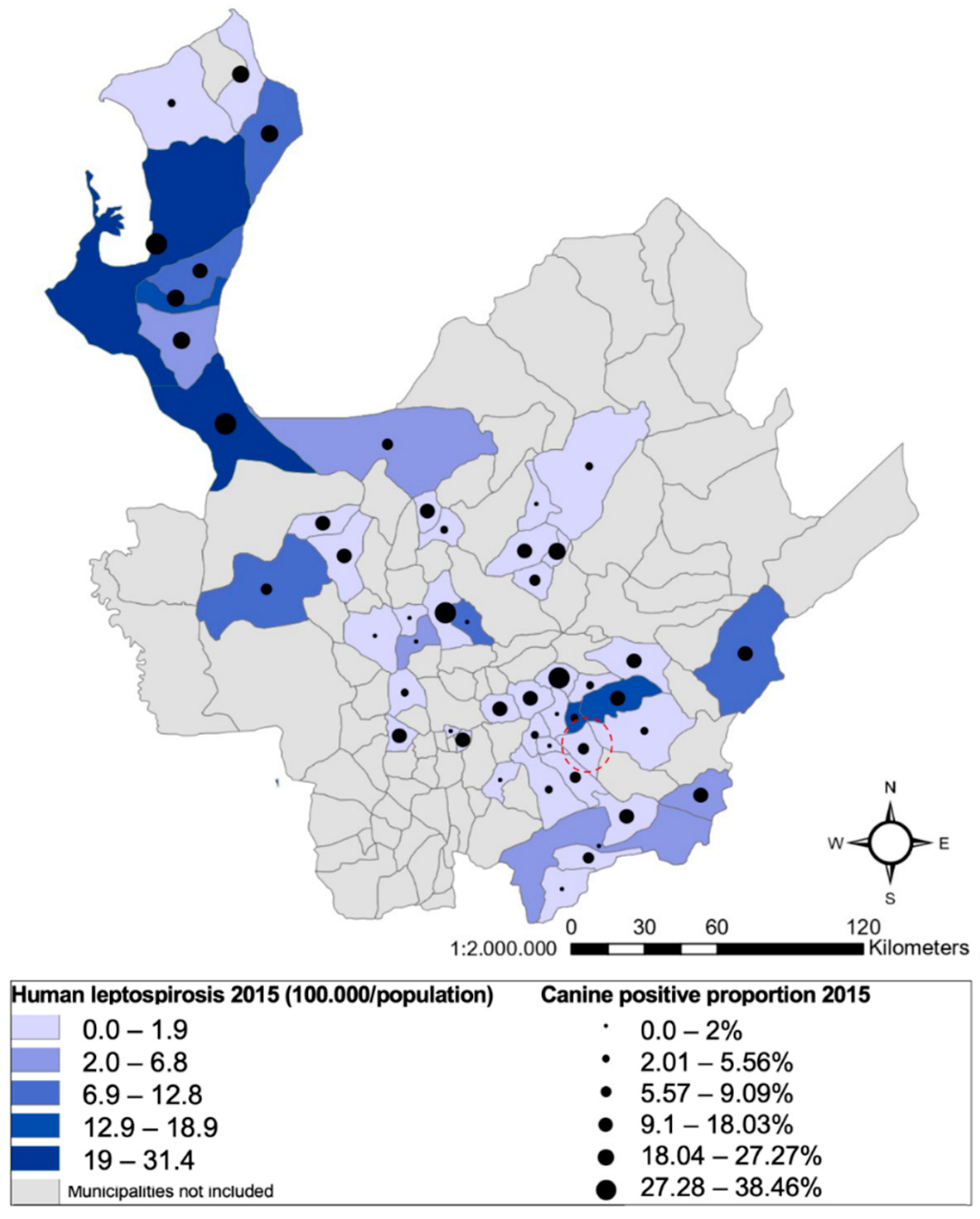
2. Results
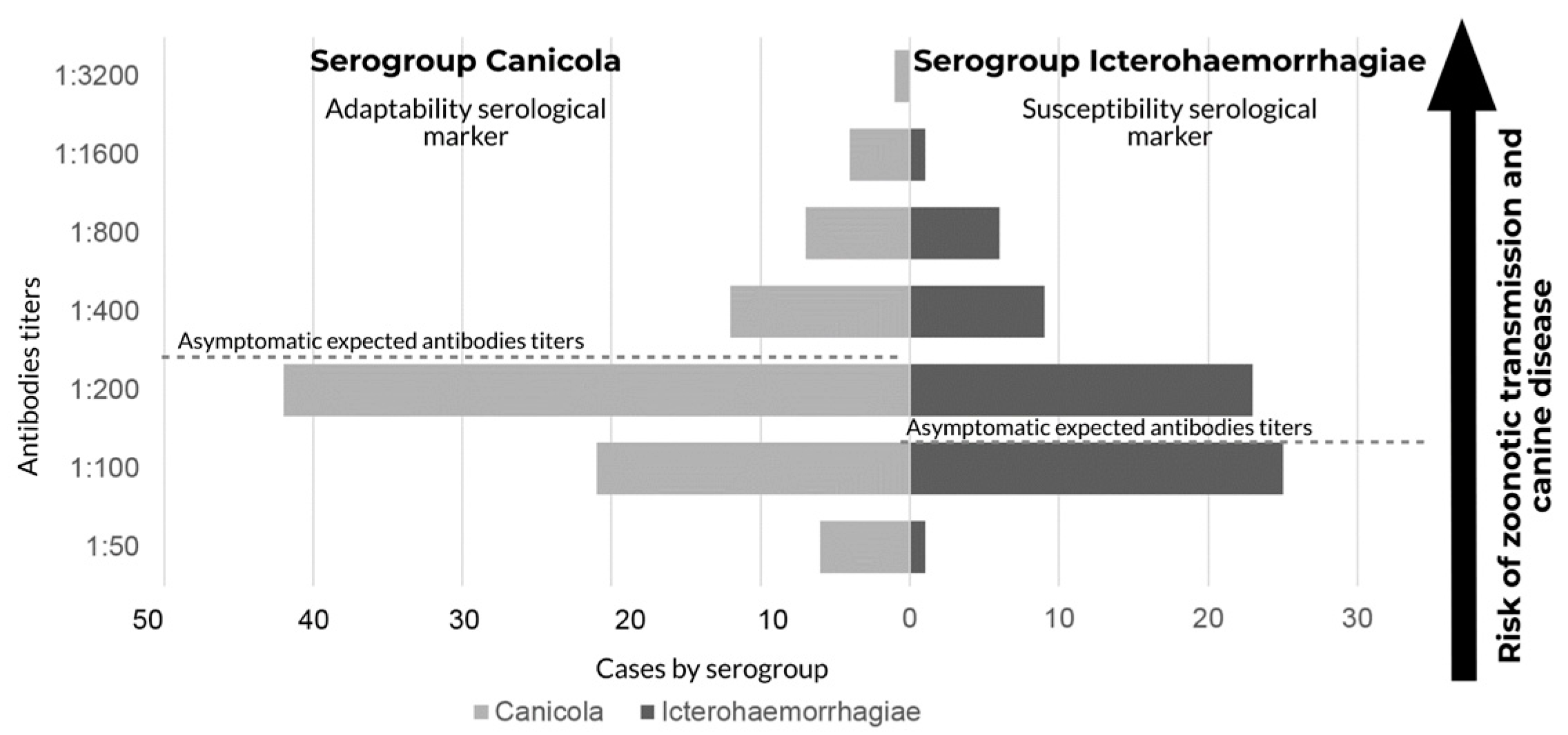
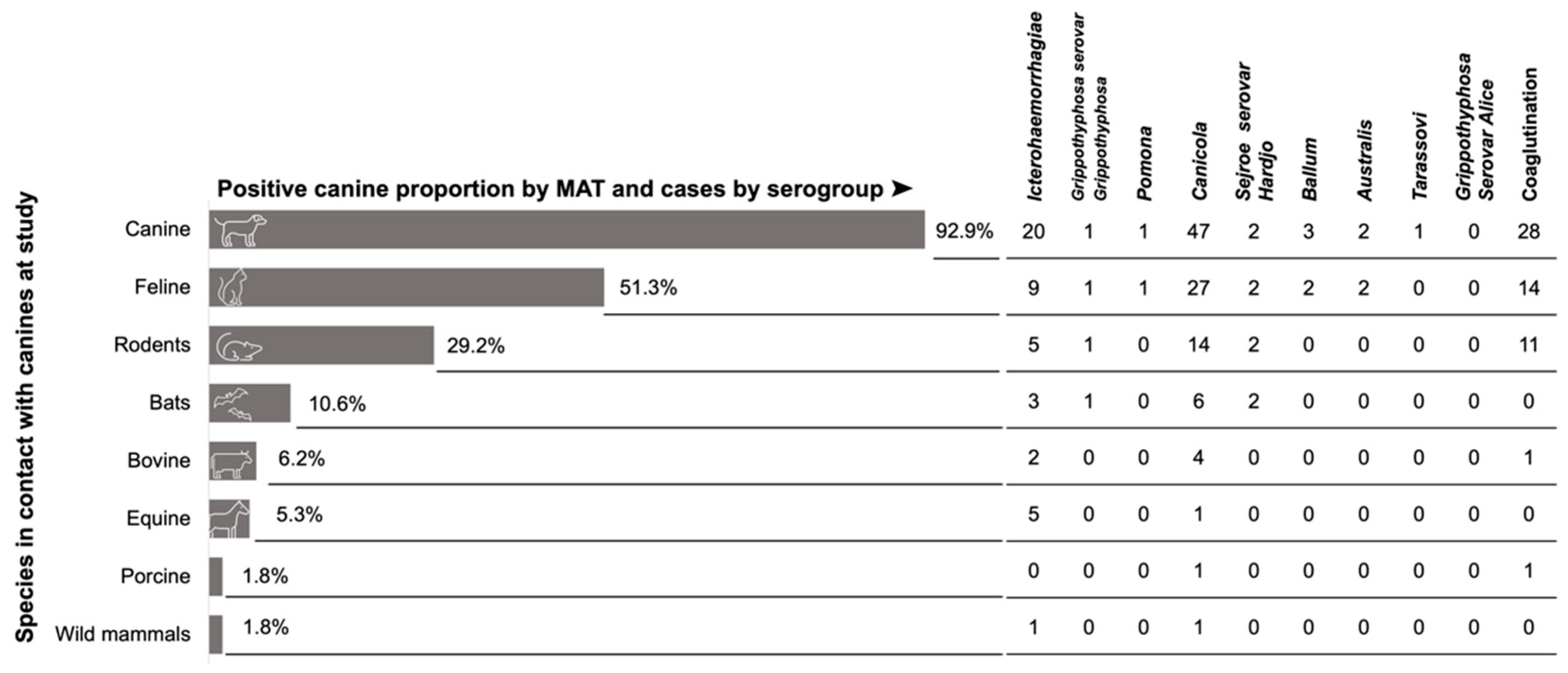
2.1. Serogroups Circulating
2.2. Molecular Characterization
2.3. Owners, Housing, and Dog Information
3. Discussion
4. Materials and Methods
4.1. Ethical Considerations
4.2. Study Area
4.3. Type of Study
4.4. Procedures
4.5. Microagglutination Test (MAT)
4.6. Molecular Characterization
4.7. Information Analysis
Author Contributions
Funding
Institutional Review Board Statement
Informed Consent Statement
Data Availability Statement
Acknowledgments
Conflicts of Interest
References
- Cilia, G.; Bertelloni, F.; Albini, S.; Fratini, F. Insight into the Epidemiology of Leptospirosis: A Review of Leptospira Isolations from “Unconventional” Hosts. Animals 2021, 11, 191. [Google Scholar] [CrossRef] [PubMed]
- Adler, B. (Ed.) Leptospira and Leptospirosis. In Current Topics in Microbiology and Immunology; Springer: Berlin/Heidelberg, Germany, 2015; Volume 387. [Google Scholar]
- Orr, B.; Westman, M.E.; Malik, R.; Purdie, A.; Craig, S.B.; Norris, J.M. Leptospirosis Is an Emerging Infectious Disease of Pig-Hunting Dogs and Humans in North Queensland. PLoS Negl. Trop. Dis. 2022, 16, e0010100. [Google Scholar] [CrossRef]
- Do Nascimento Benitez, A.; Monica, T.C.; Miura, A.C.; Romanelli, M.S.; Giordano, L.G.P.; Freire, R.L.; Mitsuka-Breganó, R.; Martins, C.M.; Biondo, A.W.; Serrano, I.M.; et al. Spatial and Simultaneous Seroprevalence of Anti-Leptospira Antibodies in Owners and Their Domiciled Dogs in a Major City of Southern Brazil. Front. Vet. Sci. 2021, 7, 580400. [Google Scholar] [CrossRef]
- Greene, C.E. (Ed.) Infectious Diseases of the Dog and Cat, 3rd ed.; Saunders/Elsevier: St. Louis, Mo, USA, 2006. [Google Scholar]
- Scanziani, E.; Origgi, F.; Giusti, A.M.; Iacchia, G.; Vasino, A.; Pirovano, G.; Scarpa, P.; Tagliabue, S. Serological Survey of Leptospiral Infection in Kennelled Dogs in Italy. J. Small Anim. Pract. 2002, 43, 154–157. [Google Scholar] [CrossRef] [PubMed]
- Suepaul, S.M.; Carrington, C.V.; Campbell, M.; Borde, G.; Adesiyun, A.A. Study on the Efficacy of Leptospira Vaccines Developed from Serovars Isolated from Trinidad and Comparison with Commercial Vaccines Using a Hamster Model. Vaccine 2010, 28, 5421–5426. [Google Scholar] [CrossRef] [PubMed]
- Ayral, F.C.; Bicout, D.J.; Pereira, H.; Artois, M.; Kodjo, A. Distribution of Leptospira Serogroups in Cattle Herds and Dogs in France. Am. J. Trop. Med. Hyg. 2014, 91, 756–759. [Google Scholar] [CrossRef] [PubMed]
- Bertasio, C.; Boniotti, M.B.; Lucchese, L.; Ceglie, L.; Bellinati, L.; Mazzucato, M.; Furlanello, T.; D’Incau, M.; Natale, A. Detection of New Leptospira Genotypes Infecting Symptomatic Dogs: Is a New Vaccine Formulation Needed? Pathogens 2020, 9, 484. [Google Scholar] [CrossRef]
- Romero, M.H.; Sánchez, J.A.; Hayek, L.C. Prevalencia de Anticuerpos Contra Leptospira En Población Urbana Humana y Canina Del Departamento Del Tolima. Rev. Salud Pública 2010, 12, 268–275. [Google Scholar] [CrossRef]
- Calderón, A.; Rodríguez, V.; Máttar, S.; Arrieta, G. Leptospirosis in Pigs, Dogs, Rodents, Humans, and Water in an Area of the Colombian Tropics. Trop. Anim. Health Prod. 2014, 46, 427–432. [Google Scholar] [CrossRef]
- Ensuncho-Hoyos, C.; Rodríguez-Rodríguez, V.; Pérez-Doria, A.; Vergara, O.; Calderón-Rangel, A. Epidemiology Behavior of Leptospirosis in Ciénaga de Oro, Córdoba (Colombia). Trop. Anim. Health Prod. 2017, 49, 1345–1351. [Google Scholar] [CrossRef]
- Cárdenas, N.C.; Infante, G.P.; Pacheco, D.A.R.; Diaz, J.P.D.; Wagner, D.C.M.; Dias, R.A.; Neto, J.S.F.; Amaku, M.; Vargas-Pinto, P.; Polo, L.; et al. Seroprevalence of Leptospira Spp Infection and Its Risk Factors among Domestic Dogs in Bogotá, Colombia. Vet. Anim. Sci. 2018, 6, 64–68. [Google Scholar] [CrossRef] [PubMed]
- Murcia, C.A.; Astudillo, M.; Romero, M.H. Prevalencia de Leptospirosis En Perros de Trabajo Vacunados y En Población Humana Con Riesgo Ocupacional. Biomédica 2020, 40 (Suppl. S1), 62–75. [Google Scholar] [CrossRef] [PubMed]
- Agudelo-Flórez, P.; Thiry, D.; Levett, P.N.; Romero-Vivas, C.M.E.; Falconar, A.K.I.; Cuello-Pérez, M. Cross-Sectional Study of Leptospira Seroprevalence in Humans, Rats, Mice, and Dogs in a Main Tropical Sea-Port City. Am. J. Trop. Med. Hyg. 2013, 88, 178–183. [Google Scholar] [CrossRef]
- Peláez Sanchez, R.G.; Lopez, J.Á.; Pereira, M.M.; Arboleda Naranjo, M.; Agudelo-Flórez, P. Genetic Diversity of Leptospira in Northwestern Colombia: First Report of Leptospira Santarosai as a Recognised Leptospirosis Agent. Memórias Inst. Oswaldo Cruz 2016, 111, 737–744. [Google Scholar] [CrossRef]
- Miotto, B.A.; Moreno, L.Z.; Guilloux, A.G.A.; de Sousa, G.O.; Loureiro, A.P.; Moreno, A.M.; Lilenbaum, W.; Vasconcellos, S.A.; Heinemann, M.B.; Hagiwara, M.K. Molecular and Serological Characterization of the First Leptospira Santarosai Strain Isolated from a Dog. Acta Trop. 2016, 162, 1–4. [Google Scholar] [CrossRef]
- Romero, M.H.; Astudillo, M.; Aguillón, D.M.; Lucio, I.D. Evidencia Serológica de Leptospirosis Canina En La Comunidad Indígena Kamentsá, Putumayo, Colombia. Rev. Investig. Vet. Perú 2018, 29, 625–634. [Google Scholar] [CrossRef]
- Castrillón Salazar, L.L.; López Diez, L.C.; Sanchez Nodarse, R.; Sanabria Gonzalez, W.; Henao, E.; Olivera Angel, M. Prevalencia de Presentación de Algunos Agentes Zoonóticos Transmitidos Por Caninos y Felinos En Medellín, Colombia. Rev. MVZ Córdoba 2018, 24, 7119–7126. [Google Scholar] [CrossRef]
- Agudelo-Flórez, P.; Restrepo-Jaramillo, B.N.; Arboleda-Naranjo, M. Situación de La Leptospirosis En El Urabá Antioqueño Colombiano: Estudio Seroepidemiológico y Factores de Riesgo En Población General Urbana. Cad. Saúde Pública 2007, 23, 2094–2102. [Google Scholar] [CrossRef]
- Yusti, D.; Arboleda, M.; Agudelo-Flórez, P. Factores de Riesgo Sociales y Ambientales Relacionados Con Casos de Leptospirosis de Manejo Ambulatorio y Hospitalario, Turbo-Colombia. Biomédica 2012, 33 (Suppl. S1), 117–129. [Google Scholar] [CrossRef]
- Pérez-García, J.; Arboleda, M.; Agudelo-Flórez, P. Leptospirosis Infantil En Pacientes Con Síndrome Febril En La Región de Urabá, Colombia. Rev. Peru. Med. Exp. Salud Pública 2016, 33, 775. [Google Scholar] [CrossRef] [Green Version]
- Carmona, S.; Arboleda, M.; Moreno, N.; Agudelo-Florez, P. Brote de Leptospirosis En Militares de La Fuerza Naval, Turbo, Antioquia, 2010. Rev. CES Med. 2010, 24, 111–112. [Google Scholar]
- Cortez, A.; dos Reis, E.A.; Gomes, N.; Souza-Filho, A.F.; Gonçales, A.P.; Pinto, C.M.; Onofrio, V.C.; Souza, G.O.; Guedes, I.B.; Lima, D.M.; et al. Leptospirose Canina Em Uma População Assintomática Da Região Sudoeste Do Estado de São Paulo, Brasil. Braz. J. Vet. Res. Anim. Sci. 2020, 57, e167893. [Google Scholar] [CrossRef]
- Miller, M.D.; Annis, K.M.; Lappin, M.R.; Lunn, K.F. Variability in Results of the Microscopic Agglutination Test in Dogs with Clinical Leptospirosis and Dogs Vaccinated against Leptospirosis: Canine Leptospirosis MAT Variability. J. Vet. Intern. Med. 2011, 25, 426–432. [Google Scholar] [CrossRef] [PubMed]
- Martin, L.E.R.; Wiggans, K.T.; Wennogle, S.A.; Curtis, K.; Chandrashekar, R.; Lappin, M.R. Vaccine-Associated Leptospira Antibodies in Client-Owned Dogs. J. Vet. Intern. Med. 2014, 28, 789–792. [Google Scholar] [CrossRef]
- Mesquita, M.O.; Trevilato, G.C.; da Silva Schons, M.; de Saraiva, L.H.; Rodrigues, R.O.; Corbellini, L.G. Percepções ambientais e fatores associados à ocorrência de anticorpos anti-Leptospira sp. em cães de um reassentamento urbano no município de Porto Alegre, estado do Rio Grande do Sul, Brasil. Rev. Pan-Amaz. Saúde 2017, 8, 23–27. [Google Scholar] [CrossRef]
- Felix, S.R.; de Oliveira Campello Felix, A.; Colonetti, K.; Seixas Neto, A.C.P.; Tillmann, M.T.; Vasconcellos, F.A.; de Oliveira Nobre, M.; Dellagostin, O.A.; da Silva, É.F. Canine Leptospirosis: An Overview of the City of Pelotas, Brazil. Res. Soc. Dev. 2020, 9, e5169108830. [Google Scholar] [CrossRef]


| Municipality | Reactive MAT | Not Reactive MAT | Sample by Municipality | Canine Population by Municipality | Proportion Sampled by Municipality | ||
|---|---|---|---|---|---|---|---|
| n | % | n | % | n | n | % | |
| Uraba region | |||||||
| Apartadó | 2 | 11.76% | 15 | 88.24% | 17 | 3369 | 0.50% |
| Arboletes | 3 | 20.00% | 12 | 80.00% | 15 | 2930 | 0.51% |
| Carepa | 3 | 27.27% | 8 | 72.73% | 11 | 1642 | 0.67% |
| Chigorodó | 4 | 25.00% | 12 | 75.00% | 16 | 4026 | 0.40% |
| Mutatá | 8 | 29.63% | 19 | 70.37% | 27 | 1280 | 2.11% |
| Necoclí | 2 | 6.67% | 28 | 93.33% | 30 | 3680 | 0.82% |
| San Pedro de Urabá | 2 | 25.00% | 6 | 75.00% | 8 | 2400 | 0.33% |
| Turbo | 11 | 34.38% | 21 | 65.63% | 32 | 4220 | 0.76% |
| Magdalena medio region | |||||||
| Puerto Berrio | 3 | 16.67% | 15 | 83.33% | 18 | 2375 | 0.76% |
| Puerto Triunfo | 5 | 15.63% | 27 | 84.38% | 32 | 1120 | 2.86% |
| Valle de Aburra region | |||||||
| Envigado | 14 | 17.50% | 66 | 82.50% | 80 | 7350 | 1.09% |
| Itagüí | 0 | 0.00% | 15 | 100.0% | 15 | 8690 | 0.17% |
| North region | |||||||
| Angostura | 3 | 11.54% | 23 | 88.46% | 26 | 1127 | 2.31% |
| Belmira | 6 | 46.15% | 7 | 53.85% | 13 | 933 | 1.39% |
| Campamento | 0 | 0.00% | 29 | 100.0% | 29 | 1130 | 2.57% |
| Carolina | 3 | 9.09% | 30 | 90.91% | 33 | 2020 | 1.63% |
| Entrerrios | 0 | 0.00% | 21 | 100.0% | 21 | 1024 | 2.05% |
| Guadalupe | 4 | 20.00% | 16 | 80.00% | 20 | 594 | 3.37% |
| Ituango | 1 | 6.25% | 15 | 93.75% | 16 | 1450 | 1.10% |
| San Andrés | 1 | 5.56% | 17 | 94.44% | 18 | 788 | 2.28% |
| Toledo | 2 | 12.50% | 14 | 87.50% | 16 | 375 | 4.27% |
| Eastern region | |||||||
| Alejandría | 1 | 2.86% | 34 | 97.14% | 35 | 594 | 5.89% |
| Argelia | 2 | 8.33% | 22 | 91.67% | 24 | 1010 | 2.38% |
| Cocorná | 4 | 12.50% | 28 | 87.50% | 32 | 936 | 3.42% |
| Concepción | 9 | 31.03% | 20 | 68.97% | 29 | 1100 | 2.64% |
| El Carmen | 2 | 5.41% | 35 | 94.59% | 37 | 2800 | 1.32% |
| El Peñol | 1 | 2.00% | 49 | 98.00% | 50 | 2669 | 1.87% |
| Granada | 2 | 8.33% | 22 | 91.67% | 24 | 600 | 4.00% |
| Guarne | 2 | 13.33% | 13 | 86.67% | 15 | 2815 | 0.53% |
| Guatapé | 1 | 3.57% | 27 | 96.43% | 28 | 843 | 3.32% |
| La Ceja | 0 | 0.00% | 19 | 100.0% | 19 | 2100 | 0.90% |
| Marinilla | 1 | 4.17% | 23 | 95.83% | 24 | 2825 | 0.85% |
| Nariño | 0 | 0.00% | 23 | 100.0% | 23 | 1250 | 1.84% |
| Santuario | 0 | 0.00% | 18 | 100.0% | 18 | 1800 | 1.00% |
| San Carlos | 2 | 5.56% | 34 | 94.44% | 36 | 1240 | 2.90% |
| San Francisco | 3 | 14.29% | 18 | 85.71% | 21 | 463 | 4.54% |
| San Rafael | 14 | 22.95% | 47 | 77.05% | 61 | 1410 | 4.33% |
| San Vicente | 6 | 16.22% | 31 | 83.78% | 37 | 1929 | 1.92% |
| Sonsón | 0 | 0.00% | 26 | 100.0% | 26 | 4357 | 0.60% |
| Northeast region | |||||||
| Anorí | 1 | 3.03% | 32 | 96.97% | 33 | 801 | 4.12% |
| San Roque | 5 | 13.89% | 31 | 86.11% | 36 | 1920 | 1.88% |
| West region | |||||||
| Armenia | 7 | 17.50% | 33 | 82.50% | 40 | 604 | 6.62% |
| Cañas Gordas | 3 | 14.29% | 18 | 85.71% | 21 | 1398 | 1.50% |
| Ebéjico | 1 | 4.35% | 22 | 95.65% | 23 | 2450 | 0.94% |
| Frontino | 3 | 7.32% | 38 | 92.68% | 41 | 1593 | 2.57% |
| Olaya | 0 | 0.00% | 22 | 100.0% | 22 | 570 | 3.86% |
| Santafé de Antioquia | 0 | 0.00% | 27 | 100.0% | 27 | 2050 | 1.32% |
| Sopetran | 0 | 0.00% | 48 | 100.0% | 48 | 1470 | 3.27% |
| Uramita | 3 | 25.00% | 9 | 75.00% | 12 | 890 | 1.35% |
| Total, municipalities | 150 | 11.24% | 1185 | 88.76% | 1335 | 97,010 | 1.38% |
| under study | |||||||
| No. | Species | Serogroup | Serovar | Strain | Number of Positives |
|---|---|---|---|---|---|
| 1 | L. interrogans | Icterohaemorrhagiae | Icterohaemorrhagiae | RGA | 43 |
| 2 | L. interrogans | Canicola | Canicola | Hond Utrecht IV | 76 |
| 3 | L. interrogans | Pomona | Pomona | Pomona | 8 |
| 4 | L. interrogans | Sejroe | Hardjo | Hardjoprajitno | 5 |
| 5 | L. interrogans | Grippothyposa | Grippothyposa | Moskva | 3 |
| 6 | L. borgpetersenii | Tarassovi | Tarassovi | Perepelitsin | 2 |
| 7 | L. borgpetersenii | Ballum | Castellonis | Castellon 3 | 6 |
| 8 | L. borgpetersenii | Australis | Bratislava | Jez Bratislava | 3 |
| 9 | L. santarosai | Grippothyposa | Alice | JET | 4 |
| Reactive MAT | Not Reactive MAT | Total † | PR (CI 95%) | p-Value | ||||
|---|---|---|---|---|---|---|---|---|
| n | % | n | % | n | % | |||
| Sex | ||||||||
| Male | 25 | 13.44% | 161 | 86.56% | 186 | 100% | 1.075 (0.79–1.63) | 0.4080 |
| Female | 84 | 12.50% | 588 | 87.50% | 672 | 100% | ||
| Breed | ||||||||
| Purebred | 25 | 11.52% | 192 | 88.48% | 217 | 100% | 0.85 (0.558–1.293) | 0.2610 |
| Cross | 81 | 13.57% | 516 | 86.43% | 597 | 100% | ||
| Housing area | ||||||||
| Rural | 44 | 13.54% | 281 | 86.46% | 325 | 100% | 1.014 (0.959–1.072) | 0.3540 |
| Urban | 54 | 12.36% | 383 | 87.64% | 437 | 100% | ||
| History of leptospirosis | ||||||||
| Yes | 0 | 0.00% | 2 | 100.00% | 2 | 100% | 0.7490 | |
| No | 102 | 13.51% | 653 | 86.49% | 755 | 100% | ||
| Vaccination against leptospirosis | ||||||||
| Yes | 3 | 4.48% | 64 | 95.52% | 67 | 100% | 0.318 (0.104–0.975) | 0.014 * |
| No | 102 | 14.09% | 622 | 85.91% | 724 | 100% | ||
| History of clinical signs | ||||||||
| Jaundice | ||||||||
| Yes | 1 | 20.00% | 4 | 80.00% | 5 | 100% | 1.504 (0.258–8.766) | 0.5120 |
| No | 98 | 13.30% | 639 | 86.70% | 737 | 100% | ||
| Hematuria | ||||||||
| Yes | 0 | 0.00% | 3 | 100.00% | 3 | 100% | 0.6530 | |
| No | 102 | 13.30% | 655 | 85.40% | 767 | 100% | ||
| Abortion | ||||||||
| Yes | 5 | 33.33% | 10 | 66.67% | 15 | 100% | 2.685 (1.272–5.668) | 0.034 * |
| No | 73 | 12.41% | 515 | 87.59% | 588 | 100% | ||
| Canine habits | ||||||||
| Most frequented place | ||||||||
| Interior | 64 | 14.71% | 371 | 85.29% | 435 | 100% | 1.37 (0.908–2.067) | 0.0800 |
| Exterior | 29 | 10.74% | 241 | 89.26% | 270 | 100% | ||
| Resting place | ||||||||
| Exterior | 43 | 11.50% | 331 | 88.50% | 374 | 100% | 0.77 (0.525–1.127) | 0.1080 |
| Interior | 49 | 14.94% | 279 | 85.06% | 328 | 100% | ||
| Reactive MAT | Not Reactive MAT | Total † | PR (CI 95%) | p-Value | ||||
|---|---|---|---|---|---|---|---|---|
| n | % | n | % | n | % | |||
| Roof materials | ||||||||
| Modern materials | 44 | 14.06% | 269 | 85.94% | 313 | 100% | 0.714 (0.408–1.48) | 0.164 |
| Traditional materials | 13 | 19.70% | 53 | 80.30% | 66 | 100% | ||
| Wall materials | ||||||||
| Modern materials | 45 | 15.15% | 252 | 84.85% | 297 | 100% | 1.010 (0.562–1.817) | 0.566 |
| Traditional materials | 12 | 15.00% | 68 | 85.00% | 80 | 100% | ||
| Floor materials | ||||||||
| Modern materials | 50 | 14.25% | 301 | 85.75% | 351 | 100% | 0.509 (0.258–1.003) | 0.066 |
| Traditional materials | 7 | 28.00% | 18 | 72.00% | 25 | 100% | ||
| History of flooding | ||||||||
| Yes | 5 | 27.78% | 13 | 72.22% | 18 | 100% | 1.670 (0.772–3.641) | 0.176 |
| No | 78 | 16.63% | 391 | 83.37% | 469 | 100% | ||
| Peridomiciliary housing | ||||||||
| Yes | 40 | 15.04% | 226 | 84.96% | 266 | 100% | 0.602 (0.347–1.043) | 0.064 |
| No | 13 | 25.00% | 39 | 75.00% | 52 | 100% | ||
| Peridomiciliary forest | ||||||||
| Yes | 12 | 23.53% | 39 | 76.47% | 51 | 100% | 1.532 (0.867–2.708) | 0.112 |
| No | 41 | 15.36% | 226 | 84.64% | 267 | 100% | ||
| Peridomiciliary crops | ||||||||
| Yes | 11 | 16.42% | 56 | 83.58% | 67 | 100% | 0.981 (0.535–1.800) | 0.558 |
| No | 42 | 16.73% | 209 | 83.27% | 251 | 100% | ||
| Peridomiciliary water | ||||||||
| Yes | 15 | 30.61% | 34 | 69.39% | 49 | 100% | 2.167 (1.296–3.624) | 0.006 * |
| No | 38 | 14.13% | 231 | 85.87% | 269 | 100% | ||
| Peridomiciliary Bare soil | ||||||||
| Yes | 16 | 34.78% | 30 | 65.22% | 46 | 100% | 2.557 (1.558–4.200) | 0.001 * |
| No | 37 | 13.60% | 235 | 86.40% | 272 | 100% | ||
| Peridomiciliary roads | ||||||||
| Yes | 21 | 23.60% | 68 | 76.40% | 89 | 100% | 1.689 (1.031–2.764) | 0.031 * |
| No | 32 | 13.97% | 197 | 86.03% | 229 | 100% | ||
| Source of drinking water | ||||||||
| Treated water | 71 | 15.71% | 381 | 84.29% | 452 | 100% | 1.055 (0.673–1.652) | 0.467 |
| Untreated water | 21 | 14.89% | 120 | 85.11% | 141 | 100% | ||
| Place of food preparation | ||||||||
| Outside of the house | 86 | 15.28% | 477 | 84.72% | 563 | 100% | 0.306 (0.134–0.696) | 0.052 |
| Inside the house | 3 | 50.00% | 3 | 50.00% | 6 | 100% | ||
| Municipal garbage collection service | ||||||||
| Yes | 72 | 15.55% | 391 | 84.45% | 463 | 100% | 0.855 (0.569–1.285) | 0.266 |
| No | 26 | 18.18% | 117 | 81.82% | 143 | 100% | ||
| Municipal sewer service | ||||||||
| Yes | 61 | 15.06% | 344 | 84.94% | 405 | 100% | 0.846 (0.577–1.240) | 0.231 |
| No | 34 | 17.80% | 157 | 82.20% | 191 | 100% | ||
Publisher’s Note: MDPI stays neutral with regard to jurisdictional claims in published maps and institutional affiliations. |
© 2022 by the authors. Licensee MDPI, Basel, Switzerland. This article is an open access article distributed under the terms and conditions of the Creative Commons Attribution (CC BY) license (https://creativecommons.org/licenses/by/4.0/).
Share and Cite
Perez-Garcia, J.; Monroy, F.P.; Agudelo-Florez, P. Canine Leptospirosis in a Northwestern Region of Colombia: Serological, Molecular and Epidemiological Factors. Pathogens 2022, 11, 1040. https://doi.org/10.3390/pathogens11091040
Perez-Garcia J, Monroy FP, Agudelo-Florez P. Canine Leptospirosis in a Northwestern Region of Colombia: Serological, Molecular and Epidemiological Factors. Pathogens. 2022; 11(9):1040. https://doi.org/10.3390/pathogens11091040
Chicago/Turabian StylePerez-Garcia, Janeth, Fernando P. Monroy, and Piedad Agudelo-Florez. 2022. "Canine Leptospirosis in a Northwestern Region of Colombia: Serological, Molecular and Epidemiological Factors" Pathogens 11, no. 9: 1040. https://doi.org/10.3390/pathogens11091040