Facile Synthesis, Characterization, and Adsorption Insights of Lanthanum Oxide Nanorods

 , ,
, ,
Abstract
:1. Introduction
2. Materials and Methods
2.1. Materials
2.2. Synthesis of La2O3 Nanorods
2.3. Analytical Methods
2.4. Adsorption Process
3. Results and Discussion
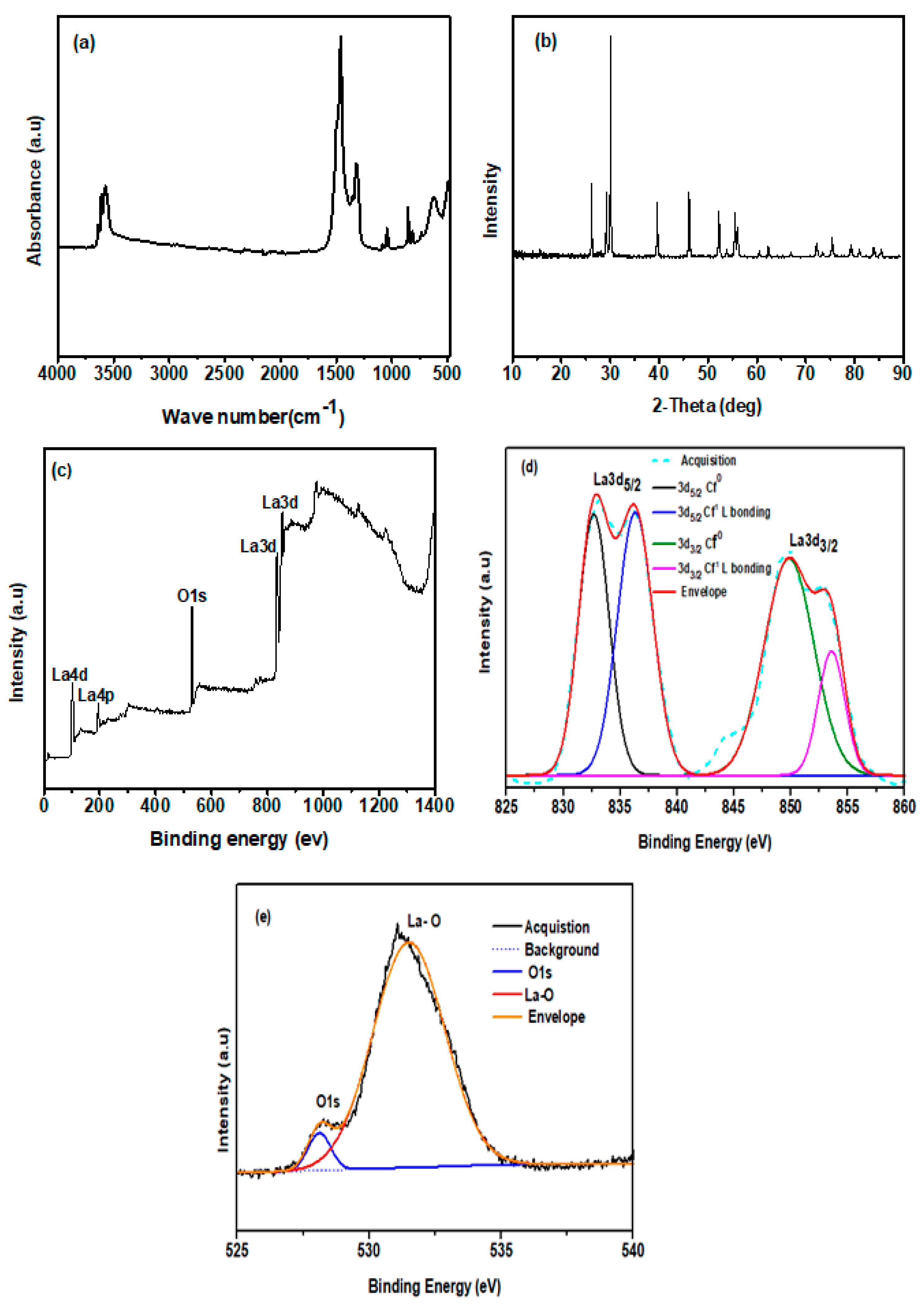
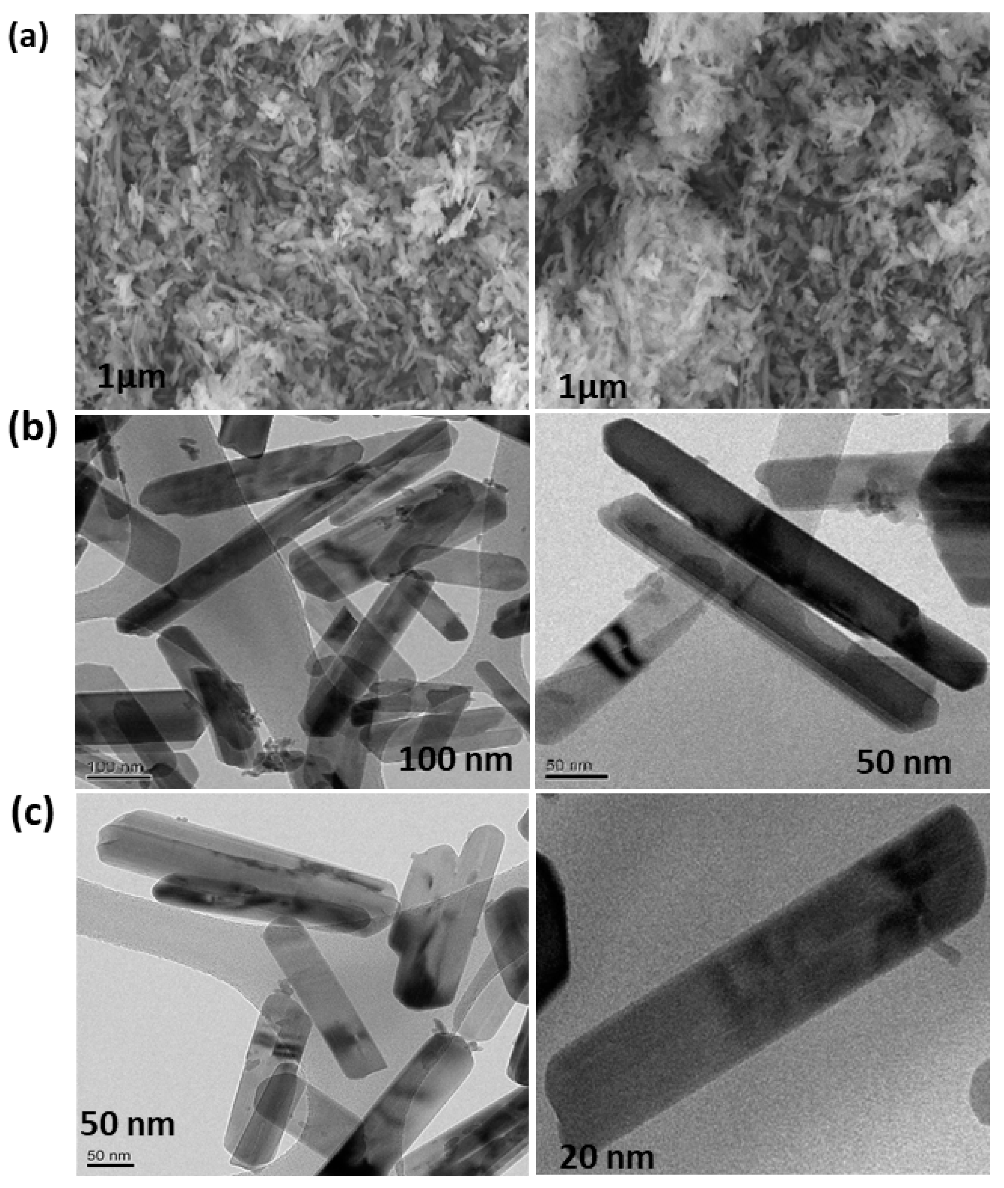
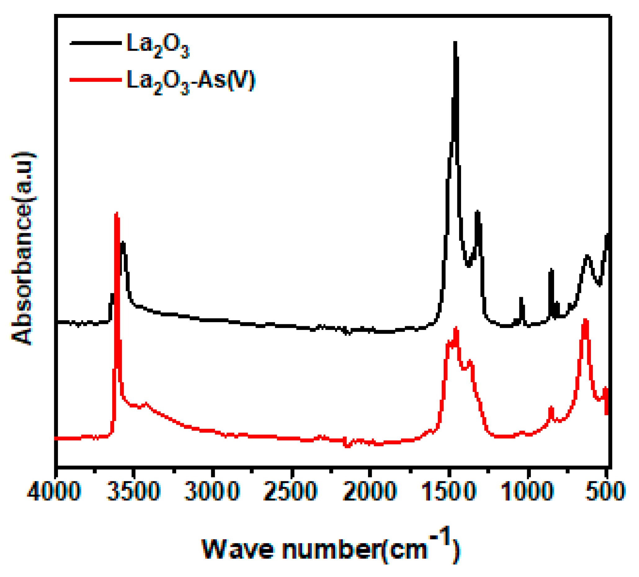
3.1. Characterization
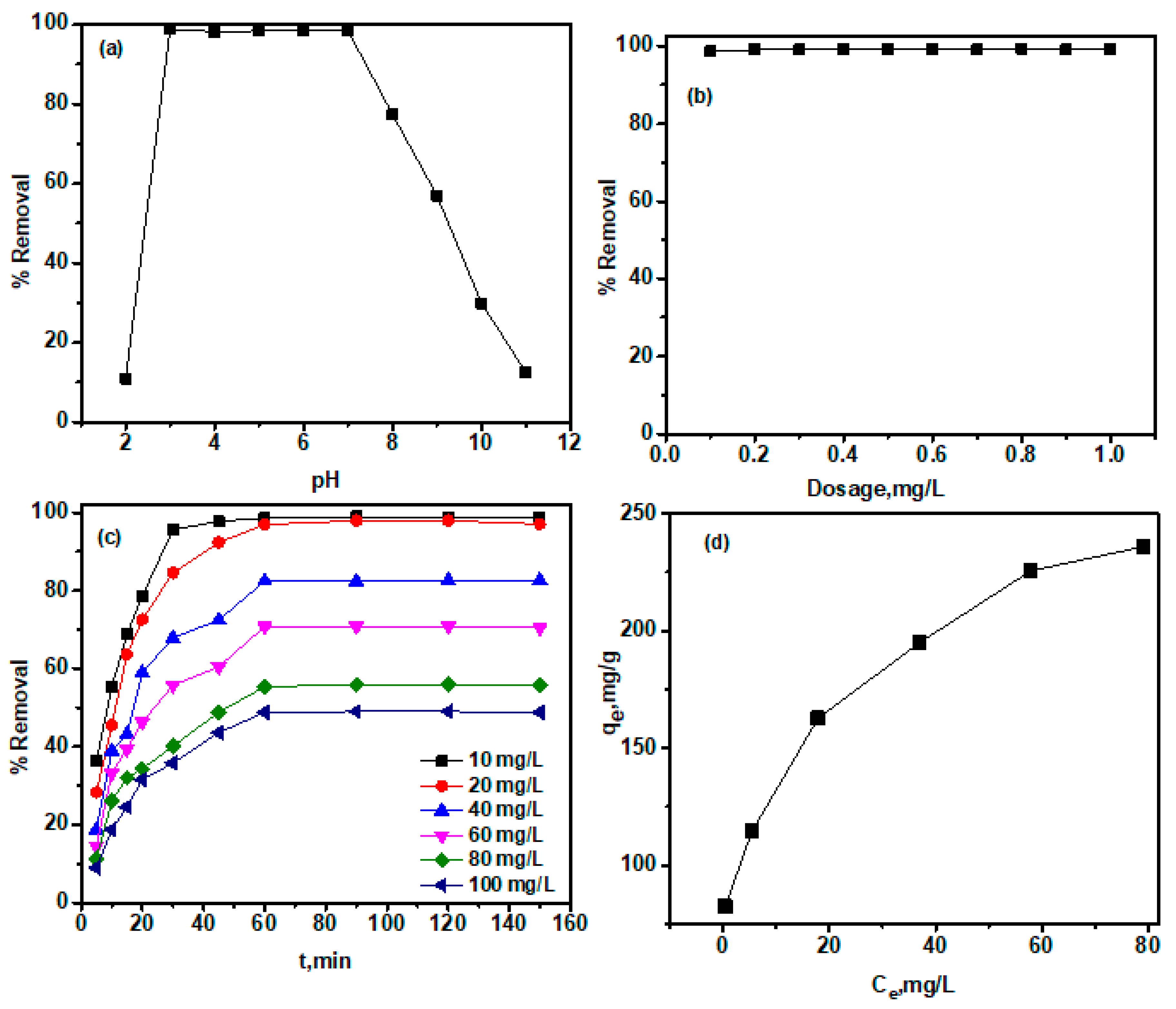
3.2. Adsorption Studies
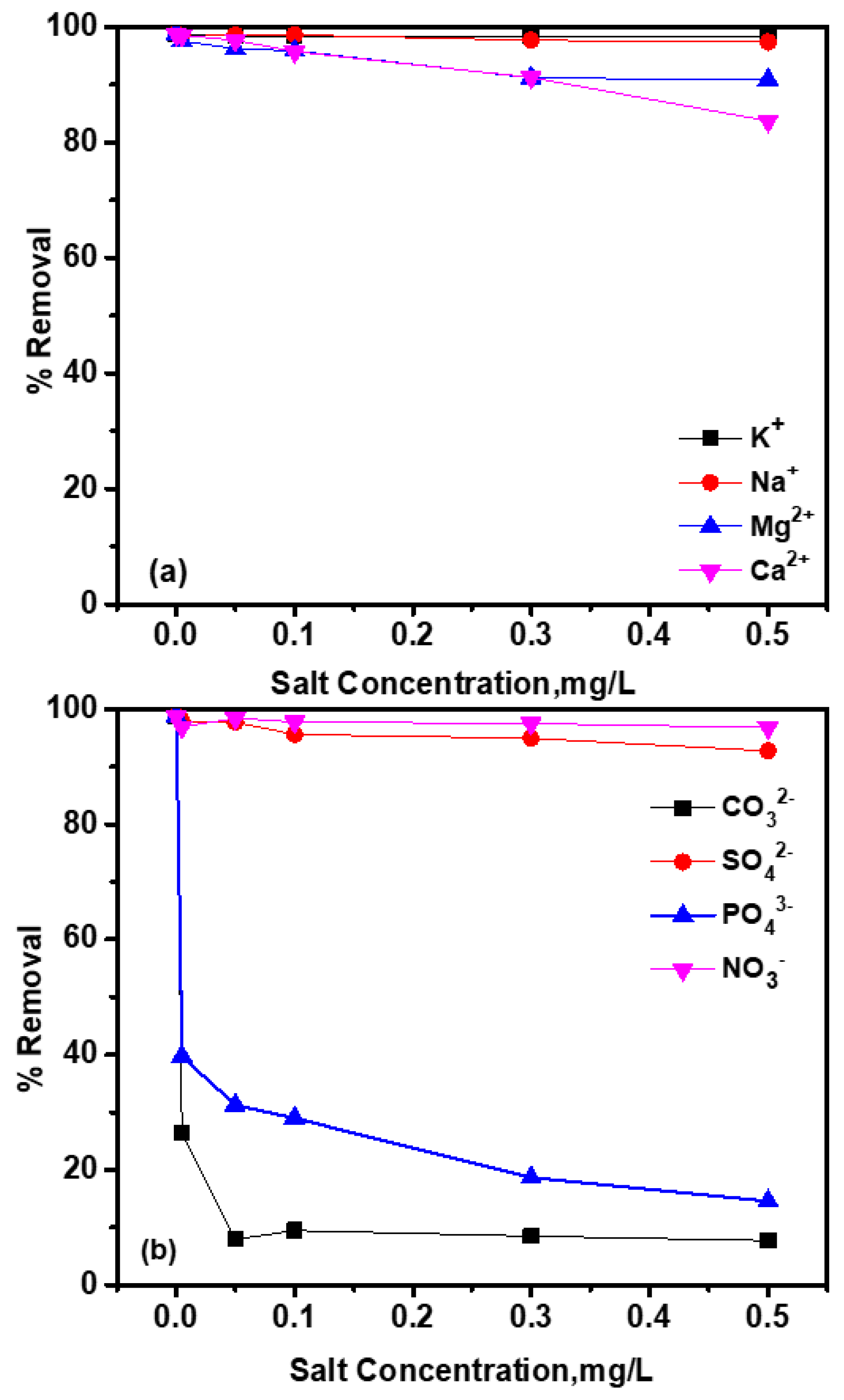
3.3. Effects of Coexisting Ions
3.4. Thermodynamic Parameters
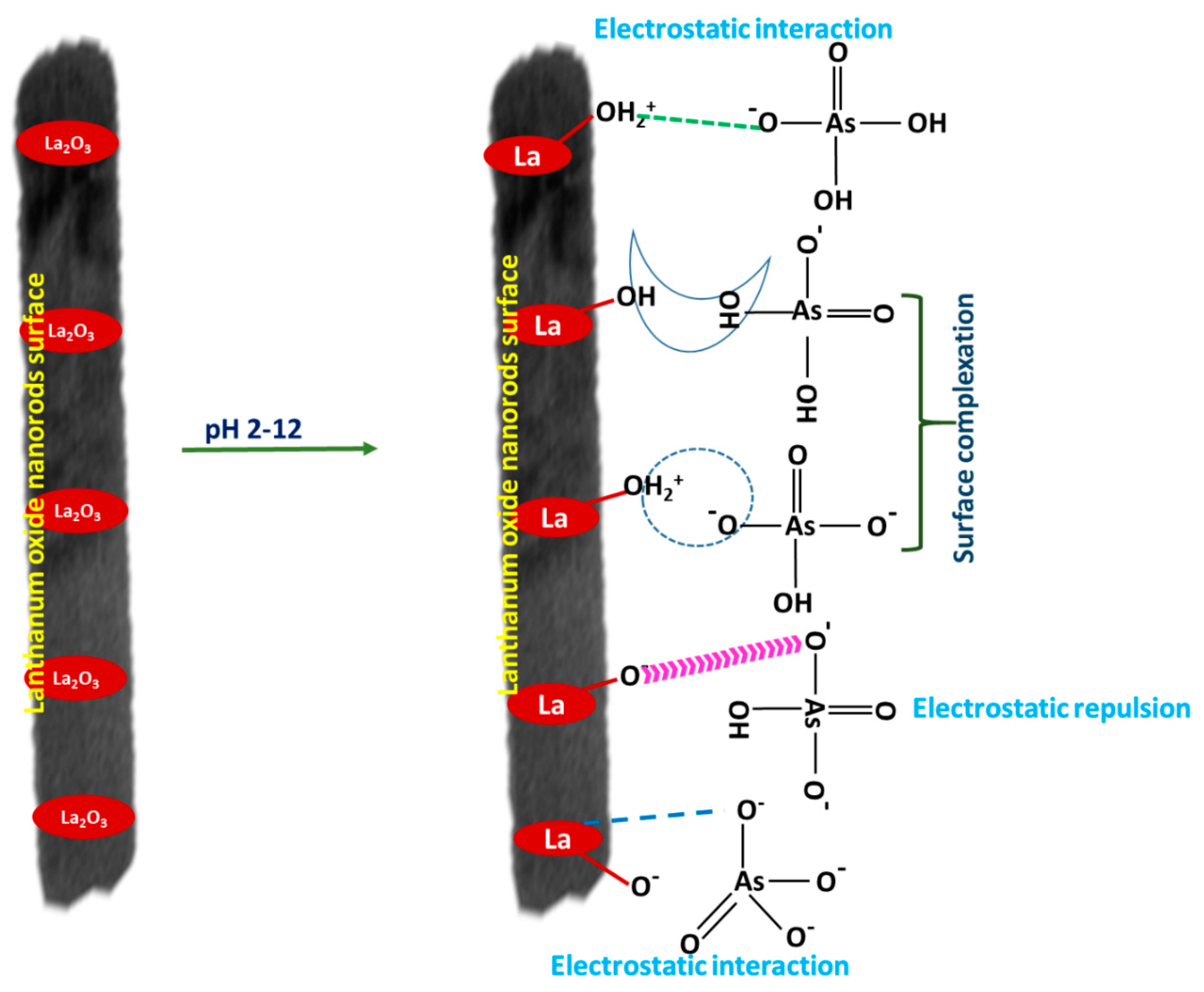
3.5. As(V) Adsorption Mechanism Using La2O3
4. Conclusions
Supplementary Materials
Author Contributions
Funding
Acknowledgments
Conflicts of Interest
References
- Boldish, S.I.; White, W.B. Vibrational spectra of crystals with the A-type rare earth oxide structure. La2O3 and Nd2O3. Spectrochim. Acta Part A 1979, 35, 1235. [Google Scholar] [CrossRef]
- Gouteron, J.; Michel, D.; Lejus, A.M.; Zarembowitch, J. Raman spectra of lanthanide sesquioxide single crystals: Correlation between A and B-type structures. J. Solid State Chem. 1981, 38, 288. [Google Scholar] [CrossRef]
- Kang, S.W.; Rhee, S.W. Deposition of La2O3 Films by Direct Liquid Injection Metallorganic Chemical Vapor Deposition. J. Electrochem. Soc. 2002, 149, 345. [Google Scholar] [CrossRef]
- De Asha, A.M.; Critchley, J.T.S.; Nix, R.M. Molecular adsorption characteristics of lanthanum oxide surfaces: The interaction of water with oxide overlayers grown on Cu. Surf. Sci. 1998, 405, 201. [Google Scholar] [CrossRef]
- Dakhel, A.A. Preparation and optical study of Au nanograins in amorphous La-oxide medium. Colloids Surf. A Physicochem. Eng. Asp. 2009, 332, 9–12. [Google Scholar] [CrossRef]
- Ferhi, M.; Horchani-Naifer, K.; Férid, M. Hydrothermal synthesis and photoluminescence of the monophosphate LaPO4:Eu(5%). J. Lumin. 2008, 128, 1777–1782. [Google Scholar] [CrossRef]
- Imanaka, N.; Okamoto, K.; Adachi, G.Y. Water-insoluble lanthanum oxychloride-based solid electrolytes with ultra-high chloride ion conductivity. Angew. Chem. Int. Ed. 2002, 41, 3890–3892. [Google Scholar] [CrossRef]
- Slagtern, Y.; Schuurman, C.; Leclercq, X.; Verykios, C. Mirodatos, Specific features concerning the mechanism of methane reforming by carbon dioxide over Ni/La2O3 catalyst. J. Catal. 1997, 172, 118–126. [Google Scholar] [CrossRef]
- Ordonez-Regil, E.; Drot, R.; Simoni, E.; Ehrhardt, J.J. Sorption of uranium (VI) onto lanthanum phosphate surfaces. Langmuir 2002, 18, 7977–7984. [Google Scholar] [CrossRef]
- Tokunaga, S.; Wasay, S.A.; Park, S.W. Removal of arsenic (V) ion from aqueous soloutions by lanthanum compounds. Water Sci. Technol. 1997, 35, 71–78. [Google Scholar] [CrossRef]
- Imanaka, N.; Okamoto, K.; Adachi, N.G. A chlorine gas sensor based on the combination of Mg2+ cation conducting and O2− anion conducting solid electrolytes with lanthanum oxychloride as an auxiliary electrode. Electrochem. Commun. 2001, 3, 49–51. [Google Scholar] [CrossRef]
- Moothedan, M.; Sherly, K.B. Synthesis, characterization and sorption studies of nano lanthanum oxide. J. Water Process Eng. 2016, 9, 29–37. [Google Scholar] [CrossRef]
- Yulin, L.; Jun, Y.; Xiaoci, L.; Huang, W.; Yu, T.; Zhang, Y. Combustion synthesis and stability of nanocrystalline La2O3 via ethanolamine-nitrate process. J. Rare Earths 2012, 30, 48–52. [Google Scholar]
- Keav, S.; Matam, S.K.; Ferri, D.; Weidenkaff, A. Structured perovskite-based catalysts and their application as three-way catalytic converters—A review. Catalysis 2014, 4, 226–255. [Google Scholar] [CrossRef] [Green Version]
- Zhao, C.; Zhao, C.Z.; Werner, M.; Taylor, S.; Chalker, P. Dielectric relaxation of high-k oxides. Nanoscale Res. Lett. 2013, 8, 1–12. [Google Scholar] [CrossRef] [Green Version]
- Tang, L.; Li, Y.M.; Xu, K.L.; Hou, X.D.; Lv, Y. Sensitive and selective acetone sensor based on its cataluminescence from nano-La2O3 surface. Sens. Actuators B 2008, 132, 243. [Google Scholar] [CrossRef]
- Cao, J.M.; Ji, H.M.; Liu, J.S.; Zheng, M.B.; Chang, X.; Ma, X.J.; Zhang, A.M.; Xu, Q.H. Controllable syntheses of hexagonal and lamellar mesostructured lanthanum oxide. Mater. Lett. 2005, 59, 408. [Google Scholar] [CrossRef]
- Wang, S.Y.; Wang, W.; Qian, Y.T. Preparation of La2O3 thin films by pulse ultrasonic spray pyrolysis method. Thin Solid Films 2000, 372, 50–53. [Google Scholar] [CrossRef]
- Imanaka, N.; Masui, T.; Kato, Y. Preparation of the cubic-type La2O3 phase by thermal decomposition of LaI3. J. Solid State Chem. 2005, 178, 395–398. [Google Scholar] [CrossRef]
- Sheng, J.; Zhang, S.; Lv, S.; Sun, W.D. Surfactant-assisted synthesis and characterization of lanthanum oxide nanostructures. J. Mater. Sci. 2007, 42, 9565–9571. [Google Scholar] [CrossRef]
- Wu, Q.Z.; Shen, Y.; Liao, J.F.; Li, Y.G. Synthesis and characterization of three-dimensionally ordered macroporous rare earth oxides. Mater. Lett. 2004, 58, 2688–2691. [Google Scholar] [CrossRef]
- Hu, C.G.; Liu, H.; Dong, W.T.; Zhang, Y.Y.; Bao, G.; Lao, C.S.; Wang, Z.L. La(OH)3 and La2O3 nanobelts-synthesis and physical properties. Adv. Mater. 2007, 19, 470–474. [Google Scholar] [CrossRef]
- Bazzi, R.; Flores-Gonzalez, M.A.; Louis, C.; Lebbou, K.; Dujardin, C.; Brenier, A.; Zhang, W.; Tillement, O.; Bernstein, E.; Perriat, P. Synthesis and luminescent properties of sub-5-nm lanthanide oxides nanoparticles. J. Lumin. 2003, 102–103, 445–450. [Google Scholar] [CrossRef]
- Vadivel Murugan, A.; Navale, S.C.; Ravi, V. Synthesis of nano-crystalline La2O3 powder at 100 °C. Mater. Lett. 2006, 60, 848–849. [Google Scholar] [CrossRef]
- Lingamdinne, L.P.; Choi, Y.L.; Kim, I.S.; Chang, Y.Y.; Koduru, J.R.; Yang, J.K. Porous graphene oxide based inverse spinel nickel ferrite nanocomposites for the enhanced adsorption removal of arsenic. RSC Adv. 2016, 6, 73776–73789. [Google Scholar] [CrossRef]
- Tuutijärvi, T.; Lu, J.; Sillanpää, M.; Chen, G. As (V) adsorption on maghemite nanoparticles. J. Hazard. Mater. 2009, 66, 1415–1420. [Google Scholar] [CrossRef] [PubMed]
- Chen, S.L.; Dzeng, S.R.; Yang, M.H.; Chiu, K.H.; Shieh, G.M.; Wai, C.M. Arsenic species in groundwaters of the black foot disease area, Taiwan. Environ. Sci. Technol. 1994, 28, 877–881. [Google Scholar] [CrossRef]
- Karim, M. Arsenic in groundwater and health problems in Bangladesh. Water Res. 2000, 34, 304–310. [Google Scholar] [CrossRef]
- Chakraborti, D.; Rahman, M.M.; Paul, K.; Chowdhury, U.K.; Sengupta, M.K.; Lodh, D.; Chanda, C.R.; Saha, K.C.; Mukherjee, S.C. Arsenic calamity in the Indian subcontinent. What lessons have been learned? Talanta 2002, 58, 3–22. [Google Scholar] [CrossRef]
- Khosrow-pour, F.; Aghazadeh, M.; Sabour, B.; Dalvand, S. Large-scale synthesis of uniform lanthanum oxide nanowires via template-free deposition followed by heat-treatment. Ceram. Int. 2013, 39, 9491–9498. [Google Scholar] [CrossRef]
- Misra, M.; Deba, C.N. Process for Removal of Selenium and Arsenic from Aqueous Streams. U.S. Patent 5,603,838, 18 February 1997. [Google Scholar]
- Davis, S.A.; Misra, M. Transport model for the adsorption of oxyanions of selenium (IV) and arsenic (V) from water onto lanthanum-and aluminum-based oxides. J. Colloid Interface Sci. 1997, 188, 340–350. [Google Scholar] [CrossRef]
- Karthikeyan, S.; Selvapandiyan, M. Effect of annealing temperature on the properties of lanthanum oxide (La2O3) nanoplates by reflux routes. Int. J. Eng. Sci. Res. Technol. 2018, 7, 559. [Google Scholar]
- Mu, Q.; Wang, Y. Synthesis, characterization, shape-preserved transformation, and optical properties of La(OH)3, La2O2CO3, and La2O3 nanorods. J. Alloys Compd. 2011, 509, 396–401. [Google Scholar] [CrossRef]
- Xiao, Y.; Zongyu, F.; Xiaowei, H.; Li, H.; Zhiqi, L.; Qiang, W.; Yongke, H. Synthesis of lanthanum oxide nanosheets by a green carbonation process. Chin. Sci. Bull. 2014, 59, 1864–1867. [Google Scholar] [CrossRef]
- Wang, K.; Wu, Y.; Li, H.; Li, M.; Guan, F.; Fan, H. A hybrid antioxidizing and antibacterial material based on Ag–La2O3 nanocomposites. J. Inorg. Biochem. 2014, 141, 36–42. [Google Scholar] [CrossRef]
- Hu, S.; Chi, B.; Pu, J.; Jian, L. Novel heterojunction photocatalysts based on lanthanum titanate nanosheets and indium oxide nanoparticles with enhanced photocatalytic hydrogen production activity. J. Mater. Chem. A 2014, 2, 19260–19267. [Google Scholar] [CrossRef]
- Sanivarapu, S.R.; Lawrence, J.B.; Sreedhar, G. Role of surface oxygen vacancies and lanthanide contraction phenomenon of Ln(OH)3 (Ln= La, Pr, and Nd) in sulfide-mediated photoelectrochemical water splitting. ACS Omega 2018, 3, 6267–6278. [Google Scholar] [CrossRef]
- Sunding, M.F.; Hadidi, K.; Diplas, S.; Løvvik, O.M.; Norby, T.E.; Gunnæs, A.E. XPS Characterization of In Situ Treated Lanthanum Oxide and Hydroxide Using Tailored Referencing and Peak Fitting Procedures. J. Electron Spectrosc. Relat. Phenom. 2011, 184, 399–409. [Google Scholar] [CrossRef]
- Chen, B.; Zhu, Z.; Ma, J.; Qiu, Y.; Chen, J. Surfactant assisted Ce–Fe mixed oxide decorated multiwalled carbon nanotubes and their arsenic adsorption performance. J. Mater. Chem. A 2013, 1, 11355–11367. [Google Scholar] [CrossRef] [Green Version]
- Sikder, M.T.; Tanaka, S.; Saito, T.; Kurasaki, M. Application of zero-valent iron impregnated chitosan-caboxymethyl β-cyclodextrin composite beads as arsenic sorbent. J. Environ. Chem. Eng. 2014, 2, 370–376. [Google Scholar] [CrossRef]
- Kyzas, G.Z.; Siafaka, P.I.; Kostoglou, M.; Bikiaris, D.N. Adsorption of As(III) and As(V) onto colloidal microparticles of commercial cross-linked polyallylamine (sevelamer) from single and binary ion solutions. J. Colloid Interface Sci. 2016, 474, 137–145. [Google Scholar] [CrossRef] [PubMed]
- Panda, A.P.; Rout, P.; Sanjukta, A.K.; Jha, U.; Swain, S.K. Enhanced performance of a core–shell structured Fe@Fe oxide and Mn@Mn oxide (ZVIM) nanocomposite towards remediation of arsenic contaminated drinking water. J. Mater. Chem. A 2020, 8, 4318–4333. [Google Scholar] [CrossRef]
- Shi, Q.; Yan, L.; Chan, T.; Jing, C. Arsenic adsorption on lanthanum-impregnated activated alumina: Spectroscopic and DFT study. ACS Appl. Mater. Interfaces 2015, 7, 26735–26741. [Google Scholar] [CrossRef] [PubMed]
- Jang, M.; Park, J.K.; Shin, E.W. Lanthanum functionalized highly ordered mesoporous media: Implications of arsenate removal. Microporous Mesoporous Mater. 2004, 75, 159–168. [Google Scholar] [CrossRef] [Green Version]
- Wang, C.; Luo, H.; Zhang, Z.; Wu, Y.; Zhang, J.; Chen, S. Removal of As (III) and As (V) from aqueous solutions using nanoscale zero valent iron-reduced graphite oxide modified composites. J. Hazard. Mater. 2014, 268, 124–131. [Google Scholar] [CrossRef]
- Yu, F.; Sun, S.; Ma, J.; Han, S. Enhanced removal performance of arsenate and arsenite by magnetic graphene oxide with high iron oxide loading. Phys. Chem. Chem. Phys. 2015, 17, 4388–4397. [Google Scholar] [CrossRef]
- Sigdel, A.; Park, J.W.; Kwak, H.; Park, P.K. Arsenic removal from aqueous solutions by adsorption onto hydrous iron oxide-impregnated alginate beads. J. Ind. Eng. Chem. 2016, 35, 277–286. [Google Scholar] [CrossRef]
- Huang, J.Y.; Wu, Z.W.; Chen, L.W.; Sun, Y.B. The sorption of Cd (II) and U (VI) on sepiolite: A combined experimental and modeling studies. J. Mol. Liq. 2015, 209, 706–712. [Google Scholar] [CrossRef]


| Metal Concentration, mg/L | qe, Th, mg/g | PFO | PSO | ||||
|---|---|---|---|---|---|---|---|
| qe, Cal, mg/g | k1 | R2 | qe, Cal, mg/g | k2 | R2 | ||
| 10 | 86.98 | 34.57 | 0.0529 | 0.801 | 84.56 | 0.00215 | 0.999 |
| 20 | 118.98 | 88.97 | 0.0370 | 0.753 | 119.56 | 0.00049 | 0.999 |
| 40 | 145.68 | 116.97 | 0.0198 | 0.867 | 142.56 | 0.00027 | 0.998 |
| 60 | 185.69 | 135.69 | 0.0396 | 0.784 | 186.31 | 0.00018 | 0.995 |
| 80 | 205.69 | 158.95 | 0.0069 | 0.472 | 203.56 | 0.00021 | 0.991 |
| 100 | 245.68 | 185.98 | 0.0313 | 0.589 | 244.56 | 0.00044 | 0.992 |
| Temperature, °C | Langmuir | Freundlich | Temkin | |||||||||
|---|---|---|---|---|---|---|---|---|---|---|---|---|
| qmax, mg/g | KL | R2 | χ2 | KF,mg/g*(L/mg)1/n | n | R2 | χ2 | B | KT | R | χ2 | |
| 25 | 260.56 | 1.88 | 0.995 | 2.16 | 185.05 | 5.30 | 0.945 | 9.87 | 98.60 | 127.96 | 0.971 | 13.45 |
| 35 | 285.98 | 2.56 | 0.999 | 1.65 | 135.08 | 6.88 | 0.917 | 12.69 | 99.88 | 212.93 | 0.948 | 18.97 |
| 50 | 305.69 | 3.12 | 0.998 | 1.98 | 131.05 | 4.06 | 0.963 | 20.76 | 56.87 | 306.12 | 0.909 | 17.89 |
| Type of Adsorbent | qmax, (mg/g) | Conditions | Reference |
|---|---|---|---|
| Initial Concentration, Dosage and pH | |||
| Reduced graphene oxide-nickel ferrite (RGONF) nanocomposite | 106.40 | 2–30 mg/L, 0.3 g/L and pH 6.5 | [25] |
| Ce–Fe oxide decorated multiwalled carbon nanotubes | 30.96 | 1–20 mg/L, 0.2 g/L and pH 4.0. | [40] |
| Sevelamer | 133.00 | 0–250 mg/L, 1 g/L and pH 6 | [42] |
| Core–shell structured Fe(0)@Fe oxide and Mn(0)@Mn oxide (ZVIM) nanocomposite | 101.95 | 0.2–1.0 mg/L, 0.2 g/L and pH 6.5 | [43] |
| Lanthanum-impregnated activated alumina | 26.30 | 0–250 mg/L, 1.0 g/L and pH 7.0 | [44] |
| Lanthanum-impregnated SBA-15 | 123.70 | 20–60 mg/L and pH 7.2 | [45] |
| La2O3 | 260.56 | 10–100 mg/L, 0.1 g/L and pH 6.0 | This work |
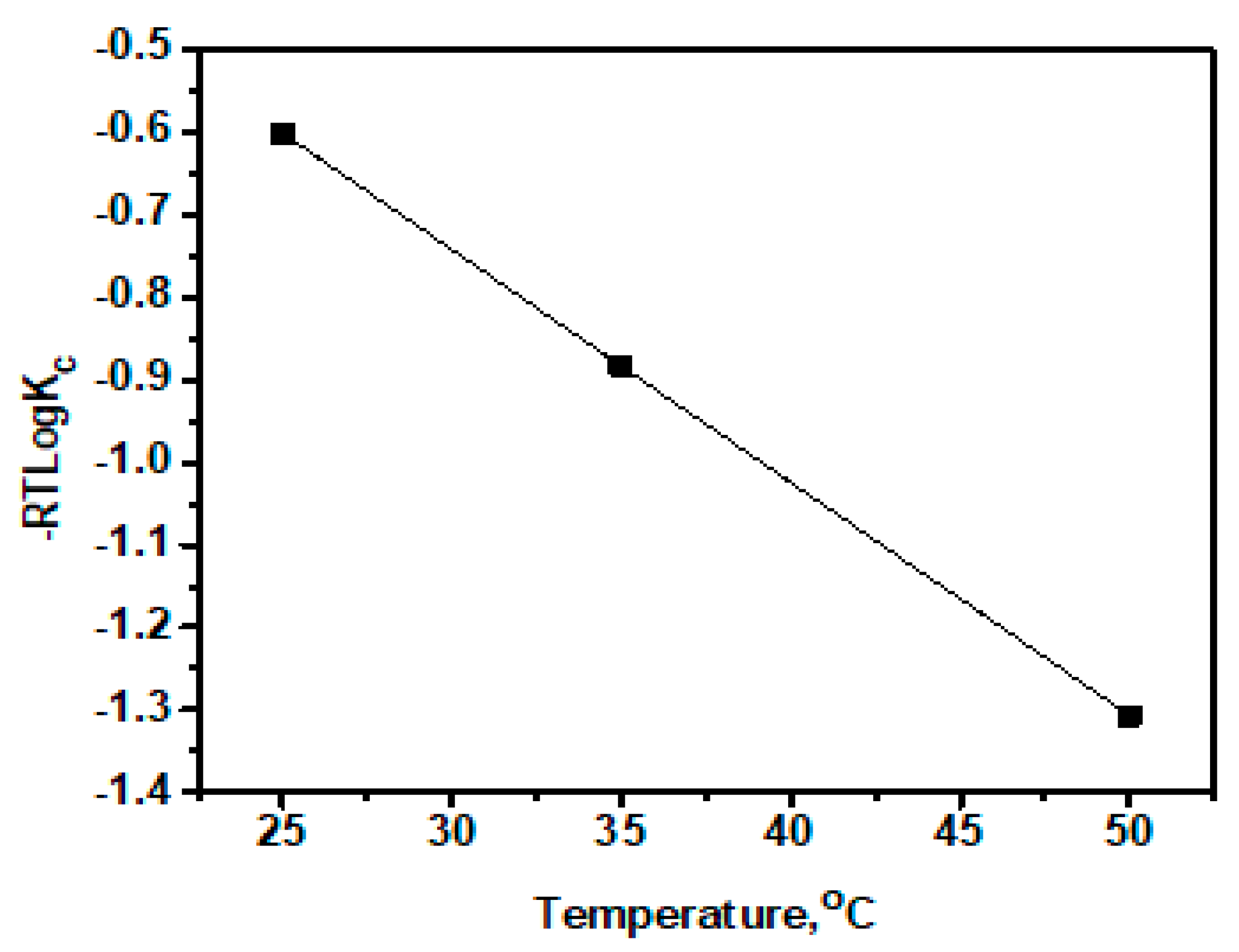
| Temperature, °C | log Kc | |||
|---|---|---|---|---|
| 25 | −601.291 | 103.93 | 28.20 | 2.892 |
| 35 | −883.134 | 3.034 | ||
| 50 | −1306.400 | 3.142 |
© 2020 by the authors. Licensee MDPI, Basel, Switzerland. This article is an open access article distributed under the terms and conditions of the Creative Commons Attribution (CC BY) license (http://creativecommons.org/licenses/by/4.0/).
Share and Cite
Lingamdinne, L.P.; Koduru, J.R.; Chang, Y.-Y.; Kang, S.-H.; Yang, J.-K. Facile Synthesis, Characterization, and Adsorption Insights of Lanthanum Oxide Nanorods. Metals 2020, 10, 1001. https://doi.org/10.3390/met10081001
Lingamdinne LP, Koduru JR, Chang Y-Y, Kang S-H, Yang J-K. Facile Synthesis, Characterization, and Adsorption Insights of Lanthanum Oxide Nanorods. Metals. 2020; 10(8):1001. https://doi.org/10.3390/met10081001
Chicago/Turabian StyleLingamdinne, Lakshmi Prasanna, Janardhan Reddy Koduru, Yoon-Young Chang, Seon-Hong Kang, and Jae-Kyu Yang. 2020. "Facile Synthesis, Characterization, and Adsorption Insights of Lanthanum Oxide Nanorods" Metals 10, no. 8: 1001. https://doi.org/10.3390/met10081001