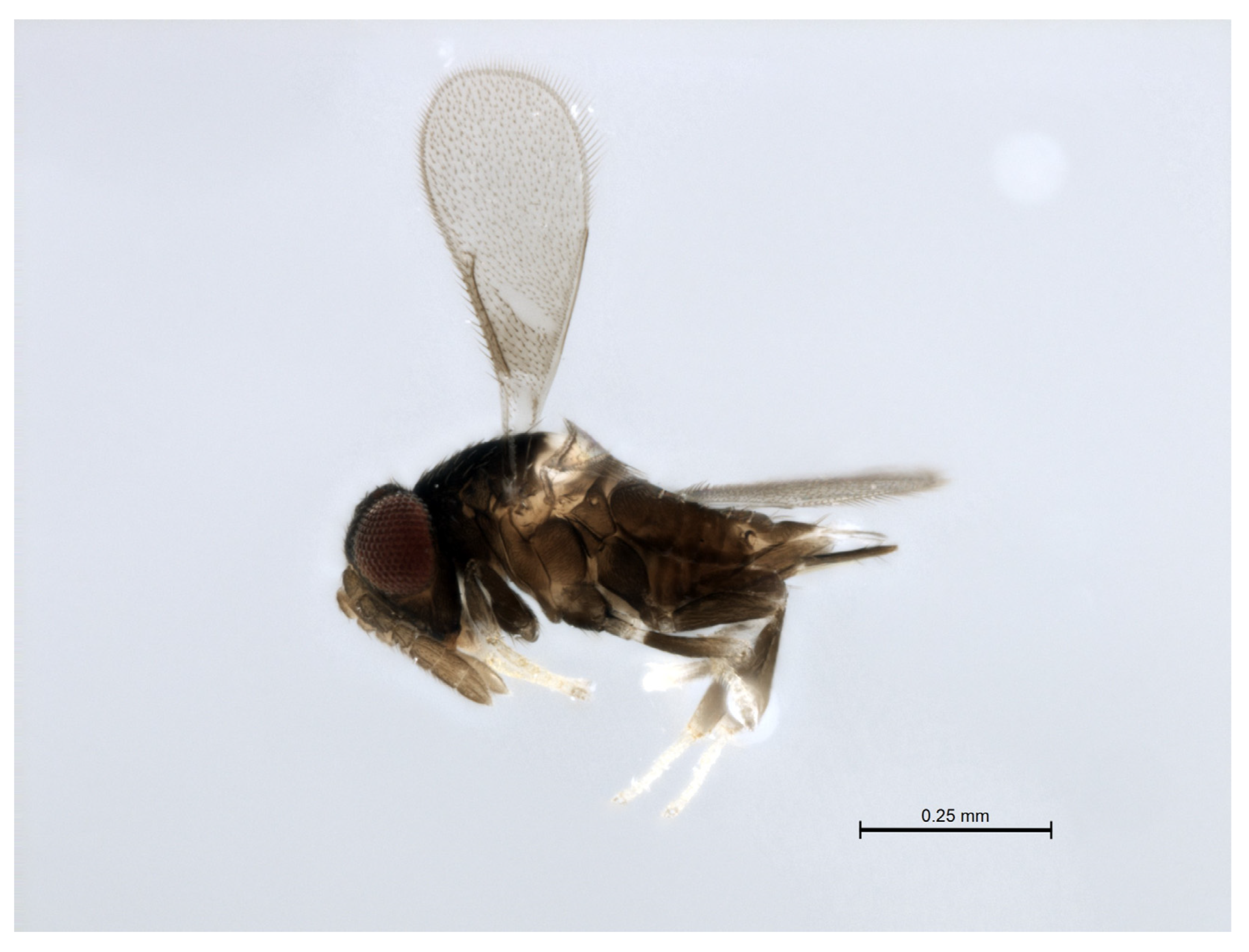
3.3. Species Descriptions
3.3.1. Encarsia acusa Polaszek and Hernández-Suárez sp. n.
urn:lsid:zoobank.org:act:F179717C-0FF0-4189-90BE-4851A8625431
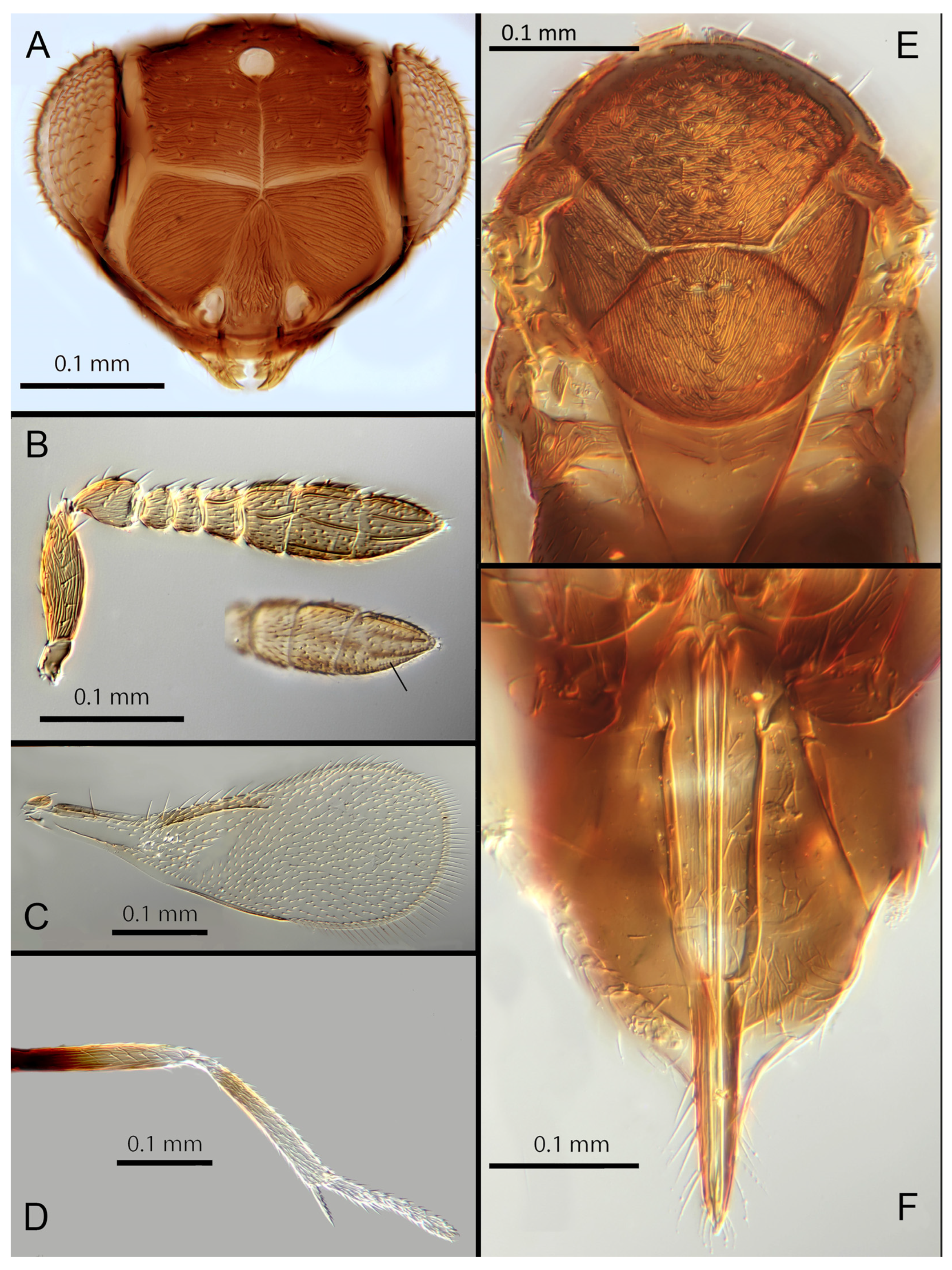
Female. Colour: Antennae light brown; radicle, scape, base of pedicel and F6 darker. Head dark brown, paler along the sutures and frons. Mesosoma and metasoma dark brown with posterior 80% of the mesoscutellum and sides of metanotum yellow. Legs yellow with most of the mid and hind femora brown, fore femora and tibiae brown, all tarsi pale. Fore wings hyaline, slightly infuscate below marginal vein, submarginal and marginal veins dark.
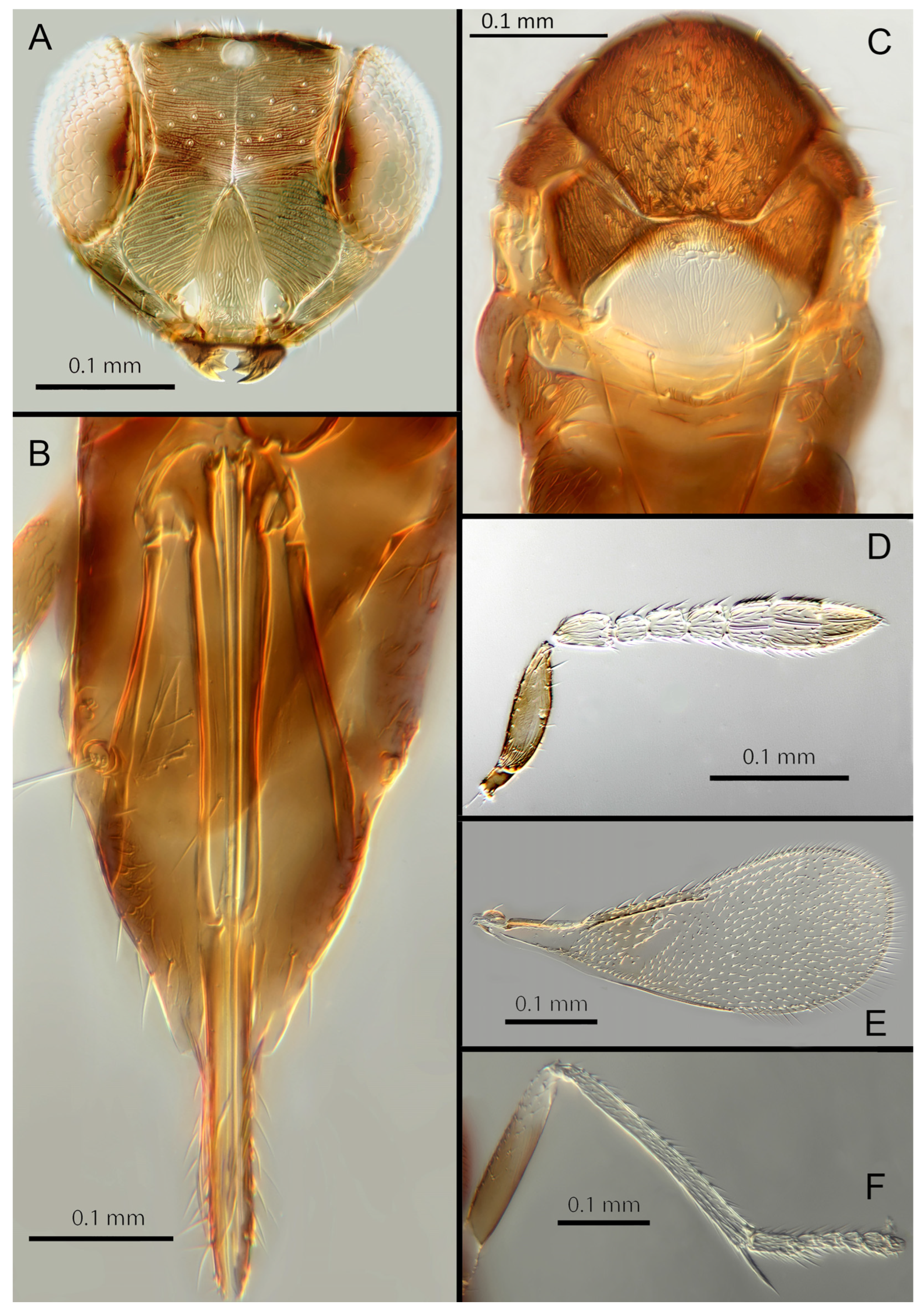
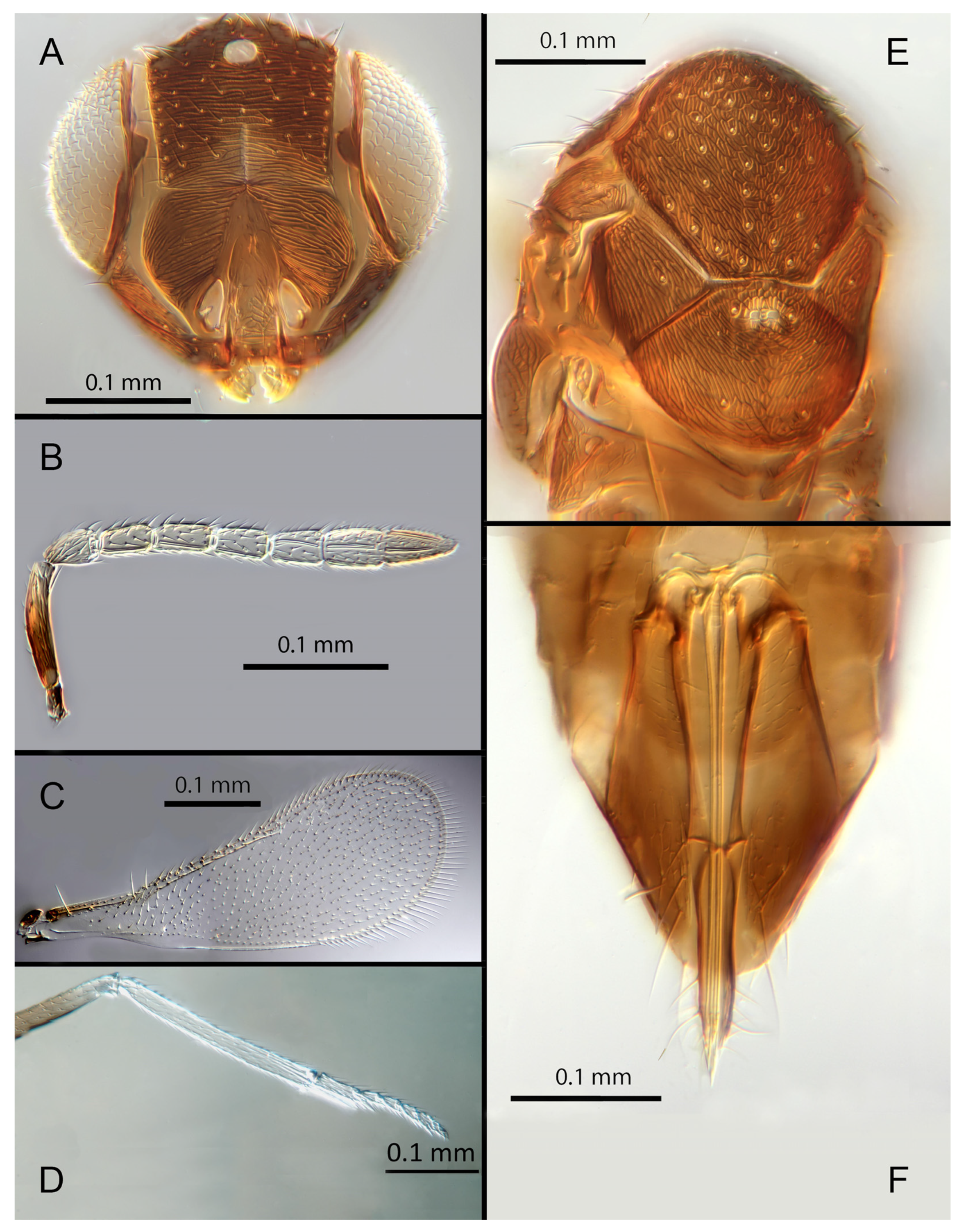
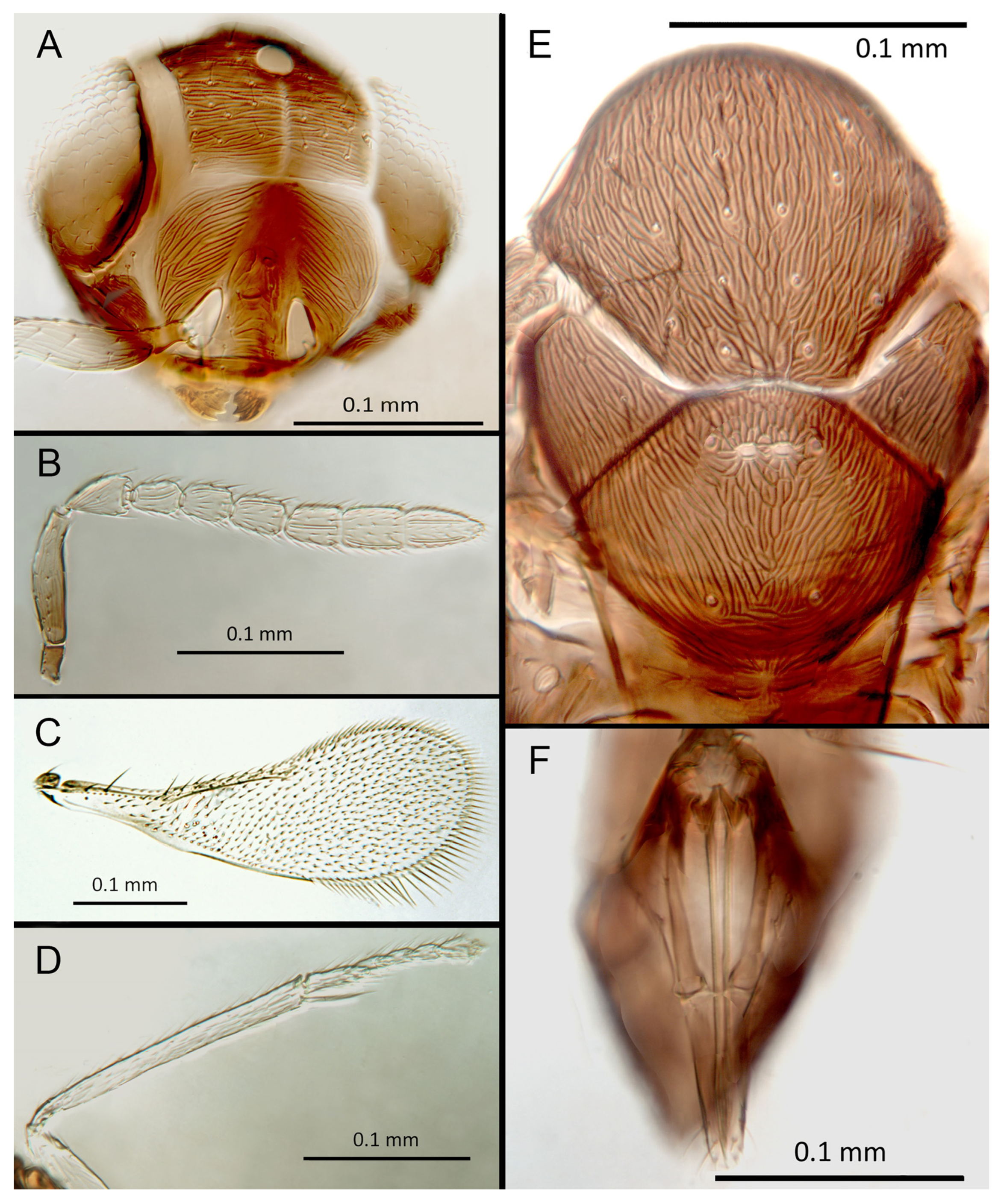
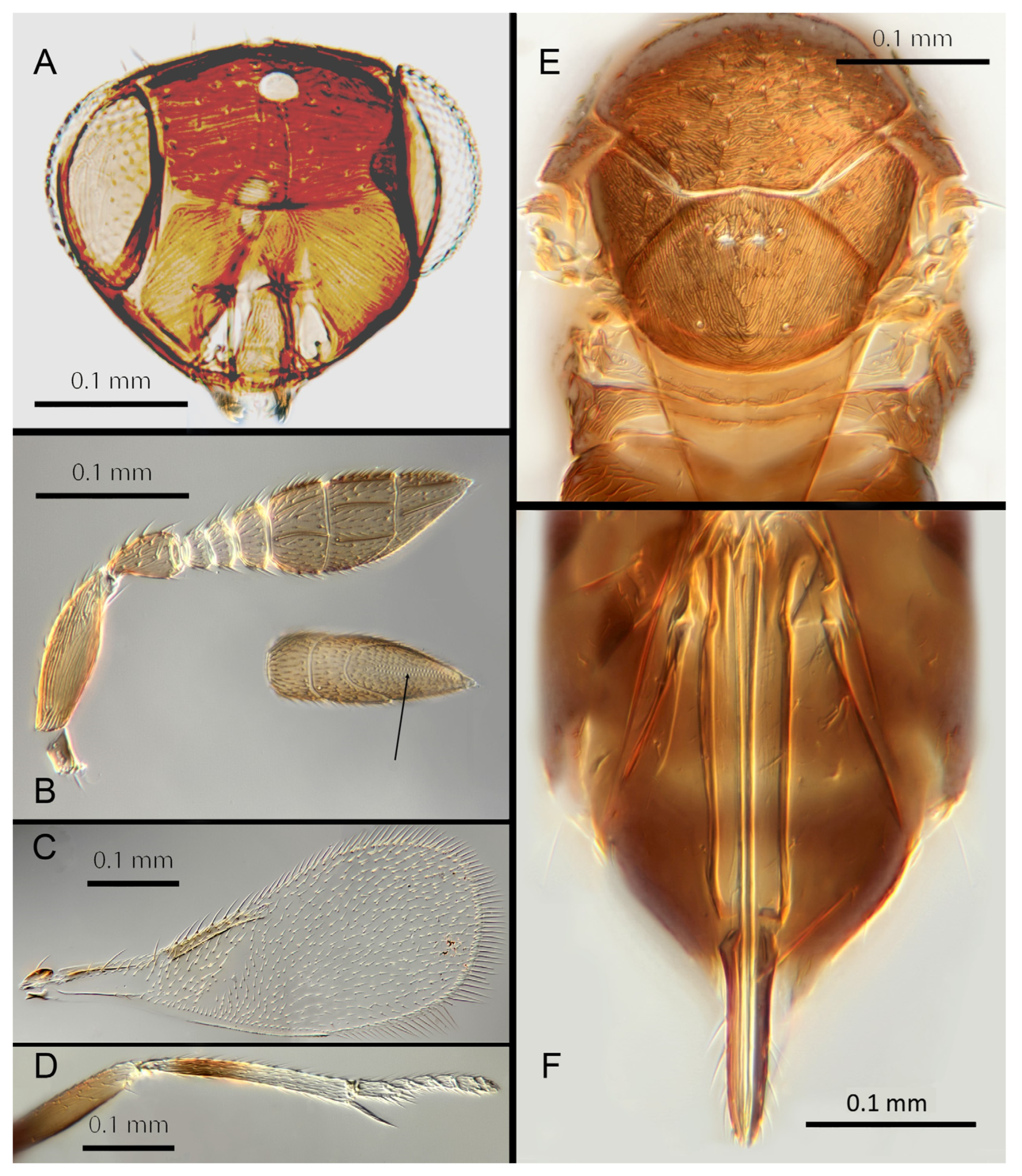
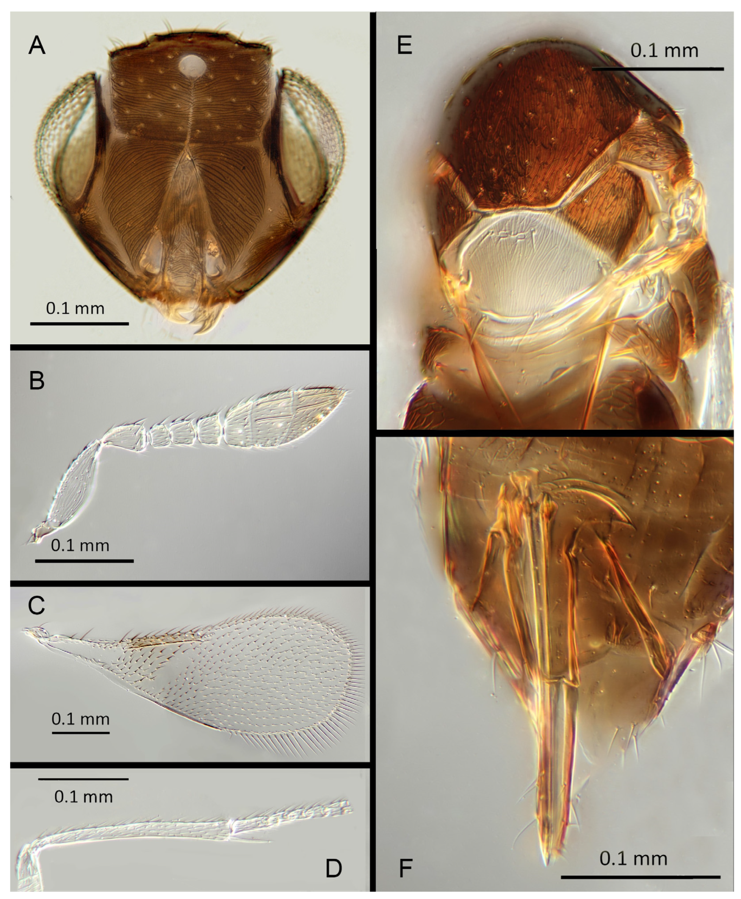
Morphology: Head (
Figure 4A) with mediofrontal line complete; transfacial line obscure; facial lines narrow. Scrobes with longitudinal aciculate sculpture. Antennal formula (
Figure 4D): 1, 1, 3, 3; scape 2.4x pedicel length; pedicel 1.9x F1; F1 0.8x F2; F2 equal to F3; funicle 0.56x clava; F6 slightly oblique, claval sensorial area present. Flagellum with the following number of longitudinal sensilla: F1: 0; F2: 1; F3: 1; F4: 3; F5: 4; F6: 3–4 (both counts present in holotype). Mandibles (
Figure 4A) with two teeth and a broad truncation. Maxillary palps two-segmented. Mid-lobe of mesoscutum (
Figure 4C) with 34–40 setae; each lateral lobe with two setae; each axilla with one seta; scutellum with four setae. Sculpture of mesoscutum longitudinal; sculpture of axillae and scutellum longitudinal. Fore wing (
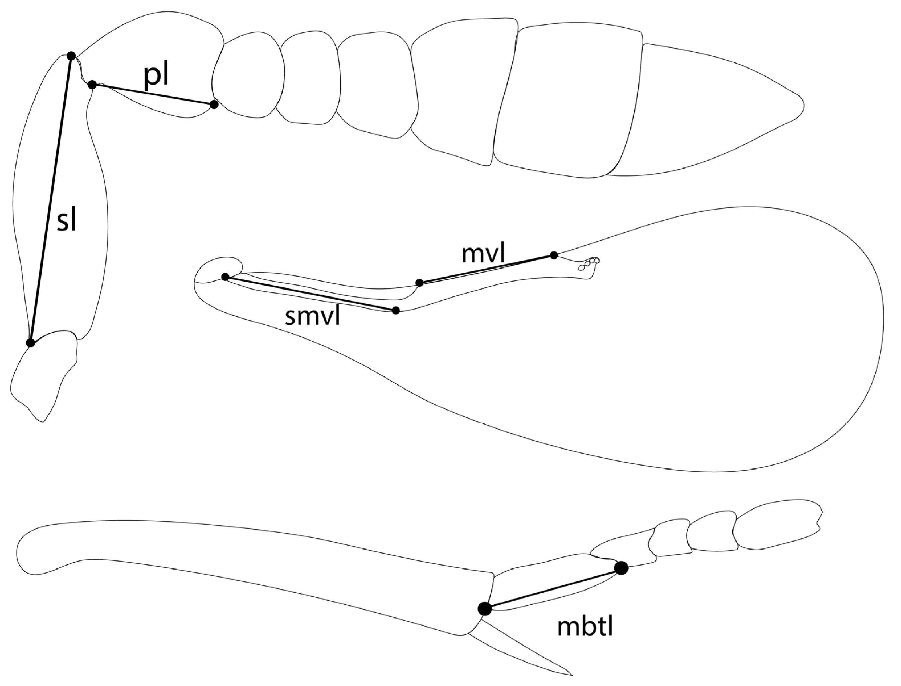
Figure 4E) with two large setae and 2–3 smaller setae on the submarginal vein, three setae in the basal cell, 6–7 setae on the anterior margin of the marginal vein, and one seta at the junction of the submarginal vein and parastigma. Linea calva present. Submarginal 0.75x marginal vein. Maximum length of fore wing 2.7x fore wing width; maximum width of wing 5.3x longest seta on marginal fringe. Ovipositor (
Figure 4B) 1.85x mid tibial length; third valvulae 0.44x ovipositor length; second valvifer 1.3x third valvula. Mid tibial spur (
Figure 4F) 0.86x corresponding basitarsus. Metasomal terga T1–T7 with 0, 2 + 2, 2 + 2, 1 + 1, 1 + 2 + 1, 1 + 2 + 1 and four setae, respectively. T7 (
Figure 4B) extremely extended, almost covering the ovipositor.
Distribution. COSTA RICA: Heredia, Limon; PERU: Iquitos.
Material examined: Holotype ♀ COSTA RICA, Heredia Estación Biológica La Selva, 75 m, 10°26′ N 84°01′ W 27–28.ii.2003 (J.S. Noyes) [DNA148: OQ683554] (NHMUK). Paratypes: 1♀ COSTA RICA, Limon RB, Hitoy-Cerere 100 m, 14–19.i.1991 (J.S. Noyes) (MZUCR). 1♀ PERU, Iquitos, Barillal, 10.ii.1984 (L. Huggert #BM 1984.337) [DNA 212] (NHMUK).
Remarks: T7 extremely extended, covering the ovipositor. Encarsia acusa appears to be most closely related to E. inbioa and E. svetlana, but is easily distinguished from those (and all other) species by the extremely long ovipositor and T7. DNA sequences from holotype deposited under GenBank accession number: OQ683554.
Etymology. From “acus” Latin for needle or pin, referring to the elongated T7.
3.3.2. Encarsia aisha Polaszek and Hernández-Suárez sp. n.
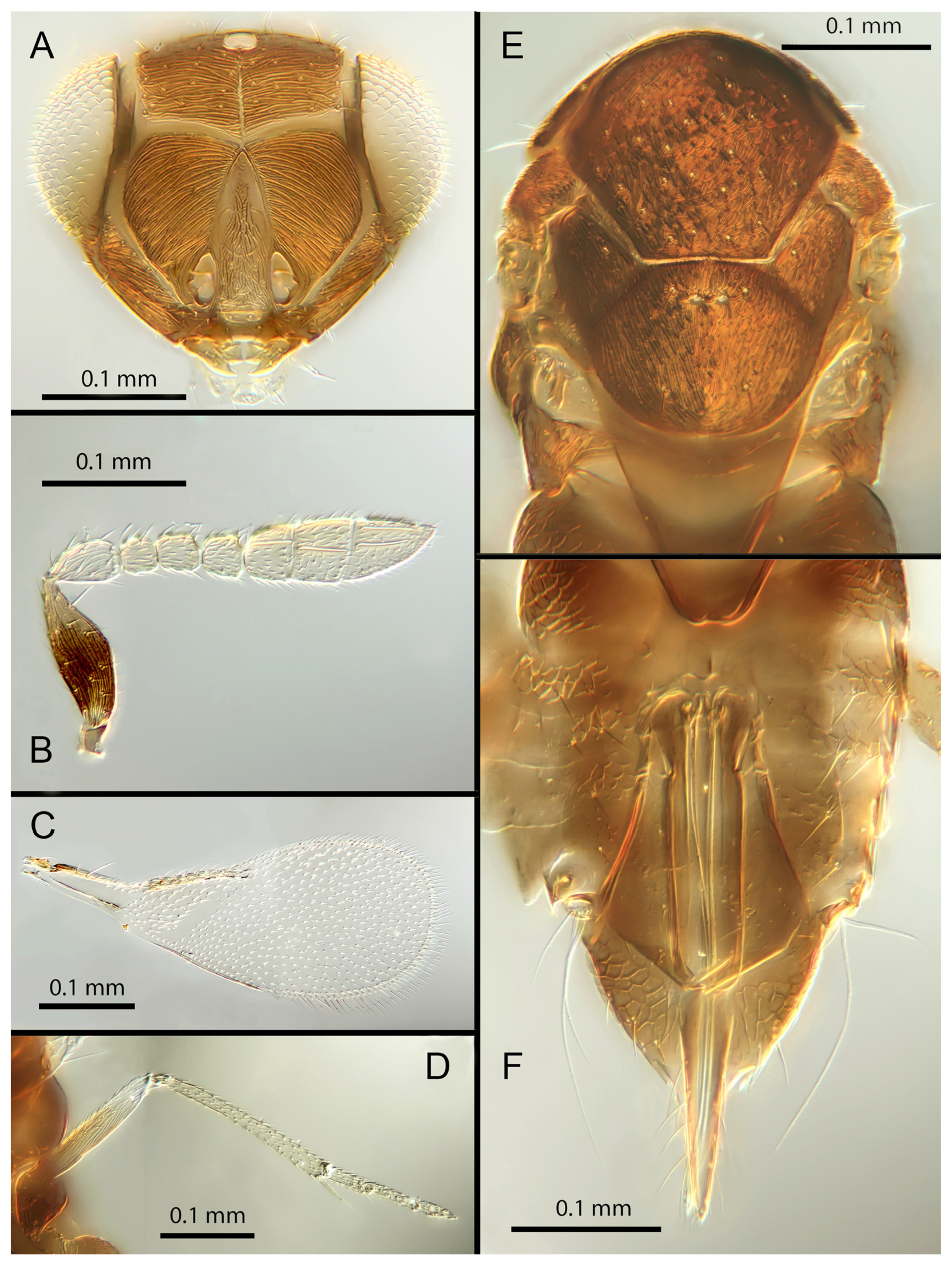
Female. Colour: Antennae brown with F1 and F2 paler. Head dark brown, paler along the sutures. Mesosoma uniformly dark brown. Legs yellow with mid and hind femora, coxae and anterior third of mid tibiae brown, all tarsi pale. Wings hyaline, slightly infuscated below marginal vein; submarginal and marginal veins darker in contrast with the paler stigmal vein.
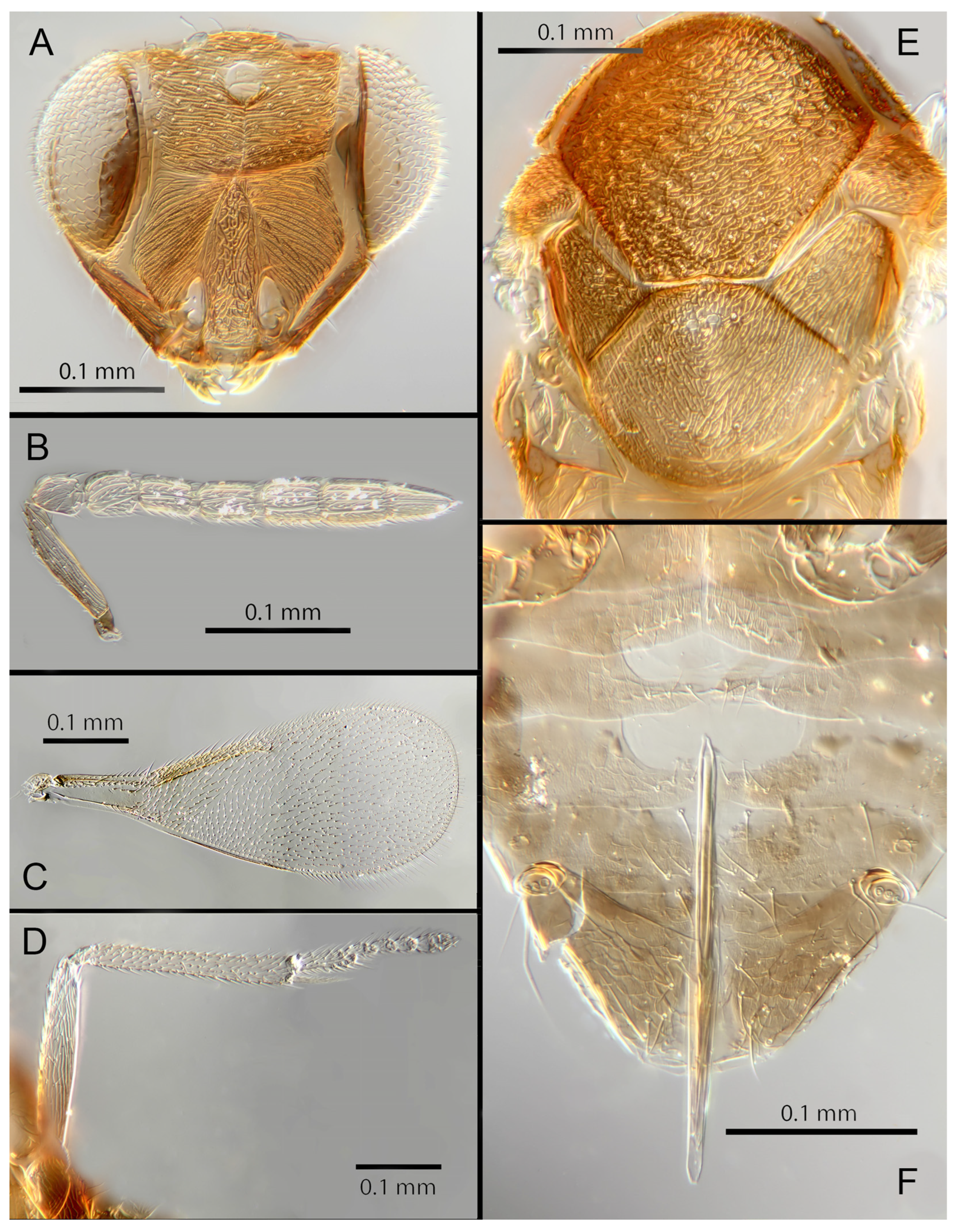
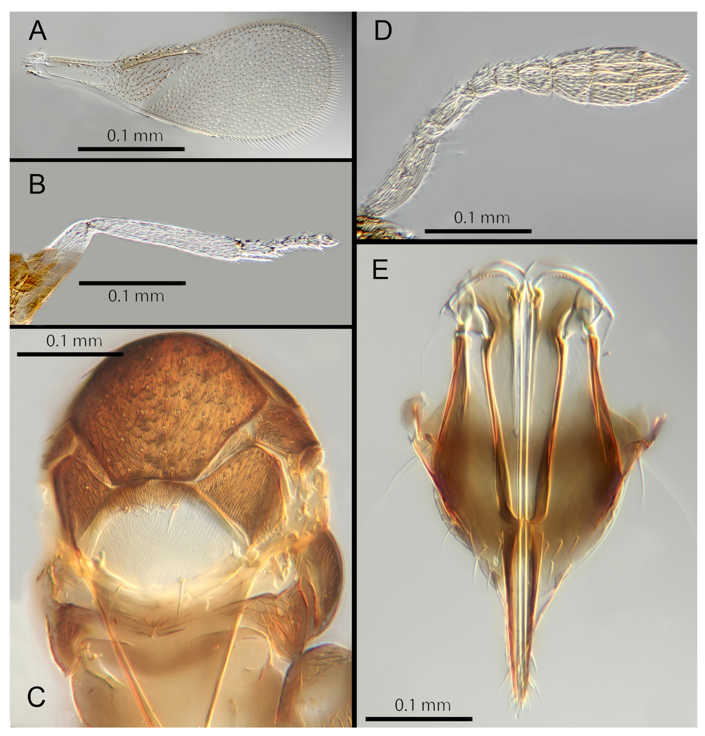
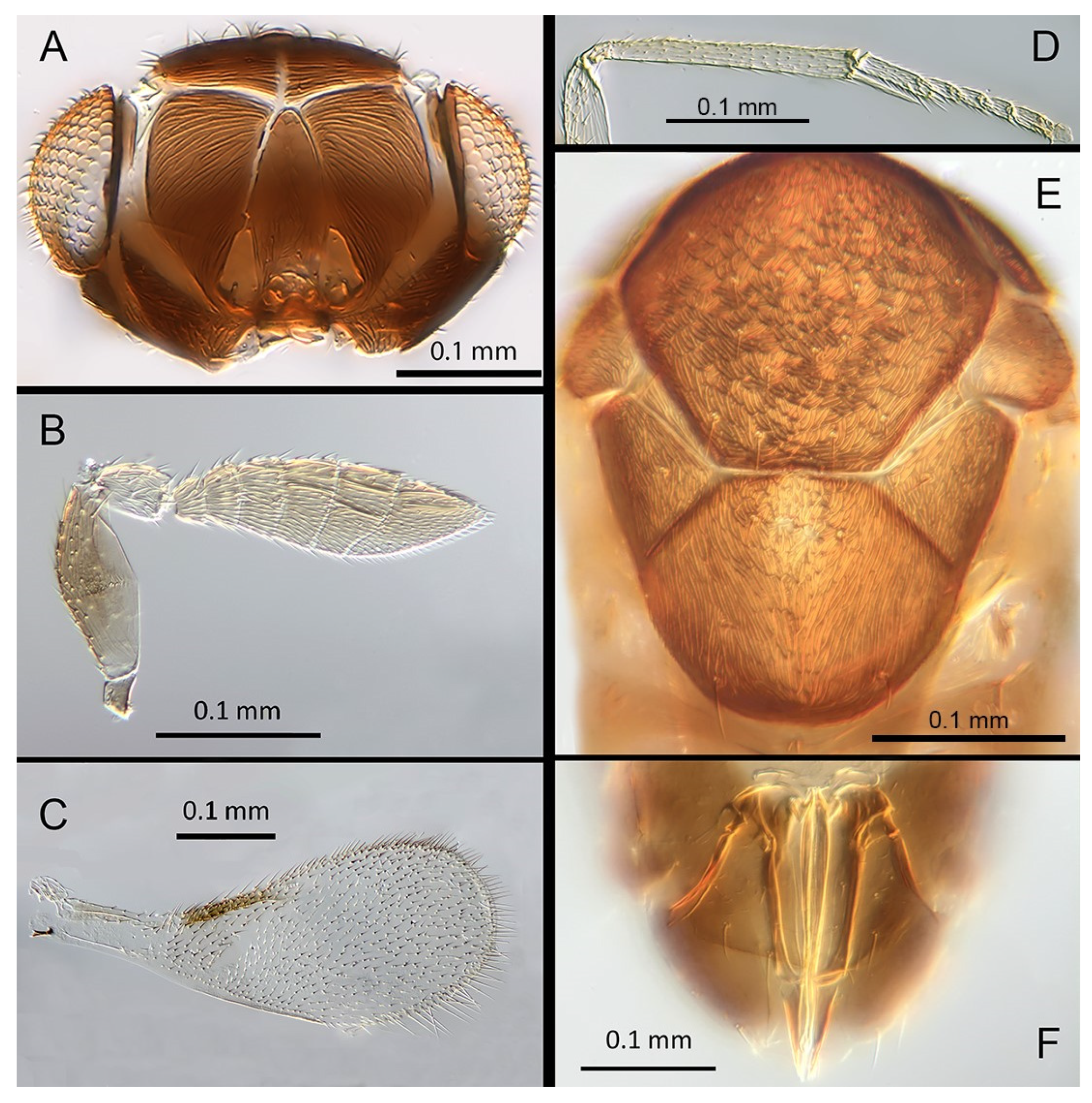
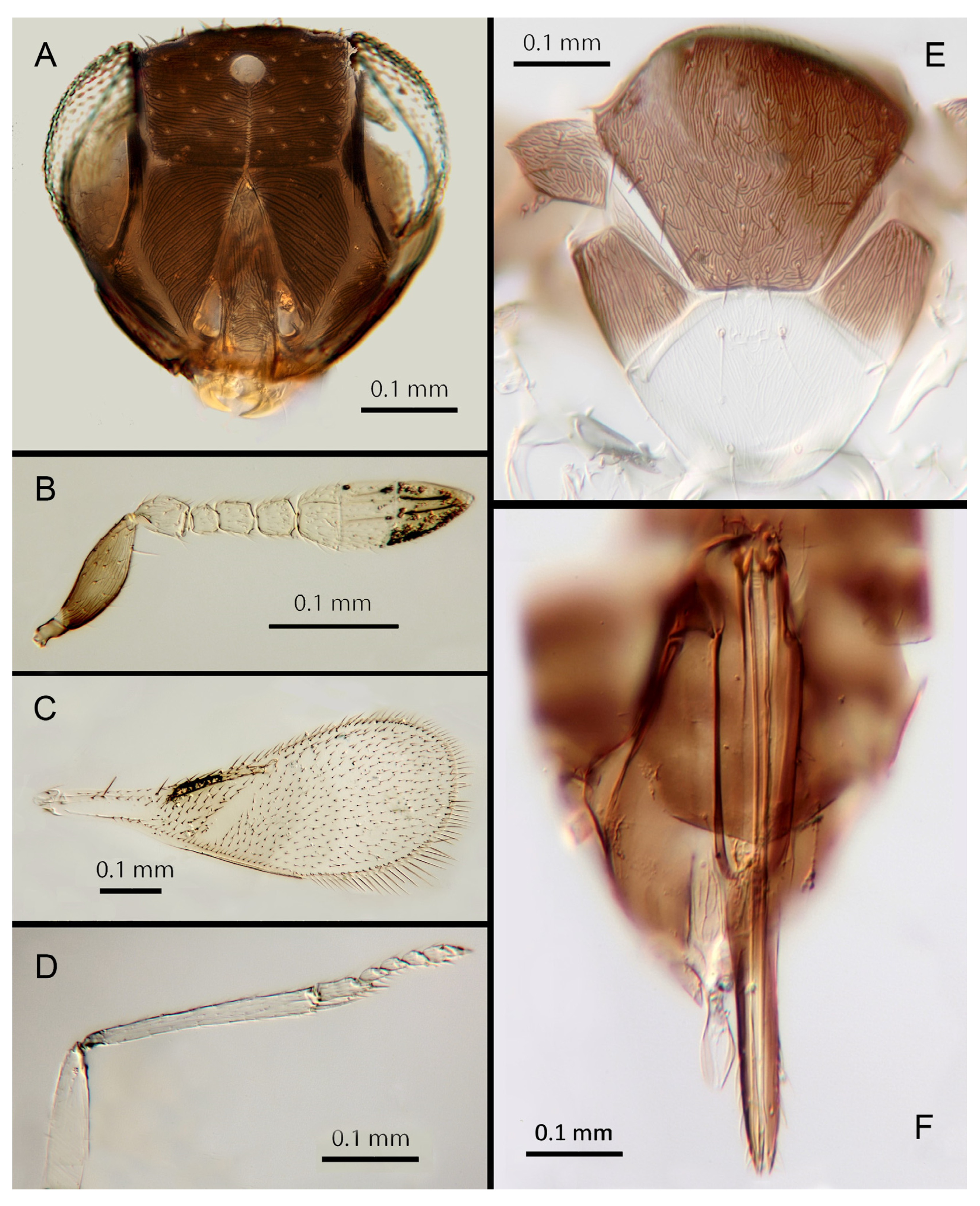
Morphology: Head (
Figure 5A) with mediofrontal line complete; transfacial line evident; facial lines very broad along their entire lengths. Scrobes with longitudinally aciculate sculpture. Antennal formula (
Figure 5B): 1, 1, 3, 3; scape 2.3x pedicel length; pedicel 2x F1; F1 1.2x F2; F2 0.75x F3; funicle 0.45x clava; F6 oblique, claval sensorial area present. Flagellum with the following number of longitudinal sensilla: F1: 0; F2: 0; F3: 1; F4: 2–3; F5: 4–5; F6: 3. Mandibles (
Figure 5A) with one large ventral tooth and a bidentate upper tooth. Maxillary palps two-segmented. Mid-lobe of mesoscutum (
Figure 5E) with about 34–40 setae; each lateral lobe with two setae; each axilla with one seta; scutellum with four setae and two apparent vestigial setal bases. Sculpture of mesoscutum aciculate; sculpture of axillae and scutellum longitudinal. Fore wing (
Figure 5C) with two large setae and four smaller setae on submarginal vein, four setae in basal cell, 6–7 setae on anterior margin of marginal vein, and one seta at the junction of the submarginal vein and parastigma. Linea calva present. Submarginal 0.85x marginal vein. Maximum length of fore wing 2.47x fore wing width, maximum width of wing 5.72x longest seta on marginal fringe. Ovipositor (
Figure 5F) 1.47x mid tibial length; third valvulae 0.42x ovipositor length; second valvifer 1.5x third valvula. Mid tibial spur (
Figure 5D) 1.07x corresponding basitarsus. Metasomal terga T1–T7 with 0, 2 + 2, 2 + 2, 2 + 2, 1 + 2 + 1, 1 + 2 + 1 and four setae, respectively. T7 (
Figure 5F) is extended, as long as ovipositor.
Distribution: COSTA RICA: Alajuela, Heredia.
Material examined: Holotype ♀ COSTA RICA, Alajuela, Est. Biol. Caribe, R. Rincon Forestal 10°53′ N 85°18′ W 400 m 19–20.ii.2003 (J.S. Noyes) [DNA 164: OQ683562] (NHMUK). 1♀ COSTA RICA, Heredia, Estacion Biologica La Selva, 10°26′ N 84°01′ W. 75 m 27–28.ii.2003 (J.S. Noyes) [DNA 146: OQ683562] (ZMUCR).
Remarks: Encarsia aisha is morphologically very similar to E. marynoyesae in many respects (though distant to it based on DNA). The species can be distinguished from E. marynoyesae by the second valvifers almost 2x (1.8) the third valvulae; while they are 1.5x as long in E. aisha. In E. marynoyesaei the clava is well over 2x the length of the funicle; in E. aisha it is less than 2x as long. DNA sequence from holotype and paratype (pooled extraction) deposited under GenBank accession number: OQ683562.
Etymology: Named for Aisha, daughter of the second author (EHS), and sister to Noora; see E. noora, below.
3.3.3. Encarsia aphania (Polaszek) 1999 (in Martin and Polaszek, 1999: 1556). comb. nov.
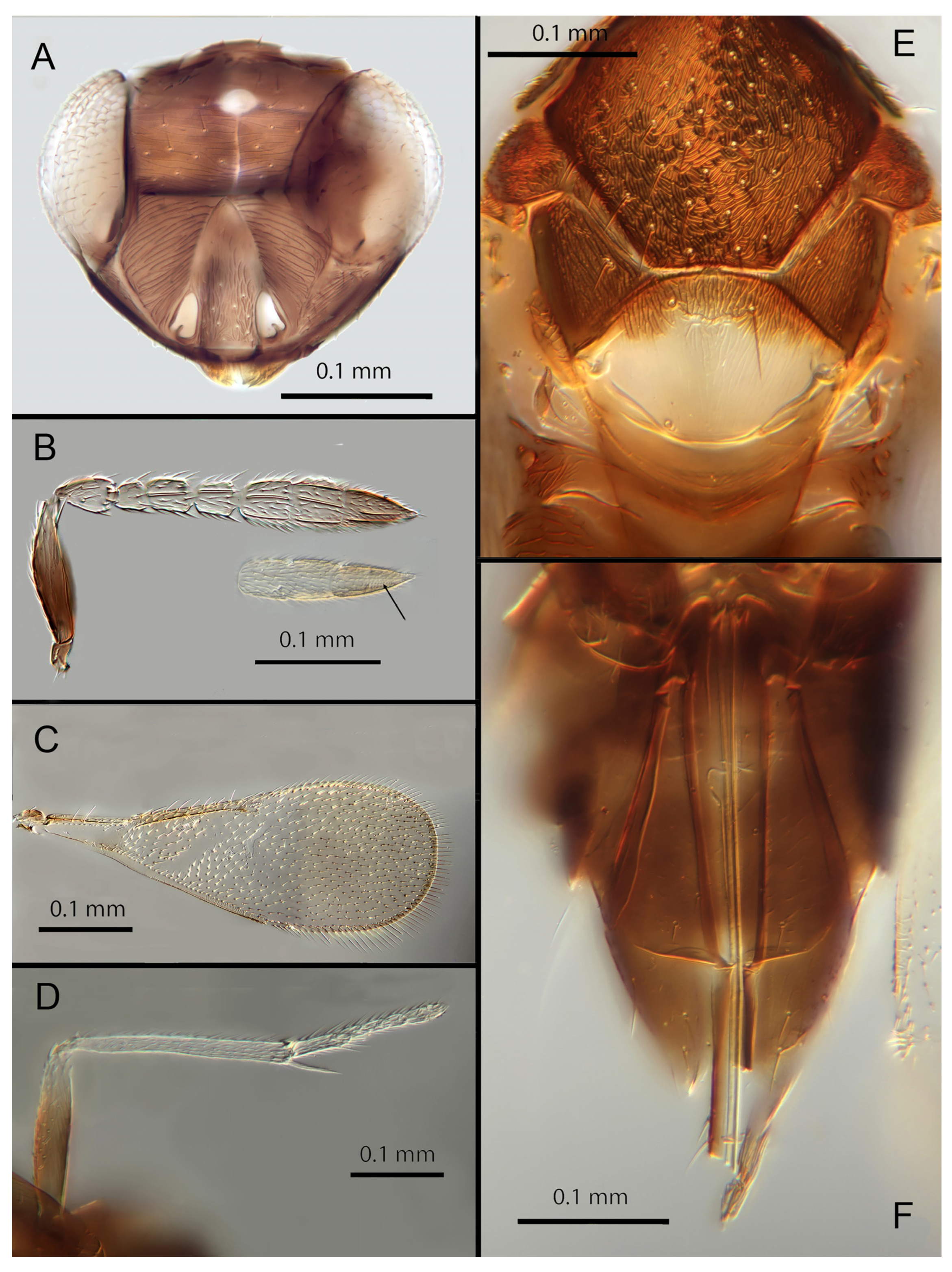
Female. Colour: Antennae pale brown with scape very dark. Head dark brown with pale lines bordering the eyes and extending along the genae towards clypeus, antennal scrobes, a line from the apex of the scrobes to the median ocellus, and a transverse line midway between the antennal scrobes. Mesosoma and metasoma uniformly dark brown. Legs yellow except all coxae and mid and hind femora which are brown. Fore wings faintly infuscate along the submarginal and the marginal veins; submarginal and marginal veins darker in contrast to the paler stigmal vein.
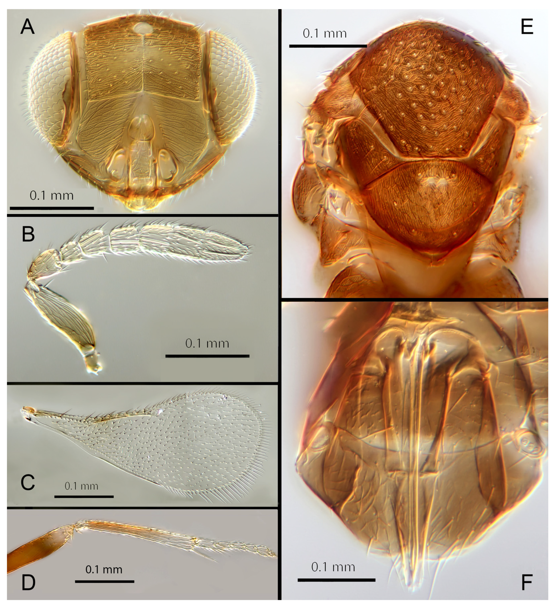
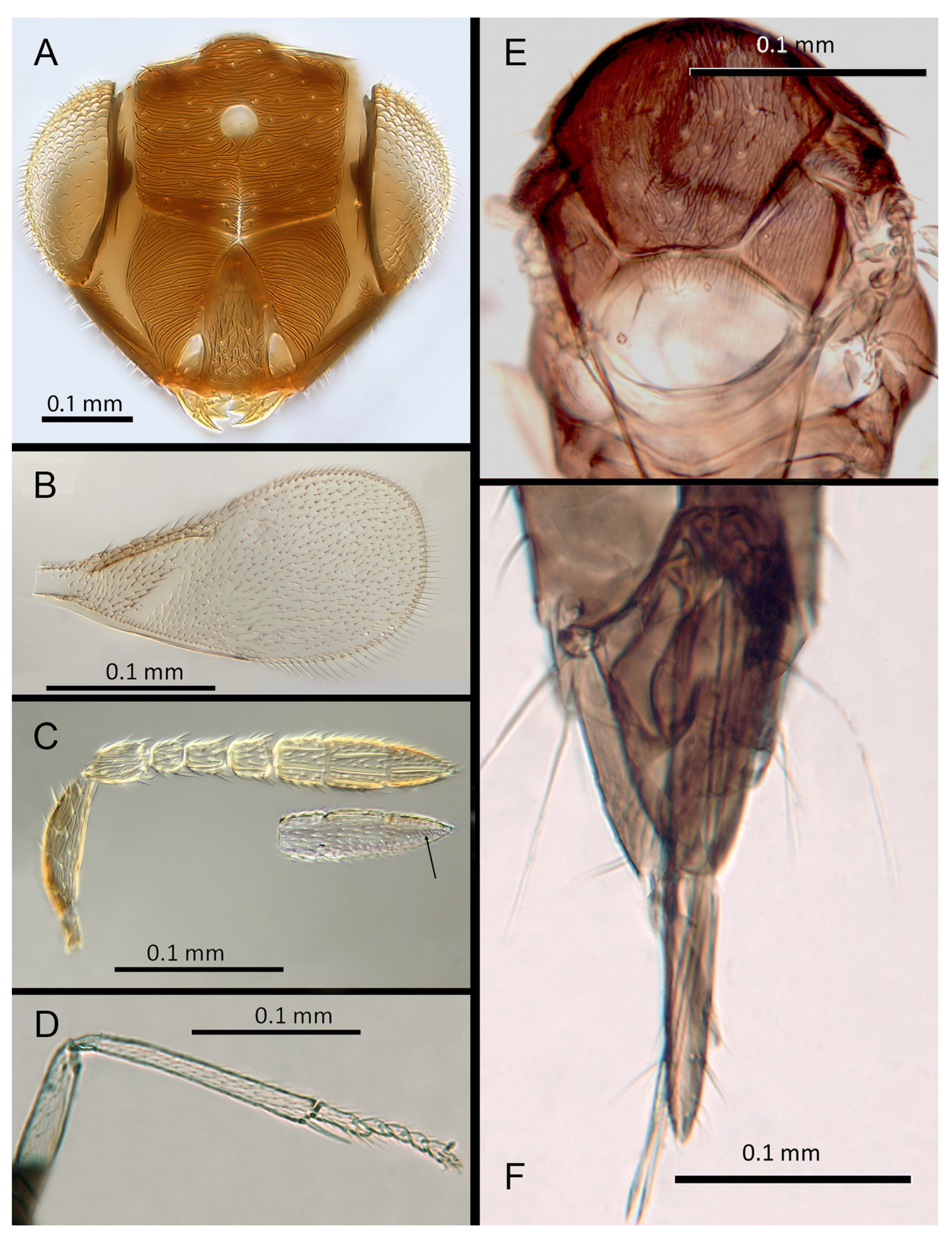
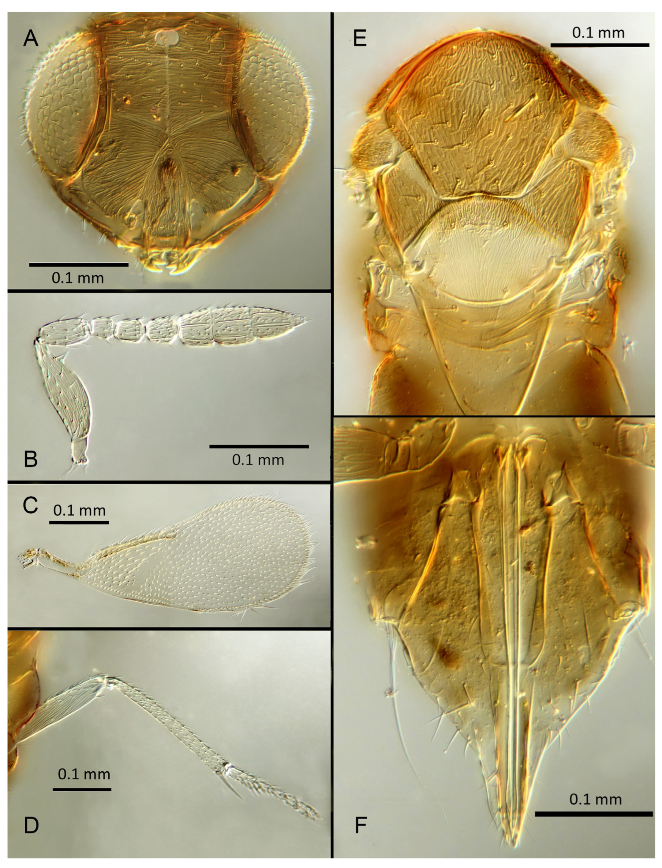
Morphology: Head (
Figure 6A) with mediofrontal line complete, though fading towards anterior ocellus; transfacial and facial lines very broad along their entire lengths. Scrobes with longitudinally aciculate sculpture centrally, smooth laterally. Antennal formula (
Figure 6B): 1, 1, 3, 3; scape 2.39x pedicel; pedicel 2x F1; F1 0.8x F2; F2 equal to F3; funicle 0.67x clava; F4 and F5 partly fused; F6 broadly oblique; claval sensorial area present, indistinct. Flagellum with the following number of longitudinal sensilla: F1: 0; F2: 1; F3: 1; F4: 2–3; F5: 4; F6: 4. Mandibles (
Figure 6A) with one large ventral tooth and a broad upper truncation; Maxillary palps two-segmented. Mid-lobe of mesoscutum (
Figure 6E) with 30–40 setae; each lateral lobe with one seta; each axilla with one seta; scutellum with four setae. Sculpture of mesoscutum aciculate; sculpture of axillae and scutellum longitudinal. Fore wing (
Figure 6C) with two large setae and 4–5 smaller setae on submarginal vein, 4–5 setae in basal cell, 5–7 setae on anterior margin of marginal vein, and 2–3 setae at the distal part of the base. Linea calva present. Submarginal 0.9x marginal vein. Maximum length of fore wing 2.6x fore wing width; maximum width of fore wing 5.2x longest setae on marginal fringe. Ovipositor (
Figure 6F) 1.6x mid tibial length; third valvula 0.4x ovipositor length; second valvifer 1.3x third valvula. Mid tibial spur (
Figure 6D) as long as corresponding basitarsus. Metasomal terga T1–T7 with 0, 2 + 2, 2 + 2, 1 + 1, 1 + 2 + 1, 1 + 2 + 1 and 5–6 setae, respectively. T7 (
Figure 6F) with a pointed, extended apex covering ovipositor.
Distribution: BELIZE: Cayo District; COSTA RICA: Puntarenas.
Hosts: Aleurodicinae: Aleurodicus pulvinatus (Maskell), Azuraleurodicus pentarthrus Martin; Nealeurodicus altissimus (Hempel).
Material examined: Holotype ♀ BELIZE, Cayo District, Chiquibul Forest Reserve, Las Cuevas-Monkey Tail trail, 5.iii.1996 (J.H. Martin #6747) ex Azuraleurodicus pentarthus (NHMUK). 13♀ BELIZE, Cayo Las Cuevas, monkey tail trail, 5.iii.1996 (J.H. Martin #6747) ex Azuraleurodicus pentarthus [s27, s22, DNA218: OQ683546]. 1♀ BELIZE, Cayo Chiquibul Fr., Monkey tail trail, 21.iii.2003 (J.H. Martin #7768) ex Nealeurodicus altissimus on Inga sp. (all NHMUK). 1♀ COSTA RICA, Puntarenas, Est. Altamira send. Los Gigantes, 9.vii.2001 (D. Rubi #63984) [DNA213: OQ683545] 1.460 m, LS 331800 572100 (MZUCR)
Remarks:
Encarsia aphania presents a unique combination of characters and appears to have no very close relatives. Morphologically it is closest to
E. larensis but is easily distinguishable by the much longer third valvulae relative to the second valvifers (compare
Figure 5F and Figure 15F). DNA sequences were obtained from two specimens from Belize (type locality) and Costa Rica, Puntarenas; deposited under GenBank accession numbers: OQ683546, OQ683545.
3.3.4. Encarsia avida Polaszek and Hernández-Suárez sp. n.
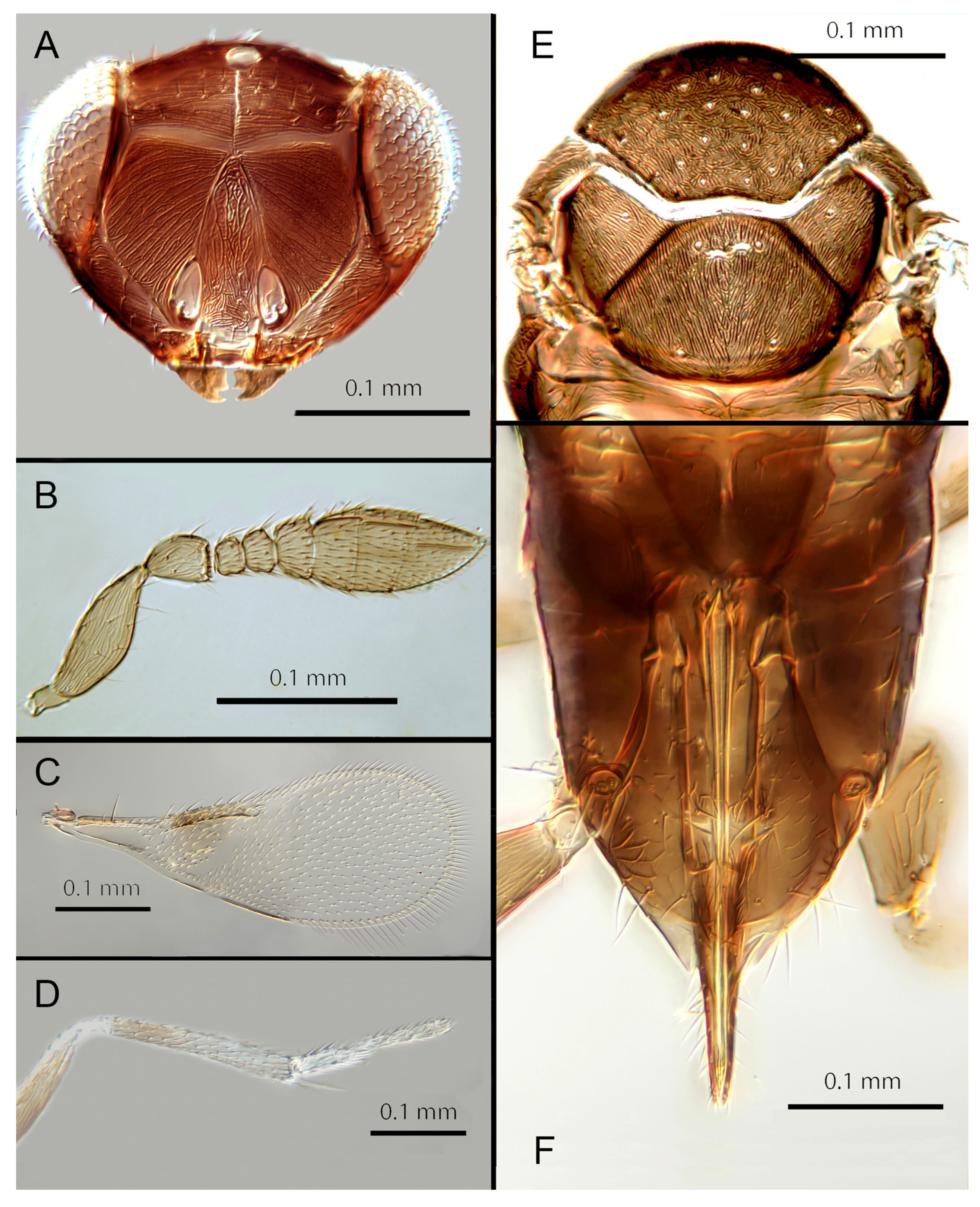
Female. Colour: Antennae pale brown, darker on F5–F6 and the scape, pedicel, and radicle. Head brown, paler along the sutures. Mesosoma dark brown with posterior three-quarters of scutellum pale. Metasoma uniformly dark brown. Legs yellow with all coxae, femora, and anterior half of fore leg tibiae brown; all tarsi pale. Wings infuscate below marginal vein; submarginal and marginal veins darker in contrast with the stigmal vein paler.
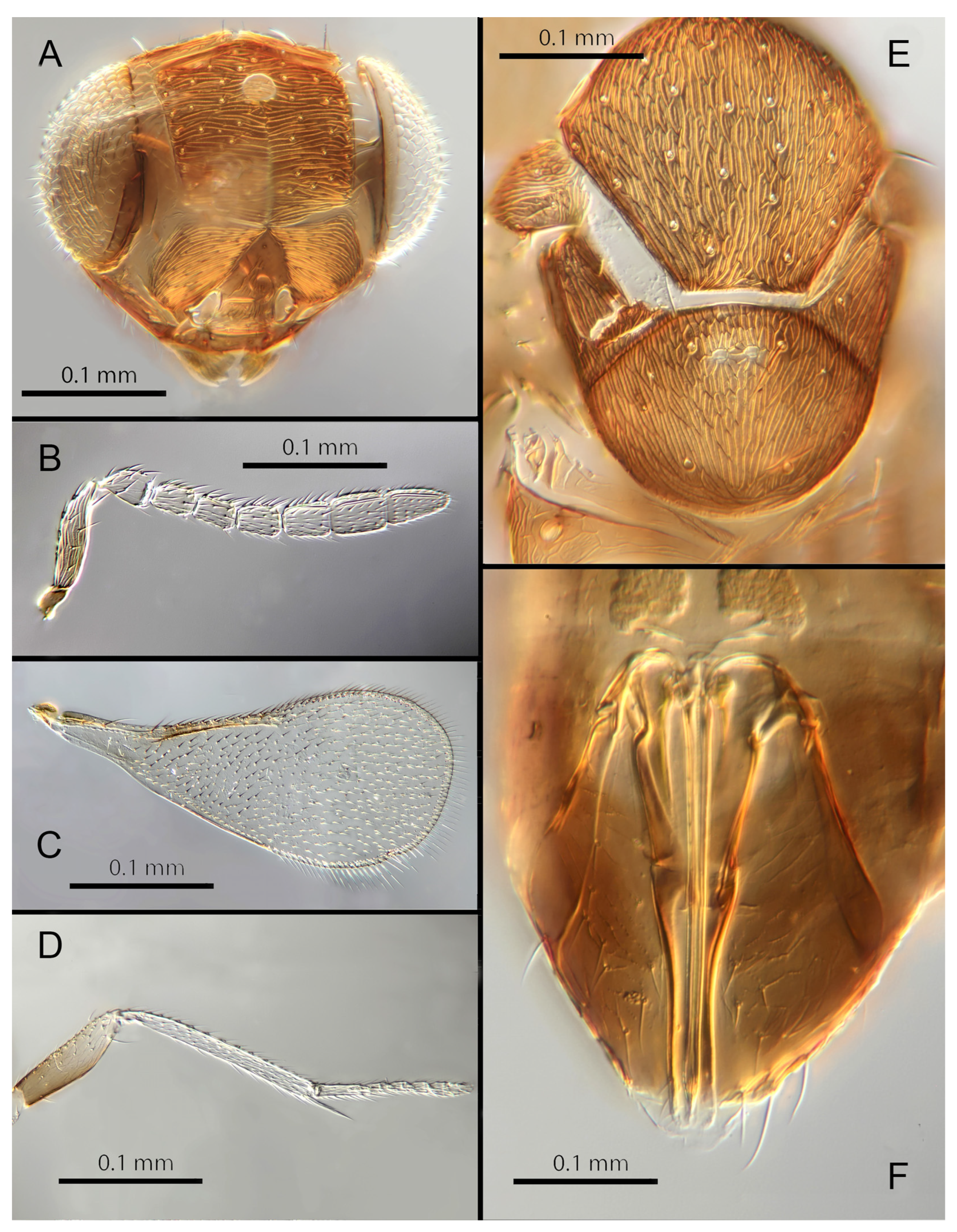
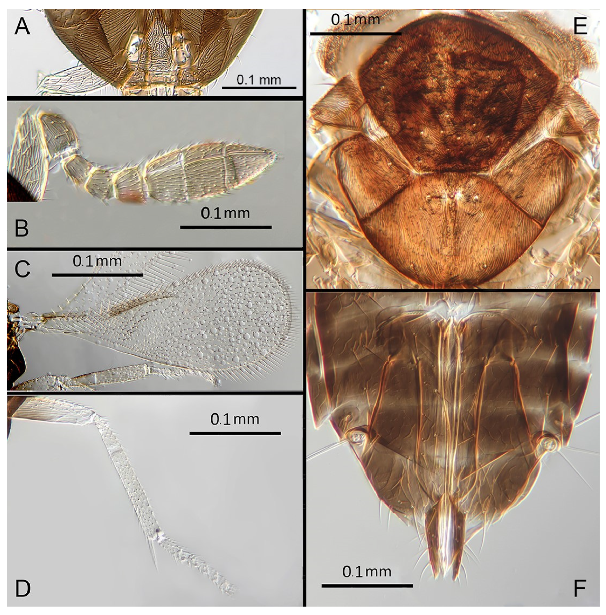
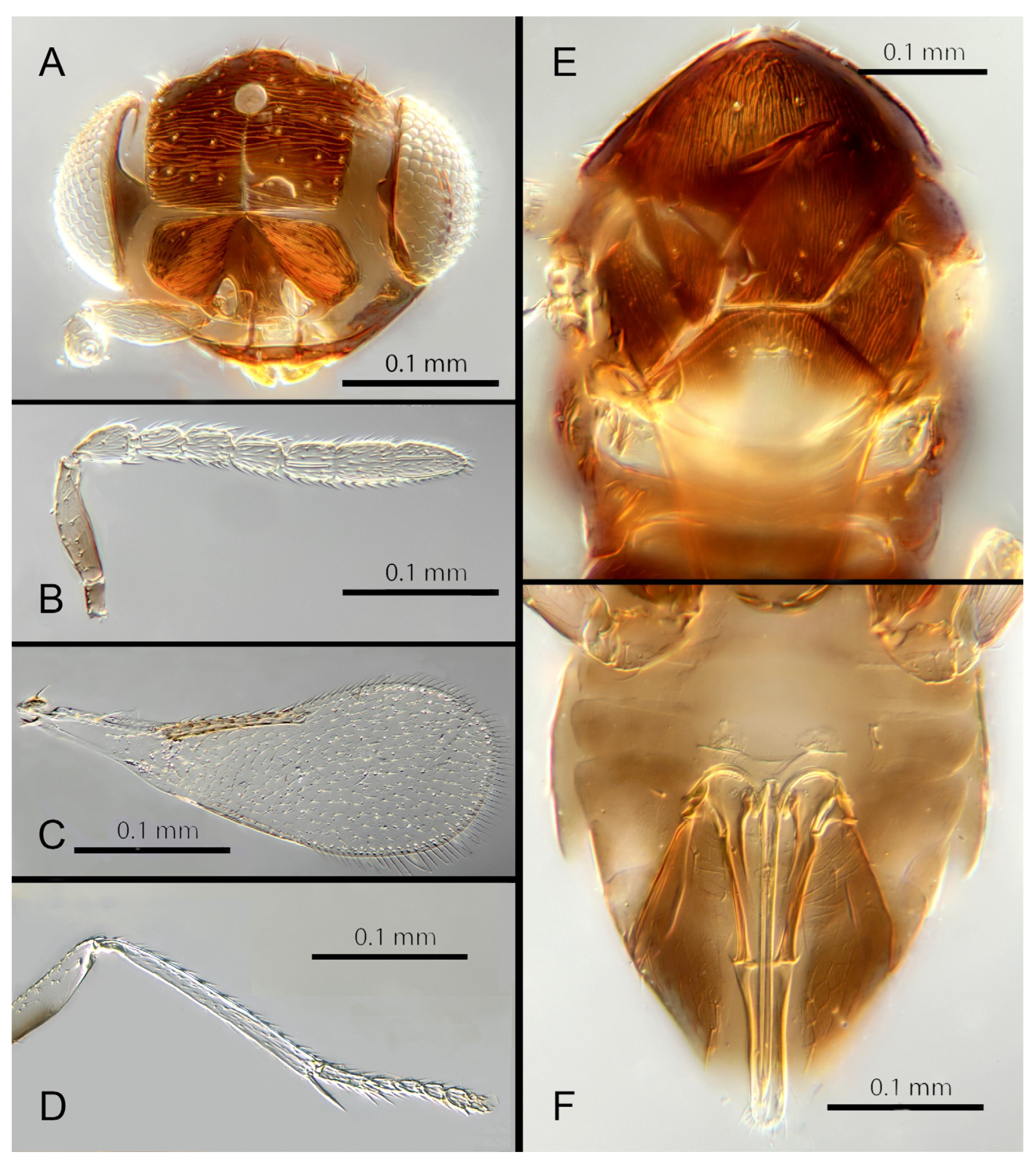
Morphology: Head (
Figure 7A) with mediofrontal line complete; transfacial line obscure; facial lines present, narrow. Scrobes with longitudinally aciculate sculpture centrally, smooth laterally. Antenna formula (
Figure 7B): 1, 1, 3, 3; scape 2.5x pedicel length; pedicel 1.95x F1; F1 0.66x F2; F2 equal to F3; funicle 0.6x clava; F6 perpendicular. Claval sensorial area present, distinct. Flagellum with the following number of longitudinal sensilla: F1: 0; F2: 1–2; F3: 1; F4: 2; F5: 3; F6: 3. Mandibles (
Figure 7A) with two small ventral teeth and a truncation. Maxillary palps two-segmented. Mid-lobe of mesoscutum (
Figure 7E) with 40–50 setae; each lateral lobe with two setae; each axilla with one seta; scutellum with four setae. Sculpture of mesoscutum aciculate; sculpture of axillae and scutellum longitudinal. Fore wing (
Figure 7C) with two large setae and four smaller setae on submarginal vein, four setae in basal cell, eight setae on anterior margin of marginal vein, and one seta at the junction of the submarginal vein and parastigma. Linea calva present. Submarginal 0.67x marginal vein. Maximum length of fore wing 2.8x fore wing width; maximum width of wing 4.7x longest setae on marginal fringe. Ovipositor (
Figure 7F) 1.6x mid tibial length; third valvulae 0.4x ovipositor length; second valvifer 1.5x third valvula. Mid tibial spur (
Figure 7D) 0.8x corresponding basitarsus. Metasomal terga T1–T7 with 0, 2 + 2, 2 + 2, 2 + 2, 1 + 2 + 1, 1 + 2 + 1 and 4 setae, respectively. T7 (
Figure 7F) extended and covering ovipositor (damaged in holotype).
Distribution: COSTA RICA: Heredia.
Material examined: Holotype ♀ COSTA RICA, Heredia, Est. Biol. La Selva, 10°26′ N 84°01′ W 75 m 27–28.ii.2003 (J.S. Noyes) [DNA143: OQ683547] (NHMUK).
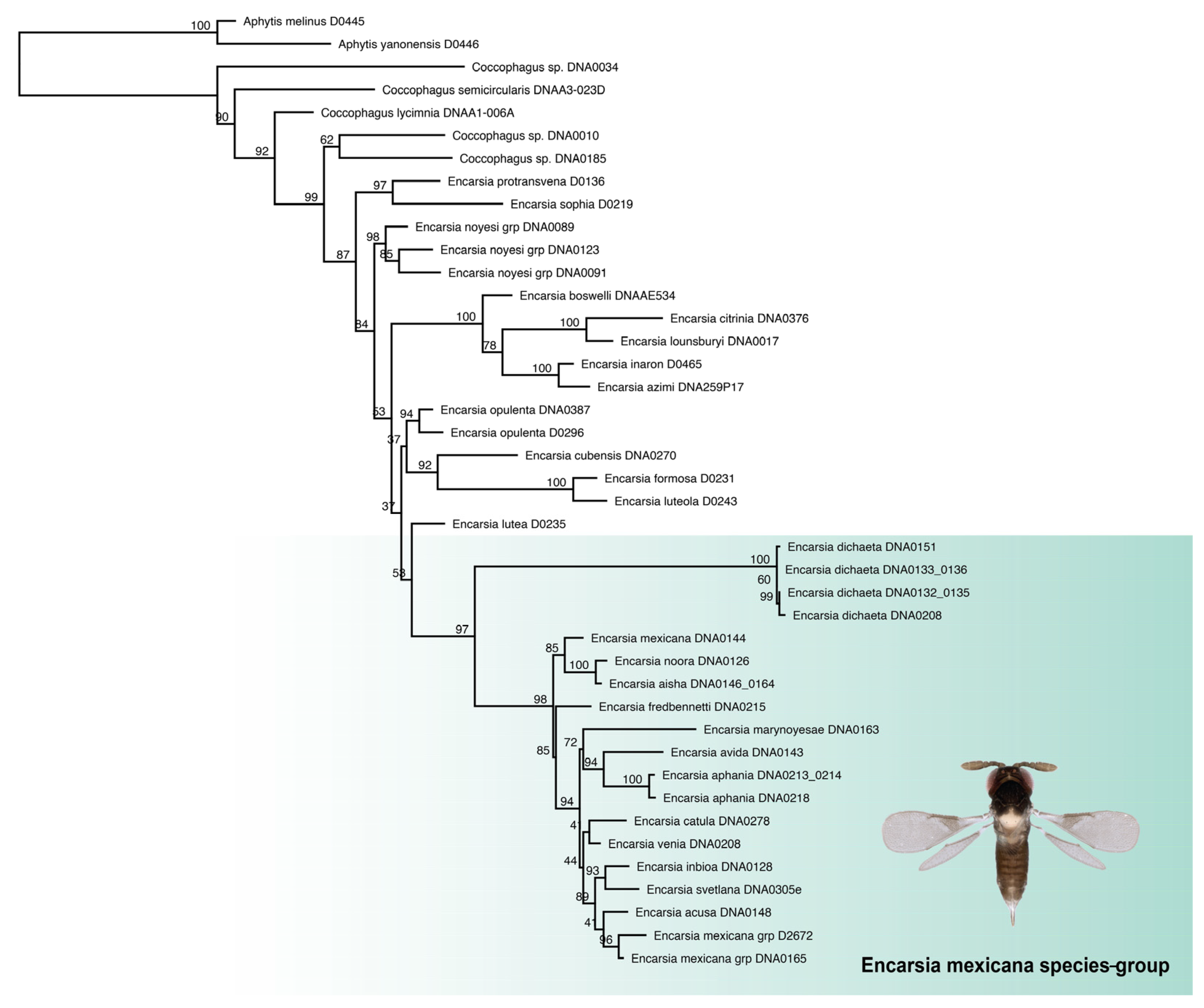
Remarks: Encarsia avida appears morphologically close to E. acusa with which it shares the color pattern (mesoscutellum anteriorly dark) and wing and antennal morphology.
The ovipositor in
E. acusa is longer (1.8x mid tibia; 1.6x in
E. avida). The most easily appreciated difference is in the sculpture of the frons:
E. avida has scattered, shallow horizontal grooves (
Figure 7A) while
E. acusa has very dense horizontal grooves (
Figure 4A). A similar difference in sculpture is evident on the lateral face. The two species are well-separated based on DNA (
Figure 1) with
E. avida coming out as sister to
E. aphania with high support (95%). DNA sequence from holotype deposited under GenBank accession number: OQ683547.
Etymology: From “avida -us” meaning “greedy” (Latin).
3.3.5. Encarsia catula Polaszek and Hernández-Suárez sp. n.
Female. Colour: Antennae brown. Head dark brown. Mesosoma uniformly dark brown. Legs yellow with mid and hind femora, coxae, and anterior half of tibiae brown, fore leg femora, coxae, and tibiae dark, all tarsi pale. Wings infuscate below marginal vein, submarginal and marginal veins dark, stigmal vein paler.
Morphology: Head (
Figure 8A) with mediofrontal line complete; transfacial line broad; facial lines present, narrow. Scrobes with longitudinally aciculate sculpture centrally, irregularly aciculate basally. Antenna formula (
Figure 8B): 1, 1, 3, 3; scape expanded, 3.1x pedicel length; pedicel 2x F1; F1 0.9x F2; F2 0.8x F3; funicle 0.48x clava; F6 oblique, claval sensorial area present. Flagellum with the following number of longitudinal sensilla: F1: 0; F2: 0; F3: 1; F4: 3; F5: 3; F6: 3. Mandibles (
Figure 8A) with one large ventral tooth and a broad truncation. Maxillary palps two-segmented. Mid-lobe of mesoscutum (
Figure 8E) with fewer than 30 setae; each lateral lobe with two setae; each axilla with one seta; scutellum with four setae. Sculpture of mesoscutum aciculate; sculpture of axillae and scutellum longitudinal. Fore wing (
Figure 8C) with two large setae and four smaller setae on submarginal vein, four setae in basal cell, six setae on anterior margin of marginal vein, and one seta at the junction of the submarginal vein and parastigma. Linea calva present. Submarginal equal to marginal vein. Maximum length of fore wing 2.38x fore wing width, maximum width of wing 4.64x longest setae on marginal fringe. Ovipositor (
Figure 8F) 1.48x mid tibial length; third valvulae 0.46x ovipositor length; third valvulae 0.46x ovipositor length; second valvifer 1.2x third valvula. Mid tibial spur (
Figure 8D) 1.1x corresponding basitarsus. Metasomal terga T1–T7 with 0, 2 + 2, 2 + 2, 2 + 2, 1 + 2 + 1, 1 + 2 + 1 and four setae, respectively. T7 (
Figure 8F) extended although apparently not covering ovipositor.
Distribution: COSTA RICA: Limon.
Material examined: Holotype ♀ COSTA RICA, Limon, Hitoy-Cerere 90°40′ N 83°02′ W, 21–22.iii.2006 (J.S. Noyes) [DNA 278: OQ683547] (NHMUK).
Remarks:
Encarsia catula shares aspects of its morphology with
E. marynoyesae, but can be distinguished by having fewer than 30 setae on the mesoscutum, and third valvulae more than ½ the length of second valvifers (less than ½ as long in
E. marynoyesae). The two species are relatively close based on DNA (
Figure 1). DNA sequence from the holotype is deposited under GenBank accession number: OQ683547.
Etymology: From “catula” meaning dog/whelp (Latin).
3.3.6. Encarsia cylindrica Polaszek and Hernández-Suárez sp. n.
Female. Colour: Antennae pale brown, slightly darker on F6, F1 and the base of the scape, pedicel, and radicle. Head dark brown. Mesosoma uniformly dark brown. Legs yellow with femora and coxae brown, fore legs with dark tibiae, all tarsi pale. Wings infuscate below submarginal vein, marginal and stigmal veins pale.
Morphology: Head (
Figure 9A) with mediofrontal line incomplete, extending halfway to anterior ocellus; transfacial line obscure; facial lines very broad along their entire lengths. Scrobes with faint longitudinal sculpture apically, irregular/lateral sculpture basally. Antennal formula (
Figure 9B): 1, 1, 3, 3; scape 2.75x pedicel length; pedicel equal to F1; F1 0.85x F2; F2 equal to F3; funicle 0.86x clava; F6 perpendicular. Flagellum with the following number of longitudinal sensilla: F1: 2; F2: 2; F3: 2; F4: 2; F5: 3; F6: 3. Mandibles (
Figure 9A) with two small teeth and a broad truncation. Maxillary palps two-segmented. Mid-lobe of mesoscutum (
Figure 9E) with about 40–50 setae; each lateral lobe with two setae; each axilla with one seta; scutellum with four setae. Sculpture of mesoscutum aciculate; sculpture of axillae and scutellum longitudinal. Fore wing (
Figure 9C) with two large setae on submarginal vein and 11 smaller setae, six setae in basal cell, 11 setae on anterior margin of marginal vein, and one seta at the junction of the submarginal vein and parastigma. Linea calva absent. Submarginal 0.62x times marginal vein. Maximum length of fore wing 2.7x fore wing width, maximum width of wing 5.87x longest setae on marginal fringe. Ovipositor (
Figure 9F) equal to mid tibial length; third valvula 0.45x ovipositor length; second valvifer 1.3x third valvula. Mid tibial spur (
Figure 9D) 1.1x corresponding basitarsus. Metasomal terga T1–T7 with 0, 1 + 1, 1 + 1, 1 + 1, 1 + 2 + 1, 1 + 2 + 1 and six setae, respectively. T7 (
Figure 9F) rounded, not extended but covering ovipositor.
Distribution: BRAZIL: Minas Gerais; COSTA RICA: Puntareñas, San Juan; JAMAICA.
Host: Aleurodicinae: Aleurodicus jamaicensis Cockerell.
Material examined: Holotype ♀ COSTA RICA, San Juan, Ciudad Colon, Heredia El Rodeo coll parataxonomist 16.ii.1991 [DNA 211] (NHMUK). Paratype 1♀ COSTA RICA, Puntareñas R.F. Golfo Dulce, 24 km W. Piedras Blancas [DNA 209] (MZUCR). 4♀ JAMAICA, Fair Prospect, xii.1968 (K. Heinze) ex Aleurodicus jamaicensis [s10] (on 1 slide, USNM). 8♀ BRAZIL, Vicosa, Minas Gerais, 6.xi.1935 (E.J. Hambleton) ex Aleurothrixus floccosus (?) (on one slide, one head missing, USNM).
Remarks: Encarsia cylindrica appears to be most closely related to E. erwini, with which it shares the elongate antenna and lack of a linea calva. It differs from E. erwini in having many more setae on the mesoscutum.
Etymology. “cylindrica” refers to the almost uniformly elongate antenna.
3.3.7. Encarsia diablejo (Polaszek and Hayat) comb. n.
Dirphys diablejo Polaszek and Hayat, 1992: 189
Female. Unknown. This species is known only from the holotype.
Male. Colour: Antennae uniformly light brown, slightly darker on the base of the scape, pedicel, and radicle. Head brown with paler areas bordering the eyes and extending along the genae towards the clypeus. Mesosoma and metasoma uniformly brown. Legs light brown, the mid and hind tibia pale in contrast to the dark femora; all tarsi pale. Fore wings hyaline, stigmal vein pale in contrast with a darker marginal vein.
Morphology:. Head (
Figure 10A) with mediofrontal line complete, though fading towards anterior ocellus; transfacial line complete, narrow; facial lines very broad along their entire lengths. Scrobes entirely with irregular aciculate sculpture. Antennal formula (
Figure 10B): 1, 1, 3, 3; scape 2.94x pedicel, F1 subequal to pedicel, F1 0.88X F2, F2 and F3 subequal; funicle 0.89x clava length. Flagellum with the following number of longitudinal sensilla: F1: 7; F2: 6; F3: 7; F4: 8; F5: 9; F6: 7. Mandibles (
Figure 10A) with two large pointed teeth and a truncation; maxillary palps two-segmented. Mid-lobe of mesoscutum (
Figure 10E) with more than 60 setae; each lateral lobe with one seta; each axilla with one seta; scutellum with four setae and two vestigial setal bases. Sculpture of mesoscutum transverse. Fore wing (
Figure 10C) with two large setae and four smaller setae on submarginal vein, five setae in basal cell, 11 setae on anterior margin of marginal vein. Linea calva absent. Submarginal 0.79x marginal vein. Maximum length of fore wing 2.48x fore wing width, maximum width of fore wing 7x longest setae on marginal fringe. Mid tibial spur (
Figure 10D) as long as corresponding basitarsus.
Distribution: PERU: Loreto.
Host: Unknown.
Material examined: Holotype ♂ PERU, Loreto, Iquitos, Granja Unap, 9.ii.1984 (L. Huggert #BM 1984-337) [s26] (NHMUK).
Remarks: For the purposes of the identification key, we have assumed that the (unknown) female of E. diablejo shares the wing and mesosomal sculpture characters with the male; the combination of which is unique in the Encarsia mexicana species-group.
3.3.8. Encarsia dichaeta Polaszek and Hernández-Suárez sp. n.
Female. Colour: Antennae light brown, slightly darker on the base of the scape, pedicel, and radicle. Head dark brown with pale lines bordering the eyes and extending along the genae towards clypeus. Mesosoma and metasoma uniformly dark brown. Legs yellow with femora and coxa brown, tibia, and all tarsi pale. Wings infuscate below submarginal and marginal veins, stigmal vein pale.
Morphology: Head (
Figure 11A) with mediofrontal line complete; transfacial line narrow; facial lines relatively narrow along their entire lengths. Scrobes with irregular aciculate sculpture. Antennal formula (
Figure 11B): antennal formula: 1, 1, 3, 3; scape 2.59x pedicel length; pedicel 1.85x F1; F1 0.85x F2; F2 0.8x F3; funicle 0.65x clava length; F6 perpendicular. Flagellum with the following number of longitudinal sensilla: F1:0; F2:1; F3:1; F4:1–2; F5:1–2; F6:1–2. Mandibles (
Figure 11A) with two small teeth and a truncation. Maxillary palps two-segmented. Mid-lobe of mesoscutum (
Figure 11E) with more than 50 setae; each lateral lobe with two setae; each axilla with two setae scutellum with four setae andtwo vestigial setal bases. Sculpture of mesoscutum aciculate; sculpture of axillae and scutellum longitudinal. Fore wing (
Figure 11C) with two large setae and 7–8 smaller setae on submarginal vein, 14 setae in basal cell, eight setae on anterior margin of marginal vein, and one seta at the junction of the submarginal vein and parastigma. Linea calva absent. Submarginal 0.85x times marginal vein. Maximum length of fore wing 2.48x fore wing width, maximum width of wing 5.45x longest setae on marginal fringe. Ovipositor (
Figure 11F) 0.93x mid tibial length; third valvulae 0.45x ovipositor; second valvifer 1.6x third valvula. Mid tibial spur (
Figure 11D) equal to corresponding basitarsus. Metasomal terga T1–T7 with 0, 1 + 1, 1 + 1, 1 + 1, 1 + 2 + 1, 1 + 2 + 1 and 12 setae, respectively. T7 (
Figure 11F) rounded, not extended but covering ovipositor.
Distribution: BRAZIL: Bahia; COSTA RICA: Guanacaste, Alajuela, Heredia; ECUADOR: Napo River.
Host: Aleurodicinae: Aleurodicus flavus Hempel, Aleurodicus sp.
Material examined: Holotype 1♀ COSTA RICA, Alajuela, P.N. Arenal Sendero Pilon, 10°27′ N 84°45′ W 600 m 25.ii.2003 (J.S. Noyes) [DNA 136: OQ683552] (NHMUK). Paratypes 3♀ COSTA RICA, Alajuela, P.N. Arenal, Sendero Pilon, 10°27′ N 84°45′ W 600 m 25.ii.2003 (J.S. Noyes), [DNA 132: OQ683550, 133: OQ683552, 135: OQ683550] (2♀ NHMUK, 1♀ MZUCR). 1♀ COSTA RICA, Heredia, Est. Biol. La Selva, 10°26′ N 84°01′ W 75 m 27–28.ii.2003 (J.S. Noyes) [DNA151: OQ683549] (UCRC); 1♀ COSTA RICA, P.N. Guanacaste, Est. Pitilla (ACG), 11°00′ N. 85°26′ W. 700 m MT/YPT (J.S. Noyes) [DNA 208: OQ683551] (NHMUK). 27♀♀ BRAZIL, Bahia (Gregorio Bondar # nº65b) ex Aleurodicus flavus (on 5 slides; USNM). 5♀ ECUADOR, Napo, Camino Añangucocha, 29.iii.04 (H. Evans) ex Aleurodicus sp. (NHMUK).
Remarks: There are some colour differences between the Costa Rican specimens and those from Brazil, the latter having the metasoma distally paler. Further studies on fresh material, in particular DNA sequencing, will be needed to confirm their status. DNA sequences were obtained from the holotype and five paratypes, deposited under GenBank accession numbers: OQ683550, OQ683552, OQ683551 and OQ683549 (three paratype specimens were pooled for extraction).
Etymology. “dichaeta” refers to the two setae on each axilla, unique for the genus.
3.3.9. Encarsia encantadora (Polaszek and Hayat) comb. n.
Dirphys encantadora Polaszek and Hayat, 1992: 191.
Female. Colour: Antennae pale brown/yellow with dark scape and radicle. Head brown with paler areas bordering the eyes and extending along the genae towards the clypeus, antennal scrobes, a line from the apex of the scrobes to the median ocellus, and a transverse line midway between the antennal sockets and the median ocellus. Mesosoma brown in holotype but with posterior three-quarters of scutellum pale in Mexican specimens. Legs light brown, the mid and hind tibiae pale in contrast to the dark femora and coxa, all tarsi pale. Wings hyaline, faintly infuscate below the marginal vein, stigmal vein pale in contrast with a darker marginal vein.
Morphology: Head (
Figure 12A) with mediofrontal line complete; transfacial line evident; facial lines very broad along their entire lengths. Scrobes with longitudinally aciculate sculpture. Antennal formula (
Figure 12B): 1, 1, 3, 3; scape 2.3x pedicel length; pedicel equal to F1; F1 to F3 funicle segments all subequal in length; funicle 0.75x clava length; F6 perpendicular. Flagellum with the following number of longitudinal sensilla: F1: 1–2; F2: 1; F3: 1; F4: 2–3; F5: 3; F6: 4. Mandibles (
Figure 12A) with two small ventral teeth and a broad truncation dorsally. Maxillary palps two-segmented. Mid-lobe of mesoscutum (
Figure 12E) with fewer than 30 setae; each lateral lobe with three setae; each axilla with one seta; scutellum with four setae. Sculpture of mesoscutum aciculate; sculpture of axillae and scutellum longitudinal. Fore wing (
Figure 12C) with two setae on submarginal vein, 7–9 setae in basal cell, 7–11 setae on anterior margin of marginal vein, and one seta at the junction of the submarginal vein and parastigma. Linea calva absent. Submarginal 0.73x times marginal vein. Maximum length of fore wing 2.52x fore wing width, maximum width of fore wing 6.9x longest setae on marginal fringe. Ovipositor (
Figure 12F) 0.82x mid tibial length; third valvula 0.55x ovipositor length; second valvifer 0.79x third valvula. Mid tibial spur (
Figure 12D) 0.87x corresponding basitarsus. Metasomal terga T1–T7 with 0, 1 + 1, 1 + 1, 1 + 1, 1 + 2 + 1, 1 + 2 + 1 and four setae, respectively. T7 (
Figure 12F) rounded and covering ovipositor third valvula.
Distribution: ECUADOR: Napo; MEXICO: Tabasco.
Host: Aleurodicinae: Nealeurodicus altissimus (Quaintance)
Material examined: Holotype ♀ ECUADOR, Napo, Sacha, 5.iii.1983 (L. Huggert) (NHMUK). 1♀, fragments of a second ♀: MEXICO, Tabasco, San Francisco del Peal, 1.vii.1897 (C.H.T. Townsend) ex Nealeurodicus altissimus (Quaintance) on Lippia myriophala Schltdl. and Cham. (USNM; on slide with E. mexicana type material).
Remarks:
Encarsia encantadora is morphologically closest to
E. erwini, differing from that species mainly in having the third valvulae longer than the second valvifers. The fore wing is also broader in
E. encantadora, especially measured relative to the longest wing fringe setae (compare
Figure 11C and
Figure 12C).
3.3.10. Encarsia erwini Polaszek and Hernández-Suárez sp. n.
Female. Colour: Antennae entirely pale, only the scape and radicle dark. Head dark brown. Mesosoma uniformly dark brown. Legs entirely pale except all coxae brown (female paratype with some infuscation on the hind femora).
Morphology: Head (
Figure 13A) with mediofrontal line complete; transfacial and facial lines very broad along their entire lengths. Scrobes with irregularly aciculate sculpture centrally. Antennal formula (
Figure 13B): 1, 1, 3, 3; scape 2.4x pedicel length; pedicel 1.2x F1 equal to F2; F2 equal to F3; funicle 0.77x clava; F6 perpendicular. Flagellum with the following number of longitudinal sensilla: F1: 1; F2: 1; F3: 1; F4: 1; F5: 2; F6: 2. Mandibles (
Figure 13A) with 2 ventral teeth and a truncation dorsally. Maxillary palps two-segmented. Mid-lobe of mesoscutum (
Figure 13E) with about 18 setae; each lateral lobe with three setae; each axilla with one seta; scutellum with four setae. Sculpture of mesoscutum and axillae longitudinally aciculate; sculpture of scutellum longitudinal, transverse apically. Fore wing (
Figure 13C) with two large setae on submarginal vein and five smaller setae above, six setae in basal cell, seven setae on anterior margin of marginal vein, and one large seta at the junction of the submarginal vein and parastigma. Linea calva absent. Submarginal vein approximately equal in length to marginal vein. Maximum length of fore wing 2.9x fore wing width, maximum width of wing 3.75x longest seta on marginal fringe.
Ovipositor (
Figure 13F) equal to mid tibial length; third valvula 0.44x ovipositor length; second valvifer 1.3x third valvula. Mid tibial spur (
Figure 13D) 1.0x corresponding basitarsus. Metasomal terga T1–T7 with 0, 1 + 1, 1 + 1, 1 + 1, 1 + 2 + 1, 2 + 2 and seven setae, respectively. T7 (
Figure 13F) conical, not extended but just covering ovipositor.
Distribution: ECUADOR: Napo.
Material examined: Holotype ♀ ECUADOR, Napo, transect ent. 1 km S. Onkone Gare Camp, Res. Etnica Waorani 220 m 0°39′10″ S 76°26′00″ W TL Erwin et al. fogging t.f. forest. Lot #1255 8.x.1995 (UCRC; 52715). Paratype ♀, same data as holotype except 4.x.1996 (NHMUK).
Remarks: Encarsia erwini appears to be most closely related to E. cylindrica, with which it shares the elongate antenna and lack of a linea calva. It differs from E. cylindrica in having far fewer setae on the mesoscutum. E. erwini is also morphologically close to E. encantadora, differing from that species mainly in having the third valvulae shorter than the second valvifers. The fore wing is also broader in E. encantadora, especially measured relative to the longest wing fringe setae.
Etymology: Named for the late Terry Erwin (1940–2020), prolific collector of insects, especially in the rain forest canopy of Ecuador.
3.3.11. Encarsia fredbennetti Polaszek and Hernández-Suárez sp. n.
Female. Colour: Antennae uniformly pale brown. Head brown with pale lines bordering the eyes and extending along the genae towards clypeus. Mesosoma dark brown with most of scutellum and post-scutellum pale; metasoma uniformly brown. Legs yellow with dark coxae, femora and anterior third of hind leg tibia. Wings infuscate below marginal vein, stigmal vein pale in contrast with darker marginal vein.
Morphology: Head (not shown) with all facial lines obscure in holotype (head absent in paratypes). Scrobes with longitudinally aciculate sculpture. Scrobes with longitudinally aciculate sculpture. Antennal formula (
Figure 14D): 1, 1, 3, 3 (though could be interpreted as 1, 1, 2, 4); scape 2.39x pedicel length; pedicel 1.9x F1; F1 0.8x F2; F2 0.9x F3; funicle 0.6x clava; F6 perpendicular. Flagellum with the following number of longitudinal sensilla: F1: 0; F2: 1; F3: 1; F4: 2–3; F5: 3; F6: 3. Mandibles with one ventral tooth and a truncation. Maxillary palps two-segmented. Mid-lobe of mesoscutum (
Figure 14C) with 30–40 setae; each lateral lobe with two setae; each axilla with one seta; scutellum with four setae. Sculpture of mesoscutum aciculate; sculpture of axillae and scutellum longitudinal. Fore wing (
Figure 14A) with two large setae and four smaller setae on submarginal vein, 4–5 setae in basal cell, seven setae on anterior margin of marginal vein. Linea calva present. Submarginal 0.93x marginal vein. Maximum length of fore wing 3.8x fore wing width, maximum width of wing 3.7x longest setae on marginal fringe. Ovipositor (
Figure 14E) 1.3x mid tibial length; third valvulae 0.5x ovipositor length; second valvifer equal to third valvula. Mid tibial spur (
Figure 14B) equal to corresponding basitarsus. Metasomal terga T1–T7 with 0, 2 + 2, 2 + 2, 2 + 2, 1 + 2 + 1, 1 + 2 + 1 and four setae, respectively. T7 (
Figure 14E) extended, covering ovipositor.
Distribution: TRINIDAD: St Augustine.
Host: Aleurodicinae.
Material examined: Holotype ♀ TRINIDAD, [St Augustine] ICTA [Imperial College of Tropical Agriculture] xii.1953 FD Bennett ex whitefly on cocoa (NHMUK); Paratype ♀ TRINIDAD, St Augustine, ex Aleurodicinae [DNA215: OQ683559] (NHMUK); Paratype ♀ TRINIDAD, Mt St Benedict, ex whitefly Coll. M. Jagroep [DNA216] (NHMUK).
Remarks: Encarsia fredbennetti is morphologically closest to E. mexicana and E. inbioa from which it differs by the enlarged clava. It is also molecularly closest to E. mexicana. Deposited under GenBank accession number: OQ683559.
Etymology: Named for the late Fred D. Bennett (1925–2021), former Director of the Commonwealth Institute of Biological Control and avid collector of parasitoids during much of his long life.
3.3.12. Encarsia inbioa Polaszek and Hernández-Suárez sp. n.
Female. Colour: Antennae light brown slightly darker at F5 and F6, the base of the scape, pedicel, and radicle. Head brown with pale lines bordering the eyes and extending along the genae towards clypeus. Mesosoma dark brown but with posterior quarter of scutellum pale; metasoma uniformly dark brown. Legs yellow with dark coxae, femora and anterior third of hind leg tibia. Wings infuscate below marginal vein, stigmal vein pale in contrast with the darker marginal vein.
Morphology: Head (
Figure 15A) with mediofrontal line incomplete, reaching to less than half the distance to the frontal ocellus; transfacial line evident, narrow; facial lines very broad along their entire lengths, particularly at the level of the lower eye. Scrobes with aciculate/reticulate sculpture basally and centrally, smooth apically and apico-laterally. Antennal formula (
Figure 15C): 1, 1, 3, 3; scape 2.39x pedicel length; pedicel 1.9x F1; F1 0.8x F2; F2 0.9x F3; funicle 0.6x clava; F6 perpendicular. Flagellum with the following number of longitudinal sensilla: F1: 0; F2: 1; F3: 1; F4: 2–3; F5: 3; F6: 3. Mandibles (
Figure 15A) with two large teeth and a truncation. Maxillary palps two-segmented. Mid-lobe of mesoscutum (
Figure 15E) with 30–40 setae; each lateral lobe with two setae; each axilla with one seta; scutellum with four setae. Sculpture of mesoscutum aciculate; sculpture of axillae and scutellum longitudinal. Fore wing (
Figure 15B) with two large setae and four smaller setae on submarginal vein, 4–5 setae in basal cell, seven setae on anterior margin of marginal vein. Linea calva is present. Submarginal 0.93x marginal vein. Maximum length of fore wing 3.8x fore wing width, maximum width of wing 3.7x longest setae on marginal fringe. Ovipositor (
Figure 15F) 1.3x mid tibial length; third valvulae 0.5x ovipositor length; second valvifer equal to third valvula. Mid tibial spur (
Figure 15D) equal to corresponding basitarsus. Metasomal terga T1–T7 with 0, 2 + 2, 2 + 2, 2 + 2, 1 + 2 + 1, 1 + 2 + 1 and four setae, respectively. T7 (
Figure 15F) extended and covering ovipositor.
Distribution: COSTA RICA: Alajuela.
Material examined: Holotype ♀ COSTA RICA, Alajuela, P.N. Arenal, Sendero Pilon, 26.ii.2003 (J.S. Noyes) [DNA 128: OQ683553] 600 m 10°27′ N 84°43′ W (NHMUK).
Remarks: Encarsia inbioa is morphologically closest to E. fredbennetti from which it differs by the non-enlarged clava. It is, perhaps surprisingly, molecularly closest to E. svetlana. Deposited under GenBank accession number: OQ683553.
Etymology: Named for INBio (Instituto Nacional de Biodiversidad) the national institute for biodiversity and conservation in Costa Rica.
3.3.13. Encarsia larensis (Chavez) comb. n.
Dirphys larensis Chavez, 1996: 11
Female. Colour:. Antennae pale brown with slightly darker clava and pedicel. Head dark brown with paler areas bordering the eyes and extending along the genae towards the clypeus. Mesosoma and metasoma uniformly dark brown, third valvulae dark brown contrasting with the rest of ovipositor. Legs pale, mid and hind femur, and coxae brown, anterior third of mid leg tibia brown. Wings infuscate below the submarginal and marginal vein; marginal and stigmal veins dark.
Morphology: Head (
Figure 16A) with mediofrontal line complete, reaching to the frontal ocellus; other facial lines obscure in paratypes examined due to mounting method. Scrobes with coarse longitudinal aciculate sculpture becoming irregular towards clypeus. Antennal formula (
Figure 16B): 1, 1, 3, 3; scape expanded, 2.4–2.5x pedicel length; pedicel 1.9x F1; F1 equal to F2; F2 0.8x F3; funicle 0.58x clava; F6 oblique.
Flagellum with the following number of longitudinal sensilla: F1: 0; F2: 0; F3: 1; F4: 4; F5: 5–6; F6: 4. Mandibles (not shown) with one large ventral tooth and a bidentate upper tooth.
Maxillary palps two-segmented. Mid-lobe of mesoscutum (
Figure 16E) with 46–60 setae; each lateral lobe with two setae; each axilla with one seta; scutellum with four setae and two vestigial setal bases. Sculpture of mesoscutum aciculate; sculpture of axillae and scutellum longitudinal. Fore wing (
Figure 16C) with two large setae and 3–4 smaller setae on submarginal vein, 7–9 setae on anterior margin of marginal vein, and one seta at the junction of the submarginal vein and parastigma. Linea calva present. Submarginal equal to marginal vein; Maximum length of fore wing 2.54x fore wing width; maximum width of fore wing 6.7x longest setae on marginal fringe. Ovipositor (
Figure 16F) 1.30x mid tibial length; third valvula 0.28x ovipositor length; second valvifer 2.4x third valvula. Mid tibial spur (
Figure 16D) 1.15x corresponding basitarsus. Metasomal terga T1–T7 with 0, 2 + 2, 2 + 2, 2 + 2, 1 + 2 + 1, 1 + 2 + 1 and four setae, respectively. T7 (
Figure 16F) rounded not covering ovipositor third valvula.
Male. Colour: Head light brown. Mesosoma and gaster uniformly brown but posterior third of mesoscutum, anterior third of axillae and scutellum yellow. Legs brown. Fore wings hyaline.
Morphology: Similar to that of female, except antennal formula, flagellum with longitudinal sensilla on all segments and funicle segments subequal in length.
Distribution: VENEZUELA: Cabudare, Lara.
Host: Aleurodicinae: Aleurodicus pulvinatus (Maskell).
Material examined: 1♀, 1♂: VENEZUELA, Cabudare, Lara, i.1994 (A. Chavez and F. Díaz) ex Aleurodicus pulvinatus on Hura crepitans L. (NHMUK).
Remarks: Encarsia larensis appears morphologically closest to E. marynoyesae from which it differs in having a much longer funicle, and shorter ovipositor. No molecular data were available for this species. Chavez (1996) recorded 16 individuals of E. larensis within a single whitefly host.
3.3.14. Encarsia marynoyesae Polaszek and Hernández-Suárez sp. n.
Female. Colour: Antennae brown with F1 and F2 paler. Head dark brown with pale lines bordering the eyes and extending along the genae towards clypeus. Mesosoma uniformly dark brown. Legs yellow with mid and hind femora, coxa and anterior third of tibia brown, all tarsi pale. Wings slightly infuscated below anterior half of submarginal vein, stigmal vein pale in contrast with a darker marginal vein.
Morphology: Head (
Figure 17A) with mediofrontal line complete; transfacial line narrow; facial lines very broad along their entire lengths. Scrobes with irregular aciculate sculpture. Antennal formula (
Figure 17B): 1, 1, 3, 3; scape slightly expanded, 2.53x pedicel length; pedicel 2.85x F1; F1 equal to F2; F2 0.8x F3; funicle 0.36x clava; F5 and F6 strongly oblique, claval sensorial complex developed. Flagellum with the following number of longitudinal sensilla: F1: 0; F2: 0–1; F3: 1; F4: 3; F5: 4; F6: 4. Mandibles (
Figure 17A) with one small ventral tooth and a bidentate upper tooth. Maxillary palps two-segmented. Mid-lobe of mesoscutum (
Figure 17E) with approximately 40 setae; each lateral lobe with two setae; each axilla with one seta; scutellum with four setae and two vestigial bases. Sculpture of mesoscutum aciculate; sculpture of axillae and scutellum longitudinal. Fore wing (
Figure 17C) with two large setae and four smaller setae on submarginal vein, four setae in basal cell, 6–7 setae on anterior margin of marginal vein, and one seta at the junction of the submarginal vein and parastigma. Linea calva present. Submarginal equal to marginal vein. Maximum length of fore wing 2.57x fore wing width, maximum width of wing 6.2x longest setae on marginal fringe. Ovipositor (
Figure 17F) 1.4x mid tibial length; third valvulae 0.35x ovipositor length; second valvifer 1.9x third valvula. Mid tibial spur (
Figure 17D) 1.07x corresponding basitarsus. Metasomal terga T1–T7 with 0, 1 + 1, 1 + 1, 1 + 1, 1 + 2 + 1, 1 + 2 + 1 and 4 setae, respectively. T7 (
Figure 17F) extended although apparently not covering ovipositor.
Distribution: COSTA RICA (Alajuela).
Material examined: Holotype ♀ COSTA RICA, Alajuela, Est. Caribe Reserva Rincón Forestal, 19–20.ii.2003 (J.S. Noyes) [DNA 163: OQ683563] 400 m, 10°53′ N 85°18′ W (NHMUK). Paratype 1♀ COSTA RICA, Alajuela, Est. Caribe Reserva Rincón Forestal, 19–20.ii.2003 (J.S. Noyes) [DNA167] (NHMUK).
Remarks: Encarsia marynoyesae is morphologically very similar to E. aisha in many respects (though distant to it based on DNA). The species can be distinguished by E. marynoyesae having the second valvifers almost 2x (1.8) the third valvulae; while they are 1.5x as long in E. aisha. In E. marynoyesae the clava is well over 2x the length of the funicle; in E. aisha it is less than 2x as long. E. marynoyesae also shares aspects of morphology with E. catula, but can be distinguished by having more than 30 setae on the mesoscutum, and V3 less than ½ the length of V2 (much more than ½ as long in E. catula). Sequence deposited under GenBank accession number: OQ683563.
Etymology. Named for Mary Noyes MBE (Member of the Order of the British Empire).
3.3.15. Encarsia mendesi (Polaszek and Hayat) comb. n.
Dirphys mendesi, Polaszek and Hayat 1992: 191
Female. Colour: Antennae brown, paler on their ventral halves. Head dark brown with pale lines bordering the eyes and extending along the genae towards clypeus, antennal scrobes, a line from the apex of the scrobes to the median ocellus, and a transverse line midway between the antennal sockets and the median ocellus centrally bordering the dorsal end of antennal scrobes. Mesosoma and metasoma uniformly dark brown. Legs pale yellow, with coxae and hind femora dark brown. Wings infuscated below the submarginal and marginal vein; stigmal vein pale in contrast with a darker marginal vein.
Morphology: Head (
Figure 18A) with mediofrontal line complete check, ocellus; transfacial line evident; facial lines very broad along their entire lengths, especially at level of lower eye and adjacent to genae. Scrobes largely smooth, some irregular sculpture centrally. Antenna (
Figure 18B): with funicle apparently absent, so the entire flagellum clavate (antennal formula therefore 1, 1, 6); scape expanded, 2.3–2.8x pedicel length; pedicel 2.4x F1; F1 0.9x F2; F2 0.8x F3; funicle 0.5x clava; F5 and F6 broadly oblique, claval sensorial complex developed. Flagellum with the following number of longitudinal sensilla: F1: 0; F2: 1; F3: 1; F4: 3; F5: 4; F6: 3. Mandibles missing from holotype; paratype (male) apparently with 2 teeth. Maxillary palps two-segmented. Mid-lobe of mesoscutum (
Figure 18E) with fewer than 30 setae; each lateral lobe with two setae; each axilla with one seta; scutellum with four setae. Sculpture of mesoscutum transverse. Fore wing (
Figure 18C) with two large setae and 2–3 smaller setae on submarginal vein, 3–5 setae in basal cell, 7–11 setae on anterior margin of marginal vein, and one seta at the junction of the submarginal vein and parastigma. Linea clava present. Submarginal equal to marginal vein; maximum width of fore wing 2.56x fore wing width, maximum width of wing 4.6x longest seta on marginal fringe. Ovipositor (
Figure 18F) 0.8x mid tibial length; third valvulae 0.3x ovipositor length; second valvifer 2.13x third valvula. Mid tibial spur (
Figure 18D) equal to corresponding basitarsus. Metasomal terga T1–T7 with 0, 1 + 1, 1 + 1, 1 + 1, 1 + 2 + 1, 1 + 2 + 1 and six setae, respectively. T7 (
Figure 18F) rounded covering ovipositor third valvula.
Male. All aspects of coloration and morphology as for female, except the antennae and genitalic characters.
Distribution: BRAZIL: São Paulo.
Host: Aleurodicinae: Aleurodicus maritimus Hempel.
Material examined: Holotype ♀ BRAZIL, São Paulo, Mogi-Guazu, 12.v.1981 (M. Cytrynowicz) 84/8 ex Aleurodicus maritimus (NHMUK). Paratype 1♂ BRAZIL, São Paulo, Mogi-Guazu, 12.v.1981 (M. Cytrynowicz) 84/8 ex Aleurodicus maritimus. 1♂ 1♀ BRAZIL, São Paulo, E.E. Mogi-Guazu, 12.v.1981 (M. Cytrynowicz) 94 ex Aleurodicus maritimus on Bauhinia holophylla (Bong.) (Fabaceae) (all NHMUK).
Remarks. Morphologically E. mendesi appears closest to E. marynoyesae having the entire flagellum more or less clavate. It differs from that species by the very short ovipositor. No molecular data were available for E. mendesi.
3.3.16. Encarsia mexicana (Howard) comb. n.
Mesidia mexicana Howard, 1907: 74
Dirphys mexicana (Howard, 1914): 81
Female. Colour: Antennae pale brown, slightly darker on the base of the scape and radicle. Head dark brown with pale lines bordering the eyes and extending along the genae towards clypeus, antennal scrobes, a line from the apex of the scrobes to the median ocellus, and a transverse line midway between the antennal sockets and the median ocellus centrally bordering the dorsal end of antennal scrobes. Mesosoma and metasoma dark brown, with the posterior two-thirds of the scutellum and sides of the metanotum yellow. Legs yellow, with mid and hind coxae and femora partly brown. Wings slightly infuscated below the marginal vein; submarginal, marginal and stigmal veins dark.
Morphology: Head (
Figure 19A) with mediofrontal line complete; transfacial line narrow; facial lines very broad adjacent to genae. Scrobes almost entirely with longitudinal sculpture. Antennal formula (
Figure 19B): 1, 1, 3, 3; scape slightly expanded, 2.45x pedicel length; pedicel 1.9x F1; F1 0.8X F2; F2 0.9x F3; funicle 0.57X clava length; F6 slightly oblique, claval sensorial complex developed. Flagellum with the following numbers of longitudinal sensilla: F1: 0; F2: 1; F3: 1; F4: 3; F5: 3–4; F6: 4–5. Mandibles (
Figure 19A) with two teeth and a truncation. Maxillary palps two-segmented. Mid-lobe of mesoscutum (
Figure 19E) with 30–40 setae; each lateral lobe with two setae; each axilla with one seta; scutellum with four setae. Sculpture of mesoscutum aciculate; sculpture of axillae and scutellum longitudinal. Fore wing (
Figure 19C) with two large setae and 3–4 smaller setae on submarginal vein, 3–7 setae in basal cell, 7–10 setae on anterior margin of marginal vein, and one seta at the junction of the submarginal vein and parastigma. Linea calva present. Submarginal 0.85x marginal vein. Maximum length of fore wing 2.7x fore wing width, maximum width of wing 4.65x longest setae on marginal fringe. Ovipositor (
Figure 19F) length 1.3x mid tibial length; third valvula 0.5x ovipositor; second valvifer 1.3x third valvula; Mid tibial spur (
Figure 19D) 0.9x corresponding basitarsus. Metasomal terga T1–T7 with 0, 2 + 2, 2 + 2, 2 + 2, 1 + 2 + 1, 1 + 2 + 1 and four setae, respectively. T7 (
Figure 19F) elongate covering third valvula of ovipositor.
Distribution: COSTA RICA: Limon, Heredia, Arenal, Alajuela; MEXICO: Tabasco.
Host: Nealeurodicus altissimus (Quaintance) (=Ceraleurodicus altissimus).
Material examined: Holotype. MEXICO, Tabasco, San Francisco del Peal. 1.vii.1887 (C.H. Townsend) (USNM). Examined. 6♀ [compared with type series AP xi.90] MEXICO, Tabasco, San Francisco del Peal, 1.vii.1897 (C.H.T. Townsend) ex Nealeurodicus altissimus [reared from Lippia myriocephala] (NHMUK, USNM). 1♀ COSTA RICA, Heredia, Est. Biol. La Selva, 27–28.ii.2003 (J.S. Noyes) 75 m 10°26′ N 84°01′ W. [DNA 144: OQ683560] (NHMUK); 1♀ COSTA RICA, Arenal, Sen. Pilon, 26.ii.2003 (J.S. Noyes) [DNA 52] (NHMUK); 1♀ COSTA RICA, Alajuela, Est. Caribe R. Rincón Forestal, 19–20.ii.2003 (J.S. Noyes) 400 m, 10°53′ N 85°18′ W [DNA 166] (NHMUK); 1♀ COSTA RICA, Alajuela, P.N. Arenal, send. Pilon, 26.ii.2003 (J.S. Noyes), 600 m 10°27′ N 84°43′ W; [DNA 129] (MZUCR).
Remarks: Encarsia mexicana is morphologically closest to E. fredbennetti from which it differs by the non-enlarged clava. It is also molecularly closest to E. fredbennetti. Sequence data deposited at GenBank accession number: OQ683560.
3.3.17. Encarsia napo Polaszek and Hernández-Suárez sp. n.
Female. Colour: Antennae pale brown, slightly darker on the base of the clava, scape, pedicel, and radicle. Head dark brown. Mesosoma and metasoma uniformly dark brown with the posterior two-thirds of the scutellum and sides of the metanotum yellow. Legs yellow, with hind femur, posterior half of mid femur and anterior half of fore femur light brown, all tarsi pale. Wings hyaline, submarginal vein pale in contrast with darker marginal and stigmal veins.
Morphology: Head (
Figure 20A) with mediofrontal line complete; transfacial line evident, broad laterally and tapering towards the middle; facial lines extremely broad along their entire lengths. Scrobes largely smooth, some irregular sculpture centrally. Antennal formula (
Figure 19B): 1, 1, 3, 3; scape 2.2x pedicel length; pedicel 1.3x F1; F1 0.88x F2; F2 equal to F3; funicle 0.7x clava; F6 perpendicular. Mandibles (
Figure 20A) with two minute teeth and a truncation. Flagellum with the following number of longitudinal sensilla: F1: 1; F2:1; F3: 1 F4: 2 F5: 3; F6: 3. Maxillary palps two-segmented. Mid-lobe of mesoscutum (
Figure 20E) with fewer than 20 setae; each lateral lobe with two setae; each axilla with one seta; scutellum with four setae and two vestigial setal bases. Sculpture of mesoscutum, axillae and scutellum longitudinal. Fore wing with two large setae and four smaller setae on submarginal vein, 4–5 setae in basal cell, 7–8 setae on anterior margin of marginal vein, and one seta at the junction of the submarginal vein and parastigma. Linea calva absent. Submarginal 0.80x times marginal vein. Maximum length of fore wing 2.68x fore wing width, maximum width of wing 4.79x longest setae on marginal fringe. Ovipositor (
Figure 20F) 0.8x mid tibial length; third valvulae 0.45x ovipositor length; second valvifer 1.2x third valvula. Mid tibial spur (
Figure 20D) 0.9x corresponding basitarsus. Metasomal terga T1–T7 with 0, 1 + 1, 1 + 1, 1 + 1, 1 + 2 + 1, 1 + 2 + 1 and six setae, respectively. T7 (
Figure 20F) rounded, not extended but covering ovipositor.
Distribution: ECUADOR: Napo River.
Material examined: Holotype ♀ ECUADOR, Napo, transect Ent. 1 km S Onkone Gare Camp, Res. Etnica Waorani, 220 m 0°39′10″ S 76°26′00″ W T.L. Erwin et al. fogging tf forest lot 1193 5.v.1995 [DNA314] (NHMUK). Paratypes 2 slides ♀ ECUADOR, Napo, transect Ent. 1 km S Onkone Gare Camp, Res. Etnica Waorani, 220 m 0°39′10″ S 76°26′00″ W T.L. Erwin et al. fogging tf forest lot 1193 5.v.1995 [DNA 312, 313] (NHMUK).
Remarks: Encarsia napo is morphologically closest to E. erwini from which it differs by the partly pale mesoscutellum. No molecular data were available for E. napo.
Etymology: Named for the Napo River (Rio Napo) on which the type locality is located.
3.3.18. Encarsia noora Polaszek and Hernández-Suárez sp. n.
Female. Colour: Antennae pale yellow. Head brown with pale lines bordering the eyes and extending along the genae towards clypeus. Mesosoma brown but with posterior three-quarters of scutellum pale. Metasoma uniformly light brown. Legs pale yellow; hind coxae and femora brown. Wings hyaline, slightly infuscated below marginal vein; stigmal vein pale in contrast with a darker marginal vein.
Morphology: Head (
Figure 21A) with mediofrontal line incomplete, reaching less than halfway to frontal ocellus, transfacial line evident; facial lines very broad along their entire lengths. Scrobes almost entirely smooth, some irregular transverse sculpture basally. Antennal formula (
Figure 21B): 1, 1, 3, 3; scape 2x pedicel length; pedicel 2.2x F1; F1 1.2x F2; F2 0.7x F3; funicle 0.6x clava; F6 slightly oblique, claval sensorial complex apparently present. Flagellum with the following number of longitudinal sensilla: F1: 0; F2: 0; F3: 1–2; F4: 3; F5: 4; F6: 4. Mandibles (
Figure 21A) with two min teeth and a truncation. Maxillary palps two-segmented. Mid-lobe of mesoscutum (
Figure 21E) with fewer than 30 setae; each lateral lobe with two setae; each axilla with one seta; scutellum with four setae. Sculpture of mesoscutum aciculate; sculpture of axillae and scutellum longitudinal. Fore wing (
Figure 21C) with two large setae and four smaller setae on submarginal vein, eight setae in basal cell, 7–8 setae on anterior margin of marginal vein, and one seta at the junction of the submarginal vein and parastigma; a group of long setae below marginal vein. Linea calva present. Submarginal 0.8x marginal vein. Maximum length of fore wing 2.58x fore wing width, maximum width of wing 6.39x longest setae on marginal fringe. Ovipositor (
Figure 21E) 1.34x mid tibial length; third valvulae 0.43x ovipositor length; second valvifer 1.2x third valvula. Mid tibial spur (
Figure 21D) 0.9x corresponding basitarsus. Metasomal terga T1–T7 with 0, 2 + 2, 2 + 2, 2 + 2, 1 + 2 + 1, 1 + 2 + 1 and four setae, respectively. T7 (
Figure 21E) extended although apparently not covering ovipositor.
Distribution: TRINIDAD: Mt St Benedict, St Augustine.
Material examined: Holotype ♀ TRINIDAD, I.C.T.A. [Imperial College of Tropical Agriculture, St. Augustine] xii.1953 (F.D. Bennett) [s9] ex whitefly on cocoa. ?Coccophagus sp. Det. FDB [identified as Coccophagus, F.D. Bennett] Imperial Parasite Service (NHMUK). Paratypes 2♀ TRINIDAD, Mt. St. Benedict, 18.x.1997 (M. Jagroep) [DNA 126: OQ683561] ex whitefly; [DNA 215b] ex Aleurodicinae (NHMUK).
Remarks: Encarsia noora is morphologically closest to E. catula, differing by the longer F1 in E. noora. Molecularly sister species to E. aisha. Sequence deposited at GenBank accession number: OQ683561.
Etymology: Named for Noora, daughter of the second author (EHS), and sister to Aisha; see E. aisha, above.
3.3.19. Encarsia svetlana Polaszek and Hernández-Suárez sp. n.
Female. Colour: Antennae light brown slightly darker on F6 and radicle. Head brown with pale lines bordering the eyes and extending along the genae towards clypeus. Mesosoma dark brown but with scutellum and sides of the metanotum yellow. Metasoma light brown. Legs uniformly pale yellow. Wings infuscated below marginal vein, stigmal vein pale in contrast with a darker marginal vein.
Morphology: Head (
Figure 22A) with mediofrontal line complete; transfacial line narrow; facial lines very broad at level of lower eyes and adjacent to genae. Scrobes with longitudinal aciculate sculpture in the upper half, transverse aciculate sculpture in the lower half. Antennal formula (
Figure 22B): 1, 1, 3, 3; scape 2.25x pedicel length; pedicel 2.36x F1; F1 0.86x F2; F2 0.9x F3; funicle 0.49x clava length; F6 broadly oblique, claval sensorial complex developed. Flagellum with the following number of longitudinal sensilla: F1: 0; F2: 1; F3: 1; F4: 1; F5: 4; F6: 4. Mandibles (
Figure 22A) with 2 very large ventral teeth and a small truncation, appearing bidentate. Maxillary palps 2-segmented. Mid-lobe of mesoscutum (
Figure 22E) with fewer than 30 setae; each lateral lobe with two setae; each axilla with one seta; scutellum with four setae. Sculpture of mesoscutum, axillae and scutellum longitudinal. Fore wing (
Figure 22C) with two large setae and four smaller setae on submarginal vein, four setae in basal cell, 6–7 setae on anterior margin of marginal vein, and one seta at the junction of the submarginal vein and parastigma. Linea calva present. Submarginal 0.9x marginal vein. Maximum length of fore wing 2.5x fore wing width, maximum width of wing 3.85x longest setae on marginal fringe. Ovipositor (
Figure 22F) 0.6x mid tibial length; third valvulae 0.95x ovipositor length; second valvifer 1.1x third valvula. Mid tibial spur (
Figure 22D) 0.9x corresponding basitarsus. Metasomal terga T1–T7 with 0, 2 + 2, 2 + 2, 1 + 1, 1 + 1 + 1, 1 + 1 and four setae, respectively. T7 (
Figure 22F) apex rounded not covering ovipositor.
Distribution: GUYANA: Dubulay Ranch.
Material examined: Holotype ♀ GUYANA, Dubulay Ranch, 17–22.i.1999 (M. Sharkey and B. Brown) [DNA 305: OQ683558] Univ. Calif. Riverside, Ent. Res. Museum UCR ENT 182858 5°40.954′ N 57°51.524′ W (UCRC).
Remarks: Encarsia svetlana is morphologically closest to E. venia from which it differs in having an F3 transverse, and its scape is entirely pale. Sequence data deposited under GenBank accession number: OQ683558.
Etymology: Named for Svetlana Myartseva, prolific describer of Mexican Aphelinidae, including many Encarsia species.
3.3.20. Encarsia venia Polaszek and Hernández-Suárez sp. n.
Female. Colour: Antennae light brown, slightly darker on scape and radicle. Head dark brown. Mesosoma and metasoma dark brown, with entire scutellum and sides of the metanotum yellow. Legs uniformly pale yellow. Wings hyaline, slightly infuscated below marginal vein.
Morphology: Head (
Figure 23A) with mediofrontal line complete, very narrow towards frontal ocellus; transfacial line narrow; facial lines broad along their entire lengths. Scrobes with longitudinal aciculate sculpture in the upper half, transverse aciculate sculpture in the lower half. Antennal formula (
Figure 23B): 1, 1, 3, 3; scape 2.4x pedicel length; pedicel 2x F1; F1 0.7x F2; F2 1.1x F3; funicle 0.6x clava; F6 slightly oblique, claval sensorial complex apparently present. Flagellum with the following number of longitudinal sensilla: F1: 0; F2: 1; F3: 1; F4: 2; F5: 3; F6: 3. Mandibles (
Figure 23A) with one very large ventral tooth and an upper bidentate tooth. Maxillary palps two-segmented. Mid-lobe of mesoscutum (
Figure 23E) with fewer than 30 setae; each lateral lobe with two setae; each axilla with 1 seta; scutellum with four setae. Sculpture of mesoscutum aciculate; sculpture of axillae and scutellum longitudinal. Fore wing with two large setae and four smaller setae on submarginal vein, 3–4 setae in basal cell, 7–8 setae on anterior margin of marginal vein, and one seta at the junction of the submarginal vein and parastigma. Linea calva present. Submarginal equal to marginal vein. Maximum length of fore wing 2.8x fore wing width, maximum width of wing 4.5x longest setae on marginal fringe. Ovipositor (
Figure 23F) 1.4x mid tibial length; third valvulae 0.4x ovipositor length; second valvifer 1.3x third valvula. Mid tibial (
Figure 23D) spur equal to corresponding basitarsus. Metasomal terga T1–T7 with 0, 1 + 1, 1 + 1, 2 + 2, 1 + 2 + 1, 1 + 2 + 1 and four setae, respectively. T7 (
Figure 23F) apex extended but not covering ovipositor entirely.
Distribution: COSTA RICA: Heredia.
Material examined: Holotype ♀ COSTA RICA, Limon, Parque Nacional Cahuita 2 m, 26.ii.2004 (J.S. Noyes) [DNA 298: OQ683557] 9°43′ N 82°49′ W (NHMUK). 1♀ COSTA RICA, Limon, Hitoy-Cerere, 21–22.iii.2006 (J.S. Noyes) [DNA 267] 90°40′ N 83°02′ W (NHMUK). 1♀ COSTA RICA, Heredia La Selva, 22.i–2.ii. 1991 (J.S. Noyes).
Remarks: Morphologically closest to E. svetlana from which it differs in having F3 quadrate (transverse in E. svetlana and a dark scape. Sequence deposited under GenBank accession number: OQ683557.
Etymology: From Latin venia meaning “kindness”.
3.3.21. Encarsia myartsevae Kresslein and Polaszek nom. nov.
Remarks: Myartseva (2007) originally described this species as Encarsia mexicana Myartseva. In synonymizing Dirphys and Encarsia, this species name becomes preoccupied by the new combination Encarsia mexicana (Howard, 1907). As a result, we rename this species Encarsia myartsevae nom. nov. in honor of the species’ author, Svetlana Myartseva, for her contributions to the systematics of Encarsia and in particular her dedication to providing accounts of their host associations. Encarsia myartsevae nom. nov. is not a member of the Encarsia mexicana species-group, instead belonging to the opulenta species-group, and is not discussed further in this revision.

 ,
, 


