Article

A Meta-Analysis on the Effectiveness of Sertaconazole 2% Cream Compared with Other Topical Therapies for Seborrheic Dermatitis

by
Simona Roxana Georgescu
1,2,
Cristina Iulia Mitran
3,*,
Madalina Irina Mitran
3,
Andreea Amuzescu
2,*,
Clara Matei
1 and
Mircea Tampa
1,2
1
Department of Dermatology, “Carol Davila” University of Medicine and Pharmacy, 020021 Bucharest, Romania
2
Department of Dermatology, “Victor Babes” Clinical Hospital for Infectious Diseases, 030303 Bucharest, Romania
3
Department of Microbiology, “Carol Davila” University of Medicine and Pharmacy, 020021 Bucharest, Romania
*
Authors to whom correspondence should be addressed.
J. Pers. Med. 2022, 12(9), 1540; https://doi.org/10.3390/jpm12091540
Submission received: 1 September 2022 / Revised: 15 September 2022 / Accepted: 15 September 2022 / Published: 19 September 2022
(This article belongs to the Special Issue Skin Inflammation—a Cornerstone in Dermatological Conditions)

Abstract

:
Seborrheic dermatitis (SD) is a relapsing inflammatory skin disorder that affects the seborrheic areas of the body. Its etiology is not completely elucidated; however, the link between disease exacerbations and the proliferation of Malassezia spp., along with the good response to antifungal agents, indicate the role of fungi in its pathophysiology. Sertaconazole nitrate is a relatively new imidazole antifungal agent with a particular structure, consisting in a benzothiophene ring similar to the indole ring of tryptophan, and it acts mainly through the inhibition of ergosterol synthesis and the formation of pores in the fungal cell membrane. The aim of our study was to evaluate the efficiency of sertaconazole 2% cream compared with other topical treatments in patients with SD. We performed an extensive literature search by browsing the PubMed database with the keyword combination “sertaconazole AND seborrheic dermatitis AND clinical trial”, which retrieved eight controlled clinical trials evaluating the effects of sertaconazole in SD. All of the clinical trials included a standard scoring index (SI). At 28 days since the beginning of the treatment, the sertaconazole regimen was associated with a significantly higher percentage of patients with mild SI and a lower percentage of patients with moderate or severe SI (odds ratio 0.51) than the other investigated treatments—hydrocortisone, ketoconazole, clotrimazole, metronidazole, pimecrolimus, and tacrolimus (odds ratio 1.95). In conclusion, treatment with sertaconazole 2% cream may represent an efficient alternative therapy for patients with SD.

1. Introduction

Seborrheic dermatitis (SD) is a chronic, recurrent inflammatory condition that affects approximately 1–3% of individuals worldwide. Its pathogenesis involves an interplay between several factors, such as genetic predisposition, altered skin barrier, excessive secretion of sebaceous glands, skin microbiome, and immune response [1,2]. A systematic review that analyzed the genetic basis of SD identified 11 gene mutations or protein deficiencies that induced an SD or SD-like phenotype in humans or mice. The majority of encoded proteins are involved in epidermal differentiation or immunity [1]. The innate immune response seems to play a critical role in the pathogenesis of SD. Individuals with impaired immunity, such as HIV-infected patients, transplant recipients, and patients with alcoholic pancreatitis or various neoplasms, are more susceptible to develop SD. Several neurologic and psychiatric disorders, including Parkinson's disease, Alzheimer’s disease, syringomyelia, epilepsy, cerebrovascular infarcts, etc., are also related to SD [3]. SD is more common in men, and its onset is more frequent at puberty; therefore, hormones, especially androgens, could play a role in its pathogenesis [4]. In recent years, the role of oxidative stress has been highlighted in many skin diseases, including SD [5,6,7,8,9].
A special role in the pathogenesis of seborrheic dermatitis is attributed to Malassezia spp. The genus Malassezia belongs to the phylum Basidiomycota and includes 17 species. Malassezia spp. normally colonize the skin of healthy individuals, but under appropriate conditions, the yeasts have the ability to invade the stratum corneum and can interact directly or indirectly with keratinocytes, melanocytes, and immune cells in the epidermis [10]. Louis-Charles Malassez was the first researcher to hypothesize that fungi are involved in the pathogenesis of SD in 1874 [11]. Eight species of Malassezia have been isolated from human skin [12]. M. globosa and M. restricta were most commonly isolated from patients with SD [13]. Malassezia spp. develop favorably in hair follicle and sebum-rich areas of the body [14].
Alterations of the skin barrier function in individuals with a genetic predisposition, along with sebum composition abnormalities, create a suitable environment for the overgrowth of Malassezia spp. [15]. Malassezia spp. are lipophilic yeasts able to secrete lipases that lead to the formation of fatty acids, such as oleic acid and arachidonic acid, and lipid peroxides that generate an inflammatory response, epidermal proliferation, and disruption of the skin barrier [16]. Malassezia spp. promote dendritic cell maturation and stimulate Th2 cells, inducing the activation of numerous inflammatory pathways and the release of a plethora of proinflammatory cytokines. Alteration of keratins 1, 10, and 11 and ceramides, which are important epidermal structural components, was also highlighted in SD [16]. Data from an in vitro study showed that Malassezia furfur isolated from SD lesions produced significantly more bioactive indolic substances compared with strains isolated from the healthy skin [10]. Indolo [3,2-b] carbazoles can cross the epidermis, reach the spinous and granular layers, and stimulate the aryl hydrocarbon receptor, a process that has been associated with the differentiation of Th17 cells and inflammation in transgenic mice [4].
However, the level of Malassezia does not correlate with the severity of the disease. The alteration of bacterial diversity seems to be a better predictor [17]. The skin microbiome can contribute to the proliferation of Malassezia through the hydrolysis of sebum, and in this way the required nutrients for fungal growth are provided. Thus, a predominance of Acinetobacter, Staphylococcus, and Streptococcus spp. on lesional skin was observed [18]. In patients with SD, a decreased level of Cutibacterium spp. was identified, demonstrating the role of this microorganism in maintaining a balance between microbial communities [2]. Recent studies have shown that fungi such as Candida and Aspergillus are also overexpressed in patients with SD [19].
The role of Malassezia yeasts in the occurrence of SD is supported by the effectiveness of antifungal treatments and by the decrease in Malassezia cell amount in patients who experience symptom relief [20]. Sertaconazole is an imidazole antifungal agent that inhibits the biosynthesis of ergosterol, an essential element of the fungal cell wall, and in addition binds to nonsterol lipids in the cell membrane, leading to impaired cell function [21]. We conducted a meta-analysis to investigate the efficacy of sertaconazole 2% cream in the treatment of SD compared with other topical therapies.

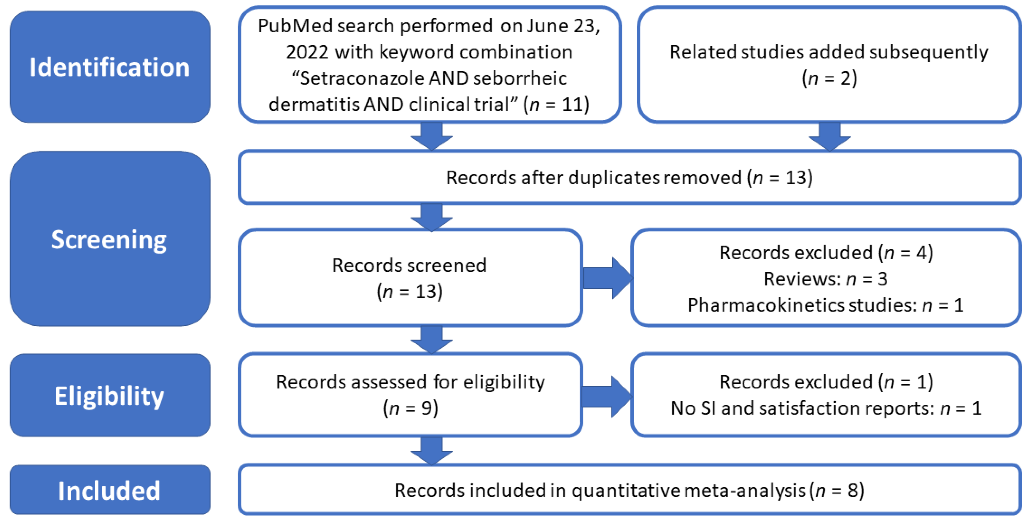
2. Materials and Methods
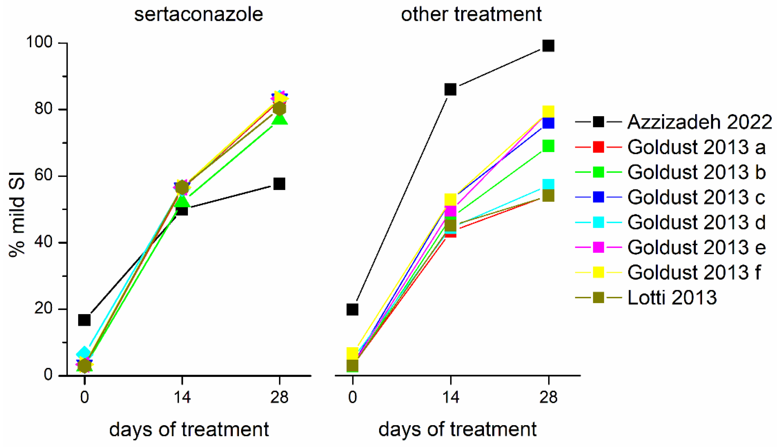
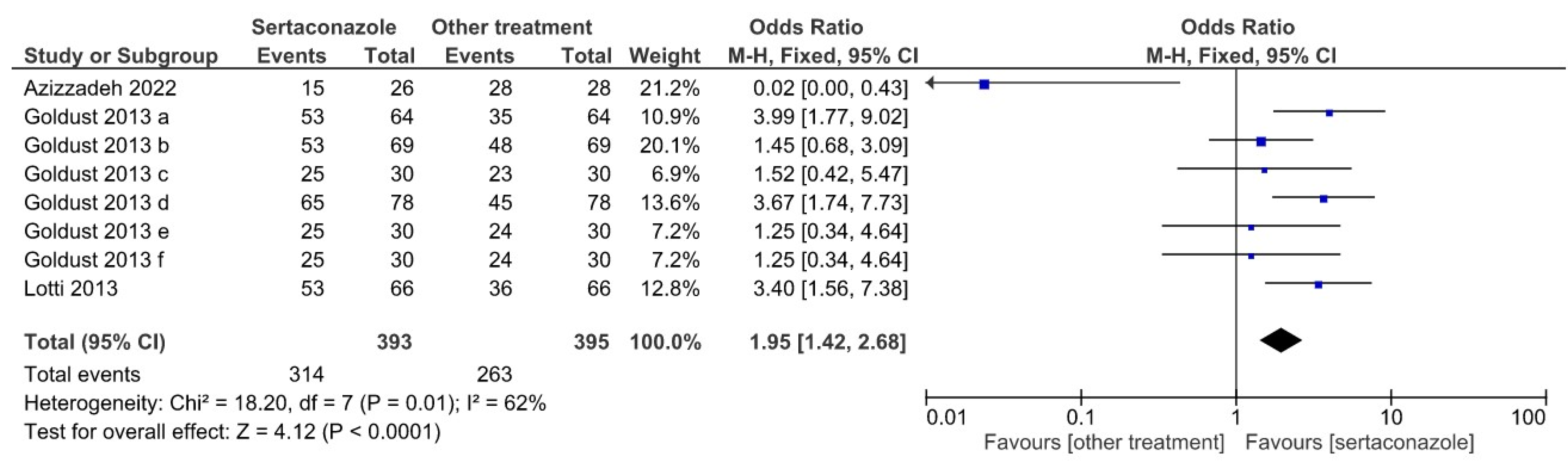
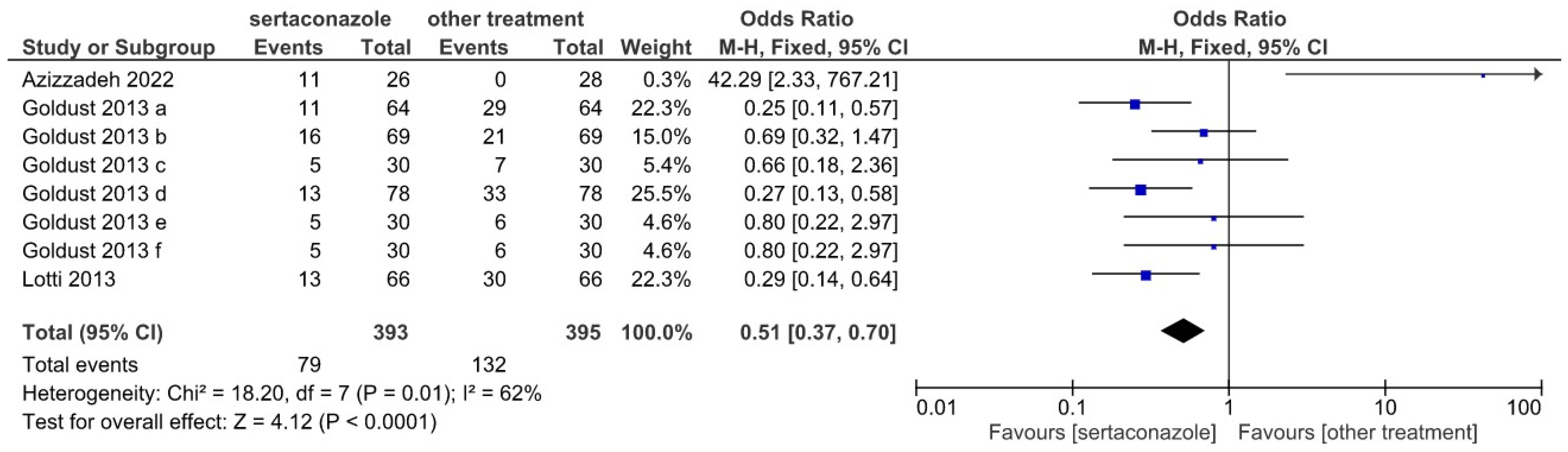
We performed a web-based search of the PubMed medical literature database (https://pubmed.ncbi.nlm.nih.gov accessed on 23 June 2022) with a specific combination of search terms: “Sertaconazole AND seborrheic dermatitis AND clinical trial”. This search retrieved 11 results, supplemented with another two papers retrieved by searching the “Similar articles” section displayed in PubMed when accessing a search hit. From this initial group of publications, we excluded two review papers, one paper addressing the pharmacokinetics and side effects of different sertaconazole nitrate formulations, and in the eligibility assessment phase, one paper that did not report adequate quantitative data, such as the scoring index (SI) assessment of treatment effects, as recommended by Koca et al. [22]. The selection procedure of the studies included in this meta-analysis was compatible with the principles and guidelines of PRISMA (Preferred Reporting Items for Systematic Re-views and Meta-Analyses) [23,24], and the consecutive selection steps are illustrated in Figure 1.
The selected and excluded studies were uploaded to a meta-analysis project created in Review Manager (RevMan) Version 5.4.1, The Cochrane Collaboration, 2020 [25]. Subsequently, we performed three quantitative comparisons for dichotomous variables (frequencies of certain outcomes in experimental vs. control group), with the same software package, selecting a Cochran–Mantel–Haenszel model with fixed effect and computation of odds ratios (OR) as a measure of effect, and a 95% confidence interval.

3. Results

For the eight studies finally included in the meta-analysis, listed in Table 1, as quantitative variables for comparison, we retrieved the scoring indexes (SI) at different time intervals over the duration of treatment (28 days), particularly the frequencies of mild SI and cumulated frequencies of moderate or severe SI in the treatment (sertaconazole) vs. control (other treatments) groups, as well as the frequencies of “good” rankings in the patient satisfaction questionnaires administered at the end of treatment (28 days).
The dynamics of the percentages of patients included in the mild SI groups at different moments of time (beginning of treatment, 14 days of treatment, and 28 days of treatment), illustrated in Figure 2, suggest differences in treatment outcomes between the sertaconazole group and control group, receiving a different treatment.
Figure 3 represents a Forrest plot showing the OR of sertaconazole vs. other treatments in the eight selected studies for the mild SI at 28 days of treatment. The cumulated number of patients was 393 in the sertaconazole groups and 395 in the other treatment groups; the OR was 1.95, favoring sertaconazole (95% confidence interval of 1.42–2.68), the heterogeneity test yielded a χ2 value of 18.20 (p = 0.01), and the overall effect yielded a z of 4.12 (p < 0.0001).
Complementary, the cumulated frequency of moderate or severe SI in the sertaconazole vs. other treatments groups (Forrest plot shown in Figure 4) yielded an OR of 0.51 (95% confidence interval 0.37–0.70), again favoring sertaconazole. The χ2 value was 18.20 (p = 0.01) and the overall effect featured a z of 4.12 (p < 0.0001).
Figure 5 illustrates a Forrest plot for the analysis of frequencies of patients indicating a “good” ranking in the satisfaction self-assessment questionnaire administered at 28 days of treatment; the OR was 3.00 (95% confidence interval 2.13–4.23), again favoring sertaconazole vs. other treatments. The heterogeneity test yielded a χ2 value of 20.12 (p = 0.005), and the overall effect a z of 6.30 (p < 0.00001).

4. Discussion

Seborrheic dermatitis is a chronic inflammatory disease with a pathogenesis closely related to the abnormal proliferation of fungi on the skin [34]. Thus, antifungals are considered first-line treatments. The most widely used topical antifungals are azoles, with very good results being obtained, especially in the case of topical ketoconazole 2%. Ciclopirox and terbinafine are other antifungals that can be used [35]. Topical corticosteroids are prescribed in SD for their anti-inflammatory effect, but patients must be carefully monitored considering the well-known adverse effects (thinning of the skin or telangiectasia), which occur especially with long-term uninterrupted use [15]. Therefore, low- to mid-potency corticosteroids are recommended. Calcineurin inhibitors (pimecrolimus and tacrolimus) can be employed to avoid the adverse effects of corticosteroids [36]. A single-blinded, randomized controlled trial that evaluated the effectiveness of hydrocortisone 1% ointment compared with tacrolimus 0.1% ointment found similar results [37]. An analysis of topical medication with an anti-inflammatory effect for SD, including studies published until September 2013, showed that there are no significant differences between corticosteroids and calcineurin inhibitors in the short term. Additionally, the results were similar in terms of their total clearance, when the group treated with corticosteroids was compared with the group treated with azoles [38]. A recent clinical trial revealed that tacrolimus 0.1% ointment is more effective than ciclopiroxolamine 1% cream for maintenance therapy in patients with a severe form of SD involving the face [39]. Okokon et al. analyzed the effectiveness of topical antifungals in SD and obtained sufficient data only in the case of ketoconazole, ciclopirox, and bifonazole. They concluded that ketoconazole and ciclopirox showed similar results regarding the alleviation of symptoms in SD. The effectiveness of ketoconazole was similar to that of topical corticosteroids, but with fewer adverse effects. The tolerability of bifonazole was lower compared with the placebo [40]. The systemic treatment is reserved for severe cases and includes itraconazole, terbinafine, fluconazole, ketoconazole, pramiconazole, prednisone, and isotretinoin. Most of the available studies focused on evaluating the effectiveness of oral antifungals [41].
Sertaconazole nitrate is a relatively new imidazole antifungal agent with a particular structure, consisting of a benzothiophene ring similar to the indole ring of tryptophan [42]. It exhibits a broad-spectrum antifungal activity against dermatophytes and yeasts, opportunistic filamentous fungi, and Gram positive bacteria [29]. The clinical efficacy of sertaconazole has been demonstrated in various skin mycoses, including candidiasis, dermatophytosis, and pityriasis versicolor [43]. Topical administration is safe, systemic absorption is low, and the cutaneous retention time is high. Sertaconazole has the advantage of being available in a variety of pharmaceutical forms (cream, gel, solution, powder, and shampoo) [44]. Depending on its concentration and the microorganism involved, sertaconazole has both a fungistatic and a fungicidal effect. The antifungal agent selectively inhibits the biosynthesis of ergosterol acting on fungal 14α-demethylase, which favors the accumulation of 14-a-methylated sterols, and subsequently cell growth and replication are dysregulated. It also has the ability to insert into the fungal cell membrane by binding to non-sterol lipids, resulting in an alteration of membrane permeability and leakage of the intracellular components [45].
Liebel et al. showed that sertaconazole inhibits the release of pro-inflammatory cytokines from activated lymphocytes in animal models of irritant contact dermatitis. Compared with other antifungal agents, the best anti-inflammatory effect was observed for sertaconazole. Only fluconazole and sertaconazole inhibited neurogenic inflammation, suggesting the possible role of these antifungal agents in the relief of itching [46]. In addition, sertaconazole exhibits anti-inflammatory and anti-itch effects through the activation of the p38-COX-2-PGE2 pathway in keratinocytes and human peripheral blood mononuclear cells [47,48]. Saki et al. revealed that sertaconazole was superior to hydrocortisone for improving the total score of disease in patients with atopic dermatitis. However, there were no differences in terms of pruritus improvement [49].
Elewski et al. conducted an open-label study that evaluated sertaconazole nitrate 2% cream in SD patients, and showed that it was effective with a good safety profile and improved scaling, erythema, induration, and pruritus [50]. Our meta-analysis included eight studies evaluating the efficacy of sertaconazole 2% cream in SD in comparison with the following treatments: hydrocortisone 1% cream (2 studies), ketoconazole 2% cream (one study), clotrimazole 1% cream (one study), calcineurin inhibitors (pimecrolimus 1% cream, two studies, and tacrolimus 0.03% cream, one study), and metronidazole gel 1% (one study). All studies highlighted a higher level of satisfaction 28 days after the initiation of treatment in the case of patients treated with sertaconazole, with the exception of the study conducted by Azzizadeh [26]. One month after stopping the treatment, no relapses were reported, regardless of the therapy administered. With respect to the level of satisfaction at 28 days, greater differences were observed when sertaconazole was compared with antifungals (clotrimazole and ketaconazole) than when it was compared with hydrocortisone or calcineurin inhibitors. Azizzadeh et al. noticed a significantly better improvement in SD in the case of patients treated with pimecrolimus 1% compared with those treated with sertaconazole 2%. In addition, relapses and adverse effects were less frequent in the group of patients treated with pimecrolimus 1%. The authors concluded that pimecrolimus 1% led to a faster improvement in symptoms than sertaconazole [26].
All studies included in the present meta-analysis evaluated the severity of the disease by the Scoring Index (SI), as recommended by Koca et al. The score is based on the evaluation of erythema, desquamation, itching, and irritation of each area, which is graded from zero to three (nonexistence = 0, mild = 1, moderate = 2, severe = 3). The sum of the scores was considered as the SD rank (classified into three ranges: mild (0–4), moderate (5–8), and severe (9–12)). At 14 days since the beginning of the treatment, no significant differences were observed between patients treated with sertaconazole and those treated with other topical treatments. At 28 days since the beginning of the treatment, the sertaconazole regimen was associated with a significantly higher percentage of patients with mild SI and a lower percentage of patients with moderate or severe SI (odds ratio 0.51) than the other investigated topical treatments (odds ratio 1.95).
This research has some limitations. The number of studies analyzed was small and the studies included reduced numbers of patients. In addition, none of the studies followed up the patients for more than one month; therefore, the data on relapses are inconclusive. Medium-/long-term studies are needed to establish if the disease-free interval is greater with one drug than with another.

5. Conclusions

The presented studies support the effectiveness of sertaconazole in SD therapy. Sertaconazole treatment was associated with a high level of satisfaction 28 days after the initiation of therapy. Additional studies including larger numbers of patients are needed to validate these results.

Author Contributions

All of the authors equally contributed to this paper. Conceptualization, M.I.M., M.T. and C.I.M.; data curation, A.A.; formal analysis, A.A. and C.M.; funding acquisition, C.M.; investigation, C.M.; methodology, M.I.M., A.A. and C.I.M.; project administration, S.R.G.; resources, M.T.; software, M.T.; supervision, S.R.G.; validation, C.I.M. and M.I.M.; visualization, C.M. and S.R.G.; writing—original draft, M.I.M., A.A. and C.I.M.; writing—review and editing, M.T. and S.R.G. All authors have read and agreed to the published version of the manuscript.

Funding

This research received no external funding.

Institutional Review Board Statement

Not applicable.

Informed Consent Statement

Not applicable.

Data Availability Statement

Not applicable.

Conflicts of Interest

The authors declare no conflict of interest.

References

  1. Karakadze, M.A.; Hirt, P.A.; Wikramanayake, T.C. The Genetic Basis of Seborrhoeic Dermatitis: A Review. J. Eur. Acad. Dermatol. Venereol. 2018, 32, 529–536. [Google Scholar] [CrossRef] [PubMed]
  2. Tao, R.; Li, R.; Wang, R. Skin Microbiome Alterations in Seborrheic Dermatitis and Dandruff: A Systematic Review. Exp. Dermatol. 2021, 30, 1546–1553. [Google Scholar] [CrossRef] [PubMed]
  3. Wikramanayake, T.C.; Borda, L.J.; Miteva, M.; Paus, R. Seborrheic Dermatitis—Looking beyond Malassezia. Exp. Dermatol. 2019, 28, 991–1001. [Google Scholar] [CrossRef] [PubMed]
  4. Dessinioti, C.; Katsambas, A. Seborrheic Dermatitis: Etiology, Risk Factors, and Treatments: Facts and controversies. Clin. Dermatol. 2013, 31, 343–351. [Google Scholar] [CrossRef]
  5. Mitran, M.I.; Nicolae, I.; Tampa, M.; Mitran, C.I.; Caruntu, C.; Sarbu, M.I.; Ene, C.D.; Matei, C.; Georgescu, S.R.; Popa, M.I. Reactive Carbonyl Species as Potential Pro-Oxidant Factors Involved in Lichen Planus Pathogenesis. Metabolites 2019, 9, 213. [Google Scholar] [CrossRef]
  6. Nakai, K.; Tsuruta, D. What Are Reactive Oxygen Species, Free Radicals, and Oxidative Stress in Skin Diseases? Int. J. Mol. Sci. 2021, 22, 10799. [Google Scholar] [CrossRef]
  7. Trüeb, R.M. Oxidative Stress and Its Impact on Skin, Scalp and Hair. Int. J. Cosmet. Sci. 2021, 43 (Suppl. 1), S9–S13. [Google Scholar] [CrossRef]
  8. Emre, S.; Kalkan, G.; Erdoğan, S.; Aktaş, A.; Ergin, M. Dynamic Thiol/Disulfide Balance in Patients with Seborrheic Dermatitis: A Case-Control Study. Saudi J. Med. Med. Sci. 2020, 8, 12–16. [Google Scholar] [CrossRef]
  9. Tampa, M.; Nicolae, I.; Ene, C.D.; Sarbu, I.; Matei, C.; Georgescu, S.R. Vitamin C and Thiobarbituric Acid Reactive Substances (TBARS) in Psoriasis Vulgaris Related to Psoriasis Area Severity Index (PASI). Rev. Chim. 2017, 68, 43–47. [Google Scholar] [CrossRef]
  10. Saunte, D.M.L.; Gaitanis, G.; Hay, R.J. Malassezia-Associated Skin Diseases, the Use of Diagnostics and Treatment. Front. Cell. Infect. Microbiol. 2020, 10, 112. [Google Scholar] [CrossRef]
  11. Malassez, L. Note Sur Le Champignon Du Pityriasis Simple. Arch. Physiol. 1874, 1, 451–459. [Google Scholar]
  12. Wang, K.; Cheng, L.; Li, W.; Jiang, H.; Zhang, X.; Liu, S.; Huang, Y.; Qiang, M.; Dong, T.; Li, Y.; et al. Susceptibilities of Malassezia Strains from Pityriasis Versicolor, Malassezia Folliculitis and Seborrheic Dermatitis to Antifungal Drugs. Heliyon 2020, 6, e04203. [Google Scholar] [CrossRef] [PubMed]
  13. Zhang, H.; Ran, Y.; Xie, Z.; Zhang, R. Identification of Malassezia Species in Patients with Seborrheic Dermatitis in China. Mycopathologia 2013, 175, 83–89. [Google Scholar] [CrossRef] [PubMed]
  14. Granger, C.; Balato, A.; Goñi-de-Cerio, F.; Garre, A.; Narda, M. Novel Non-Steroidal Facial Cream Demonstrates Antifungal and Anti-Inflammatory Properties in Ex Vivo Model for Seborrheic Dermatitis. Dermatol. Ther. 2019, 9, 571–578. [Google Scholar] [CrossRef]
  15. Piquero-Casals, J.; Hexsel, D.; Mir-Bonafé, J.F.; Rozas-Muñoz, E. Topical Non-Pharmacological Treatment for Facial Seborrheic Dermatitis. Dermatol. Ther. 2019, 9, 469–477. [Google Scholar] [CrossRef] [PubMed]
  16. Adalsteinsson, J.A.; Kaushik, S.; Muzumdar, S.; Guttman-Yassky, E.; Ungar, J. An Update on the Microbiology, Immunology and Genetics of Seborrheic Dermatitis. Exp. Dermatol. 2020, 29, 481–489. [Google Scholar] [CrossRef]
  17. Yu, Y.; Dunaway, S.; Champer, J.; Kim, J.; Alikhan, A. Changing Our Microbiome: Probiotics in Dermatology. Br. J. Dermatol. 2019, 182, 39–46. [Google Scholar] [CrossRef]
  18. Tanaka, A.; Cho, O.; Saito, C.; Saito, M.; Tsuboi, R.; Sugita, T. Comprehensive Pyrosequencing Analysis of the Bacterial Microbiota of the Skin of Patients with Seborrheic Dermatitis. Microbiol. Immunol. 2016, 60, 521–526. [Google Scholar] [CrossRef]
  19. Soares, R.C.; Camargo-Penna, P.H.; de Moraes, V.; De Vecchi, R.; Clavaud, C.; Breton, L.; Braz, A.S.; Paulino, L.C. Dysbiotic Bacterial and Fungal Communities Not Restricted to Clinically Affected Skin Sites in Dandruff. Front. Cell. Infect. Microbiol. 2016, 157. [Google Scholar] [CrossRef]
  20. Paulino, L.C. New Perspectives on Dandruff and Seborrheic Dermatitis: Lessons We Learned from Bacterial and Fungal Skin Microbiota. Eur. J. Dermatol. 2017, 27 (Suppl. 1), 4–7. [Google Scholar] [CrossRef]
  21. Carrillo-Muñoz, A.J.; Tur-Tur, C.; Giusiano, G.; Marcos-Arias, C.; Eraso, E.; Jauregizar, N.; Quindós, G. Sertaconazole: An Antifungal Agent for the Topical Treatment of Superficial Candidiasis. Expert Rev. Anti-Infect. Ther. 2013, 11, 347–358. [Google Scholar] [CrossRef] [PubMed]
  22. Koca, R.; Altinyazar, H.C.; Eştürk, E. Is Topical Metronidazole Effective in Seborrheic Dermatitis? A Double-blind Study. Int. J. Dermatol. 2003, 42, 632–635. [Google Scholar] [CrossRef] [PubMed]
  23. Moher, D.; Liberati, A.; Tetzlaff, J.; Altman, D.G.; PRISMA Group. Preferred Reporting Items for Systematic Reviews and Meta-Analyses: The PRISMA Statement. Ann. Intern. Med. 2009, 151, 264–269. [Google Scholar] [CrossRef]
  24. Page, M.J.; McKenzie, J.E.; Bossuyt, P.M.; Boutron, I.; Hoffmann, T.C.; Mulrow, C.D.; Shamseer, L.; Tetzlaff, J.M.; Akl, E.A.; Brennan, S.E. The PRISMA 2020 Statement: An Updated Guideline for Reporting Systematic Reviews. Syst. Rev. 2021, 10, 1–11. [Google Scholar] [CrossRef]
  25. Cohrane Collaboration. Review Manager (RevMan), version 5.2.3; Computer Program; The Nordic Cochrane Centre: Copenhagen, Denmark, 2012. [Google Scholar]
  26. Azizzadeh, M.; Pahlevan, D.; Bagheri, B. The Efficacy and Safety of Pimecrolimus 1% Cream vs. Sertaconazole 2% Cream in the Treatment of Patients with Facial Seborrhoeic Dermatitis: A Randomized Blinded Trial. Clin. Exp. Dermatol. 2022, 47, 926–931. [Google Scholar] [CrossRef] [PubMed]
  27. Goldust, M.; Rezaee, E.; Rouhani, S. Double Blind Study of Sertaconazole 2% Cream vs. Clotrimazole 1% Cream in Treatment of Seborrheic Dermatitis. Ann. Parasitol. 2013, 59, 25–29. [Google Scholar]
  28. Goldust, M.; Rezaee, E.; Masoudnia, S.; Raghifar, R. Clinical Study of Sertaconazole 2% Cream vs. Hydrocortisone 1% Cream in the Treatment of Seborrheic Dermatitis. Ann. Parasitol. 2013, 59, 119–123. [Google Scholar]
  29. Goldust, M.; Ranjkesh, M.R.; Amirinia, M.; Golforoushan, F.; Rezaee, E.; Rezazadeh Saatlou, M.A. Sertaconazole 2% Cream versus Hydrocortisone 1% Cream in the Treatment of Seborrheic Dermatitis. J. Dermatolog. Treat. 2013, 1–3. [Google Scholar] [CrossRef]
  30. Goldust, M.; Rezaee, E.; Raghifar, R. A Double Blind Study of the Effectiveness of Sertaconazole 2% Cream vs. Metronidazole 1% Gel in the Treatment of Seborrheic Dermatitis. Ann. Parasitol. 2013, 59, 173–177. [Google Scholar]
  31. Goldust, M.; Rezaee, E.; Raghifar, R. Treatment of Seborrheic Dermatitis: Comparison of Sertaconazole 2% Cream versus Pimecrolimus 1% Cream. Ir. J. Med. Sci. 2013, 182, 703–706. [Google Scholar] [CrossRef]
  32. Goldust, M.; Rezaee, E.; Raghifar, R.; Hemayat, S. Treatment of Seborrheic Dermatitis: The Efficiency of Sertaconazole 2% Cream vs. Tacrolimus 0.03% Cream. Ann. Parasitol. 2013, 59, 73–77. [Google Scholar] [PubMed]
  33. Lotti, T.; Goldust, M.; Rezaee, E. Treatment of Seborrheic Dermatitis, Comparison of Sertaconazole 2% Cream versus Ketoconazole 2% Cream. J. Dermatol. Treat. 2013, 1–3. [Google Scholar] [CrossRef] [PubMed]
  34. Georgescu, S.R.; Mitran, C.I.; Mitran, M.I.; Matei, C.; Popa, G.L.; Erel, O.; Tampa, M. Thiol-Disulfide Homeostasis in Skin Diseases. J. Clin. Med. 2022, 11, 1507. [Google Scholar] [CrossRef] [PubMed]
  35. Borda, L.J.; Perper, M.; Keri, J.E. Treatment of Seborrheic Dermatitis: A Comprehensive Review. J. Dermatol. Treat. 2019, 30, 158–169. [Google Scholar] [CrossRef] [PubMed]
  36. Goldenberg, G. Optimizing Treatment Approaches in Seborrheic Dermatitis. J. Clin. Aesthet. Dermatol. 2013, 6, 44–49. [Google Scholar]
  37. Papp, K.A.; Papp, A.; Dahmer, B.; Clark, C.S. Single-Blind, Randomized Controlled Trial Evaluating the Treatment of Facial Seborrheic Dermatitis with Hydrocortisone 1% Ointment Compared with Tacrolimus 0.1% Ointment in Adults. J. Am. Acad. Dermatol. 2012, 67, e11–e15. [Google Scholar] [CrossRef]
  38. Kastarinen, H.; Oksanen, T.; Okokon, E.O.; Kiviniemi, V.V.; Airola, K.; Jyrkkä, J.; Oravilahti, T.; Rannanheimo, P.K.; Verbeek, J.H. Topical Anti-Inflammatory Agents for Seborrhoeic Dermatitis of the Face or Scalp. Cochrane Database Syst. Rev. 2014, 5, CD009446. [Google Scholar] [CrossRef]
  39. Joly, P.; Tejedor, I.; Tetart, F.; Cailleux, H.C.; Barrel, A.; De Preville, P.A.; Mion-Mouton, N.; Gabison, G.; Baricault, S. Tacrolimus 0.1% versus Ciclopiroxolamine 1% for Maintenance Therapy in Patients with Severe Facial Seborrheic Dermatitis: A Multicenter, Double-Blind, Randomized Controlled Study. J. Am. Acad. Dermatol. 2021, 84, 1278–1284. [Google Scholar] [CrossRef]
  40. Okokon, E.O.; Verbeek, J.H.; Ruotsalainen, J.H. Topical Antifungals for Seborrhoeic Dermatitis. Cochrane Database Syst. Rev. 2015, 5, CD008138. [Google Scholar] [CrossRef]
  41. Gupta, A.K.; Richardson, M.; Paquet, M. Systematic Review of Oral Treatments for Seborrheic Dermatitis. J. Eur. Acad. Dermatol. Venereol. 2014, 28, 16–26. [Google Scholar] [CrossRef]
  42. Chatterjee, D.; Ghosh, S.K.; Sen, S.; Sarkar, S.; Hazra, A.; De, R. Efficacy and Tolerability of Topical Sertaconazole versus Topical Terbinafine in Localized Dermatophytosis: A Randomized, Observer-Blind, Parallel Group Study. Indian J. Pharmacol. 2016, 48, 659–664. [Google Scholar] [CrossRef] [PubMed]
  43. Torres, J.; Márquez, M.; Camps, F. Sertaconazole in the Treatment of Mycoses: From Dermatology to Gynecology. Int. J. Gynecol. Obstet. 2000, 71, 3–20. [Google Scholar] [CrossRef]
  44. Palacín, C.; Tarrago, C.; Ortiz, J. Sertaconazole: Pharmacology of a Gynecological Antifungal Agent. Int. J. Gynecol. Obstet. 2000, 71, 37–46. [Google Scholar] [CrossRef]
  45. Croxtall, J.D.; Plosker, G.L. Sertaconazole: A Review of Its Use in the Management of Superficial Mycoses in Dermatology and Gynaecology. Drugs 2009, 69, 339–359. [Google Scholar] [CrossRef] [PubMed]
  46. Liebel, F.; Lyte, P.; Garay, M.; Babad, J.; Southall, M.D. Anti-Inflammatory and Anti-Itch Activity of Sertaconazole Nitrate. Arch. Dermatol. Res. 2006, 298, 191–199. [Google Scholar] [CrossRef]
  47. Kaur, S.; Sur, R.; Liebel, F.T.; Southall, M.D. Induction of Prostaglandin D2 through the P38 MAPK Pathway Is Responsible for the Antipruritic Activity of Sertaconazole Nitrate. J. Investig. Dermatol. 2010, 130, 2448–2456. [Google Scholar] [CrossRef]
  48. Zhang, W.; Zhou, L.; Qin, S.; Jiang, J.; Huang, Z.; Zhang, Z.; Zhang, X.; Shi, Z.; Lin, J. Sertaconazole Provokes Proapoptotic Autophagy via Stabilizing TRADD in Nonsmall Cell Lung Cancer Cells. MedComm 2021, 2, 821–837. [Google Scholar] [CrossRef]
  49. Saki, N.; Jowkar, F.; Alyaseen, S. Comparison of Sertaconazole 2% Cream versus Hydrocortisone 1% Ointment in the Treatment of Atopic Dermatitis. J. Dermatol. Treat. 2013, 24, 447–449. [Google Scholar] [CrossRef]
  50. Elewski, B.E.; Cantrell, W.C. An Open-Label Study of the Safety and Efficacy of Sertaconazole Nitrate in the Treatment of Seborrheic Dermatitis. J. Drugs Dermatol. JDD 2011, 10, 895–899. [Google Scholar]
Figure 1. Retrieval and selection of publications to be included in the meta-analysis according to the PRISMA guidelines.
Jpm 12 01540 g001
Figure 2. Dynamics of percentages of patients featuring mild SI at different times (0, 14, and 28 days of treatment) in the sertaconazole vs. control groups in the eight studies included in the meta-analysis.
Jpm 12 01540 g002
Figure 3. Forrest plot illustrating the effect of sertaconazole vs. other treatments in the eight selected studies for the mild SI at 28 days of treatment (OR with 95% confidence interval, heterogeneity, and overall effect).
Jpm 12 01540 g003
Figure 4. Forrest plot illustrating the effect of sertaconazole vs. other treatments in the eight selected studies for the moderate and severe SI at 28 days of treatment (OR with 95% confidence interval, heterogeneity, and overall effect).
Jpm 12 01540 g004
Figure 5. Forrest plot illustrating the effect of sertaconazole vs. other treatments in the eight selected studies for the “good” ranking in the self-assessment and patient satisfaction questionnaire at 28 days of treatment (OR with 95% confidence interval, heterogeneity, and overall effect).
Jpm 12 01540 g005
Table 1. Main features of the clinical trials included in the present meta-analysis.
StudyDouble BlindedRando-MizedGroupsMean
Age
SexKind of LesionTreatment
Azizzadeh et al., 2022 [26]YYpimecrolimus 1% cream vs. sertaconazole 2% cream27.5M/F-twice a day for four weeks
Goldust et al., 2013 a [27]YYsertaconazole 2% cream vs. clotrimazole 1% cream36.62
±13.18
M/Flocalized: 57%
generalized: 43%
twice a day for four weeks
Goldust et al., 2013 b [28]YYsertaconazole 2% cream vs. hydrocortisone 1% cream36.45
±13.23
M/Flocalized: 59.4%
generalized: 40.6%
twice a day for four weeks
Goldust et al., 2013 c [29]YYsertaconazole 2% cream vs. hydrocortisone 1% cream32.23
±12.09
M/Flocalized: 58.3%
generalized: 41.7%
twice a day for four weeks
Goldust et al., 2013 d [30]YYsertaconazole 2% cream vs. metronidazole 1% gel32.34
±12.56
M/Flocalized: 56.4%
generalized: 43.7%
twice a day for four weeks
Goldust et al., 2013 e [31]YYsertaconazole 2% cream vs. pimecrolimus 1% cream30.12
±12.56
M/Flocalized: 60%
generalized: 40%
twice a day for four weeks
Goldust et al., 2013 f [32]YYsertaconazole 2% cream vs. tacrolimus 0.03% cream32.45
±12.78
M/Flocalized: 58.3%
generalized: 41.7%
twice a day for four weeks
Lotti et al., 2013 [33]YYsertaconazole 2% cream vs. ketoconazole 2% cream42
±14.36
M/Flocalized: 36.5%
generalized: 29.5%
twice a day for four weeks
Publisher’s Note: MDPI stays neutral with regard to jurisdictional claims in published maps and institutional affiliations.

