Next Issue
Volume 14, April-1
Previous Issue
Volume 14, March-1


Diagnostics, Volume 14, Issue 6 (March-2 2024) – 99 articles

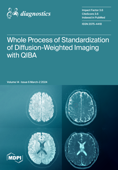
Cover Story (view full-size image): Diffusion-weighted imaging (DWI) and its derivative, the apparent diffusion coefficient (ADC), are functional sequences in magnetic resonance imaging (MRI) that reflect the diffusivity of water molecules and are well-known for demonstrating tissue cellularity. Recently, there have been efforts to use the ADC as a quantitative imaging biomarker for assessing treatment response, leading to an increased need for the validation of MRI equipment and protocols. This study provides the whole process of validating MRI equipment and protocols for DWI, using standardized phantoms and software proposed by the Quantitative Imaging Biomarker Alliance (QIBA). View this paper
  • Issues are regarded as officially published after their release is announced to the table of contents alert mailing list.
  • You may sign up for e-mail alerts to receive table of contents of newly released issues.
  • PDF is the official format for papers published in both, html and pdf forms. To view the papers in pdf format, click on the "PDF Full-text" link, and use the free Adobe Reader to open them.
Order results
Result details
Section
Select all
Export citation of selected articles as:
19 pages, 5942 KiB  
Article
Identification and Validation of T-Cell Exhaustion Signature for Predicting Prognosis and Immune Response in Pancreatic Cancer by Integrated Analysis of Single-Cell and Bulk RNA Sequencing Data
by Yaowu Zhu, Li Tan, Danju Luo and Xiong Wang
Diagnostics 2024, 14(6), 667; https://doi.org/10.3390/diagnostics14060667 - 21 Mar 2024
Viewed by 736
Abstract
Purpose: Pancreatic cancer (PACA) is one of the most fatal malignancies worldwide. Immunotherapy is largely ineffective in patients with PACA. T-cell exhaustion contributes to immunotherapy resistance. We investigated the prognostic potential of T-cell exhaustion-related genes (TEXGs). Methods: A single-cell RNA (scRNA) sequencing dataset [...] Read more.
Purpose: Pancreatic cancer (PACA) is one of the most fatal malignancies worldwide. Immunotherapy is largely ineffective in patients with PACA. T-cell exhaustion contributes to immunotherapy resistance. We investigated the prognostic potential of T-cell exhaustion-related genes (TEXGs). Methods: A single-cell RNA (scRNA) sequencing dataset from Tumor Immune Single-Cell Hub (TISCH) and bulk sequencing datasets from the Cancer Genome Atlas (TCGA) and Genotype-Tissue Expression (GTEx) were used to screen differentially expressed TEXGs. Kaplan–Meier survival, LASSO regression, and univariate/multivariate Cox regression analyses were performed to construct a TEXG risk model. This model was used to predict the prognosis, tumor immune microenvironment, and immunotherapy response. The PACA cohorts from the ICGC and GSE71729 datasets were used to validate the risk model. Pan-cancer expression of SPOCK2 was determined using the TISCH database. Results: A six-gene (SPOCK2, MT1X, LIPH, RARRES3, EMP1, and MEG3) risk model was constructed. Patients with low risk had prolonged survival times in both the training (TCGA-PAAD, n = 178) and validation (ICGC-PACA-CA, ICGC-PAAD-US, and GSE71729, n = 412) datasets. Multivariate Cox regression analysis demonstrated that the risk score was an independent prognostic variable for PACA. High-risk patients correlated with their immunosuppressive status. Immunohistochemical staining confirmed the changes in TEXGs in clinical samples. Moreover, pan-cancer scRNA sequencing datasets from TISCH analysis indicated that SPOCK2 may be a novel marker of exhausted CD8+ T-cells. Conclusion: We established and validated a T-cell exhaustion-related prognostic signature for patients with PACA. Moreover, our study suggests that SPOCK2 is a novel marker of exhausted CD8+ T cells. Full article
(This article belongs to the Section Pathology and Molecular Diagnostics)
Show Figures

Figure 1

10 pages, 415 KiB  
Article
Osteoporosis Is Associated with an Increased Risk of Colorectal Neoplasms Regardless of Sex: Nationwide Population-Based Cohort Study
by Seung Heon Yoo, Ji Hyung Nam, Dong Jun Oh, Geun U Park, Sang Hoon Kim, Hyoun Woo Kang, Jae Hak Kim and Yun Jeong Lim
Diagnostics 2024, 14(6), 666; https://doi.org/10.3390/diagnostics14060666 - 21 Mar 2024
Viewed by 633
Abstract
Vitamin D may have anticancer effects against colorectal cancer (CRC). Bone mineral density (BMD) reflects the long-term vitamin D status. This study investigated the association between osteoporosis and colorectal neoplasms (CRN). The data were obtained from the National Health Insurance Service sample cohort, [...] Read more.
Vitamin D may have anticancer effects against colorectal cancer (CRC). Bone mineral density (BMD) reflects the long-term vitamin D status. This study investigated the association between osteoporosis and colorectal neoplasms (CRN). The data were obtained from the National Health Insurance Service sample cohort, which included 60,386 osteoporosis patients and 8224 controls who underwent BMD in 2002–2019. The logistic regression models included age, sex, income level, and comorbidity. Sensitivity tests were performed using the data from the National Health Screening Program. In total, 7706 (11.2%) patients were diagnosed with CRN, and the proportion was significantly higher in osteoporosis patients than in controls (11.7% vs. 8.1%). In the multivariate analysis, osteoporosis was associated with an increased risk of CRN (odds ratio (OR) = 1.91, 95% confidence interval = 1.75–2.09, p < 0.0001), which was significant for both colorectal adenomas and CRC (OR = 1.88 and 1.83, respectively). A subgroup analysis by sex revealed a significant association between osteoporosis and CRN in both women and men (OR = 2.06 and 1.66, respectively). The sensitivity tests revealed results similar to those of the original dataset. In conclusion, osteoporosis is significantly associated with CRN risk in both sexes. In high-risk patients with low BMD, appropriate screening for CRN and vitamin D supplementation are required, regardless of sex. Full article
(This article belongs to the Section Pathology and Molecular Diagnostics)
Show Figures

Figure 1

21 pages, 2090 KiB  
Review
Ultrasound Elastography as a Diagnostic Tool for Peyronie’s Disease: A State-of-the-Art Review
by Gianni Paulis, Giovanni De Giorgio and Andrea Paulis
Diagnostics 2024, 14(6), 665; https://doi.org/10.3390/diagnostics14060665 - 21 Mar 2024
Viewed by 629
Abstract
Elastography is a noninvasive method that utilizes ultrasound imaging to assess the elasticity and stiffness of soft tissue. Peyronie’s disease (PD) is a chronic inflammatory condition that affects the male penis, causing the formation of fibrous plaques. This alters the penis’s elasticity and [...] Read more.
Elastography is a noninvasive method that utilizes ultrasound imaging to assess the elasticity and stiffness of soft tissue. Peyronie’s disease (PD) is a chronic inflammatory condition that affects the male penis, causing the formation of fibrous plaques. This alters the penis’s elasticity and can lead to changes in its shape. Ultrasound elastography (UE) is an important advancement in the diagnosis of PD. It not only identifies plaques, but it also measures their rigidity, providing crucial information to monitor changes during and after treatment. We conducted a narrative review of the scientific literature to identify articles that discuss the use of elastography in the diagnostic study of PD. The purpose of this study was to describe the “state of the art” in the diagnostic use of ultrasound in combination with elastography to highlight any benefits in the diagnosis of PD. We found 12 relevant articles after searching PubMed, Embase, and Google Scholar using the keywords “ultrasound elastography” and “Peyronie’s disease”, including eight clinical studies, two case reports, and two review articles. The results of our review indicate that UE is a useful technique for identifying Peyronie-related plaques, particularly when they are not detectable using a standard ultrasound or physical examination. It is also helpful in monitoring improvements during and after conservative treatments. More research is required to confirm the effectiveness of ultrasound elastography in diagnosing Peyronie’s disease and to determine whether it is better than traditional ultrasound. Full article
(This article belongs to the Special Issue Diagnosis and Management of Andrological Diseases)
Show Figures

Figure 1

12 pages, 275 KiB  
Article
Novel Biomarkers in Evaluating Cardiac Function in Patients on Hemodialysis—A Pilot Prospective Observational Cohort Study
by Lazar Chisavu, Viviana Mihaela Ivan, Adelina Mihaescu, Flavia Chisavu, Oana Schiller, Luciana Marc, Flaviu Bob and Adalbert Schiller
Diagnostics 2024, 14(6), 664; https://doi.org/10.3390/diagnostics14060664 - 21 Mar 2024
Viewed by 574
Abstract
Chronic kidney disease patients treated by hemodialysis present a high cardiovascular morbidity and mortality. There is an imperative need for novel biomarkers for identifying these patients and to offer possible therapeutically interventions. We performed a prospective observational cohort study on 77 patients in [...] Read more.
Chronic kidney disease patients treated by hemodialysis present a high cardiovascular morbidity and mortality. There is an imperative need for novel biomarkers for identifying these patients and to offer possible therapeutically interventions. We performed a prospective observational cohort study on 77 patients in the period of October 2021–October 2023. We measured serum plasma levels of interleukin 1-beta, galectin 3, human suppression of tumorigenicity factor 2, bone morphogenetic protein 2 and fibroblastic growth factor 23 at the inclusion site. We evaluated the correlations of these biomarkers with cardiac function and structure evaluated by echocardiography. The mean age was 61.02 (±11.81) years, with 45 (56.2%) males and with a dialysis vintage of 4.95 (2.4–7.8) years. Median ejection fraction was 51 (43–54%), and more than two-thirds of the patients presented valvular calcifications. Overall mortality was 22%. Interleukin 1-beta was correlated positively with ejection fraction and global longitudinal strain and negatively with left atrium diameter and left ventricle telesystolic diameter. Galectin 3 values were negatively correlated with aortic valve fibrosis and mitral valve calcifications, and human suppression tumorigenicity factor 2 was negatively correlated with mitral valve calcifications. Some of these novel biomarkers could be used to better assess cardiovascular disease in patients on maintenance hemodialysis. Full article
(This article belongs to the Special Issue Diagnosis and Treatment of Kidney Disease)
26 pages, 351 KiB  
Review
The Contribution of Functional Near-Infrared Spectroscopy (fNIRS) to the Study of Neurodegenerative Disorders: A Narrative Review
by Ioannis Liampas, Freideriki Danga, Panagiota Kyriakoulopoulou, Vasileios Siokas, Polyxeni Stamati, Lambros Messinis, Efthimios Dardiotis and Grigorios Nasios
Diagnostics 2024, 14(6), 663; https://doi.org/10.3390/diagnostics14060663 - 21 Mar 2024
Viewed by 827
Abstract
Functional near-infrared spectroscopy (fNIRS) is an innovative neuroimaging method that offers several advantages over other commonly used modalities. This narrative review investigated the potential contribution of this method to the study of neurodegenerative disorders. Thirty-four studies involving patients with Alzheimer’s disease (AD), mild [...] Read more.
Functional near-infrared spectroscopy (fNIRS) is an innovative neuroimaging method that offers several advantages over other commonly used modalities. This narrative review investigated the potential contribution of this method to the study of neurodegenerative disorders. Thirty-four studies involving patients with Alzheimer’s disease (AD), mild cognitive impairment (MCI), frontotemporal dementia (FTD), Parkinson’s disease (PD), or amyotrophic lateral sclerosis (ALS) and healthy controls were reviewed. Overall, it was revealed that the prefrontal cortex of individuals with MCI may engage compensatory mechanisms to support declining brain functions. A rightward shift was suggested to compensate for the loss of the left prefrontal capacity in the course of cognitive decline. In parallel, some studies reported the failure of compensatory mechanisms in MCI and early AD; this lack of appropriate hemodynamic responses may serve as an early biomarker of neurodegeneration. One article assessing FTD demonstrated a heterogeneous cortical activation pattern compared to AD, indicating that fNIRS may contribute to the challenging distinction of these conditions. Regarding PD, there was evidence that cognitive resources (especially executive function) were recruited to compensate for locomotor impairments. As for ALS, fNIRS data support the involvement of extra-motor networks in ALS, even in the absence of measurable cognitive impairment. Full article
(This article belongs to the Section Medical Imaging and Theranostics)
23 pages, 13411 KiB  
Article
Comparison of Tissue Factors in the Ontogenetic Aspects of Human Cholesteatoma
by Kristaps Dambergs, Gunta Sumeraga and Māra Pilmane
Diagnostics 2024, 14(6), 662; https://doi.org/10.3390/diagnostics14060662 - 21 Mar 2024
Viewed by 820
Abstract
Background: An acquired cholesteatoma is a benign but locally aggressive lesion in the middle ear. It is characterized by chronic inflammation and the destruction of surrounding bone. Therefore, the aim of this study was to compare defensins HβD-2 and HβD-4; pro- and anti-inflammatory [...] Read more.
Background: An acquired cholesteatoma is a benign but locally aggressive lesion in the middle ear. It is characterized by chronic inflammation and the destruction of surrounding bone. Therefore, the aim of this study was to compare defensins HβD-2 and HβD-4; pro- and anti-inflammatory cytokines IL-1α and IL-10; proliferation marker Ki-67; transcription factor NF-κβ; angiogenetic factor VEGF; Sonic hedgehog gene protein SHH; and remodeling factors MMP-2, MMP-9, TIMP-2, and TIMP-4 in adult and pediatric cholesteatoma tissue, and to compare these groups with control skin tissue. Methods: The study included 25 cholesteatoma tissue material samples from children, 25 from adults, and 7 deep external ear canal skin samples from cadavers. The tissues were stained immunohistochemically and evaluated using semi-quantitative methods. Nonparametric tests, such as the Kruskal–Wallis test and Spearman rank correlation, were used. Results: There were no statistically discernible differences between the adult and children groups when comparing the relative numbers of factor-positive cells. Conclusions: There are no histopathological differences between adult and children cholesteatoma tissues. Full article
(This article belongs to the Special Issue Advances in Diagnostic Pathology)
Show Figures

Figure 1

16 pages, 5537 KiB  
Article
A Histological Evaluation of the Efficiency of Using Periprosthetic Autologous Fat to Prevent Capsular Contracture Compared to Other Known Methods—An Experimental Study
by Mihaela Pertea, Nadia Aladari, Oxana Madalina Grosu, Stefana Luca, Raluca Tatar, Andrei-Nicolae Avadanei, Madalina Palaghia, Ana Maria Trofin, Sorinel Lunca and Nina Filip
Diagnostics 2024, 14(6), 661; https://doi.org/10.3390/diagnostics14060661 - 21 Mar 2024
Viewed by 522
Abstract
Background: Capsular contracture remains a common complication in silicone breast implantation. The etiology, formation mechanisms, predisposing and favoring factors are still subjects of research. This study aims to demonstrate the effectiveness of using autologous fat introduced periprosthetically in preventing capsular contracture compared to [...] Read more.
Background: Capsular contracture remains a common complication in silicone breast implantation. The etiology, formation mechanisms, predisposing and favoring factors are still subjects of research. This study aims to demonstrate the effectiveness of using autologous fat introduced periprosthetically in preventing capsular contracture compared to other known methods: antibiotics and corticosteroids. Methods: A cohort of 80 Wistar rats was included in the study, divided into four subgroups. All subjects received a silicone implant, implanted in a pocket created along the abdominal midline. The first subgroup served as the control group, with subjects having the implant placed without any treatment. For the second and third subgroups, the implants were treated with an antibiotic solution and intramuscular injections of dexamethasone, respectively. The subjects in the last subgroup received centrifuged autologous fat introduced periprosthetically. Results: The subgroup with autologous fat exhibited a significantly smaller capsule thickness, which was poorly represented, with a smooth surface. The use of autologous fat for treating silicone prosthesis was linked with the lack of acute inflammation around the prosthetic site. Conclusions: Autologous fat helps to minimize the “non-self” reaction, which results in the development of a periprosthetic capsule consisting of mature collagen fibers interspersed with adipocytes. Full article
(This article belongs to the Section Pathology and Molecular Diagnostics)
Show Figures

Figure 1

12 pages, 1974 KiB  
Article
Differences in Histological Subtypes of Invasive Lobular Breast Carcinoma According to Immunohistochemical Molecular Classification
by Ivan Ilić, Jana Cvetković, Ratko Ilić, Ljubiša Cvetković, Aleksandar Milićević, Stefan Todorović and Pavle Ranđelović
Diagnostics 2024, 14(6), 660; https://doi.org/10.3390/diagnostics14060660 - 21 Mar 2024
Viewed by 719
Abstract
The technical complexity of gene expression profiling in routine practice has necessitated the use of surrogate molecular classification of breast cancer, based on immunohistochemical analyses. Background and objectives: The aim of this study was to compare the differences between histological and molecular subtypes [...] Read more.
The technical complexity of gene expression profiling in routine practice has necessitated the use of surrogate molecular classification of breast cancer, based on immunohistochemical analyses. Background and objectives: The aim of this study was to compare the differences between histological and molecular subtypes of invasive lobular carcinoma (ILC) of the breast, in order to be able to predict the behavior and prognosis of the disease, as well as to effectively determine therapy. Material and Methods: This study included 263 cases of breast ILC diagnosed over a seven-year period. The diagnosis of invasive lobular carcinoma is based on the characteristic growth pattern and phenotype of cancer cells with the respective subtypes: classic, alveolar, solid, tubulolobular, pleomorphic and mixed lobular type. The examined cases were divided into five groups according to molecular classification based on the expression of ER, PR, HER2 and Ki67 immunohistochemical markers. Results: It was found that the pleomorphic subtype occurred statistically significantly less often as the luminal A subtype compared to others (p = 0.00027), and the HER2-enriched subtype occurred statistically significantly more often in the pT4 stage (p = 0.024). Conclusions: The results of this study significantly singled out the luminal A subtype, and among them classic ILC, as the subtype with the most favorable expression ratio of the investigated predictive/prognostic immunohistochemical markers. Full article
(This article belongs to the Special Issue Diagnostic Pathology of Breast Diseases)
Show Figures

Figure 1

25 pages, 606 KiB  
Article
Skin Lesions with Loss of Tissue and Cutaneous-Onset Sepsis: The Skin Infection–Sepsis Relationship
by Adriana-Ionela Pătrașcu, Dan Vâță, Doinița Temelie-Olinici, Mădălina Mocanu, Dumitrița-Lenuța Guguluș, Mădălina Marinescu, Liviu Stafie, Bogdan-Marian Tarcău, Ioana Creţu, Ioana-Adriana Popescu, Carmen-Diana Cimpoeșu and Laura Gheucă-Solovăstru
Diagnostics 2024, 14(6), 659; https://doi.org/10.3390/diagnostics14060659 - 21 Mar 2024
Viewed by 743
Abstract
Infectious and inflammatory dermatoses featuring skin lesions with loss of tissue expose skin layers to microbial invasions, disrupt the normal skin microbiome, and potentially lead to sepsis. However, literature data on the incidence of cutaneous-onset sepsis are scarce. This retrospective observational study assessed [...] Read more.
Infectious and inflammatory dermatoses featuring skin lesions with loss of tissue expose skin layers to microbial invasions, disrupt the normal skin microbiome, and potentially lead to sepsis. However, literature data on the incidence of cutaneous-onset sepsis are scarce. This retrospective observational study assessed hospital admissions for primary skin lesions without bacterial infections and sepsis during 2020–2022 in the largest emergency hospital in NE Romania. Of 509 patients, 441 had infected lesions, 78 had sepsis caused by venous ulcers from microbial eczema cellulitis, superinfected bullous dermatoses, erysipelas, and erythroderma. Cultured samples revealed S. aureus, P. aeruginosa, and E. coli; and K. pneumoniae and S. β-hemolytic associated with sepsis, even if this was rarer. Clinical manifestations included ulcerations, erosions, fissures, excoriations, bullae, vesicles, pruritus, tumefaction, edema, fever, chills, pain, adenopathy, and mildly altered mental status. Underlying chronic heart failure, atrial fibrillation, anemia, and type-1 diabetes mellitus were comorbidities associated with infection and sepsis. Significant associations and risk factors, including their combined effects, are discussed to draw attention to the need for further research and adequate management to prevent sepsis in adult patients of any age presenting with infected skin lesions (especially cellulitis) and comorbidities (especially type 1 diabetes mellitus and anemia). Full article
(This article belongs to the Section Diagnostic Microbiology and Infectious Disease)
Show Figures

Figure 1

20 pages, 2268 KiB  
Review
Comparative Review of the Current and Future Strategies to Evaluate Bone Marrow Infiltration at Diffuse Large B-Cell Lymphoma Diagnosis
by Fernando Martin-Moro, Javier Lopez-Jimenez, Jose A. Garcia-Marco and Jose A. Garcia-Vela
Diagnostics 2024, 14(6), 658; https://doi.org/10.3390/diagnostics14060658 - 21 Mar 2024
Viewed by 608
Abstract
Diffuse large B-cell lymphoma (DLBCL) requires a complete staging at diagnosis that may have prognostic and therapeutic implications. The role of bone marrow (BM) biopsy (BMB) is controversial in the era of nuclear imaging techniques. We performed a comparative review of 25 studies [...] Read more.
Diffuse large B-cell lymphoma (DLBCL) requires a complete staging at diagnosis that may have prognostic and therapeutic implications. The role of bone marrow (BM) biopsy (BMB) is controversial in the era of nuclear imaging techniques. We performed a comparative review of 25 studies focused on BM evaluation at DLBCL diagnosis, including at least two of the following techniques: BMB, flow cytometry, and positron emission tomography (PET-FDG). The report about BM involvement (BMi), diagnostic accuracy, and prognostic significance was collected and compared among techniques. A concordance analysis between BMB, FCM, and PET was also performed, and we deeply evaluated the implications of the different types of BMi: concordant by LBCL or discordant by low-grade B-cell lymphoma for both BMB and FCM, and focal or diffuse uptake pattern for PET. As a main conclusion, BMB, FCM, and PET are complementary tools that provide different and clinically relevant information in the assessment of BMi in newly diagnosed DLBCL. Full article
(This article belongs to the Special Issue Advancements in Hematopathology Diagnostic Techniques)
Show Figures

Figure 1

17 pages, 5082 KiB  
Article
Validation and Implementation of OptiView and EnVision FLEX Detection Systems for Immunocytochemical Staining Protocols of the Ten Most Commonly Used Diagnostic Markers in Routine Cytopathological Practice
by Anja Dremelj, Simona Miceska, Anamarija Kuhar, Natasa Nolde and Veronika Kloboves-Prevodnik
Diagnostics 2024, 14(6), 657; https://doi.org/10.3390/diagnostics14060657 - 21 Mar 2024
Viewed by 568
Abstract
The withdrawal of the iView detection system (iV) forced many cytopathology laboratories, including ours, to substitute immunocytochemical (ICC) staining protocols for routine practice with other detection systems. Our objective was to optimize, validate, and implement ICC protocols using OptiView (OV) and EnVision FLEX [...] Read more.
The withdrawal of the iView detection system (iV) forced many cytopathology laboratories, including ours, to substitute immunocytochemical (ICC) staining protocols for routine practice with other detection systems. Our objective was to optimize, validate, and implement ICC protocols using OptiView (OV) and EnVision FLEX (EnV) detection systems, comparing the results with those obtained using iV. Residual cytologic samples with known diagnoses were used, testing antibodies for the ten most common markers in routine cytopathology diagnostics (calretinin, Ber-EP4, MOC-31, CKAE1/AE3, CK5/6, CD68, LCA, desmin, HBME-1, and WT1). Different staining parameters were tested using OV on BenchMark ULTRA and EnV on Dako Omnis immunostainer, respectively. Optimal staining protocols were then selected and validated on 10 positive and 10 negative cases. The staining results were compared with iV protocols through evaluation of UK NEQAS and internal scores. The optimal staining protocols with OV and EnV demonstrated similar or superior results compared to the existing iV protocols, with slightly stronger intensity regarding positive cells. We have successfully established and validated optimal ICC staining protocols for commonly used markers in routine cytopathology practice. These protocols may benefit other laboratories using similar staining platforms. However, the challenge regarding standardizing ICC protocols across different cytopathology laboratories remains unresolved. Full article
(This article belongs to the Special Issue Emerging Biomarkers of Clinical Diagnosis)
Show Figures

Figure 1

16 pages, 1694 KiB  
Article
Assessing Patient Radiation Exposure in Endoscopic Retrograde Cholangiopancreatography: A Multicenter Retrospective Analysis of Procedural Complexity and Clinical Factors
by Touko Kaasalainen, Ekaterina Saukko, Outi Lindström, Marianne Udd, Sara Regnér, Arto Saarela, Ervin Toth, Gabriele Wurm Johansson, Anna-Leena Manninen, Juha Grönroos and Leena Kylänpää
Diagnostics 2024, 14(6), 656; https://doi.org/10.3390/diagnostics14060656 - 21 Mar 2024
Viewed by 822
Abstract
Background and aims: Endoscopic retrograde cholangiopancreatography (ERCP) procedures can result in significant patient radiation exposure. This retrospective multicenter study aimed to assess the influence of procedural complexity and other clinical factors on radiation exposure in ERCP. Methods: Data on kerma-area product (KAP), air-kerma [...] Read more.
Background and aims: Endoscopic retrograde cholangiopancreatography (ERCP) procedures can result in significant patient radiation exposure. This retrospective multicenter study aimed to assess the influence of procedural complexity and other clinical factors on radiation exposure in ERCP. Methods: Data on kerma-area product (KAP), air-kerma at the reference point (Ka,r), fluoroscopy time, and the number of exposures, and relevant patient, procedure, and operator factors were collected from 2641 ERCP procedures performed at four university hospitals. The influence of procedural complexity, assessed using the American Society for Gastrointestinal Endoscopy (ASGE) and HOUSE complexity grading scales, on radiation exposure quantities was analyzed within each center. The procedures were categorized into two groups based on ERCP indications: primary sclerosing cholangitis (PSC) and other ERCPs. Results: Both the ASGE and HOUSE complexity grading scales had a significant impact on radiation exposure quantities. Remarkably, there was up to a 50-fold difference in dose quantities observed across the participating centers. For non-PSC ERCP procedures, the median KAP ranged from 0.9 to 64.4 Gy·cm2 among the centers. The individual endoscopist also had a substantial influence on radiation dose. Conclusions: Procedural complexity grading in ERCP significantly affects radiation exposure. Higher procedural complexity is typically associated with increased patient radiation dose. The ASGE complexity grading scale demonstrated greater sensitivity to changes in radiation exposure compared to the HOUSE grading scale. Additionally, significant variations in dose indices, fluoroscopy times, and number of exposures were observed across the participating centers. Full article
Show Figures

Figure 1

11 pages, 841 KiB  
Article
Anterior Scleral Thickness and Anterior Segment Biometrics Measured with Swept Source Ocular Coherence Tomography in High Myopic Eyes with and without Glaucoma: A Comparative Study
by Bachar Kudsieh, Rocio Vega-González, Sofia Bryan, Elena Almazan-Alonso, Mariluz Puertas, Lucia Gutiérrez-Martin, Ignacio Flores-Moreno, Jorge Ruiz-Medrano, Muhsen Samaan and Jose Maria Ruiz-Moreno
Diagnostics 2024, 14(6), 655; https://doi.org/10.3390/diagnostics14060655 - 20 Mar 2024
Viewed by 601
Abstract
Background: To assess the anterior scleral thickness (AST), Schlemm’s canal diameter (SCD), trabecular meshwork diameter (TMD) and conjunctiva tenon capsule thickness (CTT) in high myopic (HM) subjects and HM subjects with glaucoma (HMG) compared to control eyes. Methods: One hundred and twenty [...] Read more.
Background: To assess the anterior scleral thickness (AST), Schlemm’s canal diameter (SCD), trabecular meshwork diameter (TMD) and conjunctiva tenon capsule thickness (CTT) in high myopic (HM) subjects and HM subjects with glaucoma (HMG) compared to control eyes. Methods: One hundred and twenty eyes were included, and AST at 0, 1, 2 and 3 mm from the scleral spur, SCD, TMD and CTT were measured. Results: Mean age was 64.2 ± 11.0 years, and the temporal SCD and temporal TMD were significantly longer in the HMG subjects compared to the controls (380.0 ± 62 μm vs. 316.7 ± 72 μm, p = 0.001) and (637.6 ± 113 μm vs. 512.1 ± 97 μm, p = 0.000), respectively. There were no significant differences between the HM and HMG subjects in SCD and TMD (all p > 0.025). Compared to the HM subjects, the temporal AST0 (432.5 ± 79 μm vs. 532.8 ± 99 μm, p = 0.000), temporal AST1 (383.9 ± 64 μm vs. 460.5 ± 80 μm, p = 0.000), temporal AST2 (404.0 ± 68 μm vs. 464.0 ± 88 μm, p = 0.006) and temporal AST3 (403.0 ± 80 μm vs. 458.1 ± 91 μm, p = 0.014) were significantly thinner in the HMG group. No differences were found between the CTT in the three groups (all p > 0.025). Conclusions: Our data indicate a thinner AST in HMG subjects and no differences in SCD and TMD between HM and HMG subjects. Full article
(This article belongs to the Special Issue Optical Coherence Tomography (OCT): State of the Art)
Show Figures

Figure 1

12 pages, 1836 KiB  
Article
Combined Anti-PD-1 and Anti-CTLA-4 Treatment in Stage IV Melanoma Patients: A Bicentric Analysis of Real-World Data and a Modern Treatment Scenario Proving Lactate Dehydrogenase’s Usefulness
by Alexandru Dorin Adrian Silași, Anna Carolin Sievert, Paul Danciu, Andrei Vlad Lefter, Vlad Adrian Afrasanie and Daniel Sur
Diagnostics 2024, 14(6), 654; https://doi.org/10.3390/diagnostics14060654 - 20 Mar 2024
Viewed by 535
Abstract
Background: This retrospective study evaluates patients with stage IV melanoma treated with nivolumab and ipilimumab combination therapy from two regional oncology centers in Romania from the year 2019 to the end of 2022. Methods: The data were analyzed in SAS for Windows, V9.4. [...] Read more.
Background: This retrospective study evaluates patients with stage IV melanoma treated with nivolumab and ipilimumab combination therapy from two regional oncology centers in Romania from the year 2019 to the end of 2022. Methods: The data were analyzed in SAS for Windows, V9.4. LDH means were stratified by the number of metastatic sites before treatment and compared using an independent sample T-test. The survival curves were estimated using the Kaplan–Meier method, and the survival distributions were compared with the log-rank test. The effects of the main clinical and pathological variables on OS and PFS were investigated with Cox regression. Results: The LDH mean for patients with three or more metastases before treatment was significantly higher than that for patients with only one metastatic site. The Kaplan–Meier curve of OS in all evaluable patients enrolled in the study resulted in a median OS of 346 days (95% CI: 150) and a median PFS of 211 days (95% CI: 113–430). A total of 45.3% of the patients experienced adverse events during the nivolumab + ipilimumab treatment, with some of them having multiple organ systems involved. Discussion: The OS values were lower than those reported in approved clinical trials, but the results show a marked improvement when compared to the results obtained by chemotherapy regimens previously used in these scenarios. Conclusion: This study provides real-world insights into the survival data and safety profiles of combination therapy with anti-PD-1 antibodies and anti-CTLA-4 antibodies. Full article
(This article belongs to the Section Pathology and Molecular Diagnostics)
Show Figures

Figure 1

15 pages, 8257 KiB  
Article
Evaluation of Whole Brain Intravoxel Incoherent Motion (IVIM) Imaging
by Kamil Lipiński and Piotr Bogorodzki
Diagnostics 2024, 14(6), 653; https://doi.org/10.3390/diagnostics14060653 - 20 Mar 2024
Viewed by 538
Abstract
Intravoxel Incoherent Motion (IVIM) imaging provides non-invasive perfusion measurements, eliminating the need for contrast agents. This work explores the feasibility of IVIM imaging in whole brain perfusion studies, where an isotropic 1 mm voxel is widely accepted as a standard. This study follows [...] Read more.
Intravoxel Incoherent Motion (IVIM) imaging provides non-invasive perfusion measurements, eliminating the need for contrast agents. This work explores the feasibility of IVIM imaging in whole brain perfusion studies, where an isotropic 1 mm voxel is widely accepted as a standard. This study follows the validity of a time-limited, precise, segmentation-ready whole-brain IVIM protocol suitable for clinical reality. To assess the influence of SNR on the estimation of S0, f, D*, and D IVIM parameters, a series of measurements and simulations were performed in MATLAB for the following three estimation techniques: segmented grid search, segmented curve fitting, and one-step curve fitting, utilizing known “ground truth” and noised data. Scanner-specific SNR was estimated based on a healthy subject IVIM MRI study in a 3T scanner. Measurements were conducted for 25.6 × 25.6 × 14.4 cm FOV with a 256 × 256 in-plane resolution and 72 slices, resulting in 1 × 1 × 2 mm voxel size. Simulations were performed for 36 SNR levels around the measured SNR value. For a single voxel grid, the search algorithm mean relative error Ŝ0, f^, D^*, and D^ of at the expected SNR level were 5.00%, 81.91%, 76.31%, and 18.34%, respectively. Analysis has shown that high-resolution IVIM imaging is possible, although there is significant variation in both accuracy and precision, depending on SNR and the chosen estimation method. Full article
(This article belongs to the Special Issue Advanced MRI in Clinical Diagnosis)
Show Figures

Figure 1

15 pages, 700 KiB  
Review
Recent Insights into the Role of DNA Methylation and Histone Modifications in Systemic Sclerosis: A Scoping Review
by Tsvetelina Kostova, Rositsa Karalilova, Zguro Batalov, Maria Kazakova, Victoria Sarafian and Anastas Batalov
Diagnostics 2024, 14(6), 652; https://doi.org/10.3390/diagnostics14060652 - 20 Mar 2024
Viewed by 637
Abstract
Systemic sclerosis is a complex idiopathic disease originating from an intricate interplay between genetic susceptibility, environmental factors, and epigenetic modifications. This scoping review aims to map the advancements made regarding DNA methylation abnormalities and histone modifications in systemic sclerosis in the past decade. [...] Read more.
Systemic sclerosis is a complex idiopathic disease originating from an intricate interplay between genetic susceptibility, environmental factors, and epigenetic modifications. This scoping review aims to map the advancements made regarding DNA methylation abnormalities and histone modifications in systemic sclerosis in the past decade. A literature search was conducted using three electronic databases (Scopus, Web of Science and PubMed) to identify relevant articles. A total of 44 studies were selected for this review, demonstrating the critical contribution of epigenetic perturbations in multiple cell types to disease pathogenesis. In conclusion, this scoping review has elucidated the significant discoveries made in the past decade regarding the role of DNA methylation and histone modifications in systemic sclerosis. Further progress in the field could lead to the development of novel treatment possibilities targeting epigenetic marks. Full article
(This article belongs to the Special Issue Recent Advances in Diagnosis and Management of Systemic Sclerosis)
Show Figures

Figure 1

11 pages, 2092 KiB  
Article
A Simple Clinical Scoring System to Determine the Risk of Pancreatic Cancer in the General Population
by Dai Yoshimura, Mitsuharu Fukasawa, Yoshioki Yoda, Masahiko Ohtaka, Tadao Ooka, Shinichi Takano, Satoshi Kawakami, Yoshimitsu Fukasawa, Natsuhiko Kuratomi, Shota Harai, Naruki Shimamura, Hiroyuki Hasegawa, Naoto Imagawa, Yuichiro Suzuki, Takashi Yoshida, Shoji Kobayashi, Mitsuaki Sato, Tatsuya Yamaguchi, Shinya Maekawa and Nobuyuki Enomoto
Diagnostics 2024, 14(6), 651; https://doi.org/10.3390/diagnostics14060651 - 20 Mar 2024
Viewed by 543
Abstract
This study aimed to develop and validate a simple scoring system to determine the high-risk group for pancreatic cancer (PC) in the asymptomatic general population. The scoring system was developed using data from PC cases and randomly selected non-PC cases undergoing annual medical [...] Read more.
This study aimed to develop and validate a simple scoring system to determine the high-risk group for pancreatic cancer (PC) in the asymptomatic general population. The scoring system was developed using data from PC cases and randomly selected non-PC cases undergoing annual medical checkups between 2008 and 2013. The performance of this score was validated for participants with medical checkups between 2014 and 2016. In the development set, 45 PC cases were diagnosed and 450 non-PC cases were identified. Multivariate analysis showed three changes in clinical data from 1 year before diagnosis as independent risk factors: ΔHbA1c ≥ 0.3%, ΔBMI ≤ −0.5, and ΔLDL ≤ −20 mg/dL. A simple scoring system, incorporating variables and abdominal ultrasound findings, was developed. In the validation set, 36 PC cases were diagnosed over a 3-year period from 32,877 participants. The AUROC curve of the scoring system was 0.925 (95%CI 0.877–0.973). The positive score of early-stage PC cases, including Stage 0 and I cases, was significantly higher than that of non-PC cases (80% vs. 6%, p = 0.001). The simple scoring system effectively narrows down high-risk PC cases in the general population and provides a reasonable approach for early detection of PC. Full article
(This article belongs to the Special Issue Gastroenterology and Hepatology: Diagnostics and Management)
Show Figures

Figure 1

13 pages, 2608 KiB  
Article
Comparison between Ultrasound-Guided and Palpatory Localization of the Dorsal Joint Space of the Shoulder Joint
by Stephan Stein, Andreas Weimer, Svenja Berthold, Johannes Matthias Weimer, Arnold J. Suda, Christopher Tuffs, Gerhard Schmidmaier and Christian T. Schamberger
Diagnostics 2024, 14(6), 650; https://doi.org/10.3390/diagnostics14060650 - 20 Mar 2024
Viewed by 494
Abstract
Aim of the study: Arthroscopy ranks among the frequently performed interventions in orthopedics. The aim of this study was to compare the palpation technique with the ultrasound technique for locating the dorsal glenohumeral joint space (JS) in shoulder joint punctures. Material and Methods: [...] Read more.
Aim of the study: Arthroscopy ranks among the frequently performed interventions in orthopedics. The aim of this study was to compare the palpation technique with the ultrasound technique for locating the dorsal glenohumeral joint space (JS) in shoulder joint punctures. Material and Methods: Participants inexperienced in ultrasound examinations were included. Palpatory and ultrasound finding of the joint space by the participants was performed according to current recommendations and was initially demonstrated by an instructional video. The ideal point (IP) was marked under ultrasound visualization by an experienced ultrasound examinator and shoulder–elbow surgeon. Furthermore, a corridor for a safe puncture was defined. The palpatorily determined point (pdP) was marked by the participants and evaluated by means of a coordinate system. The evaluation of the sonographically determined point (sdP) was performed similarly to that of the palpatory procedure. Results: Fifty-four participants were included in the study, and the mean length of work experience was 6.3 years. On average, participants had performed 16.5 punctures of the shoulder joint and 6.8 arthroscopies of the shoulder joint. The mean experience in performing sonographic examinations of the shoulder was 27.6 examinations. A total of 100 shoulder joints were examined (54 left, 46 right shoulders). The mean deviation from the ideal point (IP) for the palpatory approach was 17.1 mm with a maximum deviation of 59.5 mm; for the sonographic technique, the mean deviation was 10.3 mm (max. 30.2 mm). Overall, 22% of pdPs were within the defined corridor, while 42% of sdPs were within the target corridor. The average difference between palpatory and sonographic approaches was 9.0 mm in favor of the sonographic technique (max. 46.5 mm). A significantly greater deviation (p < 0.001) from the IP was observed with the palpatory approach than with the sonographic approach. Conclusion: Based on the results, the authors recommend ultrasound imaging of the shoulder joint as well as ultrasound-assisted punctures, especially for inexperienced users. Furthermore, training in ultrasound-assisted interventions should be implemented in future training curricula. Full article
(This article belongs to the Special Issue Advances in Ultrasound Imaging for Musculoskeletal Diseases)
Show Figures

Figure 1

13 pages, 1504 KiB  
Article
Correlation between Structural and Functional Changes in Patients with Raised Intraocular Pressure Due to Graves’ Orbitopathy
by Freja Bagatin, Ante Prpić, Jelena Škunca Herman, Ognjen Zrinšćak, Renata Iveković and Zoran Vatavuk
Diagnostics 2024, 14(6), 649; https://doi.org/10.3390/diagnostics14060649 - 20 Mar 2024
Viewed by 525
Abstract
This study explores the complication of secondary intraocular pressure (IOP) elevation and consequent glaucoma development in Graves’ orbitopathy (GO), an autoimmune disorder associated with hyperthyroidism. Utilizing Octopus 900 visual field testing and optical coherence tomography (OCT), the research established correlations between functional and [...] Read more.
This study explores the complication of secondary intraocular pressure (IOP) elevation and consequent glaucoma development in Graves’ orbitopathy (GO), an autoimmune disorder associated with hyperthyroidism. Utilizing Octopus 900 visual field testing and optical coherence tomography (OCT), the research established correlations between functional and structural changes in optic nerve regions in patients with GO and patients with GO with elevated IOP (GO IOP) groups. A comparison with primary open-angle glaucoma (POAG) was conducted in a cohort of 182 subjects. The study identifies optic nerve head parameters that effectively differentiate changes in GO and GO IOP groups. In the GO group, the strongest correlation between structural and functional changes was observed in sector 7, while in the GO IOP group, it was in sectors 1 and 7. For POAG, correlation was found in six sectors. Elevated IOP in GO correlates with structural and functional impairments similarly to early glaucoma. Risk factors for GO-related elevated IOP included older age, longer duration of thyroid disease, and higher anti-thyroglobulin values. The study highlights the significance of regular IOP measurements, visual field assessments, and OCT examinations in GO patients. Early antiglaucoma intervention is warranted when characteristic structural and functional changes and/or risk factors are identified. Full article
(This article belongs to the Section Pathology and Molecular Diagnostics)
Show Figures

Figure 1

9 pages, 696 KiB  
Article
Rotator Cuff Muscle Imbalance in Patients with Chronic Anterior Shoulder Instability
by Du-Han Kim, Ji-Hoon Kim and Chul-Hyun Cho
Diagnostics 2024, 14(6), 648; https://doi.org/10.3390/diagnostics14060648 - 19 Mar 2024
Viewed by 536
Abstract
(1) Background: Both intra-articular pathologies and muscle imbalance can be a cause of shoulder instability. The purpose of this study is to examine the cross-sectional areas of the rotator cuff muscle in patients with acute and chronic anterior shoulder instability and to determine [...] Read more.
(1) Background: Both intra-articular pathologies and muscle imbalance can be a cause of shoulder instability. The purpose of this study is to examine the cross-sectional areas of the rotator cuff muscle in patients with acute and chronic anterior shoulder instability and to determine the associations between imbalance and chronicity of the rotator cuff. (2) Methods: Patients with confirmed dislocation of the anterior shoulder were included. The patients were divided into two groups according to the time between the initial dislocation event and when MRI imaging was performed Measurements of the rotator cuff muscle areas were performed in the scapular Y view and glenoid face view using MRI. (3) Results: A total of 56 patients were enrolled. In the Y view, a larger area of supraspinatus muscle was observed in the chronic group compared with the acute group (17.2 ± 2.3% vs. 15.6 ± 2.2%, p = 0.006). However, a smaller area of subscapularis muscle was observed in the chronic group (47.1 ± 3.5% vs. 49.6 ± 5.3%, p = 0.044). Using the glenoid face view, a larger area of supraspinatus muscle was observed in the chronic group than in the acute group (18.5 ± 2.5% vs. 15.8 ± 2.2%, p < 0.001). However, a smaller area of subscapularis muscle was observed in the chronic group (41.6 ± 3.2% vs. 45.6 ± 4.4%, p < 0.001). (4) Conclusion: Larger areas of supraspinatus muscle compared with acute instability were observed in patients with chronic anterior shoulder instability. In contrast, a smaller area of subscapularis muscle was observed in the chronic group. Full article
(This article belongs to the Special Issue Recent Advances in the Diagnosis and Prognosis of Sports Injuries)
Show Figures

Figure 1

14 pages, 1406 KiB  
Article
Relationship of Maxillary Sinus Volume and Nasal Septum Deviation: A Cone Beam Computed Tomography Study
by Amanda B. Rodriguez Betancourt, Leidy J. Martinez Somoza, Carlos Romero Mesa, Tolga Fikret Tozum, Carlos Fernando Mourão, Jamil Awad Shibli and Lina J. Suárez
Diagnostics 2024, 14(6), 647; https://doi.org/10.3390/diagnostics14060647 - 19 Mar 2024
Viewed by 568
Abstract
The present study was designed to test the hypothesis that there would be a correlation between nasal septum deviation (NSD) and a decreased maxillary sinus volume (MSV) in a Colombian population, using Cone Beam Computed Tomography (CBCT); other sinusal anatomical structures found during [...] Read more.
The present study was designed to test the hypothesis that there would be a correlation between nasal septum deviation (NSD) and a decreased maxillary sinus volume (MSV) in a Colombian population, using Cone Beam Computed Tomography (CBCT); other sinusal anatomical structures found during the reading were described and analyzed. A retrospective analysis of 537 CBCT scans of adult patients taken between January 2014 and January 2017 included measuring the maxillary sinus diameter in the vertical, horizontal, and sagittal planes. NSD was quantified and related to MSV using the same field of view (FOV). The volume of the right and left maxillary sinuses showed a median and interquartile range (IQR) of 8.18 mm3 (IQR: 6.2–10.33) and 8.3 mm3 (IQR: 6.4–10.36). Statistically significant differences were observed between sex and right and left MSV (p = 0.000), with higher MSV in men. The presence of NSD was observed in 96.81% of the sample and was evaluated in degrees, observing a median of 11° (IQR: 7–16) where 40% of the sample had moderate angles (9–15°). There was no correlation between NSD and a decreased MSV in the population studied. Detailed CBCT analysis with a large FOV is crucial for the analysis of anatomical structures before performing surgical procedures that involve the MS as a preventive diagnostic and therapeutic step for appropriate treatment. Full article
(This article belongs to the Special Issue Advances in Oral and Maxillofacial Radiology)
Show Figures

Figure 1

11 pages, 866 KiB  
Article
Comparison of Disease Severity Classifications of Chronic Obstructive Pulmonary Disease: GOLD vs. STAR in Clinical Practice
by Koichi Nishimura, Masaaki Kusunose, Ayumi Shibayama and Kazuhito Nakayasu
Diagnostics 2024, 14(6), 646; https://doi.org/10.3390/diagnostics14060646 - 19 Mar 2024
Viewed by 661
Abstract
Background: In chronic obstructive pulmonary disease (COPD), there are two known classifications for assessing what is called disease severity. One is the Global Initiative for Chronic Obstructive Lung Disease (GOLD) classification, which is based on the post-bronchodilator value of FEV1 (% reference). [...] Read more.
Background: In chronic obstructive pulmonary disease (COPD), there are two known classifications for assessing what is called disease severity. One is the Global Initiative for Chronic Obstructive Lung Disease (GOLD) classification, which is based on the post-bronchodilator value of FEV1 (% reference). The other is the STaging of Airflow obstruction by Ratio (STAR), with four grades of severity in subjects with an FEV1/FVC ratio <0.70: STAR 1 ≥0.60 to <0.70, STAR 2 ≥0.50 to <0.60, STAR 3 ≥0.40 to <0.50, and STAR 4 <0.40. Purpose: The aim of this study was to compare the staging of COPD using the GOLD and STAR classifications in clinical practice. Methods: We reanalyzed data from our outpatient cohort study, which included 141 participants with COPD from 2015 to 2023. We compared mortality and COPD-specific health status between the GOLD 1 to 4 groups and the STAR 1 to 4 groups. Results: By simple calculation, GOLD and STAR severity classes coincided in 75 participants (53.2%). The weighted Bangdiwala B value with linear weights was 0.775. The participants were observed for up to 95 months, with a median of 54 months. Death was confirmed in 29 participants (20.5%). In univariate Cox proportional hazards analyses, there was a significant difference in mortality between the GOLD 1 and GOLD 3 + 4 groups, with the GOLD 1 group used as the reference [hazard ratio 4.222 (95% CI 1.298–13.733), p = 0.017]. However, there was no statistically significant predictive relationship between STAR 1 and STAR 2, or between STAR 1 and STAR 3 + 4. St. George’s Respiratory Questionnaire (SGRQ) Total and COPD Assessment Test (CAT) scores were significantly different between all GOLD groups, except for the CAT score between GOLD 1 and GOLD 2. The SGRQ Total and CAT scores were significantly different between STAR 1 and STAR 3 + 4, but not between STAR 1 and STAR 2. Conclusion: From the perspective of all-cause mortality and COPD-specific health status, the GOLD classification is more discriminative than STAR. Full article
Show Figures

Figure 1

8 pages, 11544 KiB  
Interesting Images
Primary Adenosquamous Carcinoma of the Prostate
by Roksolana Demianets, Dong Ren, Roozbeh Houshyar, Giovanna A. Giannico and Cary Johnson
Diagnostics 2024, 14(6), 645; https://doi.org/10.3390/diagnostics14060645 - 19 Mar 2024
Viewed by 617
Abstract
Prostate cancer accounts for 29% of malignant diagnoses among men in the United States and is the second leading cause of death from cancer. Effective screening methods and improved treatment have decreased the mortality rate significantly. This decreased mortality rate, however, does not [...] Read more.
Prostate cancer accounts for 29% of malignant diagnoses among men in the United States and is the second leading cause of death from cancer. Effective screening methods and improved treatment have decreased the mortality rate significantly. This decreased mortality rate, however, does not apply to all histologic variants. Adenosquamous carcinoma of the prostate is an extremely aggressive neoplasm with no current known curative therapy. It is often diagnosed after chemotherapy, radiation, or androgen deprivation therapy for traditional prostatic adenocarcinomas. Primary carcinomas of the prostate with squamous features include, but are not limited to, pure squamous cell carcinoma and adenocarcinoma mixed with squamous cell carcinoma (SCC). Important distinguishable clinical features of adenosquamous carcinoma include normal prostate-specific antigen (PSA) levels, even with advanced disease and osteolytic versus osteoblastic metastatic lesions in adenocarcinoma. Additional entities to consider in the differential diagnosis are squamous metaplasia of the prostate, secondary involvement of pure SCC, and urothelial carcinoma with squamous differentiation. Here, we present a de novo case of adenosquamous carcinoma in a 48-year-old man who rapidly developed extensive metastatic disease. Full article
(This article belongs to the Special Issue Detection of Prostate Cancer)
Show Figures

Figure 1

15 pages, 1783 KiB  
Review
The Role of Cardiovascular Magnetic Resonance Imaging in the Assessment of Mitral Regurgitation
by Ioannis Botis, Maria-Anna Bazmpani, Stylianos Daios, Antonios Ziakas, Vasileios Kamperidis and Theodoros D. Karamitsos
Diagnostics 2024, 14(6), 644; https://doi.org/10.3390/diagnostics14060644 - 19 Mar 2024
Viewed by 844
Abstract
Mitral regurgitation (MR), a primary cause of valvular disease in adults, affects millions and is growing due to an ageing population. Cardiovascular magnetic resonance (CMR) has emerged as an essential tool, offering insights into valvular and myocardial pathology when compared to the primary [...] Read more.
Mitral regurgitation (MR), a primary cause of valvular disease in adults, affects millions and is growing due to an ageing population. Cardiovascular magnetic resonance (CMR) has emerged as an essential tool, offering insights into valvular and myocardial pathology when compared to the primary imaging modality, echocardiography. This review highlights CMR’s superiority in high-resolution volumetric assessment and tissue characterization, including also advanced techniques like late gadolinium enhancement imaging, parametric mapping, feature tracking and 4D flow analysis. These techniques provide a deeper understanding of MR’s pathophysiology and its effect on cardiac chambers, enabling CMR to surpass echocardiography in predicting hard clinical outcomes and left ventricular (LV) remodelling post mitral valve surgery. Despite its advantages, CMR’s application faces limitations like cost, lack of standardization, and susceptibility to arrhythmia artifacts. Nonetheless, as technological advancements continue and new evidence emerges, CMR’s role in MR assessment is set to expand, offering a more nuanced and personalized approach to cardiac care. This review emphasizes the need for further research and standardized protocols to maximize CMR’s potential in MR management. Full article
(This article belongs to the Special Issue Advances in Cardiovascular and Pulmonary Imaging)
Show Figures

Figure 1

11 pages, 716 KiB  
Article
Local Diagnostic Reference Levels for Adult Computed Tomography Urography Exams
by Faruk Husremović, Orhan Muharemović, Edis Đedović, Alma Efendić, Jasmin Mušanović, Rifat Omerović, Hedim Osmanović and Mustafa Busuladžić
Diagnostics 2024, 14(6), 643; https://doi.org/10.3390/diagnostics14060643 - 19 Mar 2024
Viewed by 613
Abstract
A Computed Tomography Urography (CTU) scan is a medical imaging test that examines the urinary tract, including the bladder, kidneys, and ureters. It helps diagnose various urinary tract diseases with precision. However, patients undergoing CTU imaging receive a relatively high dose of radiation, [...] Read more.
A Computed Tomography Urography (CTU) scan is a medical imaging test that examines the urinary tract, including the bladder, kidneys, and ureters. It helps diagnose various urinary tract diseases with precision. However, patients undergoing CTU imaging receive a relatively high dose of radiation, which can be a concern. In our research paper, we analyzed the Computed Tomography Dose Index (CTDIvol) and Dose-Length Product (DLP) for 203 adult patients who underwent CTU at one of the most important regional centers in Bosnia and Herzegovina that sees a large number of patients. Our study included the distribution of age and sex, the number of phases within one examination, and different clinical indications. We compared our findings with the results available in the scientific literature, particularly the recently published results from 20 European countries. Furthermore, we established the local diagnostic reference levels (LDRLs) that can help set the national diagnostic reference levels (NDRLs). We believe our research is a significant step towards optimizing the protocols used in different hospitals in our country. Full article
(This article belongs to the Special Issue Advanced Role of Computed Tomography in Clinical Diagnosis)
Show Figures

Figure 1

14 pages, 1678 KiB  
Article
The Role of SARS-CoV-2 Nucleocapsidic Antigen and Krebs von den Lungen 6 Serum Levels in Predicting COVID-19 Pneumonia Outcome
by Stefano Sanduzzi Zamparelli, Vincenzo Fucci, Gaetano Rea, Francesco Perna, Marialuisa Bocchino and Alessandro Sanduzzi Zamparelli
Diagnostics 2024, 14(6), 642; https://doi.org/10.3390/diagnostics14060642 - 18 Mar 2024
Viewed by 730
Abstract
Background: The COVID-19 pandemic caused by SARS-CoV-2 continues to pose a significant threat worldwide, with severe cases leading to hospitalization and death. This study aims to evaluate the potential use of serum nucleocapsid antigen (NAg) and Krebs von den Lungen-6 glycoprotein (KL-6) as [...] Read more.
Background: The COVID-19 pandemic caused by SARS-CoV-2 continues to pose a significant threat worldwide, with severe cases leading to hospitalization and death. This study aims to evaluate the potential use of serum nucleocapsid antigen (NAg) and Krebs von den Lungen-6 glycoprotein (KL-6) as biomarkers of severe COVID-19 and to investigate their correlation with clinical, radiological, and biochemical parameters. Methods: This retrospective study included 128 patients with confirmed SARS-CoV-2 infection admitted to a Neapolitan hospital in Italy between October 2020 and July 2021. Demographic, clinical, and laboratory data were collected, including serum levels of NAg and KL-6. The Chung et al. Computed Tomography Severity Score (TSS) was used to assess the severity of pneumonia, and outcomes were classified as home discharge, rehabilitation, and death. Statistical analyses were performed to compare Group I (home discharge and rehabilitation) and Group II (death, sub-intensive care, and ICU stay) based on demographic data, laboratory parameters, and TSS. Results: Group II patients showed worse outcomes with higher levels of NAg, KL-6, and inflammatory markers, including interleukin-6 (IL-6), interleukin-2 receptor (IL-2R), and adrenomedullin. TSS was also significantly higher in Group II, with a positive correlation between TSS and NAg and KL-6 levels. Group I patients had higher values of hemoglobin (Hb) and platelets (PLT), while Group II patients had higher values of C-reactive protein (CRP), procalcitonin (PCT), D-Dimer, and glycemia. No significant difference was observed in gender distribution. Conclusions: Serum NAg and KL-6 levels are potential biomarkers of severe COVID-19 pneumonia, with higher levels indicating greater inflammation and organ damage. NAg may help identify infected patients at an increased risk of severe COVID-19 and ensure their admission to the most appropriate level of care. KL-6 may help predict interstitial lung damage and the severity of clinical features. Further studies are needed to establish a decision-making cut-off for these biomarkers in COVID-19. Full article
(This article belongs to the Section Pathology and Molecular Diagnostics)
Show Figures

Figure 1

14 pages, 10758 KiB  
Article
Thoracic Biometry in Patients with Congenital Diaphragmatic Hernia, a Magnetic Resonance Imaging Study
by Erick George Neștianu, Septimiu Popescu, Dragoș Ovidiu Alexandru, Laura Giurcăneanu and Radu Vlădăreanu
Diagnostics 2024, 14(6), 641; https://doi.org/10.3390/diagnostics14060641 - 18 Mar 2024
Viewed by 515
Abstract
This is a retrospective study investigating biometric measurements using magnetic resonance imaging (MRI) examinations in congenital diaphragmatic hernia (CDH). CDH is one of the more common causes of pulmonary hypoplasia, with grave consequences for the fetus. Inclusion criteria were patients diagnosed with CDH [...] Read more.
This is a retrospective study investigating biometric measurements using magnetic resonance imaging (MRI) examinations in congenital diaphragmatic hernia (CDH). CDH is one of the more common causes of pulmonary hypoplasia, with grave consequences for the fetus. Inclusion criteria were patients diagnosed with CDH as the only observed anomaly, who underwent MRI examination after the second-trimester morphology ultrasound. The patients came from three university hospitals in Bucharest, Romania. In total, 19 patients were included in the study after applying exclusion criteria. Comparing the observed values of the thoracic transverse diameter, the thoracic anterior–posterior diameter, the thoracic circumference, the thoracic area, and the thoracic volume with values from the literature, we observed a predictive alteration of these parameters, with most showing Gaussian distribution. We observed statistical significance for most of our correlations, except between the observed and expected thoracic anterior–posterior diameters and the observed and expected thoracic volume values. This is very helpful when complex studies that can calculate the pulmonary volume cannot be obtained, as in the case of movement artifacts, and allows the clinicians to better assess the severity of the disease. MRI follow-up in CDH cases is a necessity, as it offers the most accurate thoracic biometry. Full article
(This article belongs to the Special Issue Advanced MRI Imaging and Diagnostics in Lung Disease)
Show Figures

Figure 1

6 pages, 4724 KiB  
Interesting Images
Nodal Low-Grade B-Cell Lymphoma Co-Expressing CD5 and CD10 but Not CD23, IRTA1, or Cyclin D1: The Diagnostic Challenge of a Splenic Marginal Zone Lymphoma
by Khin-Than Win, Yen-Chuan Hsieh, Hung-Chang Wu and Shih-Sung Chuang
Diagnostics 2024, 14(6), 640; https://doi.org/10.3390/diagnostics14060640 - 18 Mar 2024
Viewed by 609
Abstract
The diagnosis of lymphoma is based on histopathological and immunophenotypical features. CD5 and CD10 are traditionally considered a T-cell antigen and a germinal center B-cell antigen, respectively. It is very unusual for a low-grade B-cell lymphoma (BCL) to co-express CD5 and CD10. Although [...] Read more.
The diagnosis of lymphoma is based on histopathological and immunophenotypical features. CD5 and CD10 are traditionally considered a T-cell antigen and a germinal center B-cell antigen, respectively. It is very unusual for a low-grade B-cell lymphoma (BCL) to co-express CD5 and CD10. Although the biologic basis or clinical significance of such co-expression is unclear, this rare event may pose a significant diagnostic challenge. Here, we report a case of a 63-year-old male presenting with bilateral cervical lymphadenopathy and lymphocytosis. Histologically, the nodal tumor was largely diffuse with neoplastic small atypical lymphocytes co-expressing CD5, CD10, and CD20, but not CD23 or cyclin D1. The leukemic cells in the peripheral blood exhibited hairy projections. Taking together the marked splenomegaly, involvement of lymph nodes, bone marrow, and peripheral blood, a final diagnosis of splenic marginal zone lymphoma (SMZL) was reached. The patient was alive with partial response for 10 months after immunochemotherapy. The dual expression of CD5 and CD10 is extremely unusual for low-grade BCL and may lead to an erroneous diagnosis. Integrating the findings into peripheral blood smear tests, flow cytometry, histopathology, imaging, and clinical features is mandatory to exclude other lymphoma types and to reach a correct diagnosis, particularly for a case with nodal presentation. Full article
(This article belongs to the Special Issue Recent Advances in Hematology and Oncology)
Show Figures

Figure 1

12 pages, 608 KiB  
Review
Ultrasound Biomicroscopy as a Novel, Potential Modality to Evaluate Anterior Segment Ophthalmic Structures during Spaceflight: An Analysis of Current Technology
by Benjamin Soares, Joshua Ong, Daniela Osteicoechea, Cihan Mehmet Kadipasaoglu, Ethan Waisberg, Prithul Sarker, Nasif Zaman, Alireza Tavakkoli, Gianmarco Vizzeri and Andrew G. Lee
Diagnostics 2024, 14(6), 639; https://doi.org/10.3390/diagnostics14060639 - 18 Mar 2024
Viewed by 653
Abstract
Ocular health is currently a major concern for astronauts on current and future long-duration spaceflight missions. Spaceflight-associated neuro-ocular syndrome (SANS) is a collection of ophthalmic and neurologic findings that is one potential physiologic barrier to interplanetary spaceflight. Since its initial report in 2011, [...] Read more.
Ocular health is currently a major concern for astronauts on current and future long-duration spaceflight missions. Spaceflight-associated neuro-ocular syndrome (SANS) is a collection of ophthalmic and neurologic findings that is one potential physiologic barrier to interplanetary spaceflight. Since its initial report in 2011, our understanding of SANS has advanced considerably, with a primary focus on posterior ocular imaging including fundus photography and optical coherence tomography. However, there may be changes to the anterior segment that have not been identified. Additional concerns to ocular health in space include corneal damage and radiation-induced cataract formation. Given these concerns, precision anterior segment imaging of the eye would be a valuable addition to future long-duration spaceflights. The purpose of this paper is to review ultrasound biomicroscopy (UBM) and its potential as a noninvasive, efficient imaging modality for spaceflight. The analysis of UBM for spaceflight is not well defined in the literature, and such technology may help to provide further insights into the overall anatomical changes in the eye in microgravity. Full article
Show Figures

Figure 1

3 pages, 427 KiB  
Interesting Images
Periportal Edema as an Extrarenal Manifestation of Acute Pyelonephritis
by Yu-Yun Chang and Kuei-Hong Kuo
Diagnostics 2024, 14(6), 638; https://doi.org/10.3390/diagnostics14060638 - 18 Mar 2024
Viewed by 595
Abstract
Acute pyelonephritis is a common infection of the upper urinary tract that affects approximately 250,000 adults in the United States. Individuals with acute pyelonephritis require hospitalization and intravenous antimicrobial therapy. Diagnoses of acute pyelonephritis are made on the basis of clinical and laboratory [...] Read more.
Acute pyelonephritis is a common infection of the upper urinary tract that affects approximately 250,000 adults in the United States. Individuals with acute pyelonephritis require hospitalization and intravenous antimicrobial therapy. Diagnoses of acute pyelonephritis are made on the basis of clinical and laboratory findings. Individuals with complex or severe acute pyelonephritis undergo contrast-enhanced computed tomography (CT) for the diagnosis and assessment of perirenal abnormalities. However, extrarenal manifestations, such as periportal edema and gallbladder wall thickening, may complicate the diagnostic process. We report the case of a 42-year-old woman who presented with fever, dysuria, and flank pain—the hallmarks of urosepsis. CT results confirmed acute pyelonephritis accompanied by periportal edema and elevated levels of hepatic enzymes and C-reactive protein. Despite antibiotic intervention, febrile episodes persisted for 4 days and abated over a fortnight. The patient’s blood and urine cultures yielded negative results, which may be attributed to her prior antimicrobial treatment. Recognition of extrarenal signs in acute pyelonephritis is crucial for obtaining accurate diagnoses and understanding their clinical implications. Full article
(This article belongs to the Special Issue Diagnosis and Management in Emergency Medicine)
Show Figures

Figure 1

Previous Issue
Next Issue
Back to TopTop