

Role of Machine Learning-Based CT Body Composition in Risk Prediction and Prognostication: Current State and Future Directions

Abstract
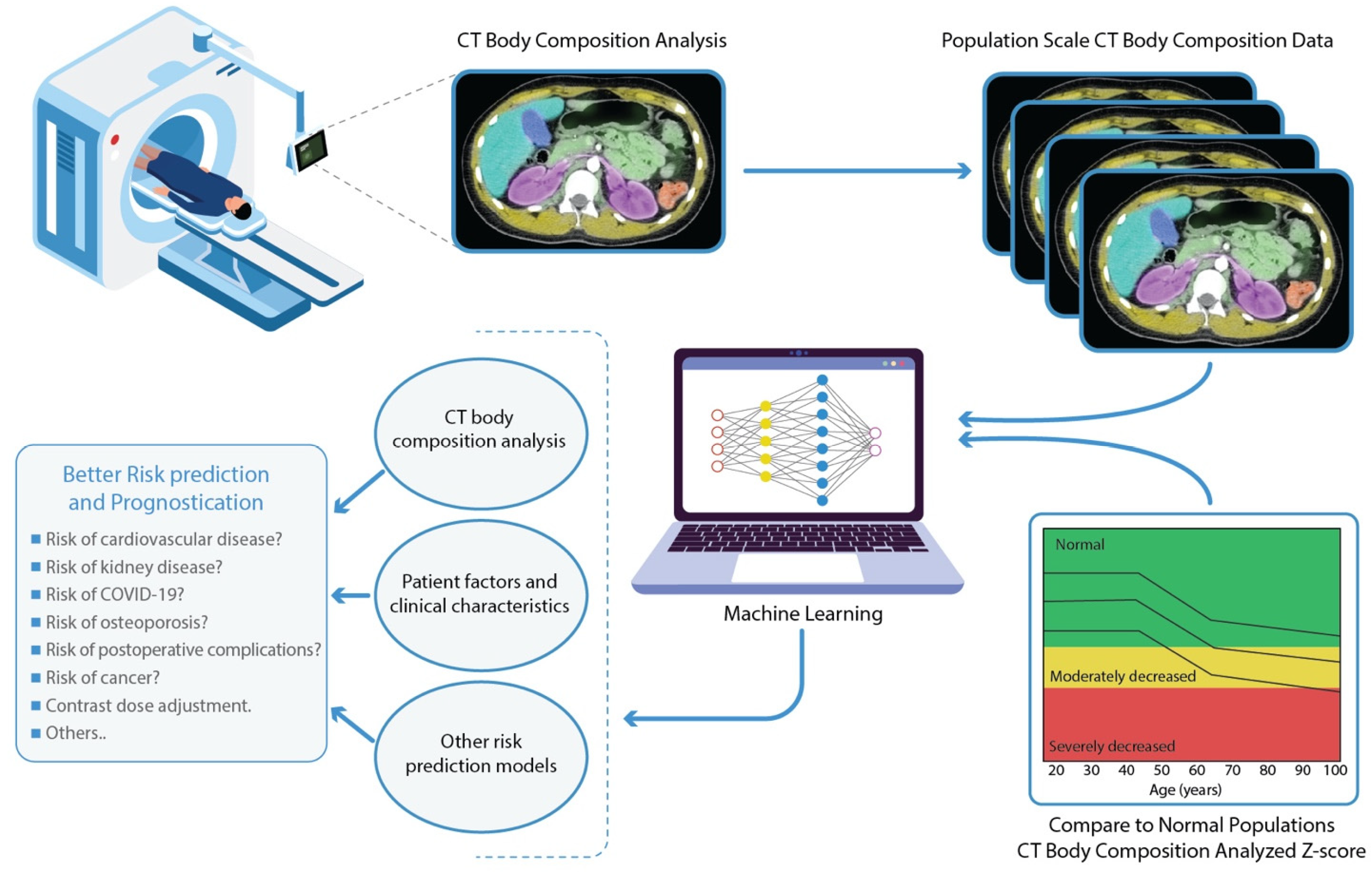
:1. Introduction
2. Imaging-Based Body Composition Analysis
2.1. Muscle Mass
2.2. Skeletal Muscle Quality
2.3. Visceral Fat Content
2.4. Bone Density
2.5. Arterial Calcifications
2.6. Other CT-Based Quantitative Metrics
3. Clinical Applications of CT Body Composition
3.1. Cancer
3.2. Liver Disease
3.3. Inflammatory Bowel Disease (IBD)
3.4. Kidney Disease
3.5. COVID-19
3.6. Cardiovascular Diseases
3.7. Critical Illness
3.8. Contrast Dose Adjustment
4. CT Body Composition Analysis—Technical Considerations
5. Future Directions
6. Conclusions
Author Contributions
Funding
Institutional Review Board Statement
Informed Consent Statement
Data Availability Statement
Conflicts of Interest
References
- Khanna, D.; Peltzer, C.; Kahar, P.; Parmar, M.S. Body Mass Index (BMI): A Screening Tool Analysis. Cureus 2022, 14, e22119. [Google Scholar] [CrossRef]
- Centers for Disease Control and Prevention. Body Mass Index: Considerations for Practitioners; CDC: Atlanta, GA, USA, 2011; pp. 1–4. [Google Scholar]
- Duren, D.L.; Sherwood, R.J.; Czerwinski, S.A.; Lee, M.; Choh, A.C.; Siervogel, R.M.; Chumlea, W.C. Body Composition Methods: Comparisons and Interpretation. J. Diabetes Sci. Technol. 2008, 2, 1139–1146. [Google Scholar] [CrossRef] [Green Version]
- Paris, M.T. Body Composition Analysis of Computed Tomography Scans in Clinical Populations: The Role of Deep Learning. Lifestyle Genom. 2020, 13, 28–31. [Google Scholar] [CrossRef] [PubMed]
- Prado, C.M.; Purcell, S.; Alish, C.; Pereira, S.L.; Deutz, N.E.; Heyland, D.K.; Goodpaster, B.H.; Tappenden, K.; Heymsfield, S.B. Implications of low muscle mass across the continuum of care: A narrative review. Ann. Med. 2018, 50, 675–693. [Google Scholar] [CrossRef] [PubMed] [Green Version]
- Hill, K.D.; Einstein, A.J. New approaches to reduce radiation exposure. Trends Cardiovasc. Med. 2016, 26, 55–65. [Google Scholar] [CrossRef] [Green Version]
- Gottumukkala, R.V.; Kalra, M.K.; Tabari, A.; Otrakji, A.; Gee, M.S. Advanced CT Techniques for Decreasing Radiation Dose, Reducing Sedation Requirements, and Optimizing Image Quality in Children. Radiographics 2019, 39, 709–726. [Google Scholar] [CrossRef]
- Center for Devices and Radiological Health. Initiative to Reduce Unnecessary RAD Exposure from Med IMG White Paper; U.S. Food and Drug Administration: Silver Spring, MD, USA, 2019. Available online: https://www.fda.gov/radiation-emitting-products/initiative-reduce-unnecessary-radiation-exposure-medical-imaging/white-paper-initiative-reduce-unnecessary-radiation-exposure-medical-imaging (accessed on 26 January 2023).
- Beaudart, C.; McCloskey, E.; Bruyère, O.; Cesari, M.; Rolland, Y.; Rizzoli, R.; Araujo De Carvalho, I.; Amuthavalli Thiyagarajan, J.; Bautmans, I.; Bertière, M.-C.; et al. Sarcopenia in daily practice: Assessment and management. BMC Geriatr. 2016, 16, 170. [Google Scholar] [CrossRef] [PubMed]
- Tolonen, A.; Pakarinen, T.; Sassi, A.; Kyttä, J.; Cancino, W.; Rinta-Kiikka, I.; Pertuz, S.; Arponen, O. Methodology, clinical applications, and future directions of body composition analysis using computed tomography (CT) images: A review. Eur. J. Radiol. 2021, 145, 109943. [Google Scholar] [CrossRef]
- Heymsfield, S.; Ross, R.; Wang, Z.; Frager, D. Imaging techniques of body composition: Advantages of measurement and new uses. In Emerging Technologies for Nutrition Research: Potential for Assessing Military Performance Capability; National Academies Press: Washington, DC, USA, 1997; pp. 127–150. [Google Scholar]
- Klopfenstein, B.J.; Kim, M.S.; Krisky, C.M.; Szumowski, J.; Rooney, W.D.; Purnell, J.Q. Comparison of 3 T MRI and CT for the measurement of visceral and subcutaneous adipose tissue in humans. Br. J. Radiol. 2012, 85, e826–e830. [Google Scholar] [CrossRef] [Green Version]
- Zaffina, C.; Wyttenbach, R.; Pagnamenta, A.; Grasso, R.F.; Biroli, M.; Del Grande, F.; Rizzo, S. Body composition assessment: Comparison of quantitative values between magnetic resonance imaging and computed tomography. Quant. Imaging Med. Surg. 2022, 12, 1450–1466. [Google Scholar] [CrossRef]
- Faron, A.; Sprinkart, A.M.; Kuetting, D.L.R.; Feisst, A.; Isaak, A.; Endler, C.; Chang, J.; Nowak, S.; Block, W.; Thomas, D.; et al. Body composition analysis using CT and MRI: Intra-individual intermodal comparison of muscle mass and myosteatosis. Sci. Rep. 2020, 10, 11765. [Google Scholar] [CrossRef] [PubMed]
- Bindman, R.; Kwan, M.L.; Marlow, E.C.; Theis, M.K.; Bolch, W.; Cheng, S.Y.; Bowles, E.J.; Duncan, J.R.; Greenlee, R.T.; Kushi, L.H.; et al. Trends in use of medical imaging in US health care systems and in Ontario, Canada, 2000–2016. Jama 2019, 322, 843–856. [Google Scholar] [CrossRef] [PubMed]
- Vrieling, A.; Kampman, E.; Knijnenburg, N.C.; Mulders, P.F.; Sedelaar, J.P.M.; Baracos, V.E.; Kiemeney, L.A. Body composition in relation to clinical outcomes in renal cell cancer: A systematic review and meta-analysis. Eur. Urol. Focus. 2018, 4, 420–434. [Google Scholar] [CrossRef] [PubMed] [Green Version]
- Kalafateli, M.; Konstantakis, C.; Thomopoulos, K.; Triantos, C. Impact of muscle wasting on survival in patients with liver cirrhosis. World J. Gastroenterol. 2015, 21, 7357. [Google Scholar] [CrossRef] [PubMed]
- Moisey, L.L.; Mourtzakis, M.; Cotton, B.A.; Premji, T.; Heyland, D.K.; Wade, C.E.; Bulger, E.; Kozar, R.A.; Nutrition and Rehabilitation Investigators Consortium (NUTRIC). Skeletal muscle predicts ventilator- free days, ICU-free days, and mortality in elderly ICU patients. Crit. Care 2013, 17, R206. [Google Scholar] [CrossRef] [PubMed] [Green Version]
- Nishimura, J.M.; Ansari, A.Z.; D’Souza, D.M.; Moffatt-Bruce, S.D.; Merritt, R.E.; Kneuertz, P.J. Computed Tomography-Assessed Skeletal Muscle Mass as a Predictor of Outcomes in Lung Cancer Surgery. Ann. Thorac. Surg. 2019, 108, 1555–1564. [Google Scholar] [CrossRef] [PubMed]
- Friedman, J.; Lussiez, A.; Sullivan, J.; Wang, S.; Englesbe, M. Implications of Sarcopenia in Major Surgery. Nutr. Clin. Pract. 2015, 30, 175–179. [Google Scholar] [CrossRef]
- Donadio, C. Body Composition Analysis Allows the Prediction of Urinary Creatinine Excretion and of Renal Function in Chronic Kidney Disease Patients. Nutrients 2017, 9, 553. [Google Scholar] [CrossRef] [Green Version]
- Toledo, D.O.; Carvalho, A.M.; Oliveira, A.M.; Toloi, J.M.; Silva, A.C.; Farah, J.F.D.M.; Prado, C.M.; Silva, J.M. The use of computed tomography images as a prognostic marker in critically ill cancer patients. Clin. Nutr. ESPEN 2018, 25, 114–120. [Google Scholar] [CrossRef]
- Weijs, P.J.; Looijaard, W.G.; Dekker, I.M.; Stapel, S.N.; Girbes, A.R.; Straaten, H.M.O.-V.; Beishuizen, A. Low skeletal muscle area is a risk factor for mortality in mechanically ventilated critically ill patients. Crit. Care 2014, 18, R12. [Google Scholar] [CrossRef] [Green Version]
- Schaffler-Schaden, D.; Mittermair, C.; Birsak, T.; Weiss, M.; Hell, T.; Schaffler, G.; Weiss, H. Skeletal muscle index is an independent predictor of early recurrence in non-obese colon cancer patients. Langenbeck’s Arch. Surg. 2020, 405, 469–477. [Google Scholar] [CrossRef] [PubMed]
- Chang, J.S.; Kim, T.H.; Kim, H.; Choi, E.H.; Kim, N.; Kong, I.D. Qualitative muscle mass index as a predictor of skeletal muscle function deficit in Asian older adults. Geriatr. Gerontol. Int. 2017, 17, 99–107. [Google Scholar] [CrossRef] [Green Version]
- Kappus, M.R.; Wegermann, K.; Bozdogan, E.; Patel, Y.A.; Janas, G.; Shropshire, E.; Parish, A.; Niedzwiecki, D.; Muir, A.J.; Bashir, M. Use of Skeletal Muscle Index as a Predictor of Wait-List Mortality in Patients With End-Stage Liver Disease. Liver Transplant. 2020, 26, 1090–1099. [Google Scholar] [CrossRef] [PubMed]
- Bai, J.; Xu, M.; Peng, F.; Gong, J.; Zhao, J.; Song, X.D.; Li, Y. Skeletal muscle mass index as a predictor of long-term cirrhosis onset in young non-cirrhotic males with acute-on-chronic liver failure. Front. Nutr. 2022, 9, 3181. [Google Scholar] [CrossRef] [PubMed]
- Zanardo, M.; Doniselli, F.M.; Esseridou, A.; Tritella, S.; Mattiuz, C.; Menicagli, L.; Di Leo, G.; Sardanelli, F. Abdominal CT: A radiologist-driven adjustment of the dose of iodinated contrast agent approaches a calculation per lean body weight. Eur. Radiol. Exp. 2018, 2, 41. [Google Scholar] [CrossRef] [PubMed]
- Ma, D.; Chow, V.; Popuri, K.; Beg, M.F. Comprehensive Validation of Automated Whole Body Skeletal Muscle, Adipose Tissue, and Bone Segmentation from 3D CT images for Body Composition Analysis: Towards Extended Body Composition. arXiv 2021, arXiv:2106.00652. [Google Scholar]
- Chu, M.P.; Lieffers, J.; Ghosh, S.; Belch, A.; Chua, N.S.; Fontaine, A.; Sangha, R.; Turner, R.A.; Baracos, V.E.; Sawyer, M.B. Skeletal muscle density is an independent predictor of diffuse large B-cell lymphoma outcomes treated with rituximab-based chemoimmunotherapy. J. Cachex Sarcopenia Muscle 2017, 8, 298–304. [Google Scholar] [CrossRef]
- Van Vugt, J.L.; Gaspersz, M.P.; Vugts, J.; Buettner, S.; Levolger, S.; De Bruin, R.W.; Polak, W.G.; De Jonge, J.; Willemssen, F.E.; Koerkamp, B.G.; et al. Low Skeletal Muscle Density Is Associated with Early Death in Patients with Perihilar Cholangiocarcinoma Regardless of Subsequent Treatment. Dig. Surg. 2019, 36, 144–152. [Google Scholar] [CrossRef] [Green Version]
- Antoun, S.; Lanoy, E.; Iacovelli, R.; Albiges-Sauvin, L.; Loriot, Y.; Merad-Taoufik, M.; Fizazi, K.; di Palma, M.; Baracos, V.E.; Escudier, B. Skeletal muscle density predicts prognosis in patients with metastatic renal cell carcinoma treated with targeted therapies. Cancer 2013, 119, 3377–3384. [Google Scholar] [CrossRef]
- Trikudanathan, G.; Vantanasiri, K.; Faizi, N.; Munigala, S.; Vanek, P.; Schat, R.; Freeman, M.L.; Chauhan, A. Decreased skeletal muscle density is an independent predictor of mortality in necrotizing pancreatitis—A single tertiary center experience in 507 patients. Pancreatology 2021, 21, 1146–1151. [Google Scholar] [CrossRef]
- Yajima, T.; Arao, M.; Yajima, K. Psoas muscle index and psoas muscle density as predictors of mortality in patients undergoing hemodialysis. Sci. Rep. 2022, 12, 10496. [Google Scholar] [CrossRef] [PubMed]
- Fujiwara, N.; Nakagawa, H.; Kudo, Y.; Tateishi, R.; Taguri, M.; Watadani, T.; Nakagomi, R.; Kondo, M.; Nakatsuka, T.; Minami, T.; et al. Sarcopenia, intramuscular fat deposition, and visceral adiposity independently predict the outcomes of hepatocellular carcinoma. J. Hepatol. 2015, 63, 131–140. [Google Scholar] [CrossRef] [Green Version]
- Brown, J.C.; Caan, B.J.; Prado, C.M.; Weltzien, E.; Xiao, J.; Feliciano, E.M.; Kroenke, C.H.; Meyerhardt, J.A. Body Composition and Cardiovascular Events in Patients With Colorectal Cancer: A Population-Based Retrospective Cohort Study. JAMA Oncol. 2019, 5, 967–972. [Google Scholar] [CrossRef] [PubMed]
- Pickhardt, P.J.; Graffy, P.M.; Zea, R.; Lee, S.J.; Liu, J.; Sandfort, V.; Summers, R.M. Automated CT biomarkers for opportunistic prediction of future cardiovascular events and mortality in an asymptomatic screening population: A retrospective cohort study. Lancet Digit. Health 2020, 2, e192–e200. [Google Scholar] [CrossRef] [PubMed]
- Smith, B.; Smith, G.; Hurria, A.; Hortobagyi, G.N.; Buchholz, T.A. Future of Cancer Incidence in the United States: Burdens Upon an Aging, Changing Nation. J. Clin. Oncol. 2009, 27, 2758–2765. [Google Scholar] [CrossRef] [PubMed]
- Pickhardt, P.J.; Graffy, P.M.; Perez, A.A.; Lubner, M.G.; Elton, D.C.; Summers, R.M. Opportunistic Screening at Abdominal CT: Use of Automated Body Composition Biomarkers for Added Cardiometabolic Value. Radiographics 2021, 41, 524–542. [Google Scholar] [CrossRef] [PubMed]
- O’Connor, S.D.; Graffy, P.M.; Zea, R.; Pickhardt, P.J. Does Nonenhanced CT-based Quantification of Abdominal Aortic Calcification Outperform the Framingham Risk Score in Predicting Cardiovascular Events in Asymptomatic Adults? Radiology 2019, 290, 108–115. [Google Scholar] [CrossRef] [Green Version]
- Guglielmo, M.; Lin, A.; Dey, D.; Baggiano, A.; Fusini, L.; Muscogiuri, G.; Pontone, G. Epicardial fat and coronary artery disease: Role of cardiac imaging. Atherosclerosis 2021, 321, 30–38. [Google Scholar] [CrossRef]
- Pieters, T.T.; Veldhuis, W.B.; Moeskops, P.; de Vos, B.D.; Verhaar, M.C.; Haitjema, S.; Huitema, A.D.R.; Rookmaaker, M.B. Deep learning body-composition analysis of clinically acquired CT-scans estimates creatinine excretion with high accuracy in patients and healthy individuals. Sci. Rep. 2022, 12, 9013. [Google Scholar] [CrossRef]
- Tallam, H.; Elton, D.C.; Lee, S.; Wakim, P.; Pickhardt, P.J.; Summers, R.M. Fully Automated Abdominal CT Biomarkers for Type 2 Diabetes Using Deep Learning. Radiology 2022, 304, 85–95. [Google Scholar] [CrossRef]
- Marasco, G.; Serenari, M.; Renzulli, M.; Alemanni, L.V.; Rossini, B.; Pettinari, I.; Dajti, E.; Ravaioli, F.; Golfieri, R.; Cescon, M.; et al. Clinical impact of sarcopenia assessment in patients with hepatocellular carcinoma undergoing treatments. J. Gastroenterol. 2020, 55, 927–943. [Google Scholar] [CrossRef] [PubMed]
- Martin, L.; Birdsell, L.; MacDonald, N.; Reiman, T.; Clandinin, M.T.; McCargar, L.J.; Murphy, R.; Ghosh, S.; Sawyer, M.B.; Baracos, V.E. Cancer Cachexia in the Age of Obesity: Skeletal Muscle Depletion Is a Powerful Prognostic Factor, Independent of Body Mass Index. J. Clin. Oncol. 2013, 31, 1539–1547. [Google Scholar] [CrossRef] [PubMed]
- Gibson, D.J.; Burden, S.T.; Strauss, B.J.; Todd, C.; Lal, S. The role of computed tomography in evaluating body composition and the influence of reduced muscle mass on clinical outcome in abdominal malignancy: A systematic review. Eur. J. Clin. Nutr. 2015, 69, 1079–1086. [Google Scholar] [CrossRef]
- Prado, C.M.; Lieffers, J.R.; McCargar, L.J.; Reiman, T.; Sawyer, M.B.; Martin, L.; Baracos, V.E. Prevalence and clinical implications of sarcopenic obesity in patients with solid tumours of the respiratory and gastrointestinal tracts: A population-based study. Lancet Oncol. 2008, 9, 629–635. [Google Scholar] [CrossRef]
- Baracos, V.E.; Arribas, L. Sarcopenic obesity: Hidden muscle wasting and its impact for survival and complications of cancer therapy. Ann. Oncol. 2018, 29, ii1–ii9. [Google Scholar] [CrossRef] [PubMed]
- Prado, C.M.; Cristina Gonzalez, M.; Heymsfield, S.B. Body composition phenotypes and obesity paradox. Curr. Opin. Clin. Nutr. Metab. Care 2015, 18, 535–551. [Google Scholar] [CrossRef] [PubMed]
- Cruz-Jentoft, A.J.; Bahat, G.; Bauer, J.; Boirie, Y.; Bruyère, O.; Cederholm, T.; Cooper, C.; Landi, F.; Rolland, Y.; Sayer, A.A.; et al. Sarcopenia: Revised European consensus on definition and diagnosis. Age Ageing 2019, 48, 16–31. [Google Scholar] [CrossRef] [PubMed] [Green Version]
- Su, H.; Ruan, J.; Chen, T.; Lin, E.; Shi, L. CT-assessed sarcopenia is a predictive factor for both long-term and short-term outcomes in gastrointestinal oncology patients: A systematic review and meta-analysis. Cancer Imaging 2019, 19, 82. [Google Scholar] [CrossRef]
- Zhuang, C.-L.; Huang, D.-D.; Pang, W.-Y.; Zhou, C.-J.; Wang, S.-L.; Lou, N.; Ma, L.-L.; Yu, Z.; Shen, X. Sarcopenia is an Independent Predictor of Severe Postoperative Complications and Long-Term Survival After Radical Gastrectomy for Gastric Cancer: Analysis from a Large-Scale Cohort. Medicine 2016, 95, e3164. [Google Scholar] [CrossRef]
- Yuji, M. The Examination Committee of Criteria for ‘Obesity Disease’ in Japan, Japan Society for the Study of Obesity. New Criteria for ‘Obesity Disease’ in Japan. Circ. J. 2002, 66, 987–992. [Google Scholar] [CrossRef] [Green Version]
- Fearon, K.; Strasser, F.; Anker, S.D.; Bosaeus, I.; Bruera, E.; Fainsinger, R.L.; Jatoi, A.; Loprinzi, C.; MacDonald, N.; Mantovani, G.; et al. Definition and classification of cancer cachexia: An international consensus. Lancet Oncol. 2011, 12, 489–495. [Google Scholar] [CrossRef]
- Meister, F.; Lurje, G.; Verhoeven, S.; Wiltberger, G.; Heij, L.; Liu, W.; Jiang, D.; Bruners, P.; Lang, S.; Ulmer, T.; et al. The Role of Sarcopenia and Myosteatosis in Short-and Long-Term Outcomes Following Curative-Intent Surgery for Hepatocellular Carcinoma in a European Cohort. Cancers 2022, 14, 720. [Google Scholar] [CrossRef] [PubMed]
- Huffman, D.M.; Barzilai, N. Role of visceral adipose tissue in aging. Biochim. Biophys. Acta (BBA) Gen. Subj. 2009, 1790, 1117–1123. [Google Scholar] [CrossRef] [Green Version]
- Neeland, I.J.; Gupta, S.; Ayers, C.R.; Turer, A.T.; Rame, J.E.; Das, S.R.; Berry, J.D.; Khera, A.; McGuire, D.K.; Vega, G.L.; et al. Relation of Regional Fat Distribution to Left Ventricular Structure and Function. Circ. Cardiovasc. Imaging 2013, 6, 800–807. [Google Scholar] [CrossRef] [PubMed] [Green Version]
- Shuster, A.; Patlas, M.; Pinthus, J.H.; Mourtzakis, M. The clinical importance of visceral adiposity: A critical review of methods for visceral adipose tissue analysis. Br. J. Radiol. 2012, 85, 1–10. [Google Scholar] [CrossRef] [PubMed] [Green Version]
- Ritchie, S.A.; Connell, J.M. The link between abdominal obesity, metabolic syndrome and cardiovascular disease. Nutr. Metab. Cardiovasc. Dis. 2007, 17, 319–326. [Google Scholar] [CrossRef]
- Lenchik, L.; Weaver, A.A.; Ward, R.J.; Boone, J.M.; Boutin, R.D. Opportunistic Screening for Osteoporosis Using Computed Tomography: State of the Art and Argument for Paradigm Shift. Curr. Rheumatol. Rep. 2018, 20, 74. [Google Scholar] [CrossRef]
- Pickhardt, P.J.; Pooler, B.D.; Lauder, T.; del Rio, A.M.; Bruce, R.J.; Binkley, N. Opportunistic Screening for Osteoporosis Using Abdominal Computed Tomography Scans Obtained for Other Indications. Ann. Intern. Med. 2013, 158, 588–595. [Google Scholar] [CrossRef] [Green Version]
- Pickhardt, P.J.; Lee, S.; Liu, J.; Yao, J.; Lay, N.; Graffy, P.M.; Summers, R.M. Population-based opportunistic osteoporosis screening: Validation of a fully automated CT tool for assessing longitudinal BMD changes. Br. J. Radiol. 2019, 92, 20180726. [Google Scholar] [CrossRef]
- Tan, S.; Yao, J.; Ward, M.M.; Yao, L.; Summers, R.M. Computer Aided Evaluation of Ankylosing Spondylitis Using High-Resolution CT. IEEE Trans. Med Imaging 2008, 27, 1252–1267. [Google Scholar] [CrossRef] [Green Version]
- Park, S.H.; Jeong, Y.M.; Lee, H.Y.; Kim, E.Y.; Kim, J.H.; Park, H.K.; Ahn, H.K. Opportunistic use of chest CT for screening osteoporosis and predicting the risk of incidental fracture in breast cancer patients: A retrospective longitudinal study. PLoS ONE 2020, 15, e0240084. [Google Scholar] [CrossRef] [PubMed]
- Azevedo, C.F.; Rochitte, C.E.; Lima, J.A. Escore de cálcio e angiotomografia coronariana na estratificação do risco cardiovascular. Arq. Bras. Cardiol. 2012, 98, 559–568. [Google Scholar] [CrossRef] [Green Version]
- Ulusoy, F.R.; Yolcu, M.; Ipek, E.; Korkmaz, A.F.; Gurler, M.Y.; Gulbaran, M. Coronary Artery Disease Risk Factors, Coronary Artery Calcification and Coronary Bypass Surgery. J. Clin. Diagn. Res. 2015, 9, OC06. [Google Scholar] [CrossRef] [PubMed]
- Takayama, Y.; Yasuda, Y.; Suzuki, S.; Shibata, Y.; Tatami, Y.; Shibata, K.; Niwa, M.; Sawai, A.; Morimoto, R.; Kato, S.; et al. Relationship between abdominal aortic and coronary artery calcification as detected by computed tomography in chronic kidney disease patients. Heart Vessel. 2016, 31, 1030–1037. [Google Scholar] [CrossRef] [PubMed]
- Zhou, X.; Han, M.; Hara, T.; Fujita, H.; Sugisaki, K.; Chen, H.; Lee, G.; Yokoyama, R.; Kanematsu, M.; Hoshi, H. Automated segmentation of mammary gland regions in non-contrast X-ray CT images. Comput. Med Imaging Graph. 2008, 32, 699–709. [Google Scholar] [CrossRef]
- Hänsch, A.; Schwier, M.; Gass, T.; Morgas, T.; Haas, B.; Dicken, V.; Meine, H.; Klein, J.; Hahn, H.K. Evaluation of deep learning methods for parotid gland segmentation from CT images. J. Med Imaging 2019, 6, 011005. [Google Scholar] [CrossRef] [Green Version]
- Lin, Z.; Cui, Y.; Liu, J.; Sun, Z.; Ma, S.; Zhang, X.; Wang, X. Automated segmentation of kidney and renal mass and automated detection of renal mass in CT urography using 3D U-Net-based deep convolutional neural network. Eur. Radiol. 2021, 31, 5021–5031. [Google Scholar] [CrossRef]
- Ma, L.; Guo, R.; Zhang, G.; Schuster, D.M.; Fei, B. A combined learning algorithm for prostate segmentation on 3D CT images. Med Phys. 2017, 44, 5768–5781. [Google Scholar] [CrossRef]
- Luo, G.; Yang, Q.; Chen, T.; Zheng, T.; Xie, W.; Sun, H. An optimized two-stage cascaded deep neural network for adrenal segmentation on CT images. Comput. Biol. Med. 2021, 136, 104749. [Google Scholar] [CrossRef]
- Rister, B.; Yi, D.; Shivakumar, K.; Nobashi, T.; Rubin, D.L. CT-ORG, a new dataset for multiple organ segmentation in computed tomography. Sci. Data 2020, 7, 381. [Google Scholar] [CrossRef]
- Chen, X.; Sun, S.; Bai, N.; Han, K.; Liu, Q.; Yao, S.; Tang, H.; Zhang, C.; Lu, Z.; Huang, Q.; et al. A deep learning-based auto-segmentation system for organs-at-risk on whole-body computed tomography images for radiation therapy. Radiother. Oncol. 2021, 160, 175–184. [Google Scholar] [CrossRef] [PubMed]
- Bortsova, G.; Bos, D.; Dubost, F.; Vernooij, M.W.; Ikram, M.K.; van Tulder, G.; de Bruijne, M. Automated Segmentation and Volume Measurement of Intracranial Internal Carotid Artery Calcification at Noncontrast CT. Radiol. Artif. Intell. 2021, 3, e200226. [Google Scholar] [CrossRef] [PubMed]
- Cui, Y.; Arimura, H.; Nakano, R.; Yoshitake, T.; Shioyama, Y.; Yabuuchi, H. Automated approach for segmenting gross tumor volumes for lung cancer stereotactic body radiation therapy using CT-based dense V-networks. J. Radiat. Res. 2021, 62, 346–355. [Google Scholar] [CrossRef] [PubMed]
- Bilic, P.; Christ, P.; Li, H.B.; Vorontsov, E.; Ben-Cohen, A.; Kaissis, G.; Szeskin, A.; Jacobs, C.; Mamani, G.E.H.; Chartrand, G.; et al. The Liver Tumor Segmentation Benchmark (LiTS). Med Image Anal. 2022, 84, 102680. [Google Scholar] [CrossRef]
- Anjanappa, M.; Corden, M.; Green, A.; Roberts, D.; Hoskin, P.; McWilliam, A.; Choudhury, A. Sarcopenia in cancer: Risking more than muscle loss. Tech. Innov. Patient Support Radiat. Oncol. 2020, 16, 50–57. [Google Scholar] [CrossRef]
- Tan, B.H.; Birdsell, L.A.; Martin, L.; Baracos, V.E.; Fearon, K.C. Sarcopenia in an Overweight or Obese Patient Is an Adverse Prognostic Factor in Pancreatic Cancer. Clin. Cancer Res. 2009, 15, 6973–6979. [Google Scholar] [CrossRef] [Green Version]
- Papaconstantinou, D.; Vretakakou, K.; Paspala, A.; Misiakos, E.P.; Charalampopoulos, A.; Nastos, C.; Patapis, P.; Pikoulis, E. The impact of preoperative sarcopenia on postoperative complications following esophagectomy for esophageal neoplasia: A systematic review and meta-analysis. Dis. Esophagus 2020, 33, doaa002. [Google Scholar] [CrossRef]
- Yang, M.; Shen, Y.; Tan, L.; Li, W. Prognostic value of sarcopenia in lung cancer: A systematic review and meta-analysis. Chest 2019, 156, 101–111. [Google Scholar] [CrossRef]
- Parkin, E.; Plumb, A.A.; O’Reilly, D.; Renehan, A.G. Body composition and outcome in patients undergoing resection of colorectal liver metastases. Br. J. Surg. 2012, 99, 1021–1022. [Google Scholar] [CrossRef]
- Sabel, M.S.; Lee, J.; Cai, S.; Englesbe, M.; Holcombe, S.; Wang, S. Sarcopenia as a Prognostic Factor among Patients with Stage III Melanoma. Ann. Surg. Oncol. 2011, 18, 3579–3585. [Google Scholar] [CrossRef]
- Best, T.D.; Mercaldo, S.F.; Bryan, D.S.; Marquardt, J.P.; Wrobel, M.M.; Bridge, C.P.; Troschel, F.M.; Javidan, C.; Chung, J.H.; Muniappan, A.; et al. Multilevel Body Composition Analysis on Chest Computed Tomography Predicts Hospital Length of Stay and Complications After Lobectomy for Lung Cancer: A Multicenter Study. Ann. Surg. 2020, 275, e708–e715. [Google Scholar] [CrossRef] [PubMed]
- Higashi, T.; Hayashi, H.; Taki, K.; Sakamoto, K.; Kuroki, H.; Nitta, H.; Hashimoto, D.; Chikamoto, A.; Beppu, T.; Baba, H. Sarcopenia, but not visceral fat amount, is a risk factor of postoperative complications after major hepatectomy. Int. J. Clin. Oncol. 2016, 21, 310–319. [Google Scholar] [CrossRef] [PubMed]
- Lim, J.; Kim, K.W.; Ko, Y.; Jang, I.-Y.; Lee, Y.S.; Chung, Y.-H.; Lee, H.C.; Lim, Y.-S.; Kim, K.M.; Shim, J.H.; et al. The role of muscle depletion and visceral adiposity in HCC patients aged 65 and over undergoing TACE. BMC Cancer 2021, 21, 1164. [Google Scholar] [CrossRef] [PubMed]
- Dello, S.A.W.G.; Lodewick, T.M.; van Dam, R.M.; Reisinger, K.W.; Broek, M.A.J.V.D.; von Meyenfeldt, M.F.; Bemelmans, M.H.A.; Damink, S.W.M.O.; Dejong, C.H.C. Sarcopenia negatively affects preoperative total functional liver volume in patients undergoing liver resection. Hpb 2013, 15, 165–169. [Google Scholar] [CrossRef] [Green Version]
- Faron, A.; Sprinkart, A.M.; Pieper, C.C.; Kuetting, D.L.; Fimmers, R.; Block, W.; Meyer, C.; Thomas, D.; Attenberger, U.; Luetkens, J.A. Yttrium-90 radioembolization for hepatocellular carcinoma: Outcome prediction with MRI derived fat-free muscle area. Eur. J. Radiol. 2020, 125, 108889. [Google Scholar] [CrossRef]
- Dixon, M.; Cruz, J.; Sarwani, N.; Gusani, N. The Future Liver Remnant. Am. Surg. 2021, 87, 276–286. [Google Scholar] [CrossRef]
- Vallati, G.; Trobiani, C.; Teodoli, L.; Lai, Q.; Cappelli, F.; Ungania, S.; Catalano, C.; Lucatelli, P. Sarcopenia Worsening One Month after Transarterial Radioembolization Predicts Progressive Disease in Patients with Advanced Hepatocellular Carcinoma. Biology 2021, 10, 728. [Google Scholar] [CrossRef]
- Yuri, Y.; Nishikawa, H.; Enomoto, H.; Ishii, A.; Iwata, Y.; Miyamoto, Y.; Ishii, N.; Hasegawa, K.; Nakano, C.; Nishimura, T.; et al. Implication of Psoas Muscle Index on Survival for Hepatocellular Carcinoma Undergoing Radiofrequency Ablation Therapy. J. Cancer 2017, 8, 1507–1516. [Google Scholar] [CrossRef] [Green Version]
- Peng, P.D.; van Vledder, M.G.; Tsai, S.; de Jong, M.C.; Makary, M.; Ng, J.; Edil, B.H.; Wolfgang, C.L.; Schulick, R.D.; Choti, M.A.; et al. Sarcopenia negatively impacts short-term outcomes in patients undergoing hepatic resection for colorectal liver metastasis. Hpb 2011, 13, 439–446. [Google Scholar] [CrossRef] [Green Version]
- Voron, T.; Tselikas, L.; Pietrasz, D.; Pigneur, F.; Laurent, A.; Compagnon, P.; Salloum, C.; Luciani, A.; Azoulay, D. Sarcopenia Impacts on Shortand Long-term Results of Hepatectomy for Hepatocellular Carcinoma. Ann. Surg. 2015, 261, 1173–1183. [Google Scholar] [CrossRef]
- Takagi, K.; Yagi, T.; Yoshida, R.; Shinoura, S.; Umeda, Y.; Nobuoka, D.; Kuise, T.; Watanabe, N.; Fujiwara, T. Sarcopenia and American Society of Anesthesiologists Physical Status in the Assessment of Outcomes of Hepatocellular Carcinoma Patients Undergoing Hepatectomy. Acta Med. Okayama 2016, 70, 363–370. [Google Scholar] [CrossRef] [PubMed]
- Yao, S.; Kamo, N.; Taura, K.; Miyachi, Y.; Iwamura, S.; Hirata, M.; Kaido, T.; Uemoto, S. Muscularity defined by the combination of muscle quantity and quality is closely related to both liver hypertrophy and postoperative outcomes following portal vein embolization in cancer patients. Ann. Surg. Oncol. 2022, 29, 301–312. [Google Scholar] [CrossRef] [PubMed]
- Chuang, Y.H.; Ou, H.Y.; Lazo, M.Z.; Chen, C.L.; Chen, M.H.; Weng, C.C.; Cheng, Y.F. Predicting post-hepatectomy liver failure by combined volumetric, functional MR image and laboratory analysis. Liver Int. 2018, 38, 868–874. [Google Scholar] [CrossRef]
- Ribero, D.; Abdalla, E.K.; Madoff, D.; Donadon, M.; Loyer, E.M.; Vauthey, J. Portal vein embolization before major hepatectomy and its effects on regeneration, resectability and outcome. Br. J. Surg. 2007, 94, 1386–1394. [Google Scholar] [CrossRef] [Green Version]
- Shindoh, J.; Tzeng, C.-W.D.; Aloia, T.A.; Curley, S.A.; Huang, S.Y.; Mahvash, A.; Gupta, S.; Wallace, M.J.; Vauthey, J.-N. Safety and Efficacy of Portal Vein Embolization Before Planned Major or Extended Hepatectomy: An Institutional Experience of 358 Patients. J. Gastrointest. Surg. 2014, 18, 45–51. [Google Scholar] [CrossRef]
- Denbo, J.W.; Kim, B.J.; Vauthey, J.-N.; Tzeng, C.-W.; Ma, J.; Huang, S.Y.; Chun, Y.S.; Katz, M.H.G.; Aloia, T.A. Overall Body Composition and Sarcopenia Are Associated with Poor Liver Hypertrophy Following Portal Vein Embolization. J. Gastrointest. Surg. 2021, 25, 405–410. [Google Scholar] [CrossRef] [PubMed]
- Hsing, J.C.; Nguyen, M.H.; Yang, B.; Min, Y.; Han, S.S.; Pung, E.; Winter, S.J.; Zhao, X.; Gan, D.; Hsing, A.W.; et al. Associations Between Body Fat, Muscle Mass, and Nonalcoholic Fatty Liver Disease: A Population-Based Study. Hepatol. Commun. 2019, 3, 1061–1072. [Google Scholar] [CrossRef] [PubMed] [Green Version]
- Shachar, S.S.; Williams, G.R.; Muss, H.B.; Nishijima, T.F. Prognostic value of sarcopenia in adults with solid tumours: A meta-analysis and systematic review. Eur. J. Cancer 2016, 57, 58–67. [Google Scholar] [CrossRef]
- Antoun, S.; Borget, I.; Lanoy, E. Impact of sarcopenia on the prognosis and treatment toxicities in patients diagnosed with cancer. Curr. Opin. Support. Palliat. Care 2013, 7, 383–389. [Google Scholar] [CrossRef]
- Nishikawa, H.; Nishijima, N.; Enomoto, H.; Sakamoto, A.; Nasu, A.; Komekado, H.; Nishimura, T.; Kita, R.; Kimura, T.; Iijima, H.; et al. Prognostic significance of sarcopenia in patients with hepatocellular carcinoma undergoing sorafenib therapy. Oncol. Lett. 2017, 14, 1637–1647. [Google Scholar] [CrossRef] [Green Version]
- Dasarathy, J.; Alkhouri, N.; Dasarathy, S. Changes in body composition after transjugular intrahepatic portosystemic stent in cirrhosis: A critical review of literature. Liver Int. 2011, 31, 1250–1258. [Google Scholar] [CrossRef] [PubMed]
- Mir, O.; Coriat, R.; Blanchet, B.; Durand, J.-P.; Boudou-Rouquette, P.; Michels, J.; Ropert, S.; Vidal, M.; Pol, S.; Chaussade, S.; et al. Sarcopenia Predicts Early Dose-Limiting Toxicities and Pharmacokinetics of Sorafenib in Patients with Hepatocellular Carcinoma. PLoS ONE 2012, 7, e37563. [Google Scholar] [CrossRef] [PubMed]
- Shachar, S.S.; Deal, A.M.; Weinberg, M.; Nyrop, K.A.; Williams, G.R.; Nishijima, T.F.; Benbow, J.M.; Muss, H.B. Skeletal Muscle Measures as Predictors of Toxicity, Hospitalization, and Survival in Patients with Metastatic Breast Cancer Receiving Taxane-Based Chemotherapy. Clin. Cancer Res. 2017, 23, 658–665. [Google Scholar] [CrossRef] [PubMed] [Green Version]
- Ariya, M.; Koohpayeh, F.; Ghaemi, A.; Osati, S.; Davoodi, S.H.; Razzaz, J.M.; Javedan, G.; Ehrampoush, E.; Homayounfar, R. Assessment of the association between body composition and risk of non-alcoholic fatty liver. PLoS ONE 2021, 16, e0249223. [Google Scholar] [CrossRef]
- Miyake, T.; Miyazaki, M.; Yoshida, O.; Kanzaki, S.; Nakaguchi, H.; Nakamura, Y.; Watanabe, T.; Yamamoto, Y.; Koizumi, Y.; Tokumoto, Y.; et al. Relationship between body composition and the histology of non-alcoholic fatty liver disease: A cross-sectional study. BMC Gastroenterol. 2021, 21, 170. [Google Scholar] [CrossRef] [PubMed]
- Ponziani, F.R.; Gasbarrini, A. Sarcopenia in Patients with Advanced Liver Disease. Curr. Protein Pept. Sci. 2018, 19, 681–691. [Google Scholar] [CrossRef] [PubMed]
- Engelmann, C.; Aehling, N.F.; Schob, S.; Nonnenmacher, I.; Handmann, L.; Macnaughtan, J.; Herber, A.; Surov, A.; Kaiser, T.; Denecke, T.; et al. Body fat composition determines outcomes before and after liver transplantation in patients with cirrhosis. Hepatol. Commun. 2022, 6, 2198–2209. [Google Scholar] [CrossRef]
- Montomoli, J. Body composition changes after transjugular intrahepatic portosystemic shunt in patients with cirrhosis. World J. Gastroenterol. 2010, 16, 348–353. [Google Scholar] [CrossRef]
- Pang, N.; Zhao, C.; Li, J.; Li, L.; Yang, X.; Yang, M.; Wu, Z.; Feng, D. Body mass index changes after transjugular intrahepatic portosystemic shunt in individuals with cirrhosis. Nutrition 2021, 84, 111095. [Google Scholar] [CrossRef]
- Artru, F.; Miquet, X.; Azahaf, M.; Labreuche, J.; Ntandja Wandji, L.C.; Sergent, G.; Nobécourt, A.; Toumelin, P.; Lassailly, G.; Dharancy, S.; et al. Consequences of TIPSS placement on the body composition of patients with cirrhosis and severe portal hypertension: A large retrospective CT-based surveillance. Aliment Pharmacol Ther. 2020, 52, 1516–1526. [Google Scholar] [CrossRef]
- Grillot, J.; D’Engremont, C.; Parmentier, A.-L.; Lakkis, Z.; Piton, G.; Cazaux, D.; Gay, C.; De Billy, M.; Koch, S.; Borot, S.; et al. Sarcopenia and visceral obesity assessed by computed tomography are associated with adverse outcomes in patients with Crohn’s disease. Clin. Nutr. 2020, 39, 3024–3030. [Google Scholar] [CrossRef] [PubMed]
- Zou, W.Y.; Enchakalody, B.E.; Zhang, P.; Shah, N.; Saini, S.D.; Wang, N.C.; Wang, S.C.; Su, G.L. Automated Measurements of Body Composition in Abdominal CT Scans Using Artificial Intelligence Can Predict Mortality in Patients With Cirrhosis. Hepatol. Commun. 2021, 5, 1901–1910. [Google Scholar] [CrossRef]
- Holt, D.Q.; Strauss, B.J.G.; Lau, K.K.; Moore, G.T. Body composition analysis using abdominal scans from routine clinical care in patients with Crohn’s Disease. Scand. J. Gastroenterol. 2016, 51, 842–847. [Google Scholar] [CrossRef] [PubMed]
- Yadav, D.P.; Kedia, S.; Madhusudhan, K.S.; Bopanna, S.; Goyal, S.; Jain, S.; Vikram, N.K.; Sharma, R.; Makharia, G.K.; Ahuja, V. Body Composition in Crohn’s Disease and Ulcerative Colitis: Correlation with Disease Severity and Duration. Can. J. Gastroenterol. Hepatol. 2017, 2017, 1215035. [Google Scholar] [CrossRef] [PubMed] [Green Version]
- Erhayiem, B.; Dhingsa, R.; Hawkey, C.J.; Subramanian, V. Ratio of Visceral to Subcutaneous Fat Area Is a Biomarker of Complicated Crohn’s Disease. Clin. Gastroenterol. Hepatol. 2011, 9, 684–687.e1. [Google Scholar] [CrossRef]
- Ding, Z.; Wu, X.-R.; Remer, E.M.; Lian, L.; Stocchi, L.; Li, Y.; McCullough, A.; Remzi, F.H.; Shen, B. Association between high visceral fat area and postoperative complications in patients with Crohn’s disease following primary surgery. Color. Dis. 2016, 18, 163–172. [Google Scholar] [CrossRef]
- Li, Y.; Zhu, W.; Gong, J.; Zhang, W.; Gu, L.; Guo, Z.; Cao, L.; Shen, B.; Li, N.; Li, J. Visceral fat area is associatedwith a high risk for early postoperative recurrence in crohn’s disease. Color. Dis. 2015, 17, 225–234. [Google Scholar] [CrossRef]
- Bamba, S.; Inatomi, O.; Takahashi, K.; Morita, Y.; Imai, T.; Ohno, M.; Kurihara, M.; Takebayashi, K.; Kojima, M.; Iida, H.; et al. Assessment of Body Composition From CT Images at the Level of the Third Lumbar Vertebra in Inflammatory Bowel Disease. Inflamm. Bowel Dis. 2021, 27, 1435–1442. [Google Scholar] [CrossRef]
- Nishikawa, H.; Nakamura, S.; Miyazaki, T.; Kakimoto, K.; Fukunishi, S.; Asai, A.; Nishiguchi, S.; Higuchi, K. Inflammatory Bowel Disease and Sarcopenia: Its Mechanism and Clinical Importance. J. Clin. Med. 2021, 10, 4214. [Google Scholar] [CrossRef]
- Fleischmann, E.; Teal, N.; Dudley, J.; May, W.; Bower, J.D.; Salahudeen, A.K. Influence of excess weight on mortality and hospital stay in 1346 hemodialysis patients. Kidney Int. 1999, 55, 1560–1567. [Google Scholar] [CrossRef] [Green Version]
- Lu, J.L.; Kalantar-Zadeh, K.; Ma, J.Z.; Quarles, L.D.; Kovesdy, C.P. Association of Body Mass Index with Outcomes in Patients with CKD. J. Am. Soc. Nephrol. 2014, 25, 2088–2096. [Google Scholar] [CrossRef] [PubMed] [Green Version]
- Chazot, C.; Gassia, J.-P.; Di Benedetto, A.; Cesare, S.; Ponce, P.; Marcelli, D. Is there any survival advantage of obesity in Southern European haemodialysis patients? Nephrol. Dial. Transplant. 2009, 24, 2871–2876. [Google Scholar] [CrossRef] [PubMed] [Green Version]
- Lin, T.-Y.; Peng, C.-H.; Hung, S.-C.; Tarng, D.-C. Body composition is associated with clinical outcomes in patients with non–dialysis-dependent chronic kidney disease. Kidney Int. 2018, 93, 733–740. [Google Scholar] [CrossRef] [PubMed]
- Sabatino, A.; D’Alessandro, C.; Regolisti, G.; di Mario, F.; Guglielmi, G.; Bazzocchi, A.; Fiaccadori, E. Muscle mass assessment in renal disease: The role of imaging techniques. Quant. Imaging Med. Surg. 2020, 10, 1672–1686. [Google Scholar] [CrossRef]
- Madabhushi, A.; Doyle, S.; Lee, G.; Basavanhally, A.; Monaco, J.; Masters, S.; Tomaszewski, J.; Feldman, M. Integrated diagnostics: A conceptual framework with examples. Clin. Chem. Lab. Med. 2010, 48, 989–998. [Google Scholar] [CrossRef]
- van Solinge, W.W.; Ten Berg, M.J.; Haitjema, S. Data-gedreven integrale diagnostiek [Data-driven integrated diagnostics: The natural evolution of clinical chemistry?]. Ned. Tijdschr. Voor Geneeskd. 2019, 163, D3512. [Google Scholar]
- Korfiatis, P.; Denic, A.; Edwards, M.E.; Gregory, A.V.; Wright, D.E.; Mullan, A.; Augustine, J.; Rule, A.D.; Kline, T.L. Automated Segmentation of Kidney Cortex and Medulla in CT Images: A Multisite Evaluation Study. J. Am. Soc. Nephrol. 2022, 33, 420–430. [Google Scholar] [CrossRef]
- Manabe, S.; Kataoka, H.; Mochizuki, T.; Iwadoh, K.; Ushio, Y.; Kawachi, K.; Watanabe, K.; Watanabe, S.; Akihisa, T.; Makabe, S.; et al. Impact of visceral fat area in patients with chronic kidney disease. Clin. Exp. Nephrol. 2021, 25, 608–620. [Google Scholar] [CrossRef]
- Kang, S.H.; Cho, K.H.; Park, J.W.; Yoon, K.W.; Do, J.Y. Association of Visceral Fat Area with Chronic Kidney Disease and Metabolic Syndrome Risk in the General Population: Analysis Using Multi-Frequency Bioimpedance. Kidney Blood Press. Res. 2015, 40, 223–230. [Google Scholar] [CrossRef] [Green Version]
- Hirai, K.; Ookawara, S.; Morishita, Y. Sarcopenia and Physical Inactivity in Patients With Chronic Kidney Disease. Nephro-Urology Mon. 2016, 8, e37443. [Google Scholar] [CrossRef] [Green Version]
- Müller, M.J.; Braun, W.; Enderle, J.; Bosy-Westphal, A. Beyond BMI: Conceptual Issues Related to Overweight and Obese Patients. Obes. Facts 2016, 9, 193–205. [Google Scholar] [CrossRef] [PubMed]
- Hocaoglu, E.; Ors, S.; Yildiz, O.; Inci, E. Correlation of Pectoralis Muscle Volume and Density with Severity of COVID-19 Pneumonia in Adults. Acad. Radiol. 2020, 28, 166–172. [Google Scholar] [CrossRef] [PubMed]
- Ufuk, F.; Demirci, M.; Sagtas, E.; Akbudak, I.H.; Ugurlu, E.; Sari, T. The prognostic value of pneumonia severity score and pectoralis muscle Area on chest CT in adult COVID-19 patients. Eur. J. Radiol. 2020, 131, 109271. [Google Scholar] [CrossRef] [PubMed]
- Chandarana, H.; Pisuchpen, N.; Krieger, R.; Dane, B.; Mikheev, A.; Feng, Y.; Kambadakone, A.; Rusinek, H. Association of body composition parameters measured on CT with risk of hospitalization in patients with COVID-19. Eur. J. Radiol. 2021, 145, 110031. [Google Scholar] [CrossRef] [PubMed]
- Bunnell, K.M.; Thaweethai, T.; Buckless, C.; Shinnick, D.J.; Torriani, M.; Foulkes, A.S.; Bredella, M.A. Body composition predictors of outcome in patients with COVID-19. Int. J. Obes. 2021, 45, 2238–2243. [Google Scholar] [CrossRef] [PubMed]
- Roth, G.A.; Mensah, G.A.; Johnson, C.O.; Addolorato, G.; Ammirati, E.; Baddour, L.M.; Barengo, N.C.; Beaton, A.Z.; Benjamin, E.J.; Benziger, C.P.; et al. Global Burden of Cardiovascular Diseases and Risk Factors, 1990–2019: Update From the GBD 2019 Study. J. Am. Coll. Cardiol. 2020, 76, 2982–3021. [Google Scholar] [CrossRef]
- Jayalath, R.; Mangan, S.; Golledge, J. Aortic Calcification. Eur. J. Vasc. Endovasc. Surg. 2005, 30, 476–488. [Google Scholar] [CrossRef] [Green Version]
- Buijs, R.V.C.; Leemans, E.L.; Greuter, M.; Tielliu, I.F.J.; Zeebregts, C.J.; Willems, T.P. Quantification of abdominal aortic calcification: Inherent measurement errors in current computed tomography imaging. PLoS ONE 2018, 13, e0193419. [Google Scholar] [CrossRef] [Green Version]
- Magudia, K.; Bridge, C.P.; Bay, C.P.; Farah, S.; Babic, A.; Fintelmann, F.J.; Brais, L.K.; Andriole, K.P.; Wolpin, B.M.; Rosenthal, M.H.; et al. Utility of Normalized Body Composition Areas, Derived From Outpatient Abdominal CT Using a Fully Automated Deep Learning Method, for Predicting Subsequent Cardiovascular Events. Am. J. Roentgenol. 2023, 220, 236–244. [Google Scholar] [CrossRef]
- Bridge, C.P.; Rosenthal, M.; Wright, B.; Kotecha, G.; Fintelmann, F.; Troschel, F.; Miskin, N.; Desai, K.; Wrobel, W.; Babic, A.; et al. Fully-automated analysis of body composition from ct in cancer patients using convolutional neural networks. In OR 2.0 Context-Aware Operating Theaters, Computer Assisted Robotic Endoscopy, Clinical Image-Based Procedures, and Skin Image Analysis; Stoyanov, D., Taylor, Z., Sarikaya, D., McLeod, J., Ballester, M.A.G., Codella, N.C.F., Martel, A., Maier-Hein, L., Malpani, A., Zenati, M.A., et al., Eds.; Lecture Notes in Computer Science, Volume 11041; Springer: Cham, Switzerland, 2018; pp. 204–213. [Google Scholar]
- Koitka, S.; Kroll, L.; Malamutmann, E.; Oezcelik, A.; Nensa, F. Fully automated body composition analysis in routine CT imaging using 3D semantic segmentation convolutional neural networks. Eur. Radiol. 2021, 31, 1795–1804. [Google Scholar] [CrossRef]
- TomoVision. sliceOmatic Alberta Protocol. Available online: http://www.tomovision.com/Sarcopenia_Help/index.htm (accessed on 11 February 2017).
- Shen, W.; Punyanitya, M.; Wang, Z.; Gallagher, D.; St.-Onge, M.-P.; Albu, J.; Heymsfield, S.B.; Heshka, S. Total body skeletal muscle and adipose tissue volumes: Estimation from a single abdominal cross-sectional image. J. Appl. Physiol. 2004, 97, 2333–2338. [Google Scholar] [CrossRef] [PubMed] [Green Version]
- Weston, A.D.; Korfiatis, P.; Kline, T.L.; Philbrick, K.A.; Kostandy, P.; Sakinis, T.; Sugimoto, M.; Takahashi, N.; Erickson, B.J. Automated Abdominal Segmentation of CT Scans for Body Composition Analysis Using Deep Learning. Radiology 2019, 290, 669–679. [Google Scholar] [CrossRef]
- Ha, J.; Park, T.; Kim, H.-K.; Shin, Y.; Ko, Y.; Kim, D.W.; Sung, Y.S.; Lee, J.; Ham, S.J.; Khang, S.; et al. Development of a fully automatic deep learning system for L3 selection and body composition assessment on computed tomography. Sci. Rep. 2021, 11, 21656. [Google Scholar] [CrossRef] [PubMed]
- Molwitz, I.; Ozga, A.K.; Gerdes, L.; Ungerer, A.; Köhler, D.; Ristow, I.; Leiderer, M.; Adam, G.; Yamamura, J. Prediction of abdominal CT body composition parameters by thoracic measurements as a new approach to detect sarcopenia in a COVID-19 cohort. Sci. Rep. 2022, 12, 6443. [Google Scholar] [CrossRef] [PubMed]
- Marquardt, J.P.; Roeland, E.J.; Van Seventer, E.E.; Best, T.D.; Horick, N.K.; Nipp, R.D.; Fintelmann, F.J. Percentile-based averaging and skeletal muscle gauge improve body composition analysis: Validation at multiple vertebral levels. J. Cachex Sarcopenia Muscle 2021, 13, 190–202. [Google Scholar] [CrossRef] [PubMed]
- Feliciano, E.M.C.; Popuri, K.; Cobzas, D.; Baracos, V.E.; Beg, M.F.; Khan, A.D.; Ma, C.; Chow, V.; Prado, C.M.; Xiao, J.; et al. Evaluation of automated computed tomography segmentation to assess body composition and mortality associations in cancer patients. J. Cachex Sarcopenia Muscle 2020, 11, 1258–1269. [Google Scholar] [CrossRef] [Green Version]
- Wyatt, S.K.; Barck, K.H.; Kates, L.; Zavala-Solorio, J.; Ross, J.; Kolumam, G.; Sonoda, J.; Carano, R.A.D. Fully-automated, high-throughput micro-computed tomography analysis of body composition enables therapeutic efficacy monitoring in preclinical models. Int. J. Obes. 2015, 39, 1630–1637. [Google Scholar] [CrossRef]
- Wang, Y.; Qiu, Y.; Thai, T.; Moore, K.; Liu, H.; Zheng, B. A two-step convolutional neural network based computer-aided detection scheme for automatically segmenting adipose tissue volume depicting on CT images. Comput. Methods Programs Biomed. 2017, 144, 97–104. [Google Scholar] [CrossRef]
- Lee, H.; Troschel, F.M.; Tajmir, S.; Fuchs, G.; Mario, J.; Fintelmann, F.J.; Do, S. Pixel-Level Deep Seg- mentation: Artificial Intelligence Quantifies Muscle on Computed Tomography for Body Morphometric Analysis. J. Digit. Imaging 2017, 30, 487–498. [Google Scholar] [CrossRef]
- Hemke, R.; Buckless, C.G.; Tsao, A.; Wang, B.; Torriani, M. Deep learning for automated segmentation of pelvic muscles, fat, and bone from CT studies for body composition assessment. Skelet. Radiol. 2020, 49, 387–395. [Google Scholar] [CrossRef]
- Burns, J.E.; Yao, J.; Chalhoub, D.; Chen, J.J.; Summers, R.M. A Machine Learning Algorithm to Estimate Sarcopenia on Abdominal CT. Acad. Radiol. 2019, 27, 311–320. [Google Scholar] [CrossRef] [PubMed]
- Paris, M.T.; Tandon, P.; Heyland, D.K.; Furberg, H.; Premji, T.; Low, G.; Mourtzakis, M. Automated body composition analysis of clinically acquired computed tomography scans using neural networks. Clin. Nutr. 2020, 39, 3049–3055. [Google Scholar] [CrossRef] [PubMed]
- Hsu, T.-M.H.; Schawkat, K.; Berkowitz, S.J.; Wei, J.L.; Makoyeva, A.; Legare, K.; DeCicco, C.; Paez, S.N.; Wu, J.S.; Szolovits, P.; et al. Artificial intelligence to assess body composition on routine abdominal CT scans and predict mortality in pancreatic cancer– A recipe for your local application. Eur. J. Radiol. 2021, 142, 109834. [Google Scholar] [CrossRef] [PubMed]
- Bridge, C.P.; Best, T.D.; Wrobel, M.M.; Marquardt, J.P.; Magudia, K.; Javidan, C.; Chung, J.H.; Kalpathy-Cramer, J.; Andriole, K.P.; Fintelmann, F.J. A Fully Automated Deep Learning Pipeline for Multi–Vertebral Level Quantification and Characterization of Muscle and Adipose Tissue on Chest CT Scans. Radiol. Artif. Intell. 2022, 4, e210080. [Google Scholar] [CrossRef]
- Nowak, S.; Faron, A.; Luetkens, J.A.; Geißler, H.L.; Praktiknjo, M.; Block, W.; Thomas, D.; Sprinkart, A.M. Fully Automated Segmentation of Connective Tissue Compartments for CT-Based Body Composition Analysis. Investig. Radiol. 2020, 55, 357–366. [Google Scholar] [CrossRef]
- Kullberg, J.; Hedström, A.; Brandberg, J.; Strand, R.; Johansson, L.; Bergström, G.; Ahlström, H. Automated analysis of liver fat, muscle and adipose tissue distribution from CT suitable for large-scale studies. Sci. Rep. 2017, 7, 10425. [Google Scholar] [CrossRef] [Green Version]
- Dabiri, S.; Popuri, K.; Feliciano, E.M.C.; Caan, B.J.; Baracos, V.E.; Beg, M.F. Muscle segmentation in axial computed tomography (CT) images at the lumbar (L3) and thoracic (T4) levels for body composition analysis. Comput. Med. Imaging Graph. 2019, 75, 47–55. [Google Scholar] [CrossRef]
- Hu, P.; Huo, Y.; Kong, D.; Carr, J.J.; Abramson, R.G.; Hartley, K.G.; Landman, B.A. Automated Characterization of Body Composition and Frailty with Clinically Acquired CT. In Computational Methods and Clinical Applications in Musculoskeletal Imaging: 5th International Workshop, MSKI 2017, Held in Conjunction with MICCAI 2017, Quebec City, QC, Canada, 10 September 2017; Springer International Publishing: Cham, Switzerland, 2018; Volume 10734, pp. 25–35. [Google Scholar] [CrossRef]
- Magudia, K.; Bridge, C.P.; Bay, C.P.; Babic, A.; Fintelmann, F.J.; Troschel, F.M.; Miskin, N.; Wrobel, W.C.; Brais, L.K.; Andriole, K.P.; et al. Population-Scale CT-based Body Composition Analysis of a Large Outpatient Population Using Deep Learning to Derive Age-, Sex-, and Race-specific Reference Curves. Radiology 2021, 298, 319–329. [Google Scholar] [CrossRef]
- Pickhardt, P.J.; Summers, R.M.; Garrett, J.W. Automated CT-Based Body Composition Analysis: A Golden Opportunity. Korean J. Radiol. 2021, 22, 1934. [Google Scholar] [CrossRef]
- Perez, A.A.; Noe-Kim, V.; Lubner, M.G.; Graffy, P.M.; Garrett, J.W.; Elton, D.C.; Summers, R.M.; Pickhardt, P.J. Deep Learning CT-based Quantitative Visualization Tool for Liver Volume Estimation: Defining Normal and Hepatomegaly. Radiology 2022, 302, 336–342. [Google Scholar] [CrossRef]
- Yin, Y.; Yakar, D.; Dierckx, R.A.J.O.; Mouridsen, K.B.; Kwee, T.C.; de Haas, R.J. Liver fibrosis staging by deep learning: A visual-based explanation of diagnostic decisions of the model. Eur. Radiol. 2021, 31, 9620–9627. [Google Scholar] [CrossRef] [PubMed]
- Jacobs, C.; Setio, A.A.A.; Scholten, E.T.; Gerke, P.K.; Bhattacharya, H.; Hoesein, F.A.M.; Brink, M.; Ranschaert, E.; de Jong, P.A.; Silva, M.; et al. Deep Learning for Lung Cancer Detection on Screening CT Scans: Results of a Large-Scale Public Competition and an Observer Study with 11 Radiologists. Radiol. Artif. Intell. 2021, 3, e210027. [Google Scholar] [CrossRef] [PubMed]
- Yan, C.; Wang, L.; Lin, J.; Xu, J.; Zhang, T.; Qi, J.; Li, X.; Ni, W.; Wu, G.; Huang, J.; et al. A fully automatic artificial intelligence–based CT image analysis system for accurate detection, diagnosis, and quantitative severity evaluation of pulmonary tuberculosis. Eur. Radiol. 2022, 32, 2188–2199. [Google Scholar] [CrossRef] [PubMed]
- Graffy, P.M.; Sandfort, V.; Summers, R.M.; Pickhardt, P.J. Automated Liver Fat Quantification at Nonenhanced Abdominal CT for Population-based Steatosis Assessment. Radiology 2019, 293, 334–342. [Google Scholar] [CrossRef]
- Beetz, N.L.; Maier, C.; Segger, L.; Shnayien, S.; Trippel, T.D.; Lindow, N.; Bousabarah, K.; Westerhoff, M.; Fehrenbach, U.; Geisel, D. First PACS-integrated artificial intelligence-based software tool for rapid and fully automatic analysis of body composition from CT in clinical routine. JCSM Clin. Rep. 2022, 7, 3–11. [Google Scholar] [CrossRef]
- Lee, J.-G.; Jun, S.; Cho, Y.-W.; Lee, H.; Kim, G.B.; Seo, J.B.; Kim, N. Deep Learning in Medical Imaging: General Overview. Korean J. Radiol. 2017, 18, 570–584. [Google Scholar] [CrossRef] [Green Version]
- Hosny, A.; Parmar, C.; Quackenbush, J.; Schwartz, L.H.; Aerts, H.J.W.L. Artificial intelligence in radiology. Nat. Rev. Cancer 2018, 18, 500–510. [Google Scholar] [CrossRef]
- Geis, J.R.; Brady, A.P.; Wu, C.C.; Spencer, J.; Ranschaert, E.; Jaremko, J.L.; Langer, S.G.; Kitts, A.B.; Birch, J.; Shields, W.F.; et al. Ethics of artificial intelligence in radiology: Summary of the joint European and North American multisociety statement. Can. Assoc. Radiol. J. 2019, 70, 329–334. [Google Scholar] [CrossRef] [Green Version]
- Ashwell, M.; Gunn, P.; Gibson, S. Waist-to-height ratio is a better screening tool than waist circumference and BMI for adult cardiometabolic risk factors: Systematic review and meta-analysis. Obes. Rev. 2012, 13, 275–286. [Google Scholar] [CrossRef]
- Alshamiri, M.Q.; A Habbab, F.M.; Al-Qahtani, S.S.; Alghalayini, K.A.; Al-Qattan, O.M.; El-Shaer, F. Waist-to-Height Ratio (WHtR) in Predicting Coronary Artery Disease Compared to Body Mass Index and Waist Circumference in a Single Center from Saudi Arabia. Cardiol. Res. Pract. 2020, 2020, 4250793. [Google Scholar] [CrossRef] [Green Version]
- Motamed, N.; Perumal, D.; Zamani, F.; Ashrafi, H.; Haghjoo, M.; Saeedian, F.; Maadi, M.; Akhavan-Niaki, H.; Rabiee, B.; Asouri, M. Conicity Index and Waist-to-Hip Ratio Are Superior Obesity Indices in Predicting 10-Year Cardiovascular Risk Among Men and Women. Clin. Cardiol. 2015, 38, 527–534. [Google Scholar] [CrossRef] [PubMed] [Green Version]
- Nkwana, M.; Monyeki, K.; Lebelo, S. Body Roundness Index, A Body Shape Index, Conicity Index, and Their Association with Nutritional Status and Cardiovascular Risk Factors in South African Rural Young Adults. Int. J. Environ. Res. Public Health 2021, 18, 281. [Google Scholar] [CrossRef] [PubMed]
- Quaye, L.; Owiredu, W.K.B.A.; Amidu, N.; Dapare, P.P.M.; Adams, Y. Comparative Abilities of Body Mass Index, Waist Circumference, Abdominal Volume Index, Body Adiposity Index, and Conicity Index as Predictive Screening Tools for Metabolic Syndrome among Apparently Healthy Ghanaian Adults. J. Obes. 2019, 2019, 8143179. [Google Scholar] [CrossRef] [PubMed]
- Nagayama, D.; Fujishiro, K.; Watanabe, Y.; Yamaguchi, T.; Suzuki, K.; Saiki, A.; Shirai, K. A Body Shape Index (ABSI) as a Variant of Conicity Index Not Affected by the Obesity Paradox: A Cross-Sectional Study Using Arterial Stiffness Parameter. J. Pers. Med. 2022, 12, 2014. [Google Scholar] [CrossRef]
- Jian, L.-Y.; Guo, S.-X.; Ma, R.-L.; He, J.; Rui, D.-S.; Ding, Y.-S.; Li, Y.; Sun, X.-Y.; Mao, Y.-D.; He, X.; et al. Comparison of obesity-related indicators for identifying metabolic syndrome among normal-weight adults in rural Xinjiang, China. BMC Public Health 2022, 22, 1730. [Google Scholar] [CrossRef]


| CT Body Composition Metrics | Analysis Method | Terminology of an Abnormal Value | Clinical Applications |
|---|---|---|---|
| Skeletal Muscle Index (SMI) (in cm2/m2) | Localization and Segmentation of Skeletal muscle at the appropriate location (commonly L3) followed by calculation of the total skeletal muscle cross-sectional area divided by height squared, resulting in SMI calculation | Sarcopenia | Predict postoperative outcomes and the risk of various disease outcomes including cancer, cirrhosis, Inflammatory bowel disease, kidney disease, Severe COVID-19 and critical illness [16,17,18,19,20,21,22,23,24,25,26,27]. |
| Skeletal Muscle Density (in HU) | After muscle segmentation, calculation of the mean muscle radiation attenuation of a muscle tissue excluding inter- and intra- muscular adipose tissue. This gives a muscle density expressed in Hounsfield units (HU). A higher attenuation indicates a low muscle density. | Myosteatosis or low muscle quality or muscle fat infiltration | Associated with poor metabolic function and worse perioperative morbidity and mortality. Can predict the risk of long-term oncological outcomes specially in those receiving treatments. It’s also an independent predictor of mortality in necrotizing pancreatitis, COVID-19 and those undergoing hemodialysis [28,29,30,31,32,33,34]. |
Adipose Tissue
| CT slice from an appropriate location is segmented and a region of interest(ROI) pass through the abdomen separating the abdominal wall from fat in a smooth manner due to the high difference in density and intensity, thus separating SAT from VAT. Automated analysis of a ROI that includes all similar grey pixels of VAT then results in a sizable area. |
| Predictor of major cardiovascular events, nonalcoholic fatty liver cirrhosis, kidney disease, cancer, metabolic syndrome, severe COVID-19 and mortality in asymptomatic screening population [28,29,35,36,37,38] |
| Bone Mineral Density (BMD) (in HU) | The mean vertebral BMD is measured by placing a ROI commonly in L1-L3 vertebral bodies at the coronal, sagittal and axial images. Automated analysis of the cortical and trabecular area/BMD is obtained in HU. |
| Can accurately screen for osteoporosis and predict future risk of osteoporotic fractures. Can also aid with measurement of syndesmophytes and predict progression of ankylosing spondylitis [39,40,41,42,43] |
Disclaimer/Publisher’s Note: The statements, opinions and data contained in all publications are solely those of the individual author(s) and contributor(s) and not of MDPI and/or the editor(s). MDPI and/or the editor(s) disclaim responsibility for any injury to people or property resulting from any ideas, methods, instructions or products referred to in the content. |
© 2023 by the authors. Licensee MDPI, Basel, Switzerland. This article is an open access article distributed under the terms and conditions of the Creative Commons Attribution (CC BY) license (https://creativecommons.org/licenses/by/4.0/).
Share and Cite
Elhakim, T.; Trinh, K.; Mansur, A.; Bridge, C.; Daye, D. Role of Machine Learning-Based CT Body Composition in Risk Prediction and Prognostication: Current State and Future Directions. Diagnostics 2023, 13, 968. https://doi.org/10.3390/diagnostics13050968
Elhakim T, Trinh K, Mansur A, Bridge C, Daye D. Role of Machine Learning-Based CT Body Composition in Risk Prediction and Prognostication: Current State and Future Directions. Diagnostics. 2023; 13(5):968. https://doi.org/10.3390/diagnostics13050968
Chicago/Turabian StyleElhakim, Tarig, Kelly Trinh, Arian Mansur, Christopher Bridge, and Dania Daye. 2023. "Role of Machine Learning-Based CT Body Composition in Risk Prediction and Prognostication: Current State and Future Directions" Diagnostics 13, no. 5: 968. https://doi.org/10.3390/diagnostics13050968