
Prediction of Wilms’ Tumor Susceptibility to Preoperative Chemotherapy Using a Novel Computer-Aided Prediction System
 , ,
, ,  , , , , , ,
Abstract
:1. Introduction
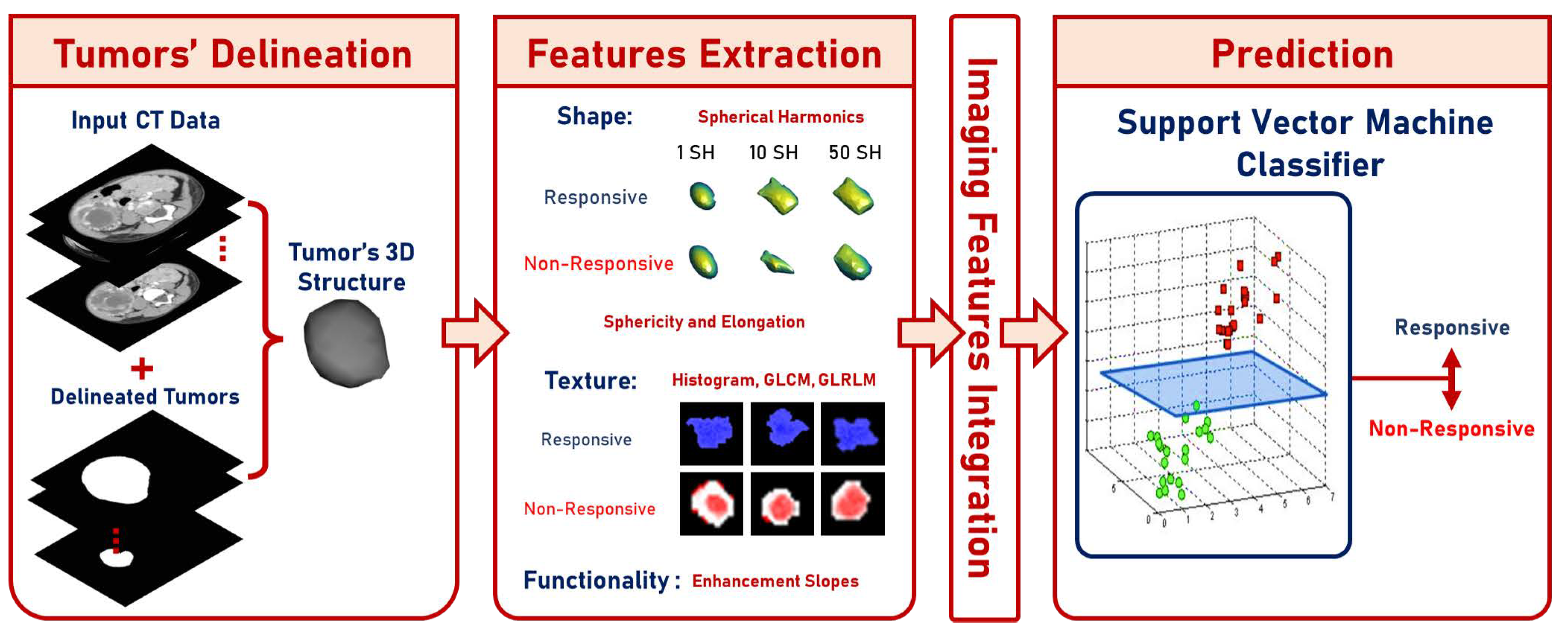
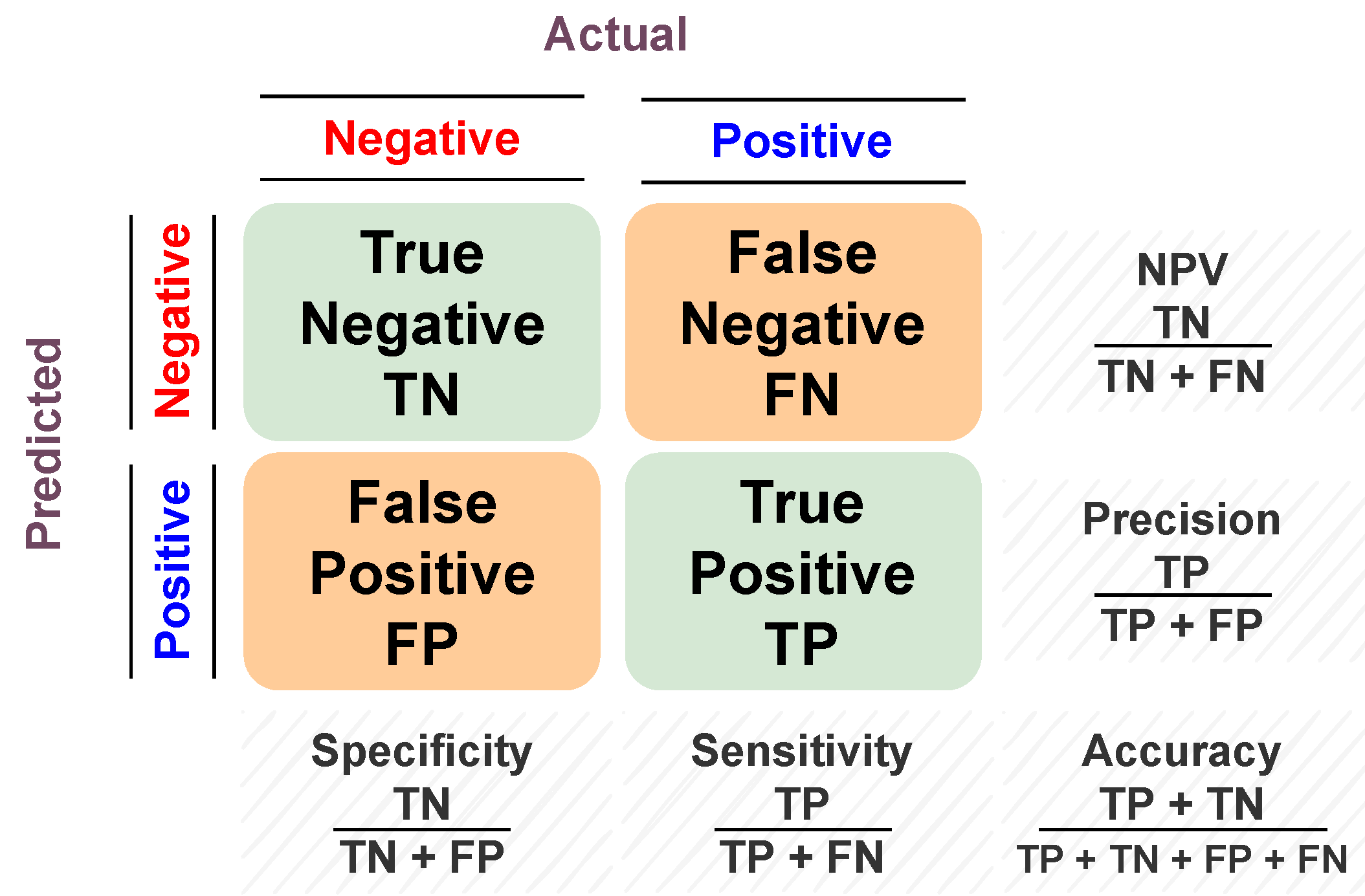
2. Materials and Methods
2.1. Data
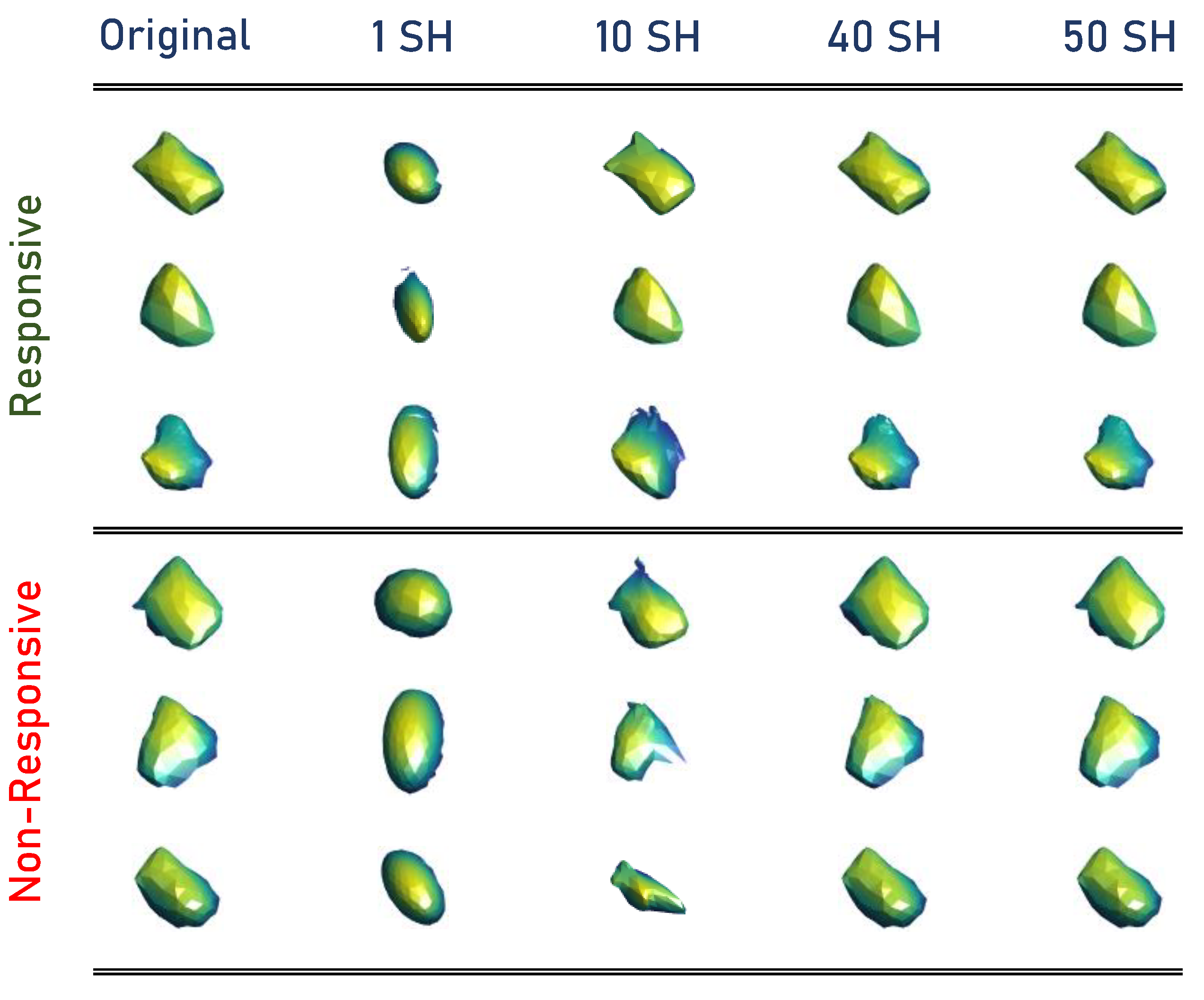
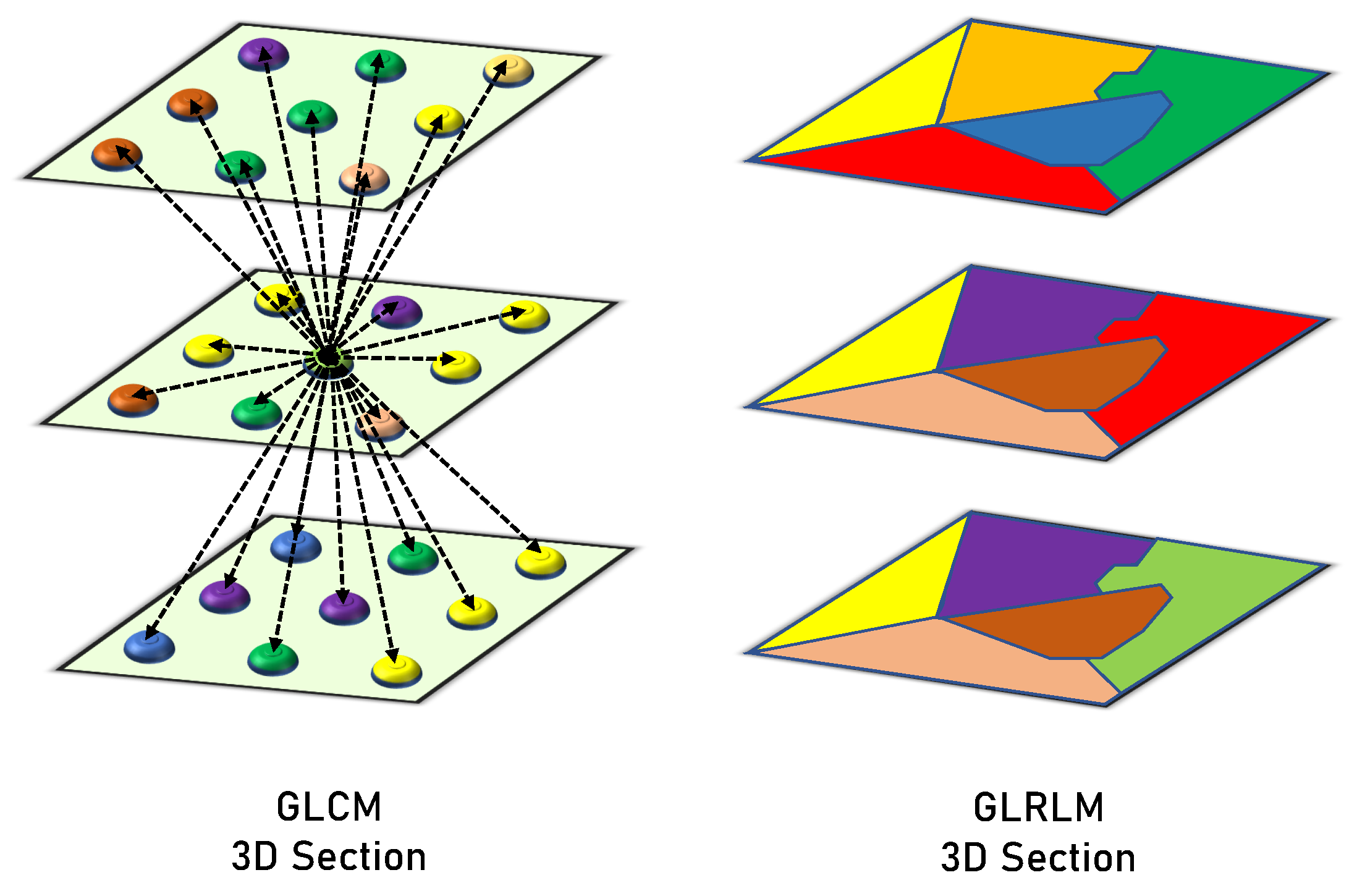
2.2. Methods
- (1)
- Initialization Process:
- −
- Triangulate the nodule surface.
- −
- Apply the Laplacian filtering to smooth the triangulated mesh.
- −
- The spherical parameterization is initialized using an arbitrary topology-preserving map onto the unit sphere.
- −
- Fix the , , , and threshold T values.
- (2)
- Attraction-repulsion Process:
3. Experiments and Results
4. Discussion
5. Limitations
6. Conclusions and Future Work
Author Contributions
Funding
Institutional Review Board Statement
Informed Consent Statement
Data Availability Statement
Conflicts of Interest
References
- Cancer.Net Editorial Board. Wilms Tumor—Childhood: Statistics. 2022. Available online: https://www.cancer.net/cancer-types/wilms-tumor-childhood/statistics (accessed on 10 November 2022).
- Cunningham, M.E.; Klug, T.D.; Nuchtern, J.G.; Chintagumpala, M.M.; Venkatramani, R.; Lubega, J.; Naik-Mathuria, B.J. Global disparities in Wilms tumor. J. Surg. Res. 2020, 247, 34–51. [Google Scholar] [CrossRef]
- Abdelhalim, A.; Helmy, T.E.; Harraz, A.M.; Abou-El-Ghar, M.E.; Dawaba, M.E.; Hafez, A.T. Can computerized tomography accurately stage childhood renal tumors? J. Urol. 2014, 192, 194–199. [Google Scholar] [CrossRef]
- Ng, Y.; Hall-Craggs, M.; Dicks-Mireaux, C.; Pritchard, J. Wilms’ tumour: pre-and post-chemotherapy CT appearances. Clin. Radiol. 1991, 43, 255–259. [Google Scholar] [CrossRef] [PubMed]
- Thomas, P.R.; Shochat, S.J.; Norkool, P.; Beckwith, J.B.; Breslow, N.E.; D’Angio, G.J. Prognostic implications of hepatic adhesion, invasion, and metastases at diagnosis of Wilms’ tumor. Cancer 1991, 68, 2486–2488. [Google Scholar] [CrossRef]
- Gleason, J.M.; Lorenzo, A.J.; Bowlin, P.R.; Koyle, M.A. Innovations in the management of Wilms’ tumor. Ther. Adv. Urol. 2014, 6, 165–176. [Google Scholar] [CrossRef]
- Taskinen, S.; Leskinen, O.; Lohi, J.; Koskenvuo, M.; Taskinen, M. Effect of Wilms tumor histology on response to neoadjuvant chemotherapy. J. Pediatr. Surg. 2019, 54, 771–774. [Google Scholar] [CrossRef] [Green Version]
- ra, I.; van Tinteren, H.; Bergeron, C.; de Kraker, J.; SIOP Nephroblastoma Study Committee. Progression of localised Wilms’ tumour during preoperative chemotherapy is an independent prognostic factor: A report from the SIOP 93–01 nephroblastoma trial and study. Eur. J. Cancer 2007, 43, 131–136. [Google Scholar]
- Elgendy, M.; Balaha, H.M.; Shehata, M.; Alksas, A.; Ghoneim, M.; Sherif, F.; Mahmoud, A.; Elgarayhi, A.; Taher, F.; Sallah, M.; et al. Role of Imaging and AI in the Evaluation of COVID-19 Infection: A Comprehensive Survey. Front. Biosci. 2022, 27, 276. [Google Scholar] [CrossRef]
- Alksas, A.; Shehata, M.; Saleh, G.A.; Shaffie, A.; Soliman, A.; Ghazal, M.; Khelifi, A.; Khalifeh, H.A.; Razek, A.A.; Giridharan, G.A.; et al. A novel computer-aided diagnostic system for accurate detection and grading of liver tumors. Sci. Rep. 2021, 11, 13148. [Google Scholar] [CrossRef]
- Ma, X.H.; Shu, L.; Jia, X.; Zhou, H.C.; Liu, T.T.; Liang, J.W.; Ding, Y.S.; He, M.; Shu, Q. Machine Learning-Based CT Radiomics Method for Identifying the Stage of Wilms Tumor in Children. Front. Pediatr. 2022, 10, 873035. [Google Scholar] [CrossRef]
- Misch, D.; Steffen, I.G.; Schönberger, S.; Voelker, T.; Furth, C.; Stöver, B.; Hautzel, H.; Henze, G.; Amthauer, H.; Denecke, T. Use of positron emission tomography for staging, preoperative response assessment and posttherapeutic evaluation in children with Wilms tumour. Eur. J. Nucl. Med. Mol. Imaging 2008, 35, 1642–1650. [Google Scholar] [CrossRef]
- Zheng, Z.; Chen, Z.; Xie, Y.; Zhong, Q.; Xie, W. Development and validation of a CT-based nomogram for preoperative prediction of clear cell renal cell carcinoma grades. Eur. Radiol. 2021, 31, 6078–6086. [Google Scholar] [CrossRef] [PubMed]
- Kim, H.M.; Byun, S.S.; Kim, J.K.; Jeong, C.W.; Kwak, C.; Hwang, E.C.; Kang, S.H.; Chung, J.; Kim, Y.J.; Ha, Y.S.; et al. Machine learning-based prediction model for late recurrence after surgery in patients with renal cell carcinoma. BMC Med. Inform. Decis. Mak. 2022, 22, 241. [Google Scholar] [CrossRef] [PubMed]
- Balaha, H.M.; Hassan, A.E.S. Skin cancer diagnosis based on deep transfer learning and sparrow search algorithm. Neural Comput. Appl. 2022, 1–39, in press. [Google Scholar] [CrossRef]
- Balaha, H.M.; Hassan, A.E.S. A variate brain tumor segmentation, optimization, and recognition framework. Artif. Intell. Rev. 2022, 1–54, in press. [Google Scholar]
- Abdel Razek, A.A.K.; Alksas, A.; Shehata, M.; AbdelKhalek, A.; Abdel Baky, K.; El-Baz, A.; Helmy, E. Clinical applications of artificial intelligence and radiomics in neuro-oncology imaging. Insights Imaging 2021, 12, 152. [Google Scholar] [CrossRef]
- Shehata, M.; Alksas, A.; Abouelkheir, R.T.; Elmahdy, A.; Shaffie, A.; Soliman, A.; Ghazal, M.; Abu Khalifeh, H.; Salim, R.; Abdel Razek, A.A.K.; et al. A comprehensive computer-assisted diagnosis system for early assessment of renal cancer tumors. Sensors 2021, 21, 4928. [Google Scholar] [CrossRef]
- Van Griethuysen, J.J.M.; Fedorov, A.; Parmar, C.; Hosny, A.; Aucoin, N.; Narayan, V.; Beets-Tan, R.G.H.; Fillion-Robin, J.C.; Pieper, S.; Aerts, H.J.W.L. Computational radiomics system to decode the radiographic phenotype. Cancer Res. 2017, 77, e104–e107. [Google Scholar] [CrossRef] [Green Version]
- Shaffie, A.; Soliman, A.; Ghazal, M.; Taher, F.; Dunlap, N.; Wang, B.; van Berkel, V.; Gimel’farb, G.; Elmaghraby, A.; El-Baz, A. A novel autoencoder-based diagnostic system for early assessment of lung cancer. In Proceedings of the 2018 25th IEEE International Conference on Image Processing (ICIP), Athens, Greece, 7–10 October 2018; IEEE: Piscataway, NJ, USA, 2018; pp. 1393–1397. [Google Scholar]
- Alksas, A.; Shehata, M.; Atef, H.; Sherif, F.; Alghamdi, N.S.; Ghazal, M.; Abdel Fattah, S.; El-Serougy, L.G.; El-Baz, A. A Novel System for Precise Grading of Glioma. Bioengineering 2022, 9, 532. [Google Scholar] [CrossRef]
- Nitzken, M.J. Shape Analysis of the Human Brain. Ph.D. Thesis, University of Louisville, Louisville, KY, USA, 2015. [Google Scholar]
- Haralick, R.M. Statistical and structural approaches to texture. Proc. IEEE 1979, 67, 786–804. [Google Scholar] [CrossRef]
- Gallowy, M. Texture analysis using gray level run length. Comput. Graph. Image Process. 1975, 4, 172–179. [Google Scholar] [CrossRef]
- Cortes, C.; Vapnik, V. Support-vector networks. Mach. Learn. 1995, 20, 273–297. [Google Scholar] [CrossRef]
- Zhang, R.; Xu, L.; Yu, Z.; Shi, Y.; Mu, C.; Xu, M. Deep-irtarget: An automatic target detector in infrared imagery using dual-domain feature extraction and allocation. IEEE Trans. Multimed. 2021, 24, 1735–1749. [Google Scholar] [CrossRef]
- Zhang, R.; Wu, L.; Yang, Y.; Wu, W.; Chen, Y.; Xu, M. Multi-camera multi-player tracking with deep player identification in sports video. Pattern Recognit. 2020, 102, 107260. [Google Scholar] [CrossRef]


| Features | Contrast-Phase | Number of Features |
|---|---|---|
| Texture Features | ||
| Histogram-based (first-order) | Venous | 26 |
| GLCM (second-order) | Pre + venous + delayed | 18 (6 per phase) |
| GLRLM (second-order) | Pre + venous + delayed | 36 (12 per phase) |
| Shape Features | ||
| Spherical harmonics | Venous | 50 |
| Descriptive | Venous | 2 |
| Functionality-Based Features | ||
| Enhancement slopes | Pre + venous + delayed | 2 |
| All | ||
| Integrated | Pre + venous + delayed | 134 |
| Features | Accuracy % | Sensitivity % | Specificity % | F1-Score |
|---|---|---|---|---|
| Texture Features | ||||
| Histogram (first-order) | 87.30 | 89.13 | 82.35 | 0.91 |
| GLCM (second-order) | 92.06 | 95.65 | 82.35 | 0.95 |
| GLRLM (second-order) | 92.06 | 93.48 | 88.24 | 0.95 |
| Shape Features | ||||
| Spherical harmonics | 90.84 | 89.13 | 94.12 | 0.93 |
| Descriptive | 90.84 | 91.30 | 88.24 | 0.93 |
| Functionality-Based Features | ||||
| Enhancement slopes | 88.89 | 89.13 | 88.24 | 0.92 |
| All | ||||
| Integrated | 95.24 | 95.65 | 94.12 | 0.97 |
| Classifier | Accuracy % | Sensitivity % | Specificity % | F1-Score |
|---|---|---|---|---|
| DT | 90.48 | 93.48 | 82.35 | 0.93 |
| KNN | 92.06 | 93.48 | 88.24 | 0.95 |
| LR | 90.48 | 91.30 | 88.24 | 0.93 |
| MLP | 92.06 | 93.48 | 88.24 | 0.95 |
| RF | 92.06 | 95.65 | 82.35 | 0.95 |
| SVM | 95.24 | 95.65 | 94.12 | 0.97 |
| Classifier | Accuracy % | Sensitivity % | Specificity % | F1-Score |
|---|---|---|---|---|
| DT | 89.95 ± 1.98 | 92.03 ± 3.69 | 84.31 ± 2.77 | 0.93 ± 0.02 |
| KNN | 89.42 ± 0.75 | 89.86 ± 2.71 | 88.24 ± 4.80 | 0.93 ± 0.01 |
| LR | 88.89 ± 0.00 | 89.13 ± 0.00 | 88.24 ± 0.00 | 0.92 ± 0.00 |
| MLP | 89.95 ± 0.75 | 92.03 ± 3.69 | 84.31 ± 7.34 | 0.93 ± 0.01 |
| RF | 91.01 ± 0.75 | 94.20 ± 2.71 | 82.35 ± 4.80 | 0.94 ± 0.01 |
| SVM | 93.65 ± 0.00 | 95.65 ± 0.00 | 88.24 ± 0.00 | 0.96 ± 0.00 |
| Classifier | Accuracy % | Sensitivity % | Specificity % | F1-Score |
|---|---|---|---|---|
| DT | 85.71 ± 2.24 | 89.13 ± 3.07 | 76.47 ± 8.32 | 0.90 ± 0.02 |
| KNN | 86.77 ± 2.70 | 86.96 ± 4.70 | 86.27 ± 7.34 | 0.91 ± 0.02 |
| LR | 84.66 ± 2.70 | 85.51 ± 2.71 | 82.35 ± 4.80 | 0.89 ± 0.02 |
| MLP | 85.71 ± 1.30 | 89.13 ± 3.07 | 76.47 ± 9.61 | 0.90 ± 0.01 |
| RF | 88.36 ± 1.50 | 91.30 ± 3.07 | 80.39 ± 7.34 | 0.92 ± 0.01 |
| SVM | 91.01 ± 1.98 | 91.30 ± 1.77 | 90.20 ± 2.77 | 0.94 ± 0.01 |
Disclaimer/Publisher’s Note: The statements, opinions and data contained in all publications are solely those of the individual author(s) and contributor(s) and not of MDPI and/or the editor(s). MDPI and/or the editor(s) disclaim responsibility for any injury to people or property resulting from any ideas, methods, instructions or products referred to in the content. |
© 2023 by the authors. Licensee MDPI, Basel, Switzerland. This article is an open access article distributed under the terms and conditions of the Creative Commons Attribution (CC BY) license (https://creativecommons.org/licenses/by/4.0/).
Share and Cite
Sharaby, I.; Alksas, A.; Nashat, A.; Balaha, H.M.; Shehata, M.; Gayhart, M.; Mahmoud, A.; Ghazal, M.; Khalil, A.; Abouelkheir, R.T.; et al. Prediction of Wilms’ Tumor Susceptibility to Preoperative Chemotherapy Using a Novel Computer-Aided Prediction System. Diagnostics 2023, 13, 486. https://doi.org/10.3390/diagnostics13030486
Sharaby I, Alksas A, Nashat A, Balaha HM, Shehata M, Gayhart M, Mahmoud A, Ghazal M, Khalil A, Abouelkheir RT, et al. Prediction of Wilms’ Tumor Susceptibility to Preoperative Chemotherapy Using a Novel Computer-Aided Prediction System. Diagnostics. 2023; 13(3):486. https://doi.org/10.3390/diagnostics13030486
Chicago/Turabian StyleSharaby, Israa, Ahmed Alksas, Ahmed Nashat, Hossam Magdy Balaha, Mohamed Shehata, Mallorie Gayhart, Ali Mahmoud, Mohammed Ghazal, Ashraf Khalil, Rasha T. Abouelkheir, and et al. 2023. "Prediction of Wilms’ Tumor Susceptibility to Preoperative Chemotherapy Using a Novel Computer-Aided Prediction System" Diagnostics 13, no. 3: 486. https://doi.org/10.3390/diagnostics13030486