
Interest of Absolute Eosinopenia as a Marker of Influenza in Outpatients during the Fall-Winter Seasons 2016–2018 in the Greater Paris Area: The SUPERFLUOUS Study
Abstract
:1. Introduction
2. Materials and Methods
2.1. Outpatients’ Selection and Objectives
2.2. Statistical Analysis
2.3. Ethics
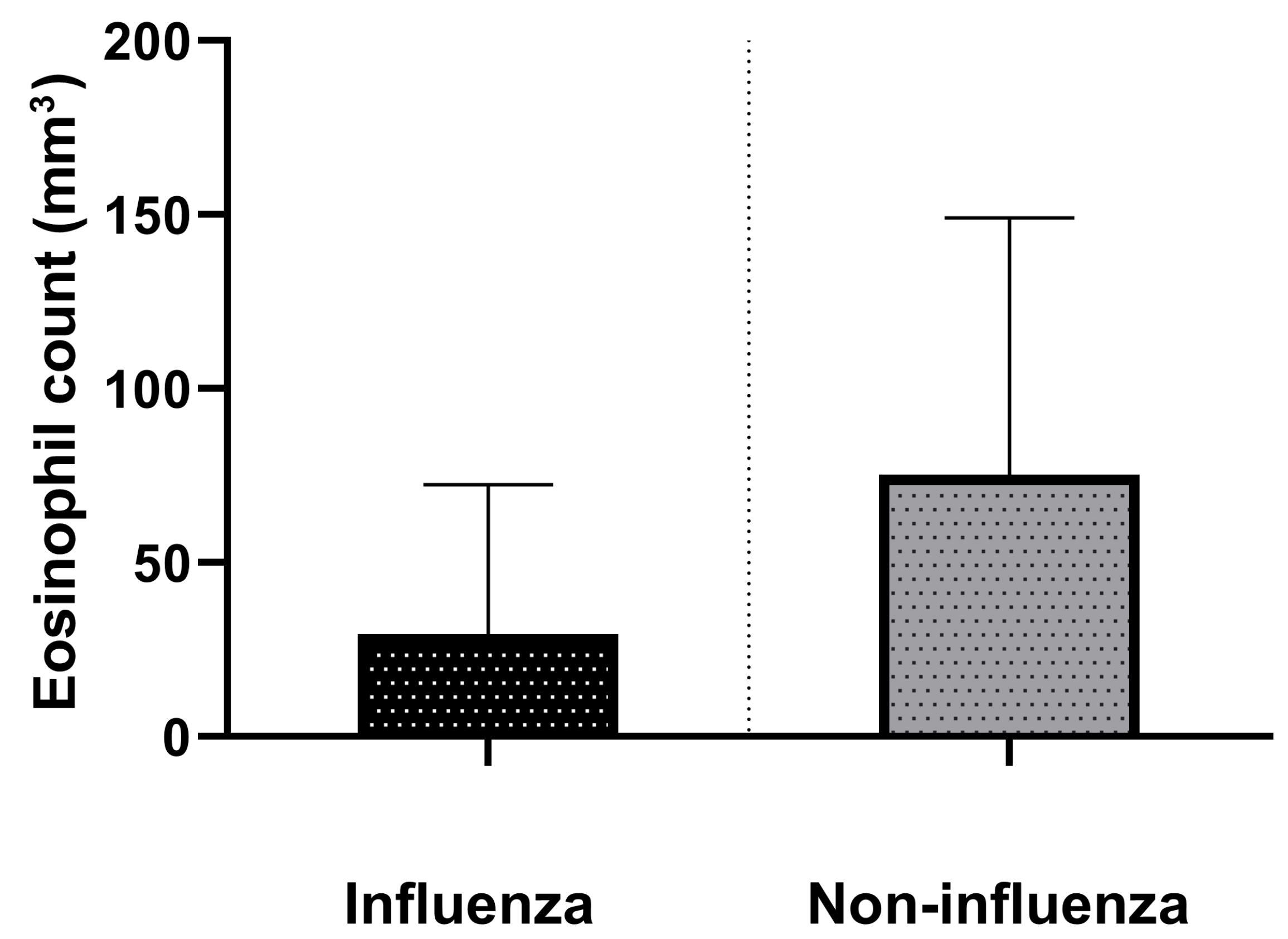
3. Results
3.1. Study Population and Infection Characteristics
3.2. Predictors of Influenza
4. Discussion
Supplementary Materials
Author Contributions
Funding
Institutional Review Board Statement
Informed Consent Statement
Data Availability Statement
Acknowledgments
Conflicts of Interest
References
- Havers, F.P.; Hicks, L.A.; Chung, J.R.; Gaglani, M.; Murthy, K.; Zimmerman, R.K.; Jackson, L.A.; Petrie, J.G.; McLean, H.Q.; Nowalk, M.P.; et al. Outpatient Antibiotic Prescribing for Acute Respiratory Infections During Influenza Seasons. JAMA Netw. Open 2018, 1, e180243. [Google Scholar] [CrossRef] [PubMed]
- Cheysson, F.; Brun-Buisson, C.; Opatowski, L.; Le Fouler, L.; Caserio-Schönemann, C.; Pontais, I.; Guillemot, D.; Watier, L. Outpatient Antibiotic Use Attributable to Viral Acute Lower Respiratory Tract Infections during the Cold Season in France, 2010–2017. Int. J. Antimicrob. Agents 2021, 57, 106339. [Google Scholar] [CrossRef] [PubMed]
- Llor, C.; Bjerrum, L. Antimicrobial Resistance: Risk Associated with Antibiotic Overuse and Initiatives to Reduce the Problem. Ther. Adv. Drug Saf. 2014, 5, 229–241. [Google Scholar] [CrossRef] [PubMed] [Green Version]
- Yang, Y.; Huang, F.; Gonzalez, R.; Wang, W.; Lu, G.; Li, Y.; Vernet, G.; Jin, Q.; Wang, J. Evaluation of Twelve Real-Time Reverse Transcriptase PCR Primer-Probe Sets for Detection of Pandemic Influenza A/H1N1 2009 Virus. J. Clin. Microbiol. 2011, 49, 1434–1440. [Google Scholar] [CrossRef] [Green Version]
- Huang, H.S.; Tsai, C.L.; Chang, J.; Hsu, T.C.; Lin, S.; Lee, C.C. Multiplex PCR System for the Rapid Diagnosis of Respiratory Virus Infection: Systematic Review and Meta-Analysis. Clin. Microbiol. Infect. 2018, 24, 1055–1063. [Google Scholar] [CrossRef] [Green Version]
- Clark, T.W.; Lindsley, K.; Wigmosta, T.B.; Bhagat, A.; Hemmert, R.B.; Uyei, J.; Timbrook, T.T. Rapid Multiplex PCR for Respiratory Viruses Reduces Time to Result and Improves Clinical Care: Results of a Systematic Review and Meta-Analysis. J. Infect. 2023, 86, 462–475. [Google Scholar] [CrossRef]
- Salonen, E.-M.; Vaheri, A. C-Reactive Protein in Acute Viral Infections. J. Med. Virol. 1981, 8, 161–167. [Google Scholar] [CrossRef]
- Le Bel, J.; Hausfater, P.; Chenevier-Gobeaux, C.; Blanc, F.-X.; Benjoar, M.; Ficko, C.; Ray, P.; Choquet, C.; Duval, X.; Claessens, Y.-E. Diagnostic Accuracy of C-Reactive Protein and Procalcitonin in Suspected Community-Acquired Pneumonia Adults Visiting Emergency Department and Having a Systematic Thoracic CT Scan. Crit. Care 2015, 19, 366. [Google Scholar] [CrossRef] [Green Version]
- Pepys, M.B.; Hirschfield, G.M. C-Reactive Protein: A Critical Update. J. Clin. Investig. 2003, 112, 299. [Google Scholar] [CrossRef]
- Samsudin, I.; Vasikaran, S.D. Clinical Utility and Measurement of Procalcitonin. Clin. Biochem. Rev. 2017, 38, 59–68. [Google Scholar]
- Abidi, K.; Khoudri, I.; Belayachi, J.; Madani, N.; Zekraoui, A.; Zeggwagh, A.A.; Abouqal, R. Eosinopenia Is a Reliable Marker of Sepsis on Admission to Medical Intensive Care Units. Crit. Care 2008, 12, R59. [Google Scholar] [CrossRef] [PubMed] [Green Version]
- Shaaban, H.; Daniel, S.; Sison, R.; Slim, J.; Perez, G. Eosinopenia: Is It a Good Marker of Sepsis in Comparison to Procalcitonin and C-Reactive Protein Levels for Patients Admitted to a Critical Care Unit in an Urban Hospital? J. Crit. Care 2010, 25, 570–575. [Google Scholar] [CrossRef] [PubMed]
- Partouche, B.; Pepin, M.; De Farcy, P.M.; Kahn, J.E.; Sawczynski, B.; Lechowski, L.; Teillet, L.; Barbot, F.; Herr, M.; Davido, B. Persistent Eosinopenia Is Associated with In-Hospital Mortality among Older Patients: Unexpected Prognostic Value of a Revisited Biomarker. BMC Geriatr. 2021, 21, 557. [Google Scholar] [CrossRef] [PubMed]
- Debray, A.; Nathanson, S.; Moulin, F.; Salomon, J.; Davido, B. Eosinopenia as a Marker of Diagnosis and Prognostic to Distinguish Bacterial from Aseptic Meningitis in Pediatrics. Eur. J. Clin. Microbiol. Infect. Dis. 2019, 38, 1821–1827. [Google Scholar] [CrossRef]
- Cortés-Vieyra, R.; Gutiérrez-Castellanos, S.; Álvarez-Aguilar, C.; Baizabal-Aguirre, V.M.; Nuñez-Anita, R.E.; Rocha-López, A.G.; Gómez-García, A. Behavior of Eosinophil Counts in Recovered and Deceased COVID-19 Patients over the Course of the Disease. Viruses 2021, 13, 1675. [Google Scholar] [CrossRef]
- Bellelli, V.; D’Ettorre, G.; Celani, L.; Borrazzo, C.; Ceccarelli, G.; Venditti, M. Clinical Significance of Lymphocytopenia in Patients Hospitalized with Pneumonia Caused by Influenza Virus. Crit. Care 2019, 23, 330. [Google Scholar] [CrossRef] [Green Version]
- Harrison, A.M.; Bonville, C.A.; Rosenberg, H.F.; Domachowske, J.B. Respiratory Syncytical Virus-Induced Chemokine Expression in the Lower Airways: Eosinophil Recruitment and Degranulation. Am. J. Respir. Crit. Care Med. 1999, 159, 1918–1924. [Google Scholar] [CrossRef]
- Phipps, S.; En Lam, C.; Mahalingam, S.; Newhouse, M.; Ramirez, R.; Rosenberg, H.F.; Foster, P.S.; Matthaei, K.I. Eosinophils Contribute to Innate Antiviral Immunity and Promote Clearance of Respiratory Syncytial Virus. Blood 2007, 110, 1578–1586. [Google Scholar] [CrossRef] [Green Version]
- Samarasinghe, A.E.; Melo, R.C.N.; Duan, S.; LeMessurier, K.S.; Liedmann, S.; Surman, S.L.; Lee, J.J.; Hurwitz, J.L.; Thomas, P.G.; McCullers, J.A. Eosinophils Promote Antiviral Immunity in Mice Infected with Influenza A Virus. J. Immunol. 2017, 198, 3214–3226. [Google Scholar] [CrossRef] [Green Version]
- Hopp, R. Eosinophils Bind Rhinovirus and Activate Virus-Specific T Cells. Pediatrics 1999, 104, 359. [Google Scholar] [CrossRef]
- Lemarie, B.; Boussaid, G.; Gault, E.; Prigent, H.; Beaune, S.; Moreau, F.; Dumoulin, J.; Pepin, M.; Greffe, S.; De Truchis, P.; et al. Predictors of Hospitalization and Superinfection in Viral Respiratory Tract Infections Between Influenza and Paramyxoviruses: The SUPERFLUOUS Study. J. Infect. Dis. 2022, 226, 1027–1035. [Google Scholar] [CrossRef] [PubMed]
- Papillard-Marechal, S.; Enouf, V.; Schnuriger, A.; Vabret, A.; Macheras, E.; Rameix-Welti, M.A.; Page, B.; Freymuth, F.; Van Der Werf, S.; Garbarg-Chenon, A.; et al. Monitoring Epidemic Viral Respiratory Infections Using One-Step Real-Time Triplex RT-PCR Targeting Influenza A and B Viruses and Respiratory Syncytial Virus. J. Med. Virol. 2011, 83, 695–701. [Google Scholar] [CrossRef] [PubMed]
- Guo, Z.; Zhang, Z.; Prajapati, M.; Li, Y. Lymphopenia Caused by Virus Infections and the Mechanisms Beyond. Viruses 2021, 13, 1876. [Google Scholar] [CrossRef] [PubMed]
- Zhao, Y.; Yu, C.; Ni, W.; Shen, H.; Qiu, M.; Zhao, Y. Peripheral Blood Inflammatory Markers in Predicting Prognosis in Patients with COVID-19. Some Differences with Influenza A. J. Clin. Lab. Anal. 2021, 35, e23657. [Google Scholar] [CrossRef] [PubMed]
- Kanda, A.; Yun, Y.; Van Bui, D.; Nguyen, L.M.; Kobayashi, Y.; Suzuki, K.; Mitani, A.; Sawada, S.; Hamada, S.; Asako, M.; et al. The Multiple Functions and Subpopulations of Eosinophils in Tissues under Steady-State and Pathological Conditions. Allergol. Int. 2021, 70, 9–18. [Google Scholar] [CrossRef] [PubMed]
- Self, W.H.; McNaughton, C.D.; Grijalva, C.G.; Zhu, Y.; Chappell, J.D.; Williams, J.V.; Talbot, H.K.; Shay, D.K.; Griffin, M.R. Diagnostic Performance of the BinaxNow Influenza A&B Rapid Antigen Test in ED Patients. Am. J. Emerg. Med. 2012, 30, 1955–1961. [Google Scholar] [CrossRef]
- Hueston, W.J.; Benich, J.J. A Cost-Benefit Analysis of Testing for Influenza A in High-Risk Adults. Ann. Fam. Med. 2004, 2, 33–40. [Google Scholar] [CrossRef] [Green Version]
- Butler, C.C.; Van der Velden, A.W.; Bongard, E.; Saville, B.R.; Holmes, J.; Coenen, S.; Cook, J.; Francis, N.A.; Lewis, R.J.; Godycki-Cwirko, M.; et al. Oseltamivir plus Usual Care versus Usual Care for Influenza-like Illness in Primary Care: An Open-Label, Pragmatic, Randomised Controlled Trial. Lancet 2020, 395, 42–52. [Google Scholar] [CrossRef]
- Flu News Europe | Home. Available online: https://www.flunewseurope.org/ (accessed on 19 March 2023).
- Davido, B.; Jaffal, K.; Gault, E.; Bourlet, S.; Beaune, S. Back to the Future of Viruses: A Case of Triple Coinfection Caused by Respiratory Syncytial Virus, Human Coronavirus OC43, and Rhinovirus. Int. J. Infect. Dis. 2023, 130, 205–207. [Google Scholar] [CrossRef]
- Teigell Muñoz, F.J.; García-Guijarro, E.; García-Domingo, P.; Pérez-Nieto, G.; Roque Rojas, F.; García-Peña, M.; Nieto Gallo, M.A.; Melero Bermejo, J.A.; De Guzman García-Monge, M.T.; Granizo, J.J. A Safe Protocol to Identify Low-Risk Patients with COVID-19 Pneumonia for Outpatient Management. Intern. Emerg. Med. 2021, 16, 1663–1671. [Google Scholar] [CrossRef]


| Baseline Characteristics | Value |
|---|---|
| Age, mean (SD), y | 59 (10) |
| Male sex, n (%) | 52 (44.8) |
| Chronic respiratory disease, n (%) | 23 (19.8) |
| CCI * < 5, n (%) | 73 (62.9) |
| Fine score, mean (SD) | 65.7 (33.3) |
| Management | |
| Consulting an ER physician, n (%) | 88 (75.8) |
| Period | |
| Season 2016–2017, n (%) | 40 (34.5) |
| Season 2017–2018, n (%) | 76 (65.5) |
| Fall, n (%) | 7 (6.0) |
| Winter, n (%) | 100 (86.2) |
| Biology and imaging (n = 96) | |
| PMN count ≥ 7000/mm3, n (%) | 20 (20.8) |
| Lymphocyte count < 800/mm3, n (%) | 44 (45.8) |
| Eosinophil count = 0/mm3, n (%) | 26 (27.1) |
| Radiological abnormalities | 10 (10.4) |
| Treatment strategies | |
| Antimicrobial therapy initiated for superinfection, n (%) | 46 (39.6) |
| Treatment duration, mean (SD) | 6.8 (2) |
| Variables | Univariate Model | Multivariable Model 1 | Multivariable Model 2 | |||
|---|---|---|---|---|---|---|
| OR [IC95%] | p Value | aOR [IC95%] | p Value | aOR [IC95%] | p Value | |
| Adjusted on Age, Sex, Eosinophil Count | Adjusted on Age, Sex, Lymphocyte Count | |||||
| Baseline characteristics | ||||||
| Age (years) | 0.97 [0.95–0.99] | 0.01 | 0.96 [0.91–1.01] | 0.12 | 0.95 [0.89–1.01] | 0.11 |
| Sex (male) | 0.90 [0.38–2.12] | 0.81 | - | - | - | - |
| Chronic respiratory disease | 0.66 [0.24–1.83] | 0.43 | - | - | - | - |
| CCI * < 5 | 3.01 [1.25–7.23] | 0.01 | 0.30 [0.07–1.26] | 0.30 | 0.29 [0.07–1.21] | 0.09 |
| Fine score (mean) | 0.99 [0.97–1.00] | 0.04 | 1.02 [0.97–1.05] | 0.46 | 1.02 [0.98–1.05] | 0.30 |
| Management | ||||||
| Consulting an ER physician | 1.36 [0.52–3.55] | 0.53 | - | - | - | - |
| Period | ||||||
| 2016–2017 vs. 2017–2018 | 1.44 [0.76–2.74] | 0.26 | - | - | - | - |
| Season: Spring | Reference | Reference | Reference | |||
| Fall | 0.5 [0.06–4.09] | 0.51 | 0.52 [0.04–7.28] | 0.62 | 0.95 [0.07–13.47] | 0.97 |
| Winter | 5.69 [1.38–23.33] | 0.02 | 7.1 [1.12–45.08] | 0.04 | 9.08 [1.49–55.40] | 0.02 |
| Biology and imaging | ||||||
| PMN count ≥ 7000/mm3 | 0.43 [0.15–1.23] | 0.12 | - | - | - | - |
| Lymphocyte count <800/mm3 | 6.77 [2.11–21.77] | 0.001 | - | - | 7.37 [1.86–29.20] | 0.004 |
| Eosinophil count = 0/mm3 | 5.87 [1.28–27.05] | 0.02 | 6.16 [1.14–33.24] | 0.03 | - | - |
| Radiological abnormalities | 0.72 [0.17–2.99] | 0.65 | - | - | - | - |
| Treatment strategies | ||||||
| Antitbiotics for superinfection | 0.58 [0.25–1.38] | 0.21 | - | - | - | - |
| Treatment duration (mean) | 0.94 [0.84–1.06] | 0.36 | - | - | - | - |
Disclaimer/Publisher’s Note: The statements, opinions and data contained in all publications are solely those of the individual author(s) and contributor(s) and not of MDPI and/or the editor(s). MDPI and/or the editor(s) disclaim responsibility for any injury to people or property resulting from any ideas, methods, instructions or products referred to in the content. |
© 2023 by the authors. Licensee MDPI, Basel, Switzerland. This article is an open access article distributed under the terms and conditions of the Creative Commons Attribution (CC BY) license (https://creativecommons.org/licenses/by/4.0/).
Share and Cite
Davido, B.; Lemarie, B.; Gault, E.; Dumoulin, J.; D’anglejan, E.; Beaune, S.; De Truchis, P. Interest of Absolute Eosinopenia as a Marker of Influenza in Outpatients during the Fall-Winter Seasons 2016–2018 in the Greater Paris Area: The SUPERFLUOUS Study. Diagnostics 2023, 13, 2115. https://doi.org/10.3390/diagnostics13122115
Davido B, Lemarie B, Gault E, Dumoulin J, D’anglejan E, Beaune S, De Truchis P. Interest of Absolute Eosinopenia as a Marker of Influenza in Outpatients during the Fall-Winter Seasons 2016–2018 in the Greater Paris Area: The SUPERFLUOUS Study. Diagnostics. 2023; 13(12):2115. https://doi.org/10.3390/diagnostics13122115
Chicago/Turabian StyleDavido, Benjamin, Benoit Lemarie, Elyanne Gault, Jennifer Dumoulin, Emma D’anglejan, Sebastien Beaune, and Pierre De Truchis. 2023. "Interest of Absolute Eosinopenia as a Marker of Influenza in Outpatients during the Fall-Winter Seasons 2016–2018 in the Greater Paris Area: The SUPERFLUOUS Study" Diagnostics 13, no. 12: 2115. https://doi.org/10.3390/diagnostics13122115