Next Issue
Volume 13, July-1
Previous Issue
Volume 13, June-1


Diagnostics, Volume 13, Issue 12 (June-2 2023) – 149 articles

Cover Story (view full-size image): 
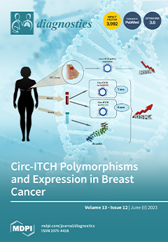
Breast cancer (BC) is one of the leading causes of cancer-related mortality worldwide. The association between circ-ITCH gene polymorphisms, circ-ITCH expression, and their effect on β-catenin level correlates with the development of BC. rs10485505 and rs4911154 polymorphisms are related to the risk and prognosis of BC by affecting the level of circ-ITCH mRNA expression in BC tissues and serum levels of β-catenin. The relative expression of circ-ITCH was found to be remarkably decreased, while the β-catenin level significantly increased in patients carrying the A allele (rs4911154) and T allele (rs10485505). Kaplan–Meier analysis showed that the expression of circ-ITCH was associated with the prognosis of BC and correlated with tumor size, grade, TNM stage, and clinical stage, pointing to its possible role as a biomarker in prognosis. View this paper
  • Issues are regarded as officially published after their release is announced to the table of contents alert mailing list.
  • You may sign up for e-mail alerts to receive table of contents of newly released issues.
  • PDF is the official format for papers published in both, html and pdf forms. To view the papers in pdf format, click on the "PDF Full-text" link, and use the free Adobe Reader to open them.
Order results
Result details
Section
Select all
Export citation of selected articles as:
19 pages, 3832 KiB  
Article
Quantitative Evaluation of COVID-19 Pneumonia CT Using AI Analysis—Feasibility and Differentiation from Other Common Pneumonia Forms
by Una Ebong, Susanne Martina Büttner, Stefan A. Schmidt, Franziska Flack, Patrick Korf, Lynn Peters, Beate Grüner, Steffen Stenger, Thomas Stamminger, Hans Kestler, Meinrad Beer and Christopher Kloth
Diagnostics 2023, 13(12), 2129; https://doi.org/10.3390/diagnostics13122129 - 20 Jun 2023
Viewed by 1279
Abstract
PURPOSE: To implement the technical feasibility of an AI-based software prototype optimized for the detection of COVID-19 pneumonia in CT datasets of the lung and the differentiation between other etiologies of pneumonia. METHODS: This single-center retrospective case–control-study consecutively yielded 144 patients (58 female, [...] Read more.
PURPOSE: To implement the technical feasibility of an AI-based software prototype optimized for the detection of COVID-19 pneumonia in CT datasets of the lung and the differentiation between other etiologies of pneumonia. METHODS: This single-center retrospective case–control-study consecutively yielded 144 patients (58 female, mean age 57.72 ± 18.25 y) with CT datasets of the lung. Subgroups including confirmed bacterial (n = 24, 16.6%), viral (n = 52, 36.1%), or fungal (n = 25, 16.6%) pneumonia and (n = 43, 30.7%) patients without detected pneumonia (comparison group) were evaluated using the AI-based Pneumonia Analysis prototype. Scoring (extent, etiology) was compared to reader assessment. RESULTS: The software achieved an optimal sensitivity of 80.8% with a specificity of 50% for the detection of COVID-19; however, the human radiologist achieved optimal sensitivity of 80.8% and a specificity of 97.2%. The mean postprocessing time was 7.61 ± 4.22 min. The use of a contrast agent did not influence the results of the software (p = 0.81). The mean evaluated COVID-19 probability is 0.80 ± 0.36 significantly higher in COVID-19 patients than in patients with fungal pneumonia (p < 0.05) and bacterial pneumonia (p < 0.001). The mean percentage of opacity (PO) and percentage of high opacity (PHO ≥ −200 HU) were significantly higher in COVID-19 patients than in healthy patients. However, the total mean HU in COVID-19 patients was −679.57 ± 112.72, which is significantly higher than in the healthy control group (p < 0.001). CONCLUSION: The detection and quantification of pneumonia beyond the primarily trained COVID-19 datasets is possible and shows comparable results for COVID-19 pneumonia to an experienced reader. The advantages are the fast, automated segmentation and quantification of the pneumonia foci. Full article
(This article belongs to the Special Issue Advances in Diagnostic Medical Imaging in 2023)
Show Figures

Figure 1

11 pages, 1395 KiB  
Article
Determination of Anti-Xa Inhibitor Plasma Concentrations Using a Universal Edoxaban Calibrator
by Annika Burger, Jan-Dirk Studt, Adriana Mendez, Lorenzo Alberio, Pierre Fontana, Walter A. Wuillemin, Adrian Schmidt, Lukas Graf, Bernhard Gerber, Cédric Bovet, Thomas C. Sauter, Nikolaus B. Binder and Michael Nagler
Diagnostics 2023, 13(12), 2128; https://doi.org/10.3390/diagnostics13122128 - 20 Jun 2023
Cited by 1 | Viewed by 1080
Abstract
A universal calibrator for the determination of all anti-Xa inhibitors would support laboratory processes. We aimed to test the clinical performance of an anti-Xa assay utilizing a universal edoxaban calibrator to determine clinically relevant concentrations of all anti-Xa inhibitors. Following a pilot study, [...] Read more.
A universal calibrator for the determination of all anti-Xa inhibitors would support laboratory processes. We aimed to test the clinical performance of an anti-Xa assay utilizing a universal edoxaban calibrator to determine clinically relevant concentrations of all anti-Xa inhibitors. Following a pilot study, we enrolled 553 consecutive patients taking rivaroxaban, edoxaban, or apixaban from nine study centers in a prospective cross-sectional study. The Technochrom® anti-Xa assay was conducted using the Technoview® edoxaban calibrator. Using ultra-high-performance liquid chromatography-tandem mass spectrometry (LC-MS/MS), anti-Xa inhibitor drug concentrations were determined. Sensitivities and specificities to detect three clinically relevant drug concentrations (30 µgL−1, 50 µgL−1, 100 µgL−1) were determined. Overall, 300 patients treated with rivaroxaban, 221 with apixaban, and 32 with edoxaban were included. The overall correlation coefficient (rs) was 0.95 (95% CI 0.94, 0.96). An area under the receiver operating characteristic curve of 0.96 for 30 µgL−1, 0.98 for 50 µgL−1, and 0.99 for 100 µgL−1 was found. The sensitivities were 92.3% (95% CI 89.2, 94.6), 92.7% (89.4, 95.1), and 94.8% (91.1, 97.0), respectively (specificities 82.2%, 93.7%, and 94.4%). In conclusion, the clinical performance of a universal, edoxaban-calibrated anti-Xa assay was solid and most drug concentrations were predicted correctly. Full article
(This article belongs to the Section Clinical Laboratory Medicine)
Show Figures

Figure 1

11 pages, 2824 KiB  
Article
Esophagogastroduodenal Findings in Patients with Intraductal Papillary Mucinous Neoplasms
by Dana Zelnik Yovel, Erwin Santo, Majd Khader, Roie Tzadok, Nir Bar, Asaf Aizic, Oren Shibolet and Dana Ben-Ami Shor
Diagnostics 2023, 13(12), 2127; https://doi.org/10.3390/diagnostics13122127 - 20 Jun 2023
Viewed by 1020
Abstract
The association between intraductal papillary mucinous neoplasms (IPMNs) and extra-pancreatic malignancies is controversial. This cross-sectional study compared esophagogastroduodenal findings in 340 IPMN patients to those of age- and gender-matched controls without known IPMNs who underwent esophagogastroduodenoscopies (EGDs) for similar clinical reasons. The presence [...] Read more.
The association between intraductal papillary mucinous neoplasms (IPMNs) and extra-pancreatic malignancies is controversial. This cross-sectional study compared esophagogastroduodenal findings in 340 IPMN patients to those of age- and gender-matched controls without known IPMNs who underwent esophagogastroduodenoscopies (EGDs) for similar clinical reasons. The presence of gastric and esophageal cancer, Barrett’s esophagus, neuroendocrine tumors (NETs), gastrointestinal stromal tumors (GISTs), gastric adenomas, and ampullary tumors was assessed. The results showed that 4/340 (1.2%) of the IPMN patients had gastric cancer and 1/340 (0.3%) had esophageal cancer. The matched control group had a similar incidence of gastric cancer (5/340) (1.5%), with no esophageal cancer cases (p > 0.999). The overall incidence of other esophagogastroduodenal conditions did not significantly differ between the IPMN patients and the controls. However, the incidence of gastric cancer in the IPMN patients was higher than expected based on national cancer registry data (standardized incidence ratio of 31.39; p < 0.001; CI 8.38–78.76). In conclusion, IPMN patients have a significantly higher incidence of gastric cancer compared to the general population. However, the incidence of esophagogastroduodenal findings, including gastric and esophageal cancer, is similar between IPMN patients and those who undergo an EGD for similar clinical indications. Further research is needed to determine optimal surveillance strategies for IPMN patients regarding their risk of developing gastric cancer. Full article
(This article belongs to the Special Issue Advances in the Diagnostic Imaging of Gastrointestinal Diseases)
Show Figures

Figure 1

13 pages, 1788 KiB  
Article
Identifying Postural Instability in Children with Cerebral Palsy Using a Predictive Model: A Longitudinal Multicenter Study
by Carlo Marioi Bertoncelli, Domenico Bertoncelli, Sikha S. Bagui, Subhash C. Bagui, Stefania Costantini and Federico Solla
Diagnostics 2023, 13(12), 2126; https://doi.org/10.3390/diagnostics13122126 - 20 Jun 2023
Viewed by 1385
Abstract
Insufficient postural control and trunk instability are serious concerns in children with cerebral palsy (CP). We implemented a predictive model to identify factors associated with postural impairments such as spastic or hypotonic truncal tone (TT) in children with CP. We conducted a longitudinal, [...] Read more.
Insufficient postural control and trunk instability are serious concerns in children with cerebral palsy (CP). We implemented a predictive model to identify factors associated with postural impairments such as spastic or hypotonic truncal tone (TT) in children with CP. We conducted a longitudinal, double-blinded, multicenter, descriptive study of 102 teenagers with CP with cognitive impairment and severe motor disorders with and without truncal tone impairments treated in two specialized hospitals (60 inpatients and 42 outpatients; 60 males, mean age 16.5 ± 1.2 years, range 12 to 18 yrs). Clinical and functional data were collected between 2006 and 2021. TT-PredictMed, a multiple logistic regression prediction model, was developed to identify factors associated with hypotonic or spastic TT following the guidelines of “Transparent Reporting of a multivariable prediction model for Individual Prognosis or Diagnosis”. Predictors of hypotonic TT were hip dysplasia (p = 0.01), type of etiology (postnatal > perinatal > prenatal causes; p = 0.05), male gender, and poor manual (p = 0.01) and gross motor function (p = 0.05). Predictors of spastic TT were neuromuscular scoliosis (p = 0.03), type of etiology (prenatal > perinatal > postnatal causes; p < 0.001), spasticity (quadri/triplegia > diplegia > hemiplegia; p = 0.05), presence of dystonia (p = 0.001), and epilepsy (refractory > controlled, p = 0.009). The predictive model’s average accuracy, sensitivity, and specificity reached 82%. The model’s accuracy aligns with recent studies on applying machine learning models in the clinical field. Full article
(This article belongs to the Section Machine Learning and Artificial Intelligence in Diagnostics)
Show Figures

Figure 1

14 pages, 1447 KiB  
Article
Diagnosis of Chest Pneumonia with X-ray Images Based on Graph Reasoning
by Cheng Wang, Chang Xu, Yulai Zhang and Peng Lu
Diagnostics 2023, 13(12), 2125; https://doi.org/10.3390/diagnostics13122125 - 20 Jun 2023
Cited by 1 | Viewed by 2100
Abstract
Pneumonia is an acute respiratory infection that affects the lungs. It is the single largest infectious disease that kills children worldwide. According to a 2019 World Health Organization survey, pneumonia caused 740,180 deaths in children under 5 years of age, accounting for 14% [...] Read more.
Pneumonia is an acute respiratory infection that affects the lungs. It is the single largest infectious disease that kills children worldwide. According to a 2019 World Health Organization survey, pneumonia caused 740,180 deaths in children under 5 years of age, accounting for 14% of all deaths in children under 5 years of age but 22% of all deaths in children aged 1 to 5 years. This shows that early recognition of pneumonia in children is particularly important. In this study, we propose a pneumonia binary classification model for chest X-ray image recognition based on a deep learning approach. We extract features using a traditional convolutional network framework to obtain features containing rich semantic information. The adjacency matrix is also constructed to represent the degree of relevance of each region in the image. In the final part of the model, we use graph inference to complete the global modeling to help classify pneumonia disease. A total of 6189 children’s X-ray films containing 3319 normal cases and 2870 pneumonia cases were used in the experiment. In total, 20% was selected as the test data set, and 11 common models were compared using 4 evaluation metrics, of which the accuracy rate reached 89.1% and the F1-score reached 90%, achieving the optimum. Full article
(This article belongs to the Section Medical Imaging and Theranostics)
Show Figures

Figure 1

5 pages, 1414 KiB  
Interesting Images
Cytological Features of a Metastatic Angiosarcoma in the Lymph Node Diagnosed via Liquid-Based Cytology
by Jie-Yang Jhuang, Chih-Yi Liu, Min-Hui Tseng and Shih-Sung Chuang
Diagnostics 2023, 13(12), 2124; https://doi.org/10.3390/diagnostics13122124 - 20 Jun 2023
Viewed by 1066
Abstract
Angiosarcoma is a soft tissue sarcoma of vascular origin, with more than half of the cases arising in the skin and affecting primarily the face and scalp of elderly males. Furthermore, cutaneous angiosarcoma exhibits a higher incidence of lymph node metastases than other [...] Read more.
Angiosarcoma is a soft tissue sarcoma of vascular origin, with more than half of the cases arising in the skin and affecting primarily the face and scalp of elderly males. Furthermore, cutaneous angiosarcoma exhibits a higher incidence of lymph node metastases than other types of sarcomas. Angiosarcomas are rarely aspirated and are occasionally encountered on cytological samples. It is a diagnostic challenge in evaluating fine needle aspiration (FNA) from a metastatic angiosarcoma without the knowledge of prior history. We present a case of scalp angiosarcoma with disease progression to erythroderma and cervical lymphadenopathy 20 months after. FNA of the cervical node revealed vasoformative features, including hemophagocytosis, formation of an intracytoplasmic lumen/vacuole, endothelial wrapping, and cell grasping. The diagnosis of nodal metastasis by angiosarcoma was confirmed with immunohistochemistry (IHC) using two vascular markers on cell block sections. Our case demonstrates the recognizable cytomorphologic clues for this rare metastatic malignancy. Full article
(This article belongs to the Section Pathology and Molecular Diagnostics)
Show Figures

Figure 1

4 pages, 1550 KiB  
Interesting Images
Failed Dental Implant: When Titanium Fractures
by João Paulo Mendes Tribst, Arie Werner and Erik J. Blom
Diagnostics 2023, 13(12), 2123; https://doi.org/10.3390/diagnostics13122123 - 20 Jun 2023
Cited by 2 | Viewed by 1230
Abstract
Despite the widespread use of titanium implants in orthopedic and dental surgeries, concerns have recently emerged regarding potential deformations and fractures after osseointegration. In a recent clinical case, a titanium implant fractured after successful osseointegration. This fracture occurred despite the absence of any [...] Read more.
Despite the widespread use of titanium implants in orthopedic and dental surgeries, concerns have recently emerged regarding potential deformations and fractures after osseointegration. In a recent clinical case, a titanium implant fractured after successful osseointegration. This fracture occurred despite the absence of any significant trauma or excessive external force applied to the area. The fracture was attributed to a combination of factors, including abutment design flaws, material fatigue, and biomechanical stress imposed on the implant during functional loading. This raises concerns about the long-term durability and reliability of titanium implants, particularly in high-stress areas such as the posterior region or weight-bearing bones. An image was made with scanning electron microscopy showing the fracture region near the prosthetic platform and highlighting the knowledge that despite their ductility, titanium implants can fracture. Full article
(This article belongs to the Collection Interesting Images)
Show Figures

Figure 1

12 pages, 599 KiB  
Article
Brixia Chest X-ray Score, Laboratory Parameters and Vaccination Status for Prediction of Mortality in COVID-19 Hospitalized Patients
by Jusuf A. Nukovic, Valentina Opancina, Nebojsa Zdravkovic, Nikola Prodanovic, Ana Pejcic, Miljan Opancina, Jasmin J. Nukovic, Radisa Vojinovic, Dragan Dulovic, Fehim Jukovic, Nedim Hamzagic, Merisa Nukovic and Nenad V. Markovic
Diagnostics 2023, 13(12), 2122; https://doi.org/10.3390/diagnostics13122122 - 20 Jun 2023
Viewed by 1241
Abstract
Chest X-ray has verified its role as a crucial tool in COVID-19 assessment due to its practicability, especially in emergency units, and Brixia score has proven as a useful tool for COVID-19 pneumonia grading. The aim of our study was to investigate correlations [...] Read more.
Chest X-ray has verified its role as a crucial tool in COVID-19 assessment due to its practicability, especially in emergency units, and Brixia score has proven as a useful tool for COVID-19 pneumonia grading. The aim of our study was to investigate correlations between main laboratory parameters, vaccination status, and Brixia score, as well as to confirm if Brixia score is a significant independent predictor of unfavorable outcome (death) in COVID-19 patients. The study was designed as a cross-sectional multicentric study. It included patients with a diagnosed COVID-19 infection who were hospitalized. This study included a total of 279 patients with a median age of 62 years. The only significant predictor of unfavorable outcome (death) was Brixia score (adjusted odds ratio 1.148, p = 0.022). In addition, the results of the multiple linear regression analysis (R2 = 0.334, F = 19.424, p < 0.001) have shown that male gender (B = 0.903, p = 0.046), severe COVID-19 (B = 1.970, p < 0.001), and lactate dehydrogenase (B = 0.002, p < 0.001) were significant positive predictors, while albumin level (B = −0.211, p < 0.001) was a significant negative predictor of Brixia score. Our results provide important information about factors influencing Brixia score and its usefulness in predicting the unfavorable outcome (death) of COVID-19 patients. These findings have clinical relevance, especially in epidemic circumstances. Full article
(This article belongs to the Special Issue Diagnostic Modalities in Critical Care -Volume 2)
Show Figures

Figure 1

19 pages, 3306 KiB  
Article
ResRandSVM: Hybrid Approach for Acute Lymphocytic Leukemia Classification in Blood Smear Images
by Adel Sulaiman, Swapandeep Kaur, Sheifali Gupta, Hani Alshahrani, Mana Saleh Al Reshan, Sultan Alyami and Asadullah Shaikh
Diagnostics 2023, 13(12), 2121; https://doi.org/10.3390/diagnostics13122121 - 20 Jun 2023
Cited by 3 | Viewed by 1668
Abstract
Acute Lymphocytic Leukemia is a type of cancer that occurs when abnormal white blood cells are produced in the bone marrow which do not function properly, crowding out healthy cells and weakening the immunity of the body and thus its ability to resist [...] Read more.
Acute Lymphocytic Leukemia is a type of cancer that occurs when abnormal white blood cells are produced in the bone marrow which do not function properly, crowding out healthy cells and weakening the immunity of the body and thus its ability to resist infections. It spreads quickly in children’s bodies, and if not treated promptly it may lead to death. The manual detection of this disease is a tedious and slow task. Machine learning and deep learning techniques are faster than manual detection and more accurate. In this paper, a deep feature selection-based approach ResRandSVM is proposed for the detection of Acute Lymphocytic Leukemia in blood smear images. The proposed approach uses seven deep-learning models: ResNet152, VGG16, DenseNet121, MobileNetV2, InceptionV3, EfficientNetB0 and ResNet50 for deep feature extraction from blood smear images. After that, three feature selection methods are used to extract valuable and important features: analysis of variance (ANOVA), principal component analysis (PCA), and Random Forest. Then the selected feature map is fed to four different classifiers, Adaboost, Support Vector Machine, Artificial Neural Network and Naïve Bayes models, to classify the images into leukemia and normal images. The model performs best with a combination of ResNet50 as a feature extractor, Random Forest as feature selection and Support Vector Machine as a classifier with an accuracy of 0.900, precision of 0.902, recall of 0.957 and F1-score of 0.929. Full article
(This article belongs to the Special Issue Deep Learning for Early Detection of Cancer)
Show Figures

Figure 1

13 pages, 11856 KiB  
Review
The Spectrum of Solitary Benign Splenic Lesions—Imaging Clues for a Noninvasive Diagnosis
by Sofia Gourtsoyianni, Michael Laniado, Luis Ros-Mendoza, Giancarlo Mansueto and Giulia A. Zamboni
Diagnostics 2023, 13(12), 2120; https://doi.org/10.3390/diagnostics13122120 - 20 Jun 2023
Cited by 1 | Viewed by 3796
Abstract
Cross-sectional imaging of the upper abdomen, especially if intravenous contrast has been administered, will most likely reveal any acute or chronic disease harbored in the spleen. Unless imaging is performed with the specific purpose of evaluating the spleen or characterizing a known splenic [...] Read more.
Cross-sectional imaging of the upper abdomen, especially if intravenous contrast has been administered, will most likely reveal any acute or chronic disease harbored in the spleen. Unless imaging is performed with the specific purpose of evaluating the spleen or characterizing a known splenic lesion, incidentally discovered splenic lesions pose a small challenge. Solitary benign splenic lesions include cysts, hemangiomas, sclerosing angiomatous nodular transformation (SANT), hamartomas, and abscesses, among others. Sarcoidosis and tuberculosis, although predominantly diffuse micronodular disease processes, may also present as a solitary splenic mass lesion. In addition, infarction and rupture, both traumatic and spontaneous, may take place in the spleen. This review aims to describe the imaging features of the most common benign focal splenic lesions, with emphasis on the imaging findings as these are encountered on routine cross-sectional imaging from a multicenter pool of cases that, coupled with clinical information, can allow a definite diagnosis. Full article
(This article belongs to the Special Issue Imaging Diagnosis in Abdomen)
Show Figures

Figure 1

13 pages, 5834 KiB  
Article
Automated Opportunistic Trabecular Volumetric Bone Mineral Density Extraction Outperforms Manual Measurements for the Prediction of Vertebral Fractures in Routine CT
by Sophia S. Goller, Jon F. Rischewski, Thomas Liebig, Jens Ricke, Sebastian Siller, Vanessa F. Schmidt, Robert Stahl, Julian Kulozik, Thomas Baum, Jan S. Kirschke, Sarah C. Foreman and Alexandra S. Gersing
Diagnostics 2023, 13(12), 2119; https://doi.org/10.3390/diagnostics13122119 - 20 Jun 2023
Cited by 1 | Viewed by 1147
Abstract
Opportunistic osteoporosis screening using multidetector CT-scans (MDCT) and convolutional neural network (CNN)-derived segmentations of the spine to generate volumetric bone mineral density (vBMD) bears the potential to improve incidental osteoporotic vertebral fracture (VF) prediction. However, the performance compared to the established manual opportunistic [...] Read more.
Opportunistic osteoporosis screening using multidetector CT-scans (MDCT) and convolutional neural network (CNN)-derived segmentations of the spine to generate volumetric bone mineral density (vBMD) bears the potential to improve incidental osteoporotic vertebral fracture (VF) prediction. However, the performance compared to the established manual opportunistic vBMD measures remains unclear. Hence, we investigated patients with a routine MDCT of the spine who had developed a new osteoporotic incidental VF and frequency matched to patients without incidental VFs as assessed on follow-up MDCT images after 1.5 years. Automated vBMD was generated using CNN-generated segmentation masks and asynchronous calibration. Additionally, manual vBMD was sampled by two radiologists. Automated vBMD measurements in patients with incidental VFs at 1.5-years follow-up (n = 53) were significantly lower compared to patients without incidental VFs (n = 104) (83.6 ± 29.4 mg/cm3 vs. 102.1 ± 27.7 mg/cm3, p < 0.001). This comparison was not significant for manually assessed vBMD (99.2 ± 37.6 mg/cm3 vs. 107.9 ± 33.9 mg/cm3, p = 0.30). When adjusting for age and sex, both automated and manual vBMD measurements were significantly associated with incidental VFs at 1.5-year follow-up, however, the associations were stronger for automated measurements (β = −0.32; 95% confidence interval (CI): −20.10, 4.35; p < 0.001) compared to manual measurements (β = −0.15; 95% CI: −11.16, 5.16; p < 0.03). In conclusion, automated opportunistic measurements are feasible and can be useful for bone mineral density assessment in clinical routine. Full article
(This article belongs to the Section Machine Learning and Artificial Intelligence in Diagnostics)
Show Figures

Figure 1

14 pages, 1027 KiB  
Systematic Review
Prognostic Models in Growth-Hormone- and Prolactin-Secreting Pituitary Neuroendocrine Tumors: A Systematic Review
by Roxana-Ioana Dumitriu-Stan, Iulia-Florentina Burcea, Teodor Salmen and Catalina Poiana
Diagnostics 2023, 13(12), 2118; https://doi.org/10.3390/diagnostics13122118 - 19 Jun 2023
Viewed by 1394
Abstract
Growth-hormone (GH)- and prolactin (PRL)-secreting PitNETs (pituitary neuroendocrine tumors) are divided into multiple histological subtypes, which determine their clinical and biological variable behavior. Proliferation markers alone have a questionable degree of prediction, so we try to identify validated prognostic models as accurately as [...] Read more.
Growth-hormone (GH)- and prolactin (PRL)-secreting PitNETs (pituitary neuroendocrine tumors) are divided into multiple histological subtypes, which determine their clinical and biological variable behavior. Proliferation markers alone have a questionable degree of prediction, so we try to identify validated prognostic models as accurately as possible. (1) Background: The data available so far show that the use of staging and clinical–pathological classification of PitNETs, along with imaging, are useful in predicting the evolution of these tumors. So far, there is no consensus for certain markers that could predict tumor evolution. The application of the WHO (World Health Organisation) classification in practice needs to be further evaluated and validated. (2) Methods: We performed the CRD42023401959 protocol in Prospero with a systematic literature search in PubMed and Web of Science databases and included original full-text articles (randomized control trials and clinical trials) from the last 10 years, published in English, and the search used the following keywords: (i) pituitary adenoma AND (prognosis OR outcome OR prediction), (ii) growth hormone pituitary adenoma AND (prognosis OR outcome OR prediction), (iii) prolactin pituitary adenoma AND (prognosis OR outcome OR prediction); (iv) mammosomatotroph adenoma AND (prognosis OR outcome OR prediction). (3) Results: Two researchers extracted the articles of interest and if any disagreements occurred in the selection process, these were settled by a third reviewer. The articles were then assessed using the ROBIS bias assessment and 75 articles were included. (4) Conclusions: the clinical–pathological classification along with factors such as GH, IGF-1, prolactin levels both preoperatively and postoperatively offer valuable information. Full article
(This article belongs to the Section Pathology and Molecular Diagnostics)
Show Figures

Figure 1

19 pages, 2056 KiB  
Review
Intravascular Imaging versus Physiological Assessment versus Biomechanics—Which Is a Better Guide for Coronary Revascularization
by Miłosz Starczyński, Stanisław Dudek, Piotr Baruś, Emilia Niedzieska, Mateusz Wawrzeńczyk, Dorota Ochijewicz, Adam Piasecki, Karolina Gumiężna, Krzysztof Milewski, Marcin Grabowski, Janusz Kochman and Mariusz Tomaniak
Diagnostics 2023, 13(12), 2117; https://doi.org/10.3390/diagnostics13122117 - 19 Jun 2023
Viewed by 1497
Abstract
Today, coronary artery disease (CAD) continues to be a prominent cause of death worldwide. A reliable assessment of coronary stenosis represents a prerequisite for the appropriate management of CAD. Nevertheless, there are still major challenges pertaining to some limitations of current imaging and [...] Read more.
Today, coronary artery disease (CAD) continues to be a prominent cause of death worldwide. A reliable assessment of coronary stenosis represents a prerequisite for the appropriate management of CAD. Nevertheless, there are still major challenges pertaining to some limitations of current imaging and functional diagnostic modalities. The present review summarizes the current data on invasive functional and intracoronary imaging assessment using optical coherence tomography (OCT), and intravascular ultrasound (IVUS). Amongst the functional parameters—on top of fractional flow reserve (FFR) and instantaneous wave-free ratio (iFR)—we point to novel angiography-based measures such as quantitative flow ratio (QFR), vessel fractional flow reserve (vFFR), angiography-derived fractional flow reserve (FFRangio), and computed tomography-derived flow fractional reserve (FFR-CT), as well as hybrid approaches focusing on optical flow ratio (OFR), computational fluid dynamics and attempts to quantify the forces exaggerated by blood on the coronary plaque and vessel wall. Full article
(This article belongs to the Section Optical Diagnostics)
Show Figures

Figure 1

21 pages, 5903 KiB  
Review
Dual-Energy CT in Cardiothoracic Imaging: Current Developments
by Leona S. Alizadeh, Thomas J. Vogl, Stephan S. Waldeck, Daniel Overhoff, Tommaso D’Angelo, Simon S. Martin, Ibrahim Yel, Leon D. Gruenewald, Vitali Koch, Florian Fulisch and Christian Booz
Diagnostics 2023, 13(12), 2116; https://doi.org/10.3390/diagnostics13122116 - 19 Jun 2023
Cited by 7 | Viewed by 2436
Abstract
This article describes the technical principles and clinical applications of dual-energy computed tomography (DECT) in the context of cardiothoracic imaging with a focus on current developments and techniques. Since the introduction of DECT, different vendors developed distinct hard and software approaches for generating [...] Read more.
This article describes the technical principles and clinical applications of dual-energy computed tomography (DECT) in the context of cardiothoracic imaging with a focus on current developments and techniques. Since the introduction of DECT, different vendors developed distinct hard and software approaches for generating multi-energy datasets and multiple DECT applications that were developed and clinically investigated for different fields of interest. Benefits for various clinical settings, such as oncology, trauma and emergency radiology, as well as musculoskeletal and cardiovascular imaging, were recently reported in the literature. State-of-the-art applications, such as virtual monoenergetic imaging (VMI), material decomposition, perfused blood volume imaging, virtual non-contrast imaging (VNC), plaque removal, and virtual non-calcium (VNCa) imaging, can significantly improve cardiothoracic CT image workflows and have a high potential for improvement of diagnostic accuracy and patient safety. Full article
(This article belongs to the Special Issue Leading Diagnosis on Chest Imaging)
Show Figures

Figure 1

9 pages, 417 KiB  
Brief Report
Interest of Absolute Eosinopenia as a Marker of Influenza in Outpatients during the Fall-Winter Seasons 2016–2018 in the Greater Paris Area: The SUPERFLUOUS Study
by Benjamin Davido, Benoit Lemarie, Elyanne Gault, Jennifer Dumoulin, Emma D’anglejan, Sebastien Beaune and Pierre De Truchis
Diagnostics 2023, 13(12), 2115; https://doi.org/10.3390/diagnostics13122115 - 19 Jun 2023
Viewed by 850
Abstract
Introduction: Prior to the emergence of COVID-19, when influenza was the predominant cause of viral respiratory tract infections (VRTIs), this study aimed to analyze the distinct biological abnormalities associated with influenza in outpatient settings. Methods: A multicenter retrospective study was conducted among outpatients, [...] Read more.
Introduction: Prior to the emergence of COVID-19, when influenza was the predominant cause of viral respiratory tract infections (VRTIs), this study aimed to analyze the distinct biological abnormalities associated with influenza in outpatient settings. Methods: A multicenter retrospective study was conducted among outpatients, with the majority seeking consultation at the emergency department, who tested positive for VRTIs using RT-PCR between 2016 and 2018. Patient characteristics were compared between influenza (A and B types) and non-influenza viruses, and predictors of influenza were identified using two different models focusing on absolute eosinopenia (0/mm3) and lymphocyte count <800/mm3. Results: Among 590 VRTIs, 116 (19.7%) were identified as outpatients, including 88 cases of influenza. Multivariable logistic regression analysis revealed the following predictors of influenza: in the first model, winter season (adjusted odds ratio [aOR] 7.1, 95% confidence interval [CI] 1.12–45.08) and absolute eosinopenia (aOR 6.16, 95% CI 1.14–33.24); in the second model, winter season (aOR 9.08, 95% CI 1.49–55.40) and lymphocyte count <800/mm3 (aOR 7.37, 95% CI 1.86–29.20). Absolute eosinopenia exhibited the highest specificity and positive predictive value (92% and 92.3%, respectively). Conclusion: During the winter season, specific biological abnormalities can aid physicians in identifying influenza cases and guide the appropriate use of antiviral therapy when rapid molecular tests are not readily available. Full article
(This article belongs to the Special Issue Diagnosis of Viral Respiratory Infections)
Show Figures

Figure 1

19 pages, 668 KiB  
Review
Medical Applications of Molecular Biotechnologies in the Context of Hashimoto’s Thyroiditis
by Maria Trovato and Andrea Valenti
Diagnostics 2023, 13(12), 2114; https://doi.org/10.3390/diagnostics13122114 - 19 Jun 2023
Viewed by 1248
Abstract
Hashimoto’s thyroiditis (HT) is a gender autoimmune disease that is manifested by chronic inflammation of the thyroid. Clinical trial studies (CTSs) use molecular biotechnologies (MB) to approach HT appearance. The aims of this study were to analyze the applications of MB in CTSs [...] Read more.
Hashimoto’s thyroiditis (HT) is a gender autoimmune disease that is manifested by chronic inflammation of the thyroid. Clinical trial studies (CTSs) use molecular biotechnologies (MB) to approach HT appearance. The aims of this study were to analyze the applications of MB in CTSs carried out in HT populations (HT-CTSs). Further, to evaluate the role of MB in the context of the hygiene hypothesis (HH). From 75 HT-CTSs found at clinicaltrials.gov web place, forty-five were considered for this investigation. Finally, six HT-CTSs were reported as molecular HT-CTSs (mHT-CTSs) because these were planning to utilize MB. Two of mHT-CTSs were programmed on the French population to isolate DNA viral sequences. Blood, urine, and thyroid tissue biospecimens were analyzed to pick out the parvo and polyoma viruses. Two mHT-CTSs carried out in China aimed to identify oral and fecal microbiotas by measuring PCR sequencing of the 16S rRNA gene. Two mHT-CTSs were programmed in the USA and Greece, respectively, for interception of DNA polymorphisms to associate with genetic susceptibility to HT. In conclusion, MB are mainly employed in HT-CTSs for infective pathogenesis and genetic fingerprinting of HT. Furthermore, MB do not provide evidence of HH; however, they are useful for providing direct evidence of the presence of viruses. Full article
(This article belongs to the Special Issue The Biomarkers in Thyroid Disease)
Show Figures

Figure 1

11 pages, 5101 KiB  
Article
Four-Dimensional Flow MRI for the Evaluation of Aortic Endovascular Graft: A Pilot Study
by Paolo Righini, Francesco Secchi, Daniela Mazzaccaro, Daniel Giese, Marina Galligani, Dor Avishay, Davide Capra, Caterina Beatrice Monti and Giovanni Nano
Diagnostics 2023, 13(12), 2113; https://doi.org/10.3390/diagnostics13122113 - 19 Jun 2023
Viewed by 1351
Abstract
We aimed to explore the feasibility of 4D flow magnetic resonance imaging (MRI) for patients undergoing thoracic aorta endovascular repair (TEVAR). We retrospectively evaluated ten patients (two female), with a mean (±standard deviation) age of 61 ± 20 years, undergoing MRI for a [...] Read more.
We aimed to explore the feasibility of 4D flow magnetic resonance imaging (MRI) for patients undergoing thoracic aorta endovascular repair (TEVAR). We retrospectively evaluated ten patients (two female), with a mean (±standard deviation) age of 61 ± 20 years, undergoing MRI for a follow-up after TEVAR. All 4D flow examinations were performed using a 1.5-T system (MAGNETOM Aera, Siemens Healthcare, Erlangen, Germany). In addition to the standard examination protocol, a 4D flow-sensitive 3D spatial-encoding, time-resolved, phase-contrast prototype sequence was acquired. Among our cases, flow evaluation was feasible in all patients, although we observed some artifacts in 3 out of 10 patients. Three individuals displayed a reduced signal within the vessel lumen where the endograft was placed, while others presented with turbulent or increased flow. An aortic endograft did not necessarily hinder the visualization of blood flow through 4D flow sequences, although the graft could generate flow artifacts in some cases. A 4D Flow MRI may represent the ideal tool to follow up on both healthy subjects deemed to be at an increased risk based on their anatomical characteristics or patients submitted to TEVAR for whom a surveillance protocol with computed tomography angiography would be cumbersome and unjustified. Full article
(This article belongs to the Collection Vascular Diseases Diagnostics)
Show Figures

Figure 1

12 pages, 1061 KiB  
Review
Inflammatory Bowel Diseases: Does One Histological Score Fit All?
by Vincenzo Villanacci, Rachele Del Sordo, Tommaso Lorenzo Parigi, Giuseppe Leoncini and Gabrio Bassotti
Diagnostics 2023, 13(12), 2112; https://doi.org/10.3390/diagnostics13122112 - 19 Jun 2023
Cited by 1 | Viewed by 3039
Abstract
Mucosal healing (MH) is the main treatment target in ulcerative colitis (UC) and Crohn’s disease, and it is defined by the combination of complete endoscopic and histologic remission. The complete resolution of mucosal inflammation should be confirmed by histology but its assessment is [...] Read more.
Mucosal healing (MH) is the main treatment target in ulcerative colitis (UC) and Crohn’s disease, and it is defined by the combination of complete endoscopic and histologic remission. The complete resolution of mucosal inflammation should be confirmed by histology but its assessment is not always univocal. Neutrophil infiltration represents the unique histological marker in discriminating the active vs. quiescent phases of the disease, together with crypt injuries (cryptitis and crypt abscesses), erosions, and ulcerations. On the contrary, basal plasmacytosis is not indicative of activity or the remission of inflammatory bowel diseases (IBDs) but instead represents a diagnostic clue, mostly at the onset. Several histological scoring systems have been developed to assess grade severity, particularly for UC. However, most are complex and/or subjective. The aim of this review was to summarize available scores, their characteristics and limitations, and to present the advantages of a simplified mucosa healing scheme (SHMHS) based on neutrophils and their distribution in the gut mucosa. Finally, we overview future developments including artificial intelligence models for standardization of disease assessments and novel molecular markers of inflammation with potential application in diagnostic practice. Full article
(This article belongs to the Special Issue IBD: New Trends in Diagnosis and Management)
Show Figures

Figure 1

15 pages, 1597 KiB  
Systematic Review
Artificial Intelligence for Automated DWI/FLAIR Mismatch Assessment on Magnetic Resonance Imaging in Stroke: A Systematic Review
by Cecilie Mørck Offersen, Jens Sørensen, Kaining Sheng, Jonathan Frederik Carlsen, Annika Reynberg Langkilde, Akshay Pai, Thomas Clement Truelsen and Michael Bachmann Nielsen
Diagnostics 2023, 13(12), 2111; https://doi.org/10.3390/diagnostics13122111 - 19 Jun 2023
Cited by 1 | Viewed by 1891
Abstract
We conducted this Systematic Review to create an overview of the currently existing Artificial Intelligence (AI) methods for Magnetic Resonance Diffusion-Weighted Imaging (DWI)/Fluid-Attenuated Inversion Recovery (FLAIR)—mismatch assessment and to determine how well DWI/FLAIR mismatch algorithms perform compared to domain experts. We searched PubMed [...] Read more.
We conducted this Systematic Review to create an overview of the currently existing Artificial Intelligence (AI) methods for Magnetic Resonance Diffusion-Weighted Imaging (DWI)/Fluid-Attenuated Inversion Recovery (FLAIR)—mismatch assessment and to determine how well DWI/FLAIR mismatch algorithms perform compared to domain experts. We searched PubMed Medline, Ovid Embase, Scopus, Web of Science, Cochrane, and IEEE Xplore literature databases for relevant studies published between 1 January 2017 and 20 November 2022, following the Preferred Reporting Items for Systematic Reviews and Meta-Analyses guidelines. We assessed the included studies using the Quality Assessment of Diagnostic Accuracy Studies 2 tool. Five studies fit the scope of this review. The area under the curve ranged from 0.74 to 0.90. The sensitivity and specificity ranged from 0.70 to 0.85 and 0.74 to 0.84, respectively. Negative predictive value, positive predictive value, and accuracy ranged from 0.55 to 0.82, 0.74 to 0.91, and 0.73 to 0.83, respectively. In a binary classification of ±4.5 h from stroke onset, the surveyed AI methods performed equivalent to or even better than domain experts. However, using the relation between time since stroke onset (TSS) and increasing visibility of FLAIR hyperintensity lesions is not recommended for the determination of TSS within the first 4.5 h. An AI algorithm on DWI/FLAIR mismatch assessment focused on treatment eligibility, outcome prediction, and consideration of patient-specific data could potentially increase the proportion of stroke patients with unknown onset who could be treated with thrombolysis. Full article
(This article belongs to the Section Medical Imaging and Theranostics)
Show Figures

Figure 1

16 pages, 4623 KiB  
Article
In-Domain Transfer Learning Strategy for Tumor Detection on Brain MRI
by Duygu Sinanc Terzi and Nuh Azginoglu
Diagnostics 2023, 13(12), 2110; https://doi.org/10.3390/diagnostics13122110 - 19 Jun 2023
Cited by 5 | Viewed by 1562
Abstract
Transfer learning has gained importance in areas where there is a labeled data shortage. However, it is still controversial as to what extent natural image datasets as pre-training sources contribute scientifically to success in different fields, such as medical imaging. In this study, [...] Read more.
Transfer learning has gained importance in areas where there is a labeled data shortage. However, it is still controversial as to what extent natural image datasets as pre-training sources contribute scientifically to success in different fields, such as medical imaging. In this study, the effect of transfer learning for medical object detection was quantitatively compared using natural and medical image datasets. Within the scope of this study, transfer learning strategies based on five different weight initialization methods were discussed. A natural image dataset MS COCO and brain tumor dataset BraTS 2020 were used as the transfer learning source, and Gazi Brains 2020 was used for the target. Mask R-CNN was adopted as a deep learning architecture for its capability to effectively handle both object detection and segmentation tasks. The experimental results show that transfer learning from the medical image dataset was found to be 10% more successful and showed 24% better convergence performance than the MS COCO pre-trained model, although it contains fewer data. While the effect of data augmentation on the natural image pre-trained model was 5%, the same domain pre-trained model was measured as 2%. According to the most widely used object detection metric, transfer learning strategies using MS COCO weights and random weights showed the same object detection performance as data augmentation. The performance of the most effective strategies identified in the Mask R-CNN model was also tested with YOLOv8. Results showed that even if the amount of data is less than the natural dataset, in-domain transfer learning is more efficient than cross-domain transfer learning. Moreover, this study demonstrates the first use of the Gazi Brains 2020 dataset, which was generated to address the lack of labeled and qualified brain MRI data in the medical field for in-domain transfer learning. Thus, knowledge transfer was carried out from the deep neural network, which was trained with brain tumor data and tested on a different brain tumor dataset. Full article
(This article belongs to the Special Issue Application of Deep Learning in the Diagnosis of Brain Diseases)
Show Figures

Figure 1

12 pages, 1252 KiB  
Article
Artificial Intelligence in Dementia: A Bibliometric Study
by Chieh-Chen Wu, Chun-Hsien Su, Md. Mohaimenul Islam and Mao-Hung Liao
Diagnostics 2023, 13(12), 2109; https://doi.org/10.3390/diagnostics13122109 - 19 Jun 2023
Cited by 1 | Viewed by 1643
Abstract
The applications of artificial intelligence (AI) in dementia research have garnered significant attention, prompting the planning of various research endeavors in current and future studies. The objective of this study is to provide a comprehensive overview of the research landscape regarding AI and [...] Read more.
The applications of artificial intelligence (AI) in dementia research have garnered significant attention, prompting the planning of various research endeavors in current and future studies. The objective of this study is to provide a comprehensive overview of the research landscape regarding AI and dementia within scholarly publications and to suggest further studies for this emerging research field. A search was conducted in the Web of Science database to collect all relevant and highly cited articles on AI-related dementia research published in English until 16 May 2023. Utilizing bibliometric indicators, a search strategy was developed to assess the eligibility of titles, utilizing abstracts and full texts as necessary. The Bibliometrix tool, a statistical package in R, was used to produce and visualize networks depicting the co-occurrence of authors, research institutions, countries, citations, and keywords. We obtained a total of 1094 relevant articles published between 1997 and 2023. The number of annual publications demonstrated an increasing trend over the past 27 years. Journal of Alzheimer’s Disease (39/1094, 3.56%), Frontiers in Aging Neuroscience (38/1094, 3.47%), and Scientific Reports (26/1094, 2.37%) were the most common journals for this domain. The United States (283/1094, 25.86%), China (222/1094, 20.29%), India (150/1094, 13.71%), and England (96/1094, 8.77%) were the most productive countries of origin. In terms of institutions, Boston University, Columbia University, and the University of Granada demonstrated the highest productivity. As for author contributions, Gorriz JM, Ramirez J, and Salas-Gonzalez D were the most active researchers. While the initial period saw a relatively low number of articles focusing on AI applications for dementia, there has been a noticeable upsurge in research within this domain in recent years (2018–2023). The present analysis sheds light on the key contributors in terms of researchers, institutions, countries, and trending topics that have propelled the advancement of AI in dementia research. These findings collectively underscore that the integration of AI with conventional treatment approaches enhances the effectiveness of dementia diagnosis, prediction, classification, and monitoring of treatment progress. Full article
(This article belongs to the Special Issue Artificial Intelligence in Clinical Medical Imaging Analysis)
Show Figures

Figure 1

4 pages, 791 KiB  
Interesting Images
Congenital Anterior Dislocation of the Sacrococcygeal Bone in a Newborn
by Artur Fabijan, Bartosz Polis, Krzysztof Zakrzewski, Agnieszka Zawadzka-Fabijan, Sara Korabiewska-Pluta and Emilia Nowosławska
Diagnostics 2023, 13(12), 2108; https://doi.org/10.3390/diagnostics13122108 - 19 Jun 2023
Viewed by 1342
Abstract
We present a case of a child who was transported to the Neurosurgery Clinic from another hospital for the purpose of performing a surgical procedure of the spinal myelomeningocele. On the first day of the stay, a set of tests was performed, including [...] Read more.
We present a case of a child who was transported to the Neurosurgery Clinic from another hospital for the purpose of performing a surgical procedure of the spinal myelomeningocele. On the first day of the stay, a set of tests was performed, including an anterior-posterior (AP) projection X-ray, which clearly showed a developmental defect in the lumbar-sacral section of the spine. In the follow-up physical examination, there was a depression of the skin on the right side of the surgical scar after closing the open myelomeningocele. In the follow-up MRI of the lumbar-sacral section, an extremely rare congenital anterior dislocation of the sacrococcygeal bone was unexpectedly visualized. Despite recommendations for further diagnostics, the patient did not attend the required follow-up examinations. In the final section, we provide a general summary of the literature on rare developmental defects of the spine in children. Full article
(This article belongs to the Section Medical Imaging and Theranostics)
Show Figures

Figure 1

22 pages, 6530 KiB  
Article
Infant Cry Signal Diagnostic System Using Deep Learning and Fused Features
by Yara Zayed, Ahmad Hasasneh and Chakib Tadj
Diagnostics 2023, 13(12), 2107; https://doi.org/10.3390/diagnostics13122107 - 19 Jun 2023
Cited by 3 | Viewed by 2572
Abstract
Early diagnosis of medical conditions in infants is crucial for ensuring timely and effective treatment. However, infants are unable to verbalize their symptoms, making it difficult for healthcare professionals to accurately diagnose their conditions. Crying is often the only way for infants to [...] Read more.
Early diagnosis of medical conditions in infants is crucial for ensuring timely and effective treatment. However, infants are unable to verbalize their symptoms, making it difficult for healthcare professionals to accurately diagnose their conditions. Crying is often the only way for infants to communicate their needs and discomfort. In this paper, we propose a medical diagnostic system for interpreting infants’ cry audio signals (CAS) using a combination of different audio domain features and deep learning (DL) algorithms. The proposed system utilizes a dataset of labeled audio signals from infants with specific pathologies. The dataset includes two infant pathologies with high mortality rates, neonatal respiratory distress syndrome (RDS), sepsis, and crying. The system employed the harmonic ratio (HR) as a prosodic feature, the Gammatone frequency cepstral coefficients (GFCCs) as a cepstral feature, and image-based features through the spectrogram which are extracted using a convolution neural network (CNN) pretrained model and fused with the other features to benefit multiple domains in improving the classification rate and the accuracy of the model. The different combination of the fused features is then fed into multiple machine learning algorithms including random forest (RF), support vector machine (SVM), and deep neural network (DNN) models. The evaluation of the system using the accuracy, precision, recall, F1-score, confusion matrix, and receiver operating characteristic (ROC) curve, showed promising results for the early diagnosis of medical conditions in infants based on the crying signals only, where the system achieved the highest accuracy of 97.50% using the combination of the spectrogram, HR, and GFCC through the deep learning process. The finding demonstrated the importance of fusing different audio features, especially the spectrogram, through the learning process rather than a simple concatenation and the use of deep learning algorithms in extracting sparsely represented features that can be used later on in the classification problem, which improves the separation between different infants’ pathologies. The results outperformed the published benchmark paper by improving the classification problem to be multiclassification (RDS, sepsis, and healthy), investigating a new type of feature, which is the spectrogram, using a new feature fusion technique, which is fusion, through the learning process using the deep learning model. Full article
(This article belongs to the Section Machine Learning and Artificial Intelligence in Diagnostics)
Show Figures

Figure 1

27 pages, 9172 KiB  
Article
A Novel Hybrid Approach for Classifying Osteosarcoma Using Deep Feature Extraction and Multilayer Perceptron
by Md. Tarek Aziz, S. M. Hasan Mahmud, Md. Fazla Elahe, Hosney Jahan, Md Habibur Rahman, Dip Nandi, Lassaad K. Smirani, Kawsar Ahmed, Francis M. Bui and Mohammad Ali Moni
Diagnostics 2023, 13(12), 2106; https://doi.org/10.3390/diagnostics13122106 - 18 Jun 2023
Cited by 4 | Viewed by 2580
Abstract
Osteosarcoma is the most common type of bone cancer that tends to occur in teenagers and young adults. Due to crowded context, inter-class similarity, inter-class variation, and noise in H&E-stained (hematoxylin and eosin stain) histology tissue, pathologists frequently face difficulty in osteosarcoma tumor [...] Read more.
Osteosarcoma is the most common type of bone cancer that tends to occur in teenagers and young adults. Due to crowded context, inter-class similarity, inter-class variation, and noise in H&E-stained (hematoxylin and eosin stain) histology tissue, pathologists frequently face difficulty in osteosarcoma tumor classification. In this paper, we introduced a hybrid framework for improving the efficiency of three types of osteosarcoma tumor (nontumor, necrosis, and viable tumor) classification by merging different types of CNN-based architectures with a multilayer perceptron (MLP) algorithm on the WSI (whole slide images) dataset. We performed various kinds of preprocessing on the WSI images. Then, five pre-trained CNN models were trained with multiple parameter settings to extract insightful features via transfer learning, where convolution combined with pooling was utilized as a feature extractor. For feature selection, a decision tree-based RFE was designed to recursively eliminate less significant features to improve the model generalization performance for accurate prediction. Here, a decision tree was used as an estimator to select the different features. Finally, a modified MLP classifier was employed to classify binary and multiclass types of osteosarcoma under the five-fold CV to assess the robustness of our proposed hybrid model. Moreover, the feature selection criteria were analyzed to select the optimal one based on their execution time and accuracy. The proposed model achieved an accuracy of 95.2% for multiclass classification and 99.4% for binary classification. Experimental findings indicate that our proposed model significantly outperforms existing methods; therefore, this model could be applicable to support doctors in osteosarcoma diagnosis in clinics. In addition, our proposed model is integrated into a web application using the FastAPI web framework to provide a real-time prediction. Full article
(This article belongs to the Special Issue Deep Learning for Early Detection of Cancer)
Show Figures

Figure 1

15 pages, 887 KiB  
Article
A Prospective Analysis of the Retinopathy of Prematurity Correlated with the Inflammatory Status of the Extremely Premature and Very Premature Neonates
by Claudia Ioana Borțea, Ileana Enatescu, Mirabela Dima, Manuela Pantea, Emil Radu Iacob, Catalin Dumitru, Alin Popescu, Florina Stoica, Rodica Elena Heredea and Daniela Iacob
Diagnostics 2023, 13(12), 2105; https://doi.org/10.3390/diagnostics13122105 - 18 Jun 2023
Cited by 2 | Viewed by 1182
Abstract
Retinopathy of Prematurity (ROP) is a major cause of blindness in premature infants. This study aimed to evaluate the association between inflammatory markers and ROP development in extremely premature and very premature neonates and identify potential inflammatory biomarkers for ROP risk prediction. This [...] Read more.
Retinopathy of Prematurity (ROP) is a major cause of blindness in premature infants. This study aimed to evaluate the association between inflammatory markers and ROP development in extremely premature and very premature neonates and identify potential inflammatory biomarkers for ROP risk prediction. This prospective study was conducted from January 2021 to January 2023 in two clinical hospitals associated with the “Victor Babes” University of Medicine and Pharmacy Timisoara. The study population comprised neonates with a gestational age of less than 32 weeks. Various inflammatory markers, including total white blood cell count, polymorphonuclear leukocytes, C-reactive protein, interleukin-6, and lactate dehydrogenase, were analyzed from blood samples collected at birth and three days postnatally. ROP was diagnosed and classified following the International Classification of Retinopathy of Prematurity. The study included 48 neonates, 12 Extremely Premature Infants (EPI), and 36 Very Premature Infants (VPI). The EPI group had significantly higher mean interleukin-6 and lactate dehydrogenase levels at birth and three days postnatally than the VPI group. C-reactive protein levels at three days were significantly higher in the VPI group. Umbilical cord inflammation and ROP severity were found to have a statistically significant positive correlation. Half of the EPIs had moderate to severe ROP, significantly more than in the VPI group. The duration of oxygen supplementation, mechanical ventilation, Continuous Positive Airway Pressure (CPAP), gestational age less than 28 weeks, and umbilical cord inflammation at or above stage 3 were significant risk factors for developing ROP stage 2 or above. Elevated CRP and IL-6 were also significantly associated with an increased risk of developing ROP stage 2 or above, highlighting their potential as biomarkers for ROP risk prediction. This study suggests a significant association between inflammatory markers and ROP development in extremely premature and very premature neonates. These findings could contribute to the identification of potential inflammatory biomarkers for ROP risk prediction, improving early diagnosis and intervention strategies for this condition. Full article
(This article belongs to the Special Issue Diagnosis and Management of Preterm Infants and Neonates)
Show Figures

Figure 1

11 pages, 1794 KiB  
Review
Local Recurrences in Rectal Cancer: MRI vs. CT
by Giulia Grazzini, Ginevra Danti, Giuditta Chiti, Caterina Giannessi, Silvia Pradella and Vittorio Miele
Diagnostics 2023, 13(12), 2104; https://doi.org/10.3390/diagnostics13122104 - 17 Jun 2023
Cited by 1 | Viewed by 1923
Abstract
Rectal cancers are often considered a distinct disease from colon cancers as their survival and management are different. Particularly, the risk for local recurrence (LR) is greater than in colon cancer. There are many factors predisposing to LR such as postoperative histopathological features [...] Read more.
Rectal cancers are often considered a distinct disease from colon cancers as their survival and management are different. Particularly, the risk for local recurrence (LR) is greater than in colon cancer. There are many factors predisposing to LR such as postoperative histopathological features or the mesorectal plane of surgical resection. In addition, the pattern of LR in rectal cancer has a prognostic significance and an important role in the choice of operative approach and. Therefore, an optimal follow up based on imaging is critical in rectal cancer. The aim of this review is to analyse the risk and the pattern of local recurrences in rectal cancer and to provide an overview of the role of imaging in early detection of LRs. We performed a literature review of studies published on Web of Science and MEDLINE up to January 2023. We also reviewed the current guidelines of National Comprehensive Cancer Network (NCCN) and the European Society for Medical Oncology (ESMO). Although the timing and the modality of follow-up is not yet established, the guidelines usually recommend a time frame of 5 years post surgical resection of the rectum. Computed Tomography (CT) scans and/or Magnetic Resonance Imaging (MRI) are the main imaging techniques recommended in the follow-up of these patients. PET-CT is not recommended by guidelines during post-operative surveillance and it is generally used for problem solving. Full article
(This article belongs to the Special Issue Advanced MRI in Clinical Diagnosis)
Show Figures

Figure 1

15 pages, 722 KiB  
Article
Pulmonary Nodules in Juvenile Systemic Sclerosis: A Case-Series from the National Registry for Childhood Onset Scleroderma (NRCOS)
by Jonathan C. Li, Sameh Tadros, Franziska Rosser and Kathryn S. Torok
Diagnostics 2023, 13(12), 2103; https://doi.org/10.3390/diagnostics13122103 - 17 Jun 2023
Cited by 1 | Viewed by 1454
Abstract
Background: Juvenile systemic sclerosis (jSSc) is a systemic inflammatory and fibrotic autoimmune disease. Adult guidelines recommend obtaining a screening high-resolution computed tomography scan (CT) at diagnosis. As these recommendations are adopted as standard of care for jSSc, increased screening with CT may lead [...] Read more.
Background: Juvenile systemic sclerosis (jSSc) is a systemic inflammatory and fibrotic autoimmune disease. Adult guidelines recommend obtaining a screening high-resolution computed tomography scan (CT) at diagnosis. As these recommendations are adopted as standard of care for jSSc, increased screening with CT may lead to increased detection of nodules. The implications of nodules identified in jSSc are unclear and unreported. Methods: A retrospective chart review was performed on the prospectively enrolled National Registry for Childhood-Onset Scleroderma (NRCOS) cohort over an enrollment period of 20 years. Clinical associations with presence of nodules and nodule characteristics were investigated. Results: In this jSSc cohort, the prevalence of pulmonary nodules was 31% (n = 17 of 54). Nodule characteristics were heterogeneous, and most displayed stability over time. More participants with nodules had structural esophageal abnormalities, restriction, and reduced diffusing capacity on lung function tests, and follow-up imaging. Most participants had multiple nodules, and although most nodules were <5 mm, most participants had at least one nodule >5 mm. Conclusions: Pulmonary nodules are seen in children with jSSc and may be related to more severe disease and/or esophageal dysfunction. More work is needed to provide guidance on radiologic follow-up and clinical management of pulmonary nodules in jSSc. Full article
(This article belongs to the Special Issue Advances in Identification and Management of Systemic Sclerosis)
Show Figures

Figure 1

17 pages, 2364 KiB  
Review
Beyond Aortic Stenosis: Addressing the Challenges of Multivalvular Disease Assessment
by Sara Bombace, Maria Chiara Meucci, Federico Fortuni, Federica Ilardi, Rachele Manzo, Grazia Canciello, Giovanni Esposito, Paul A. Grayburn, Maria Angela Losi and Anna Sannino
Diagnostics 2023, 13(12), 2102; https://doi.org/10.3390/diagnostics13122102 - 17 Jun 2023
Cited by 1 | Viewed by 2417
Abstract
Aortic stenosis (AS) can often coexist with other valvular diseases or be combined with aortic regurgitation (AR), leading to unique pathophysiological conditions. The combination of affected valves can vary widely, resulting in a lack of standardized diagnostic or therapeutic approaches. Echocardiography is crucial [...] Read more.
Aortic stenosis (AS) can often coexist with other valvular diseases or be combined with aortic regurgitation (AR), leading to unique pathophysiological conditions. The combination of affected valves can vary widely, resulting in a lack of standardized diagnostic or therapeutic approaches. Echocardiography is crucial in assessing patients with valvular heart disease (VHD), but careful consideration of the hemodynamic interactions between combined valvular defects is necessary. This is important as it may affect the reliability of commonly used echocardiographic parameters, making the diagnosis challenging. Therefore, a multimodality imaging approach, including computed tomography or cardiac magnetic resonance, is often not just beneficial but crucial. It represents the future of diagnostics in this intricate field due to its unprecedented capacity to quantify and comprehend valvular pathology. The absence of definitive data and guidelines for the therapeutic management of AS in the context of multiple valve lesions makes this condition particularly challenging. As a result, an individualized, case-by-case approach is necessary, guided primarily by the recommendations for the predominant valve lesion. This review aims to summarize the pathophysiology of AS in the context of multiple and mixed valve disease, with a focus on the hemodynamic implications, diagnostic challenges, and therapeutic options. Full article
(This article belongs to the Section Medical Imaging and Theranostics)
Show Figures

Figure 1

20 pages, 2279 KiB  
Case Report
Paget’s Disease of the Bone and Lynch Syndrome: An Exceptional Finding
by Ana-Maria Gheorghe, Laura-Semonia Stanescu, Eugenia Petrova, Mara Carsote, Claudiu Nistor and Adina Ghemigian
Diagnostics 2023, 13(12), 2101; https://doi.org/10.3390/diagnostics13122101 - 17 Jun 2023
Cited by 1 | Viewed by 1484
Abstract
Our objective is to present an exceptional case of a patient diagnosed with Paget’s disease of the bone (PDB) while being confirmed with Lynch syndrome (LS). A 44-year-old woman was admitted for progressive pain in the left forearm 2 years ago, and was [...] Read more.
Our objective is to present an exceptional case of a patient diagnosed with Paget’s disease of the bone (PDB) while being confirmed with Lynch syndrome (LS). A 44-year-old woman was admitted for progressive pain in the left forearm 2 years ago, and was partially relieved since admission by non-steroidal anti-inflammatory drugs. Suggestive imaging findings and increased blood bone turnover markers helped the diagnosis of PDB. She was offered zoledronate 5 mg. She had two more episodes of relapse, and a decision of new medication was taken within the following years (a second dose of zoledronate, as well as denosumab 60 mg). Her family history showed PDB (mother) and colorectal cancer (father). Whole exome sequencing was performed according to the manufacturer’s standard procedure (Ion AmpliSeq™ Exome RDY S5 Kit). A heterozygous pathogenic variant in the SQSTM1 gene (c.1175C>T, p.Pro392Leu) was confirmed, consistent with the diagnosis of PDB. Additionally, a heterozygous pathogenic variant of MSH2 gene (c.2634+1G>T) was associated with LS. The patient’s first-degree relatives (her brother, one of her two sisters, and her only daughter) underwent specific genetic screening and found negative results, except for her daughter, who tested positive for both pathogenic variants while being clinically asymptomatic. The phenotype influence of either mutation is still an open issue. To our current knowledge, no similar case has been published before. Both genetic defects that led to the two conditions appeared highly transmissible in the patient’s family. The patient might have an increased risk of osteosarcoma and chondrosarcoma, both due to PDB and LS, and a review of the literature was introduced in this particular matter. The phenotypic expression of the daughter remains uncertain and is yet to be a lifelong follow-up as the second patient harbouring this unique combination of gene anomalies. Full article
(This article belongs to the Section Pathology and Molecular Diagnostics)
Show Figures

Figure 1

11 pages, 2559 KiB  
Protocol
A Multicentric, Single Arm, Prospective, Stratified Clinical Investigation to Confirm MammoWave’s Ability in Breast Lesions Detection
by Daniel Álvarez Sánchez-Bayuela, Navid Ghavami, Cristina Romero Castellano, Alessandra Bigotti, Mario Badia, Lorenzo Papini, Giovanni Raspa, Gianmarco Palomba, Mohammad Ghavami, Riccardo Loretoni, Massimo Calabrese, Alberto Tagliafico and Gianluigi Tiberi
Diagnostics 2023, 13(12), 2100; https://doi.org/10.3390/diagnostics13122100 - 17 Jun 2023
Cited by 1 | Viewed by 1437
Abstract
Novel techniques, such as microwave imaging, have been implemented in different prototypes and are under clinical validation, especially for breast cancer detection, due to their harmless technology and possible clinical advantages over conventional imaging techniques. In the prospective study presented in this work, [...] Read more.
Novel techniques, such as microwave imaging, have been implemented in different prototypes and are under clinical validation, especially for breast cancer detection, due to their harmless technology and possible clinical advantages over conventional imaging techniques. In the prospective study presented in this work, we aim to investigate through a multicentric European clinical trial (ClinicalTrials.gov Identifier NCT05300464) the effectiveness of the MammoWave microwave imaging device, which uses a Huygens-principle-based radar algorithm for image reconstruction and comprises dedicated image analysis software. A detailed clinical protocol has been prepared outlining all aspects of this study, which will involve adult females having a radiologist study output obtained using conventional exams (mammography and/or ultrasound and/or magnetic resonance imaging) within the previous month. A maximum number of 600 volunteers will be recruited at three centres in Italy and Spain, where they will be asked to sign an informed consent form prior to the MammoWave scan. Conductivity weighted microwave images, representing the homogeneity of the tissues’ dielectric properties, will be created for each breast, using a conductivity = 0.3 S/m. Subsequently, several microwave image parameters (features) will be used to quantify the images’ non-homogenous behaviour. A selection of these features is expected to allow for distinction between breasts with lesions (either benign or malignant) and those without radiological findings. For all the selected features, we will use Welch’s t-test to verify the statistical significance, using the gold standard output of the radiological study review. Full article
(This article belongs to the Special Issue Advances in Breast Cancer Imaging and Treatment)
Show Figures

Figure 1

Previous Issue
Next Issue
Back to TopTop