Review

Endoscopic Vacuum Therapy of Upper Gastrointestinal Anastomotic Leaks: How to Deal with the Challenges (with Video)

Department of Gastroenterology & Hepatology, Cliniques Universitaires Saint-Luc, Avenue Hippocrate 10, B-1200 Brussels, Belgium
*
Author to whom correspondence should be addressed.
Life 2023, 13(6), 1412; https://doi.org/10.3390/life13061412
Submission received: 13 March 2023 / Revised: 19 May 2023 / Accepted: 14 June 2023 / Published: 19 June 2023
(This article belongs to the Special Issue Advances in Endoscopic Therapy for Gastrointestinal Disease)

Abstract

:
Anastomotic leaks after gastrointestinal surgery have an important impact on surgical outcomes because of the high morbidity and mortality rates. Multiple treatment options exist requiring an individualized patient-tailored treatment plan after multidisciplinary discussion. Endoscopic vacuum therapy (EVT) is a novel treatment option that is nowadays recognized as an effective and useful endoscopic approach to treat leaks or perforations in both the upper and lower gastrointestinal tract. EVT has a very good safety profile. However, it is a time-consuming endeavour requiring engagement from the endoscopist and understanding from the patient. To the unexperienced, the EVT technique may be prone to several hurdles which may deter endoscopists from using it and depriving patients from a potentially life-saving therapeutic option. The current review highlights the possible difficulties of the EVT procedure and aims to provide some practical solutions to facilitate its use in daily clinical practice. Personal tips and tricks are shared to overcome the pre-, intra- and post-procedural hurdles. An instructive video of the procedure helps to illustrate the technique of EVT.

1. Introduction

Upper gastrointestinal tract surgery for both benign and malignant indications is prone to postoperative adverse events, with anastomotic leakage being one of the most challenging to treat. The risk and the location of the anastomotic leak depends on the type of surgical intervention and different classifications of anastomotic leaks exist [1,2]. The site of the leak and the extent of the corresponding extraluminal collection depends on the type of surgical intervention [2]. Despite new developments such as minimally invasive and robotic-assisted surgical techniques, anastomotic leak remains a frequent adverse event, occurring in up to one out of three patients and usually appearing in the early postoperative period [3,4,5]. Leaks may vary from a minor anastomotic fistula to complete dehiscence of the anastomosis, greatly impacting the surgical outcome, with high morbidity and mortality up to 25% [5,6,7]. Apart from the major clinical implications, postoperative anastomotic leaks after oesophageal surgery also have an important economic impact, nearly doubling the amount of the admission cost compared to non-complicated surgery [8]. A patient-tailored approach is required, and different therapeutic options are currently available, ranging from redo surgery, interventional radiology and endoscopy, often with a combination of techniques, depending on local availability and expertise, but also on size and type of the defect [9,10]. None of these rescue techniques is currently superior over the other ones. Therefore, possible therapeutic options should be discussed on a multidisciplinary base with the surgeons, radiologists and endoscopists to optimize the treatment plan [11]. The patient should be informed about the duration of the treatment, the potential risks and the expected outcome.
With the advent of new techniques and therapeutic options, endoscopy has become an important player in the treatment of anastomotic leaks in the upper gastrointestinal tract [12]. The endoscopic approach has been reviewed recently and encompasses internal drainage techniques of extramural collections using double-pigtail stents and closure techniques of the anastomotic defect using covered metallic stents, through-the-scope or over-the-scope clips, and endoluminal suturing [9,12,13,14]. A newly adopted option is endoscopic vacuum therapy (EVT), which has been shown to be very effective in draining extramural collections, reducing their size to even complete closure of the anastomotic defect, or improving the inflammatory tissue environment before redo surgery [15,16,17,18]. Prospective randomized comparative studies are currently missing due to the often life-threatening condition of the patients with postoperative leaks, requiring an individualized patient-tailored approach. Research data are therefore based on retrospective case series, with a possible selection bias when trying to compare the efficacy and safety of the different available techniques. EVT is currently accepted as a valuable endoscopic treatment option of large-sized anastomotic leaks in both the upper and the lower gastrointestinal tract and appears to be more effective and less burdened by adverse events as compared to metallic stenting according to a recent meta-analysis published in this journal [19,20]. It was also shown to be useful for the treatment of duodenal perforations and defects after biliopancreatic surgery [12].

2. Literature Overview of EVT in the Upper Gastrointestinal Tract

The principle of EVT is based on continuous negative pressure applied to the walls of the extramural collection with an open-pore sponge, resulting in local arteriolar dilation, the promotion of granulation, and consecutive wound healing leading to fistula closure [21]. Although the beneficial application of wound negative-pressure suction dates back to the 1960s, the endoscopic use of vacuum therapy to treat anastomotic leaks was first reported in 2008 [22,23]. Table 1 provides an historical overview of the literature of EVT in the upper gastrointestinal tract. Despite the fact that the available research data are based on retrospective case series, prone to selection bias, Table 1 clearly demonstrates that EVT reaches very high success rates (85% in the largest case series including 156 patients) to treat postoperative leaks and perforations in the upper gastrointestinal tract [24]. EVT turns out to be more effective in fistula closure and in the reduction in leak-related mortality than the use of fully covered metal stents, which was long considered the standard endoscopic approach of anastomotic leak closure [16,17,20]. Prospective randomized trials comparing EVT and stenting are now initiated, and the results are awaited [19]. According to the currently available data reviewed in a recent meta-analysis, EVT may lead to a paradigm shift in the treatment of large-sized postoperative anastomotic leaks in the upper gastrointestinal tract, being more efficacious than stenting [20]. However, Table 1 also shows that the mortality risk is still present in this group of critically ill patients under EVT treatment, the risk relating to both the underlying clinical condition and to the endoscopic therapy itself. This warrants continuous critical evaluation of the EVT indication before and during the therapy. In addition to the treatment of postoperative leaks in the upper gastrointestinal tract, EVT was shown to be effective to treat spontaneous and iatrogenic perforations in the upper gastrointestinal tract, including the duodenum (Table 1). Additionally, postoperative leaks after biliopancreatic, bariatric and colorectal surgery have been treated successfully using EVT [9,12,15,16,17,18,25,26]. A few retrospective studies indicate the beneficial effects of the combined use of EVT and stenting as a rescue technique for complex uncontained leaks in the upper gastrointestinal tract [27,28].
Despite the fact that EVT has been in use now for almost 15 years and with very good clinical results, it remains a challenging multi-step procedure with possible difficulties at different levels, hampering its use in daily clinical practice. The current review focusses on these difficulties and highlights the hurdles of EVT to treat anastomotic leaks in the upper gastrointestinal tract. Ready-to-use practical solutions for these caveats are provided where possible.

3. Endoscopic Vacuum Therapy Principle

The principle of EVT relies on the well-known negative-pressure therapy for external wound healing by secondary intention (per secundam) [21,74]. The negative pressure stimulates local tissue perfusion while decreasing tissue oedema. Continuous aspiration of secretions, pus and necrotic debris cleans the wound surface and stimulates vital granulation tissue, rendering the local environment healthier, ultimately leading to secondary wound healing. When transposing this principle of negative-pressure therapy for internal wound healing, such as an anastomotic leak after digestive surgery, endoscopy comes into play. The endoscope facilitates access to the fistula and the extramural collection to apply the negative-pressure therapy. Endoscopy also allows us to evaluate the size of the leak and the extension of the collection, as well as perform endoscopic debridement of necrotic tissue. To apply internal EVT, a flexible perforated drainage tube loaded with an open-pore element (sponge or compress) is placed endoscopically within the extramural cavity through the fistula and connected to a vacuum source [75]. With the active aspiration of necrotic debris and pus into the open-pore element, the cavity walls collapse, and the pores of the sponge become gradually saturated, resulting in less effective negative-pressure application. The success of the technique relies on the continuous negative-pressure suction on the cavity walls. Therefore, the drainage tube with the attached sponge needs to be replaced every 3 to 5 days. Since negative-pressure therapy results in a slow healing process per secundam, the total treatment duration usually takes several weeks, with a mean of 3 to 7 sponge exchange procedures, along with an endoscopic evaluation of the progressive reduction in cavity volume [15].

4. Endoscopic Vacuum Therapy Procedure

Video S1 shows the procedure of EVT in a patient with an anastomotic leak after oncological oesophagectomy. Under general anaesthesia with endotracheal intubation, diagnostic upper gastrointestinal endoscopy is performed to evaluate the surgical anastomosis, the location and size of the leak and the extent of the extramural cavity. Therapeutic endoscopy should always be performed using carbon dioxide insufflation to reduce luminal and extraluminal distension and to reduce the risk of air embolism in case of perforation [76]. Endoscopic debridement of necrotic tissue is performed whenever possible in order to optimize local healing conditions and to facilitate the optimal use of EVT. Next, the flexible overtube is placed over the endoscope outside the patient, and the overtube-loaded endoscope is reintroduced into the oesophagus and the cavity with or without fluoroscopic guidance. The overtube is gently pushed forward into the cavity using the endoscope as guidance. The endoscope is removed from the overtube, and the sponge-loaded drainage tube is pushed through the overtube into the cavity. The overtube is removed, leaving the sponge inside the cavity. The draining tube is then repositioned from the mouth into the nose with the rendezvous technique and fixed behind the ear like a conventional nasogastric tube. Endoscopic control of the correct position of the sponge inside the cavity may be warranted. Finally, the drainage tube is connected to the vacuum source, which, in general, is an electronic pump with a negative pressure of −75 mm Hg to −200 mm Hg. A vacuum redon drain or bottle can be used as an alternative for the electronic pump.

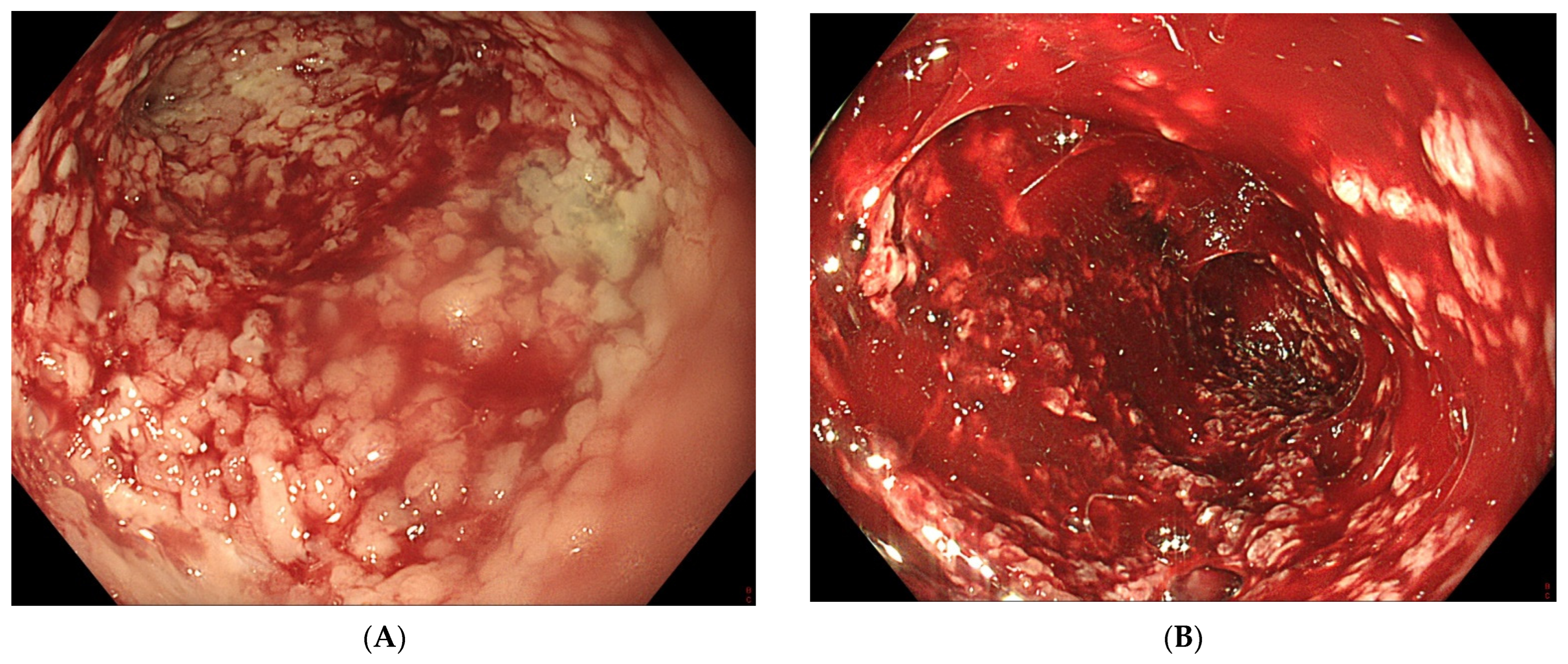
5. Difficulties of Endoscopic Vacuum Therapy in the Upper Gastrointestinal Tract

Despite the fact that EVT is nowadays a standardized procedure, it remains an enterprise prone to several hurdles and difficulties. They can be encountered in the pre-, intra- and post-procedure period, and also during the removal of the sponge and drainage tube.

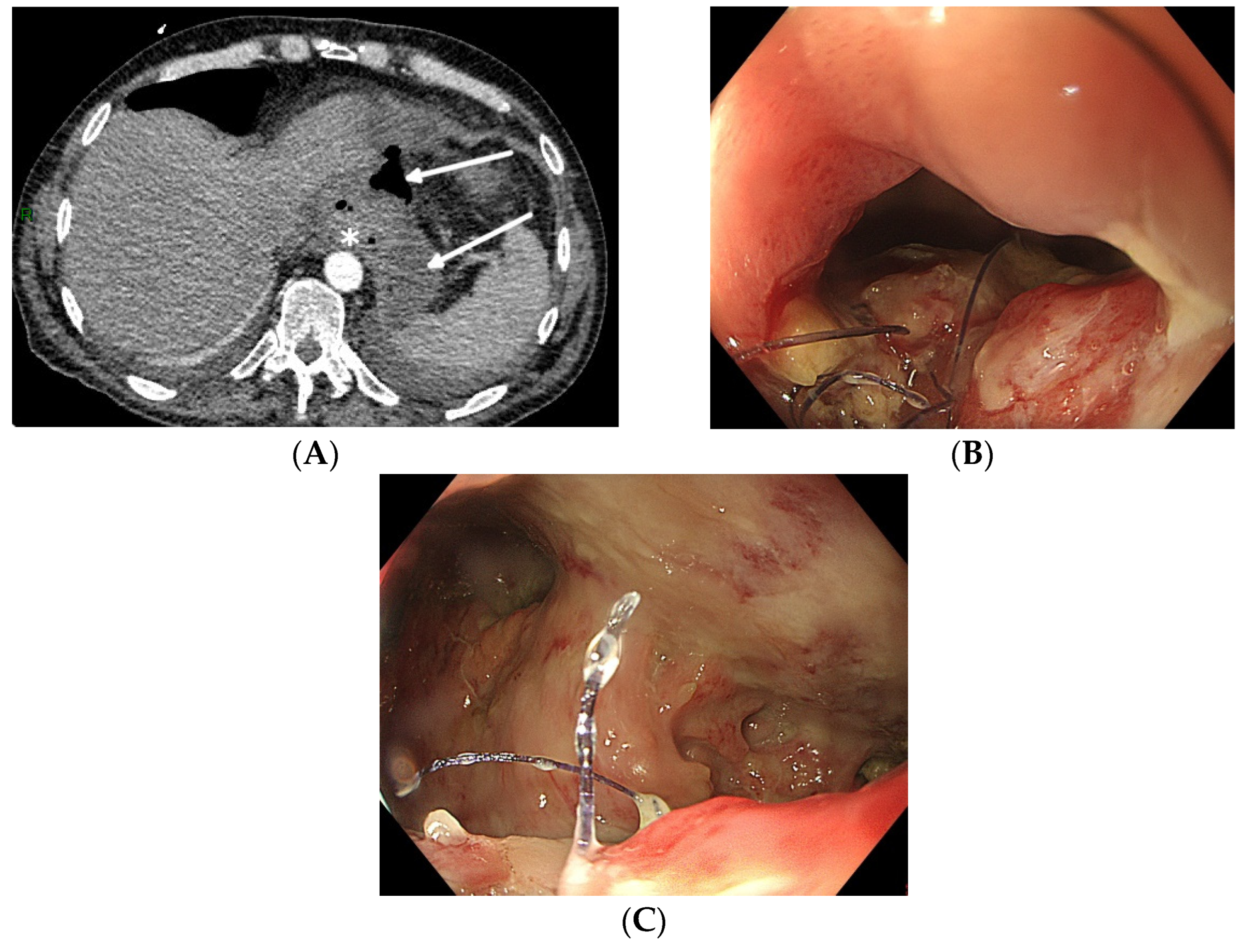
5.1. Pre-Procedure Difficulties

Not all oesophageal perforations can be treated using EVT [77]. The technique relies on the collapse of the extramural collection under negative pressure. The presence of an oesophageal fistula to the respiratory system does not permit the necessary build-up of negative pressure, thus rendering EVT unsuccessful. Therefore, EVT should not be used to treat broncho-oesophageal fistulas, although there are a few case reports of successful EVT treatment [42]. Secondly, since the device stimulates local tissue perfusion, it is considered contra-indicated in a malignant environment because of the risk of tumour growth induction. Finally, complete anastomotic dehiscence due to ischemic conduit necrosis necessitates redo surgery, and EVT should not be attempted in these particular situations (beyond EVT). Pre-procedure evaluation with CT scan (with oral contrast) and upper gastrointestinal endoscopy is mandatory to correctly evaluate the indication for EVT and to facilitate a patient-tailored multidisciplinary treatment plan (Figure 1).
Although ready-to-use sets are commercially available (Endo-Sponge, Eso-Sponge; B. Braun Melsungen AG; Melsungen, Germany), their availability is not widespread, and their financial cost is significant. However, since the design of the device is not very complicated, it can be self-assembled by attaching an open-pore drainage film or a size-cut polyurethane sponge to a nasogastric tube with multiple side-holes around the distal tip [78,79]. It can be introduced into the cavity through the commercially available overtube, or it can be endoscopically manoeuvred using rat tooth forceps or a polypectomy snare, taking care not to damage the sponge during the introduction. In the absence of an electronic vacuum pump with alarm function, a vacuum redon drain or bottle can be used as a continuous vacuum source. However, neither the redon drain nor bottle allows us to pre-set the vacuum force precisely (high–medium–low), and regular evaluation of the vacuum force is mandatory when using these alternative vacuum sources. Whenever the vacuum suction force drops because the redon drain is filled with aspirated fluids and debris, it should be replaced immediately. The loss of continuous vacuum aspiration negatively impacts EVT efficacy.

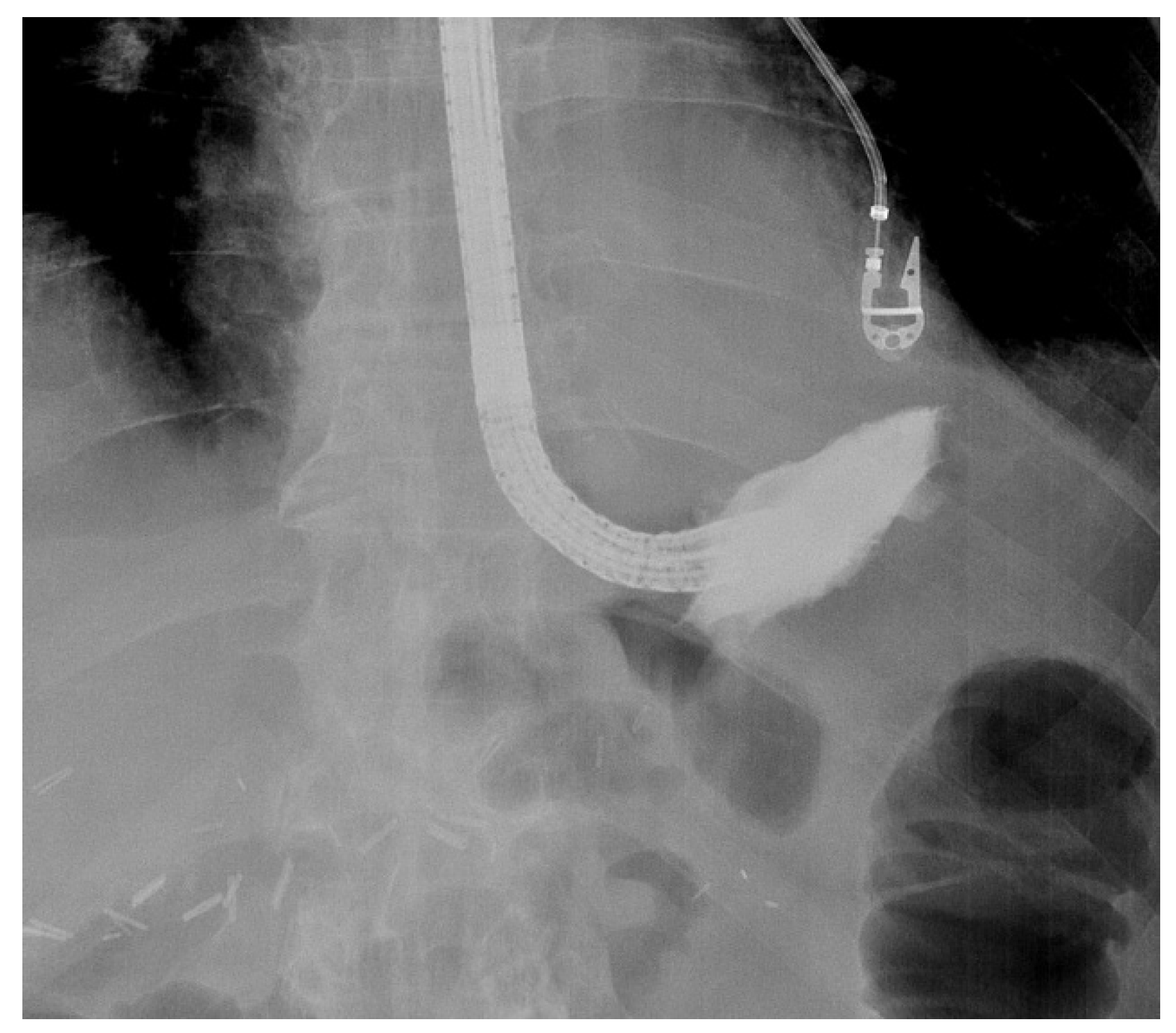
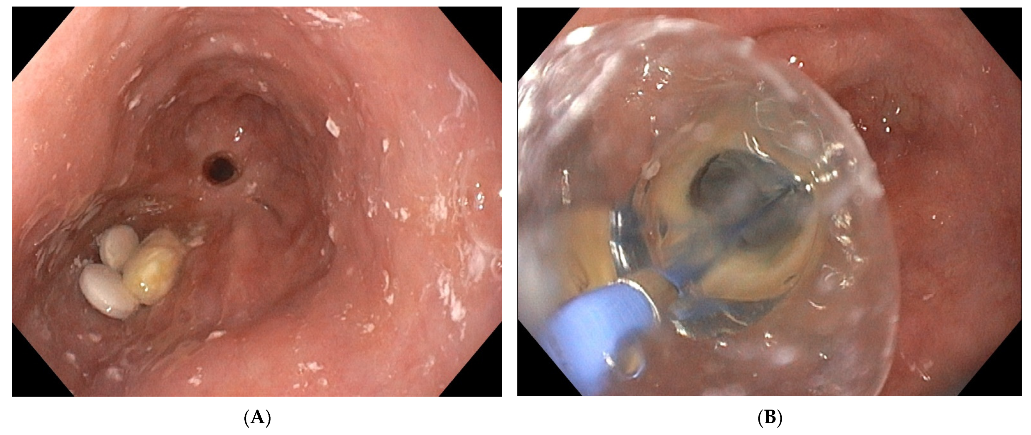
5.2. Intra-Procedure Difficulties

When engaging into the EVT procedure of oesophageal anastomotic leaks, several difficulties can be encountered. Since this is a time-consuming procedure with multiple introductions of the endoscope, it is best performed under general anaesthesia with endotracheal intubation, to prevent secretions and debris from the collection entering the respiratory system and to improve working conditions for both the endoscopist as for the patient. Fluoroscopy may also help to evaluate the depth and the extent of the extramural collection at the beginning of the EVT treatment, without being mandatory for future sponge exchanges (Figure 2).
The size of the anastomotic fistula may range from a millimetric orificium to a nearly complete dehiscence of the anastomosis [7]. A small-sized fistula can be closed using clips or stents or endoscopic suturing. However, external drainage of an extramural collection is mandatory when closing the fistula. Internal drainage using double pigtail stents can be an option, as well as EVT. However, when the fistula diameter does not allow the introduction of the overtube and the sponge, balloon dilatation of the fistula tract is possible. Additionally, the size of the sponge can be adapted to the small volume cavity by cutting it to a smaller and compatible size. Finally, positioning of the sponge into the oesophageal lumen covering the fistula orificium is another option to treat small-sized fistula [80]. The latter is usually a less efficacious solution, since the majority of the negative-pressure force will be lost in the lumen of the gastrointestinal tract, and other closure techniques with clips or endoscopic suturing may be more appropriate in case of small-sized fistula [9]. Intraluminal positioning of the sponge can also be applied at the end of successful EVT treatment with collapse of the extramural cavity to close the remaining small-sized fistula tract. The intraluminal sponge placement provokes stasis of saliva above the sponge. As already mentioned, the size of the sponge can be adapted to the volume of a small collection by cutting the sponge to its correct size. Large-sized fistulas and collections or near-complete anastomotic dehiscence may need the introduction of more than one sponge in order to optimize vacuum therapy and to promote the collapse of the collection (Figure 3).
When opting for more than one EVT device, each device should be connected to its proper vacuum source to avoid the loss of aspiration through communicating vessels. In case of anastomotic dehiscence without extraluminal collection, the sponge should be positioned in the anastomosis maintaining a local negative pressure of −75 mm Hg to −125 mm Hg. However, this approach can induce an anastomotic fibrotic stricture as a result of successful EVT (Figure 4).
Large collections may contain necrotic debris, pus and fibrine. To avoid early saturation of the sponge with necrotic debris and a loss of negative-pressure force, the collection should be cleaned as much as possible before positioning the sponge. This can be performed via removal of the debris using rat tooth forceps, a polypectomy snare or mere rinsing and aspiration through the working channel of the endoscope. This cleaning procedure is indicated at every new sponge exchange.
Successful sponge positioning relies on the correct introduction of the overtube into the collection. This can be challenging when the access to the collection requires an angulation of the tip of the endoscope (Figure 5).
Moreover, introducing the sponge through the overtube comes with a lot of friction, and it increases the risk of dislocation of the overtube from the collection into the oesophageal lumen. To reduce the friction, it is advised to use a silicon spray to lubricate the inner lumen of the overtube and to apply lubrification gel onto the sponge to facilitate its introduction through the overtube. Endoscopic control of the correct position of the sponge into the collection is warranted, since dislocation of both the overtube and the sponge is possible during the introduction process. In case of incorrect positioning of the sponge after removal of the overtube, the sponge should be removed, and the procedure should be restarted. As an alternative, the sponge can be endoscopically manoeuvred from the oesophageal lumen into the collection using rat tooth forceps, taking care not to damage the sponge while trying to reposition it. Additionally, in case of a self-assembled device lacking the overtube, the sponge-loaded tube can be taken by rat tooth forceps or a polypectomy snare before it is introduced endoscopically through the mouth and taken down into the oesophagus and the collection [78]. Correct positioning of the sponge into the collection or intraluminally adjacent to the fistula is mandatory for successful EVT. Migration of the sponge will negatively affect EVT efficacy. Endoscopic control of the correct position is therefore mandatory at the beginning and at the end of each sponge exchange procedure.
Although the manufacturer’s product information defines the position of the sponge-loaded flexible tube in the mouth, it is much more comfortable for the patient to position it through the nose like a classical nasogastric tube. Transnasal position of the tube will also avoid occlusion of the tube (and thus a loss of negative pressure) by biting it. The tube can be redirected from the mouth to the nose using the rendezvous technique with a naso-oral catheter. Transnasal position does not prevent the removal of the tube and the soft sponge through the nose, but this should only be performed under general anaesthesia with endotracheal intubation, in order to prevent aspiration of debris adherent to the sponge and to optimize the patient’s clinical comfort. The majority of patients undergoing EVT in the upper gastrointestinal tract are hospitalized until the end of the endoscopic treatment, but only a minority needs admission to the Intensive Care Unit.

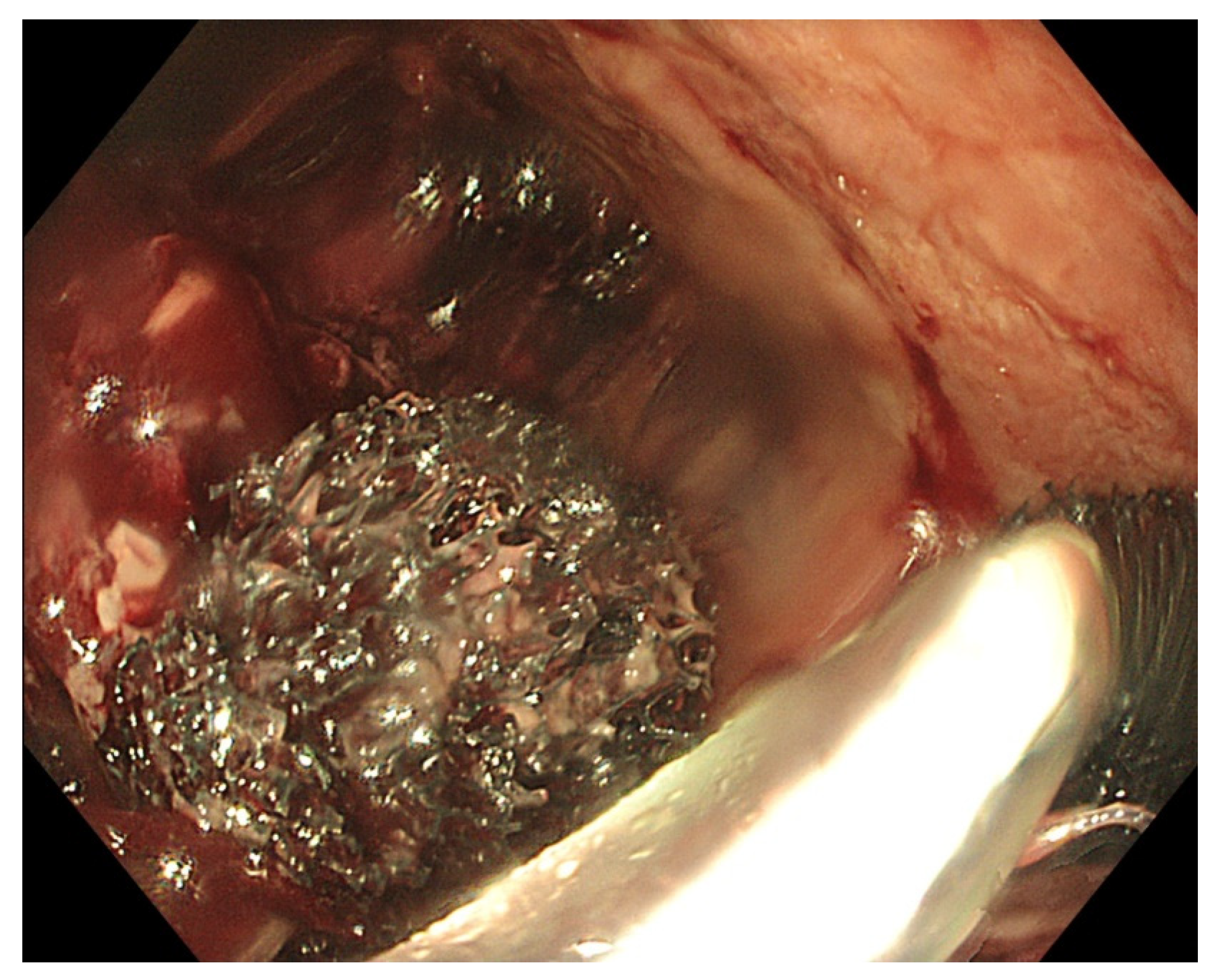
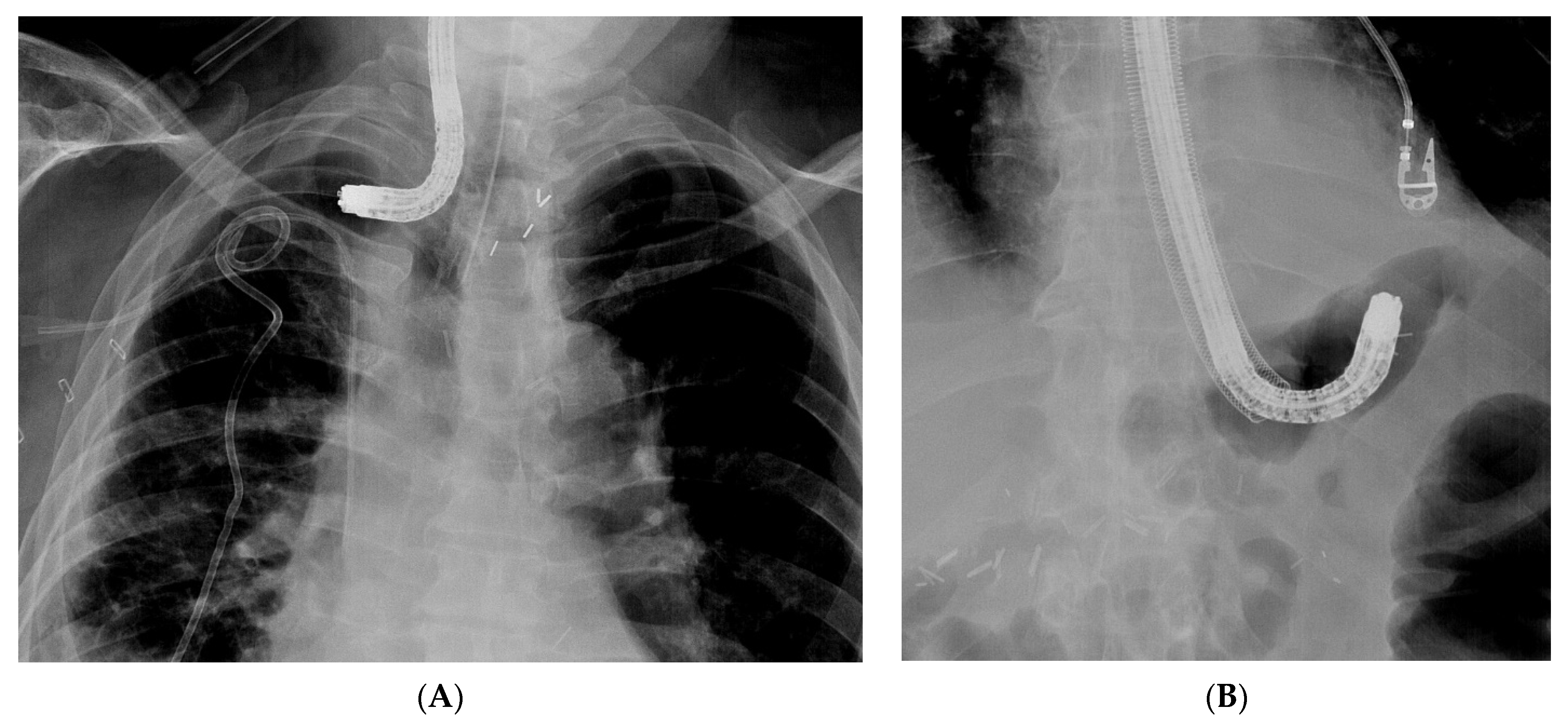
5.3. Post-Procedure Difficulties

After exteriorisation of the tube through the nose, gentle traction should be applied before fixation of the tube, in order to maintain the sponge in the correct position and to avoid distal migration of the sponge. As mentioned before, endoscopic control of the correct final sponge position is warranted.
Connecting the vacuum source to the aspiration tube requires adjusted connectors depending on the type of vacuum source. As for the EVT device, a compatible electronic vacuum pump is not always available, and it can be replaced by a surgical vacuum redon drain or a vacuum bottle. These non-electronic vacuum sources usually allow high–medium–low aspiration force through a physical switch, in contrast to the electronic pump with an adjustable negative force up to −200 mm Hg. The use of a vacuum redon drain or bottle requires frequent evaluation of the negative pressure since there is no electronic alarm when the negative pressure drops too low. When the redon contains too much aspiration fluids from the collection, it should be replaced immediately in order to maintain effective negative pressure. Leaks at the connectors should also be looked for in case of a loss of negative pressure.
As mentioned before, EVT requires multiple replacements of the device in order to maintain a continuous negative pressure at the level of the anastomotic leak. Therefore, the sponge should be replaced every 3 to 4 days. A delay in the replacement of the sponge may, on the one hand, lead to a loss of negative pressure and, on the other hand, to gradual tissue ingrowth into the open pores of the sponge. This may render sponge removal more difficult, introducing the risk of sponge rupture [29]. Since multiple sponge replacements are required (usually during a procedure under general anaesthesia), the necessary endoscopy time slots in the upcoming weeks should be reserved once EVT is initiated and the first sponge is put in place. Reserved time slots every Monday and Thursday or every Tuesday and Friday are very practical, in order to avoid scheduling replacements during the weekends.
Transnasal removal of the sponge is feasible and safe, but should only be performed under general anaesthesia with endotracheal intubation to avoid aspiration of secretions, blood and debris in the respiratory system. Endoscopic evaluation of the correct position of the sponge is important before its removal to assure efficient EVT. It might be necessary to dislodge the sponge from the granulating tissue using the endoscope or rat tooth forceps to facilitate atraumatic dissection and removal. Endoscopic inspection of the residual cavity is not only mandatory to evaluate the reduction in its volume and the aspect of the granulation tissue, but also to exclude and remove retained fragments of the sponge, if any (Figure 6). Intracavitary bleeding may occur after sponge removal. It is usually diffuse oozing bleeding and represents a sign of favourable tissue healing (Figure 6). It generally stops after endoscopic rinsing and cleaning of the cavity. However, it is advised to stop anticoagulant therapy before removal of the sponge [77].

6. Adverse Events Relating to EVT in the Upper Gastrointestinal Tract

Patients requiring EVT to treat postoperative anastomotic leaks in the upper gastrointestinal tract are critically ill, presenting with a septic condition due to mediastinal infection with cardiorespiratory repercussion and a high mortality risk. They may require admission to the Intensive Care Unit. Despite the multiple EVT-related endoscopic interventions in these critically ill patients, the adverse event rate of EVT is low with mainly minor adverse events (Table 1). A recent meta-analysis reviewed the EVT-related adverse events in the upper gastrointestinal tract [81]. The overall mortality of patients undergoing EVT for oesophageal perforations or leaks was 7.1%, relating to the underlying septic condition, not to the EVT procedure itself. This mortality rate illustrates the severe clinical condition induced by an anastomotic leak in the upper gastrointestinal tract, rather than the EVT-related risks. However, a few cases of mortality directly relating to the EVT procedure itself have been described. Death was caused by uncontrollable bleeding after sponge removal due to the proximity of the thoracic aorta leading to a lethal aortic fistula [31,48]. Total EVT-related adverse event rate was 13.6%, characterized by EVT-induced anastomotic stricture (Figure 4), local bleeding after sponge removal (Figure 6) and sponge dislocation. Sponge rupture during removal can be avoided by respecting the foreseen time schedule of sponge replacement every 3 to 5 days. In general, EVT-related adverse events are minor and can be managed endoscopically (AGREE IIIa classification of endoscopic adverse events) [82]. On only very rare occasions, visceral injury such as spleen injury or bowel ischemia have been described as relating to the EVT procedure [83]. Despite the multiple endoscopic interventions in critically ill patients, EVT can be considered as a safe therapeutic procedure.

7. Conclusions

Anastomotic leaks after digestive surgery have an important impact on surgical outcome [5,6,7]. They represent a real therapeutic challenge because of the high morbidity and mortality rates. Multiple treatment options exist, often combining interventional radiology and endoscopy and even redo surgery [9,10]. To improve patient outcomes, it is of the utmost importance to provide an individualized patient-tailored treatment plan after multidisciplinary discussion [11]. EVT is nowadays recognized as an effective and useful endoscopic approach to treat leaks or perforations in both the upper and lower gastrointestinal tract. Moreover, it has become one of the most effective treatment options overall with a very good safety profile [20,81]. However, it is a time-consuming endeavour requiring engagement from the endoscopist and understanding from the patient. Correct information and communication about the outcome and expectations are mandatory. The current review highlights the possible difficulties of the technique of EVT and provides practical solutions.
Pre-procedural evaluation of the postoperative leak using radiology and endoscopy is important in order to define the correct indication for EVT and to estimate the expected duration of the treatment. Although EVT has shown very good clinical outcomes, there are some specific contra-indications to consider: malignant perforation, broncho-oesophageal fistula and ischemic conduit necrosis with complete anastomotic dehiscence. Since the device (open-pore sponge attached to a drainage tube connected to a vacuum source) is not complex, it can be self-assembled if not commercially available, and a vacuum redon or bottle can be an alternative to the more sophisticated electronic vacuum pump [79,80]. Lubrification of the device helps to push the sponge through the overtube to its correct position. Endoscopic control of the correct positioning is advised in order to avoid sponge migration and ineffective EVT. Regular replacements should be scheduled at the initiation of EVT for the upcoming weeks.
EVT has shown to be a real asset in the endoscopic management of postoperative digestive defects [20]. However, the technique is prone to several challenges and endeavours, hampering its use in daily clinical practice and depriving critically ill patients of a potentially life-saving treatment. This review aims to provide practical solutions to the aforementioned difficulties. Next to these ‘tips and tricks’ for daily EVT practice, new developments are emerging not only to further improve clinical EVT outcomes, but also to facilitate the labour-intensive EVT procedure. Additionally, the prophylactic or pre-emptive use of EVT in case of high-risk or redo oesophagectomy is under study but has shown conflicting results so far, so further study is awaited [49,84,85]. Case series of successful EVT in young infants using small calibre self-assembled sponge devices are also emerging, illustrating the expanding use and indications of EVT [55,66,86]. One of the latest developments is the hybrid VacStent combining the advantages of EVT and a self-expandable metal stent to treat oesophageal perforation and anastomotic leak (VacStent, Micro-Tech, Düsseldorf, Germany; VacStent, Medech AG, Steinhausen, Switzerland) [87,88]. However, this device is placed inside the oesophageal lumen, not into the extraluminal cavity, and it does not require replacement every 3 to 5 days. Further study is required to define its optimal indications and to compare it with EVT and other endoscopic techniques such as stenting, clips and suturing.

8. Core Tip

Endoscopic vacuum therapy (EVT) is a new, safe and effective technique to treat anastomotic leaks and perforations in the upper and lower gastrointestinal tract. Although EVT has shown very good clinical outcomes, the procedure may be difficult to perform, hampering its use in daily clinical practice and depriving critically ill patients of a potentially life-saving therapy. The current review highlights the possible hurdles of EVT and provides practical solutions to facilitate its use in daily clinical practice.

Supplementary Materials

The following supporting information can be downloaded at: https://www.mdpi.com/article/10.3390/life13061412/s1, Video S1: Procedure of EVT in a patient with anastomotic leak after oesophagectomy.

Author Contributions

L.M. and T.G.M.: substantial contribution to the described endoscopic procedures, the concept and drafting of the manuscript, and the illustrations with video and final approval of the submitted manuscript. All authors have read and agreed to the published version of the manuscript.

Funding

This research received no external funding.

Conflicts of Interest

The authors declare no conflict of interest.

References

  1. Low, D.E.; Alderson, D.; Cecconello, I.; Chang, A.C.; Darling, G.; D’journo, X.B.; Griffin, S.M.; Hölscher, A.H.; Hofstetter, W.L.; Jobe, B.A.; et al. International Consensus on Standardization of Data Collection for Complications Associated With Esophagectomy: Esophagectomy Complications Consensus Group (ECCG). Ann. Surg. 2015, 262, 286–294. [Google Scholar] [CrossRef]
  2. Ross, S.L.; Veluswamy, B.; Craig, E.V.; Miller, F.H.; Horowitz, J.M.; Kelahan, L.C. Optimizing detection of postoperative leaks on upper gastrointestinal fluoroscopy: A step-by-step guide. Abdom. Imaging 2021, 46, 3019–3032. [Google Scholar] [CrossRef]
  3. Vetter, D.; Gutschow, C.A. Strategies to prevent anastomotic leakage after esophagectomy and gastric conduit reconstruction. Langenbeck’s Arch. Surg. 2020, 405, 1069–1077. [Google Scholar] [CrossRef] [PubMed]
  4. Mils, K.; Miró, M.; Farran, L.; Videla, S.; Alba, E.; Estremiana, F.; Bettonica, C.; Aranda, H. A pilot randomized controlled trial on the utility of gastric conditioning in the prevention of esophagogastric anastomotic leak after Ivor Lewis esophagectomy. The APIL_2013 Trial. Int. J. Surg. 2022, 106, 106921. [Google Scholar] [CrossRef] [PubMed]
  5. Baiocchi, G.L.; Giacopuzzi, S.; Vittimberga, G.; De Pascale, S.; Pastorelli, E.; Gelmini, R.; Viganò, J.; Graziosi, L.; Vagliasindi, A.; Rosa, F.; et al. Clinical outcomes of patients with complicated post-operative course after gastrectomy for cancer: A GIRCG study using the GASTRODATA registry. Updat. Surg. 2023, 75, 419–427. [Google Scholar] [CrossRef] [PubMed]
  6. Ubels, S.; Verstegen, M.; Klarenbeek, B.; Bouwense, S.; Berge Henegouwen, M.; Daams, F.; van Det, M.J.; Griffiths, E.A.; Haveman, J.W.; Heisterkamp, J.; et al. Severity of oEsophageal Anastomotic Leak in patients after oesophagectomy: The SEAL score. Br. J. Surg. 2022, 109, 864–871. [Google Scholar] [CrossRef]
  7. Bachmann, J.; Feith, M.; Schlag, C.; Abdelhafez, M.; Martignoni, M.E.; Friess, H. Anastomotic leakage following resection of the esophagus—Introduction of an endoscopic grading system. World J. Surg. Oncol. 2022, 20, 104. [Google Scholar] [CrossRef]
  8. Browning, A.F.; Chong, L.; Read, M.; Hii, M.W. Economic burden of complications and readmission following oesophageal cancer surgery. ANZ J. Surg. 2022, 92, 2901–2906. [Google Scholar] [CrossRef]
  9. Chan, S.M.; Auyeung, K.K.Y.; Lam, S.F.; Chiu, P.W.Y.; Teoh, A.Y.B. Current status in endoscopic management of upper gastrointestinal perforations, leaks and fistulas. Dig. Endosc. 2022, 34, 43–62. [Google Scholar] [CrossRef]
  10. Ubels, S.; Lubbers, M.; Verstegen, M.H.P.; Bouwense, S.A.W.; van Daele, E.; Ferri, L.; Gisbertz, S.S.; Griffiths, E.A.; Grimminger, P.; Hanna, G.; et al. Treatment of anastomotic leak after esophagectomy: Insights of an international case vignette survey and expert discussions. Dis. Esophagus 2022, 35, doac020. [Google Scholar] [CrossRef]
  11. Stavropoulos, S.N.; Modayil, R.; Friedel, D. Closing Perforations and Postperforation Management in Endoscopy. Gastrointest. Endosc. Clin. N. Am. 2015, 25, 29–45. [Google Scholar] [CrossRef] [PubMed]
  12. Binda, C.; Jung, C.F.M.; Fabbri, S.; Giuffrida, P.; Sbrancia, M.; Coluccio, C.; Gibiino, G.; Fabbri, C. Endoscopic Management of Postoperative Esophageal and Upper GI Defects—A Narrative Review. Medicina 2023, 59, 136. [Google Scholar] [CrossRef]
  13. Goenka, M.K.; Goenka, U. Endotherapy of leaks and fistula. World J. Gastrointest. Endosc. 2015, 7, 702. [Google Scholar] [CrossRef]
  14. Cereatti, F.; Grassia, R.; Drago, A.; Conti, C.B.; Donatelli, G. Endoscopic management of gastrointestinal leaks and fistulae: What option do we have? World J. Gastroenterol. 2020, 26, 4198–4217. [Google Scholar] [CrossRef]
  15. Jung, D.H.; Yun, H.-R.; Lee, S.J.; Kim, N.W.; Huh, C.W. Endoscopic Vacuum Therapy in Patients with Transmural Defects of the Upper Gastrointestinal Tract: A Systematic Review with Meta-Analysis. J. Clin. Med. 2021, 10, 2346. [Google Scholar] [CrossRef]
  16. Junior, E.S.D.M.; De Moura, D.T.H.; Ribeiro, I.B.; Hathorn, K.E.; Farias, G.F.A.; Turiani, C.V.; Medeiros, F.S.; Bernardo, W.M.; De Moura, E.G.H. Endoscopic vacuum therapy versus endoscopic stenting for upper gastrointestinal transmural defects: Systematic review and meta-analysis. Dig. Endosc. 2021, 33, 892–902. [Google Scholar] [CrossRef]
  17. Scognamiglio, P.; Reeh, M.; Melling, N.; Kantowski, M.; Eichelmann, A.-K.; Chon, S.-H.; El-Sourani, N.; Höller, A.; Izbicki, J.R.; Tachezy, M. Management of intra-thoracic anastomotic leakages after esophagectomy: Updated systematic review and meta-analysis of endoscopic vacuum therapy versus stenting. BMC Surg. 2022, 22, 309. [Google Scholar] [CrossRef]
  18. Intriago, J.M.V.; de Moura, D.T.H.; Junior, E.S.D.M.; Proença, I.M.; Ribeiro, I.B.; Sánchez-Luna, S.A.; Bernardo, W.M.; de Moura, E.G.H. Endoscopic Vacuum Therapy (EVT) for the Treatment of Post-Bariatric Surgery Leaks and Fistulas: A Systematic Review and Meta-analysis. Obes. Surg. 2022, 32, 3435–3451. [Google Scholar] [CrossRef] [PubMed]
  19. Tachezy, M.; Chon, S.-H.; Rieck, I.; Kantowski, M.; Christ, H.; Karstens, K.; Gebauer, F.; Goeser, T.; Rösch, T.; Izbicki, J.R.; et al. Endoscopic vacuum therapy versus stent treatment of esophageal anastomotic leaks (ESOLEAK): Study protocol for a prospective randomized phase 2 trial. Trials 2021, 22, 377. [Google Scholar] [CrossRef]
  20. Mandarino, F.V.; Barchi, A.; D’amico, F.; Fanti, L.; Azzolini, F.; Viale, E.; Esposito, D.; Rosati, R.; Fiorino, G.; Bemelman, W.A.; et al. Endoscopic Vacuum Therapy (EVT) versus Self-Expandable Metal Stent (SEMS) for Anastomotic Leaks after Upper Gastrointestinal Surgery: Systematic Review and Meta-Analysis. Life 2023, 13, 287. [Google Scholar] [CrossRef]
  21. Loske, G. Endoscopic negative pressure therapy of the upper gastrointestinal tract. Der Chir. 2019, 90 (Suppl. S1), 1–6. [Google Scholar] [CrossRef]
  22. McLean, W.C. The role of closed wound negative pressure suction in radical surgical procedures of the head and neck. Laryngoscope 1964, 74, 70–94. [Google Scholar] [CrossRef]
  23. Wedemeyer, J.; Schneider, A.; Manns, M.P.; Jackobs, S. Endoscopic vacuum-assisted closure of upper intestinal anastomotic leaks. Gastrointest. Endosc. 2008, 67, 708–711. [Google Scholar] [CrossRef]
  24. Reimer, S.; Seyfried, F.; Flemming, S.; Brand, M.; Weich, A.; Widder, A.; Plaßmeier, L.; Kraus, P.; Döring, A.; Hering, I.; et al. Evolution of endoscopic vacuum therapy for upper gastrointestinal leakage over a 10-year period: A quality improvement study. Surg. Endosc. 2022, 36, 9169–9178. [Google Scholar] [CrossRef]
  25. Markus, A.; Henrik, B.J.; Benedikt, R.; Alexander, H.; Thomas, B.; Clemens, S.; Jan-Hendrik, E. Endoscopic vacuum therapy in salvage and standalone treatment of gastric leaks after bariatric surgery. Langenbeck’s Arch. Surg. 2022, 407, 1039–1046. [Google Scholar] [CrossRef]
  26. Munshi, E.; Dahlbäck, C.; Johansson, S.; Lydrup, M.-L.; Jutesten, H.; Buchwald, P. Long-term Outcomes of Endoscopic Vacuum Therapy and Transanal Drainage for Anastomotic Leakage After Anterior Resection. In Vivo 2022, 36, 2275–2278. [Google Scholar] [CrossRef]
  27. Gubler, C.; Schneider, P.M.; Bauerfeind, P. Complex anastomotic leaks following esophageal resections: The new stent over sponge (SOS) approach. Dis. Esophagus 2013, 26, 598–602. [Google Scholar] [CrossRef]
  28. Valli, P.V.; Mertens, J.C.; Kröger, A.; Gubler, C.; Gutschow, C.; Schneider, P.M.; Bauerfeind, P. Stent-over-sponge (SOS): A novel technique complementing endosponge therapy for foregut leaks and perforations. Endoscopy 2018, 50, 148–153. [Google Scholar] [CrossRef] [PubMed]
  29. Loske, G.; Schorsch, T.; Müller, C. Endoscopic vacuum sponge therapy for esophageal defects. Surg. Endosc. 2010, 24, 2531–2535. [Google Scholar] [CrossRef] [PubMed]
  30. Loske, G.; Schorsch, T.; Müller, C. Endoscopic intracavitary vacuum therapy of Boerhaave’s syndrome: A case report. Endoscopy 2010, 42, E144–E145. [Google Scholar] [CrossRef] [PubMed]
  31. Ahrens, M.; Schulte, T.; Egberts, J.; Schafmayer, C.; Hampe, J.; Fritscher-Ravens, A.; Broering, D.C.; Schniewind, B. Drainage of esophageal leakage using endoscopic vacuum therapy: A prospective pilot study. Endoscopy 2010, 42, 693–698. [Google Scholar] [CrossRef]
  32. Weidenhagen, R.; Hartl, W.H.; Gruetzner, K.U.; Eichhorn, M.E.; Spelsberg, F.; Jauch, K.W. Anastomotic Leakage After Esophageal Resection: New Treatment Options by Endoluminal Vacuum Therapy. Ann. Thorac. Surg. 2010, 90, 1674–1681. [Google Scholar] [CrossRef]
  33. Loske, G.; Schorsch, T.; Müller, C. Intraluminal and intracavitary vacuum therapy for esophageal leakage: A new endoscopic minimally invasive approach. Endoscopy 2011, 43, 540–544. [Google Scholar] [CrossRef]
  34. Kuehn, F.; Schiffmann, L.; Rau, B.M.; Klar, E. Surgical Endoscopic Vacuum Therapy for Anastomotic Leakage and Perforation of the Upper Gastrointestinal Tract. J. Gastrointest. Surg. 2012, 16, 2145–2150. [Google Scholar] [CrossRef]
  35. Schorsch, T.; Müller, C.; Loske, G. Endoscopic vacuum therapy of anastomotic leakage and iatrogenic perforation in the esophagus. Surg. Endosc. 2013, 27, 2040–2045. [Google Scholar] [CrossRef]
  36. Schniewind, B.; Schafmayer, C.; Voehrs, G.; Egberts, J.; von Schoenfels, W.; Rose, T.; Kurdow, R.; Arlt, A.; Ellrichmann, M.; Jürgensen, C.; et al. Endoscopic endoluminal vacuum therapy is superior to other regimens in managing anastomotic leakage after esophagectomy: A comparative retrospective study. Surg. Endosc. 2013, 27, 3883–3890. [Google Scholar] [CrossRef] [PubMed]
  37. Brangewitz, M.; Voigtländer, T.; Helfritz, F.A.; Lankisch, T.O.; Winkler, M.; Klempnauer, J.; Manns, M.P.; Schneider, A.S.; Wedemeyer, J. Endoscopic closure of esophageal intrathoracic leaks: Stent versus endoscopic vacuum-assisted closure, a retrospective analysis. Endoscopy 2013, 45, 433–438. [Google Scholar] [CrossRef]
  38. Lenzen, H.; Negm, A.A.; Erichsen, T.J.; Manns, M.P.; Wedemeyer, J.; Lankisch, T.O. Successful treatment of cervical esophageal leakage by endoscopic-vacuum assisted closure therapy. World J. Gastrointest. Endosc. 2013, 5, 340–345. [Google Scholar] [CrossRef]
  39. Bludau, M.; Hölscher, A.H.; Herbold, T.; Leers, J.M.; Gutschow, C.; Fuchs, H.; Schröder, W. Management of upper intestinal leaks using an endoscopic vacuum-assisted closure system (E-VAC). Surg. Endosc. 2014, 28, 896–901. [Google Scholar] [CrossRef] [PubMed] [Green Version]
  40. Heits, N.; Stapel, L.; Reichert, B.; Schafmayer, C.; Schniewind, B.; Becker, T.; Hampe, J.; Egberts, J.-H. Endoscopic Endoluminal Vacuum Therapy in Esophageal Perforation. Ann. Thorac. Surg. 2014, 97, 1029–1035. [Google Scholar] [CrossRef] [PubMed]
  41. Schorsch, T.; Muller, C.; Loske, G. Endoscopic vacuum therapy of perforations and anastomotic insufficiency of the esophagus. Chirurg 2014, 85, 1081–1093. [Google Scholar] [CrossRef] [PubMed]
  42. Lee, H.J.; Lee, H. Endoscopic Vacuum-assisted Closure With Sponge for Esophagotracheal Fistula After Esophagectomy. Surg. Laparosc. Endosc. Percutaneous Tech. 2015, 25, e76–e77. [Google Scholar] [CrossRef] [PubMed]
  43. Mennigen, R.; Harting, C.; Lindner, K.; Vowinkel, T.; Rijcken, E.; Palmes, D.; Senninger, N.; Laukoetter, M.G. Comparison of Endoscopic Vacuum Therapy Versus Stent for Anastomotic Leak After Esophagectomy. J. Gastrointest. Surg. 2015, 19, 1229–1235. [Google Scholar] [CrossRef]
  44. Loske, G.; Schorsch, T.; Dahm, C.; Martens, E.; Müller, C. Iatrogenic perforation of esophagus successfully treated with Endoscopic Vacuum Therapy (EVT). Endosc. Int. Open 2015, 3, E547–E551. [Google Scholar] [CrossRef] [PubMed] [Green Version]
  45. Möschler, O.; Nies, C.; Mueller, M.K. Endoscopic vacuum therapy for esophageal perforations and leakages. Endosc. Int. Open 2015, 03, E554–E558. [Google Scholar] [CrossRef] [Green Version]
  46. Smallwood, N.R.; Fleshman, J.W.; Leeds, S.G.; Burdick, J.S. The use of endoluminal vacuum (E-Vac) therapy in the management of upper gastrointestinal leaks and perforations. Surg. Endosc. 2016, 30, 2473–2480. [Google Scholar] [CrossRef] [PubMed]
  47. Kuehn, F.; Schiffmann, L.; Janisch, F.; Schwandner, F.; Alsfasser, G.; Gock, M.C.; Klar, E. Surgical Endoscopic Vacuum Therapy for Defects of the Upper Gastrointestinal Tract. J. Gastrointest. Surg. 2016, 20, 237–243. [Google Scholar] [CrossRef]
  48. Laukoetter, M.G.; Mennigen, R.; Neumann, P.A.; Dhayat, S.; Horst, G.; Palmes, D.; Senninger, N.; Vowinkel, T. Successful closure of defects in the upper gastrointestinal tract by endoscopic vacuum therapy (EVT): A prospective cohort study. Surg. Endosc. 2017, 31, 2687–2696. [Google Scholar] [CrossRef]
  49. Neumann, P.-A.; Mennigen, R.; Palmes, D.; Senninger, N.; Vowinkel, T.; Laukoetter, M.G. Pre-emptive endoscopic vacuum therapy for treatment of anastomotic ischemia after esophageal resections. Endoscopy 2017, 49, 498–503. [Google Scholar] [CrossRef]
  50. Bludau, M.; Fuchs, H.F.; Herbold, T.; Maus, M.K.H.; Alakus, H.; Popp, F.; Leers, J.M.; Bruns, C.J.; Hölscher, A.H.; Schröder, W.; et al. Results of endoscopic vacuum-assisted closure device for treatment of upper GI leaks. Surg. Endosc. 2018, 32, 1906–1914. [Google Scholar] [CrossRef]
  51. Pournaras, D.J.; Hardwick, R.H.; Safranek, P.M.; Sujendran, V.; Bennett, J.; Macaulay, G.D.; Hindmarsh, A. Endoluminal Vacuum Therapy (E-Vac): A Treatment Option in Oesophagogastric Surgery. World J. Surg. 2018, 42, 2507–2511. [Google Scholar] [CrossRef] [Green Version]
  52. Heits, N.; Bernsmeier, A.; Reichert, B.; Hauser, C.; Hendricks, A.; Seifert, D.; Richter, F.; Schafmayer, C.; Ellrichmann, M.; Schniewind, B.; et al. Long-term quality of life after endovac-therapy in anastomotic leakages after esophagectomy. J. Thorac. Dis. 2018, 10, 228–240. [Google Scholar] [CrossRef] [PubMed] [Green Version]
  53. Noh, S.M.; Ahn, J.Y.; Lee, J.H.; Jung, H.-Y.; Alghamdi, Z.; Kim, H.R.; Kim, Y.-H. Endoscopic Vacuum-Assisted Closure Therapy in Patients with Anastomotic Leakage after Esophagectomy: A Single-Center Experience. Gastroenterol. Res. Prac. 2018, 2018, 1697968. [Google Scholar] [CrossRef] [PubMed]
  54. Still, S.; Mencio, M.; Ontiveros, E.; Burdick, J.; Leeds, S.G. Primary and Rescue Endoluminal Vacuum Therapy in the Management of Esophageal Perforations and Leaks. Ann. Thorac. Cardiovasc. Surg. 2018, 24, 173–179. [Google Scholar] [CrossRef] [PubMed] [Green Version]
  55. Manfredi, M.A.; Clark, S.; Staffa, S.J.; Ngo, P.D.; Smithers, C.J.; Hamilton, T.E.; Jennings, R.W. Endoscopic Esophageal Vacuum Therapy: A Novel Therapy for Esophageal Perforations in Pediatric Patients. J. Pediatr. Gastroenterol. Nutr. 2018, 67, 706–712. [Google Scholar] [CrossRef]
  56. Berlth, F.; Bludau, M.; Plum, P.S.; Herbold, T.; Christ, H.; Alakus, H.; Kleinert, R.; Bruns, C.J.; Hölscher, A.H.; Chon, S.-H. Self-Expanding Metal Stents Versus Endoscopic Vacuum Therapy in Anastomotic Leak Treatment After Oncologic Gastroesophageal Surgery. J. Gastrointest. Surg. 2019, 23, 67–75. [Google Scholar] [CrossRef] [PubMed]
  57. Min, Y.W.; Kim, T.; Lee, H.; Min, B.-H.; Kim, H.K.; Choi, Y.S.; Lee, J.H.; Rhee, P.-L.; Kim, J.J.; Zo, J.I.; et al. Endoscopic vacuum therapy for postoperative esophageal leak. BMC Surg. 2019, 19, 37. [Google Scholar] [CrossRef] [PubMed] [Green Version]
  58. Alakkari, A.; Sood, R.; Everett, S.M.; Rembacken, B.J.; Hayden, J.; Sarela, A.; Mohammed, N. First UK experience of endoscopic vacuum therapy for the management of oesophageal perforations and postoperative leaks. Front. Gastroenterol. 2019, 10, 200–203. [Google Scholar] [CrossRef] [PubMed]
  59. Sendino, O.; Loras, C.; Mata, A.; Momblán, D.; Andujar, X.; Cruz, M.; Cárdenas, A.; Marquez, I.; Uchima, H.; Cordova, H.; et al. Eficacia y seguridad de la terapia de vacío endoscópica para el tratamiento de perforaciones y dehiscencias anastomóticas del tracto digestivo superior. Gastroenterol. Hepatol. 2020, 43, 431–438. [Google Scholar] [CrossRef]
  60. Nijhuis, R.A.B.O.; Bergman, J.J.G.H.M.; Takkenberg, R.B.; Fockens, P.; Bredenoord, A.J. Non-surgical treatment of esophageal perforation after pneumatic dilation for achalasia: A case series. Scand. J. Gastroenterol. 2020, 55, 1248–1252. [Google Scholar] [CrossRef]
  61. Rubicondo, C.; Lovece, A.; Pinelli, D.; Indriolo, A.; Lucianetti, A.; Colledan, M. Endoluminal vacuum-assisted closure (E-Vac) therapy for postoperative esophageal fistula: Successful case series and literature review. World J. Surg. Oncol. 2021, 18, 301. [Google Scholar] [CrossRef]
  62. Jung, C.F.M.; Müller-Dornieden, A.; Gaedcke, J.; Kunsch, S.; Gromski, M.A.; Biggemann, L.; Hosseini, A.S.A.; Ghadimi, M.; Ellenrieder, V.; Wedi, E. Impact of Endoscopic Vacuum Therapy with Low Negative Pressure for Esophageal Perforations and Postoperative Anastomotic Esophageal Leaks. Digestion 2021, 102, 469–479. [Google Scholar] [CrossRef] [PubMed]
  63. De Pasqual, C.A.; Mengardo, V.; Tomba, F.; Veltri, A.; Sacco, M.; Giacopuzzi, S.; Weindelmayer, J.; de Manzoni, G. Effectiveness of endoscopic vacuum therapy as rescue treatment in refractory leaks after gastro-esophageal surgery. Updat. Surg. 2020, 73, 607–614. [Google Scholar] [CrossRef]
  64. Zhang, C.C.; Liesenfeld, L.; Klotz, R.; Koschny, R.; Rupp, C.; Schmidt, T.; Diener, M.K.; Müller-Stich, B.P.; Hackert, T.; Sauer, P.; et al. Feasibility, effectiveness, and safety of endoscopic vacuum therapy for intrathoracic anastomotic leakage following transthoracic esophageal resection. BMC Gastroenterol. 2021, 21, 72. [Google Scholar] [CrossRef] [PubMed]
  65. Book, T.; Wortmann, N.; Winkler, M.; Kirstein, M.M.; Heidrich, B.; Wedemeyer, H.; Voigtländer, T. Endoscopic vacuum assisted closure (E-VAC) of upper gastrointestinal leakages. Scand. J. Gastroenterol. 2021, 56, 1376–1379. [Google Scholar] [CrossRef] [PubMed]
  66. Ritz, L.A.; Hajji, M.S.; Schwerd, T.; Koletzko, S.; von Schweinitz, D.; Lurz, E.; Hubertus, J. Esophageal Perforation and EVAC in Pediatric Patients: A Case Series of Four Children. Front. Pediatr. 2021, 9, 727472. [Google Scholar] [CrossRef]
  67. Mastoridis, S.; Chana, P.; Singh, M.; Akbari, K.; Shalaby, S.; Maynard, N.D.; Sgromo, B. Endoscopic vacuum therapy (EVT) in the management of oesophageal perforations and post-operative leaks. Minim. Invasive Ther. Allied Technol. 2022, 31, 380–388. [Google Scholar] [CrossRef] [PubMed]
  68. El-Sourani, N.; Miftode, S.; Bockhorn, M.; Arlt, A.; Meinhardt, C. Endoscopic Management of Anastomotic Leakage after Esophageal Surgery: Ten Year Analysis in a Tertiary University Center. Clin. Endosc. 2022, 55, 58–66. [Google Scholar] [CrossRef] [PubMed]
  69. Richter, F.; Hendricks, A.; Schniewind, B.; Hampe, J.; Heits, N.; von Schönfels, W.; Reichert, B.; Eberle, K.; Ellrichmann, M.; Baumann, P.; et al. Eso-Sponge® for anastomotic leakage after oesophageal resection or perforation: Outcomes from a national, prospective multicentre registry. BJS Open 2022, 6, zrac030. [Google Scholar] [CrossRef] [PubMed]
  70. Stathopoulos, P.; Zumblick, M.; Wächter, S.; Schiffmann, L.; Gress, T.M.; Bartsch, D.; Seitz, G.; Denzer, U.W. Endoscopic vacuum therapy (EVT) for acute esophageal perforation: Could it replace surgery? Endosc. Int. Open 2022, 10, E686–E693. [Google Scholar] [CrossRef]
  71. Chon, S.-H.; Brunner, S.; Müller, D.T.; Lorenz, F.; Stier, R.; Streller, L.; Eckhoff, J.; Straatman, J.; Babic, B.; Schiffmann, L.M.; et al. Time to endoscopic vacuum therapy—Lessons learned after > 150 robotic-assisted minimally invasive esophagectomies (RAMIE) at a German high-volume center. Surg. Endosc. 2023, 37, 741–748. [Google Scholar] [CrossRef] [PubMed]
  72. Maier, J.; Kandulski, A.; Donlon, N.E.; Werner, J.M.; Mehrl, A.; Müller, M.; Doenecke, A.; Schlitt, H.J.; Hornung, M.; Weiss, A.R.R. Endoscopic vacuum therapy significantly improves clinical outcomes of anastomotic leakages after 2-stage, 3-stage, and transhiatal esophagectomies. Langenbeck’s Arch. Surg. 2023, 408, 90. [Google Scholar] [CrossRef]
  73. Panneerselvam, K.; Jacob, J.S.; Samuel, R.E.; Tau, A.; Ketwaroo, G.A.; Abidi, W.M.; Sealock, R.J. Endoscopic vacuum therapy for treatment of spontaneous and iatrogenic upper gastrointestinal defects: A case series. Clin. Endosc. 2023. [Google Scholar] [CrossRef] [PubMed]
  74. Moura, D.; De Moura, B.F.B.H.; Manfredi, M.A.; Hathorn, K.E.; Bazarbashi, A.N.; Ribeiro, I.B.; de Moura, E.; Thompson, C.C. Role of endoscopic vacuum therapy in the management of gastrointestinal transmural defects. World J. Gastrointest. Endosc. 2019, 11, 329–344. [Google Scholar] [CrossRef]
  75. Jansen, K.T.; Hetzel, J.; Schulte, C.; Düzenli, N.; Fusco, S.; Zerabruck, E.; Schmider, E.; Malek, N.P.; Königsrainer, A.; Stüker, D.; et al. Differences in fluid removal of different open-pore elements for endoscopic negative pressure therapy in the upper gastrointestinal tract. Sci. Rep. 2022, 12, 13889. [Google Scholar] [CrossRef] [PubMed]
  76. Lord, A. Is the type of insufflation a key issue in gastro-intestinal endoscopy? World J. Gastroenterol. 2014, 20, 2193–2199. [Google Scholar] [CrossRef]
  77. Gutschow, C.A.; Schlag, C.; Vetter, D. Endoscopic vacuum therapy in the upper gastrointestinal tract: When and how to use it. Langenbeck’s Arch. Surg. 2022, 407, 957–964. [Google Scholar] [CrossRef]
  78. Leeds, S.G.; Mencio, M.; Ontiveros, E.; Ward, M.A. Endoluminal Vacuum Therapy: How I Do It. J. Gastrointest. Surg. 2019, 23, 1037–1043. [Google Scholar] [CrossRef] [PubMed]
  79. de Moura, D.T.H.; Hirsch, B.S.; McCarty, T.R.; dos Santos, M.E.L.; Guedes, H.G.; Gomes, G.F.; de Medeiros, F.S.; de Moura, E.G.H. Homemade endoscopic vacuum therapy device for the management of transmural gastrointestinal defects. Dig. Endosc. 2023; online ahead of print. [Google Scholar] [CrossRef]
  80. Loske, G.; Müller, C.T. Tips and tricks for endoscopic negative pressure therapy. Der Chir. 2019, 90 (Suppl. S1), 7–14. [Google Scholar] [CrossRef] [PubMed] [Green Version]
  81. Aziz, M.; Haghbin, H.; Sharma, S.; Weissman, S.; Saleem, S.; Lee-Smith, W.; Kobeissy, A.; Nawras, A.; Alastal, Y. Safety and effectiveness of endoluminal vacuum-assisted closure for esophageal defects: Systematic review and meta-analysis. Endosc. Int. Open 2021, 9, E1371–E1380. [Google Scholar] [CrossRef]
  82. Nass, K.J.; Zwager, L.W.; van der Vlugt, M.; Dekker, E.; Bossuyt, P.M.; Ravindran, S.; Thomas-Gibson, S.; Fockens, P. Novel classification for adverse events in GI endoscopy: The AGREE classification. Gastrointest. Endosc. 2022, 95, 1078–1085.e8. [Google Scholar] [CrossRef]
  83. Ooi, G.; Burton, P.; Packiyanathan, A.; Loh, D.; Chen, R.; Shaw, K.; Brown, W.; Nottle, P. Indications and efficacy of endoscopic vacuum-assisted closure therapy for upper gastrointestinal perforations. ANZ J. Surg. 2018, 88, E257–E263. [Google Scholar] [CrossRef]
  84. Chon, S.-H.; Berlth, F.; Dratsch, T.; Plum, P.S.; Lorenz, F.; Goeser, T.; Bruns, C.J. Outcome of prophylactic endoscopic vacuum therapy for high-risk anastomosis after esophagectomy. Minim. Invasive Ther. Allied Technol. 2022, 31, 1079–1085. [Google Scholar] [CrossRef] [PubMed]
  85. Müller, P.C.; Morell, B.; Vetter, D.; Raptis, D.A.M.; Kapp, J.R.; Gubler, C.; Gutschow, C.A. Preemptive Endoluminal Vacuum Therapy to Reduce Morbidity After Minimally Invasive Ivor Lewis Esophagectomy. Ann. Surg. 2021, 274, 751–757. [Google Scholar] [CrossRef]
  86. Kaczmarek, D.J.; Heling, D.J.; Strassburg, C.P.; Katzer, D.; Düker, G.; Strohm, J.; Müller, A.; Heydweiller, A.; Weismüller, T.J. Management of esophageal perforations in infants by endoscopic vacuum therapy: A single center case series. BMC Gastroenterol. 2022, 22, 282. [Google Scholar] [CrossRef]
  87. Lange, J.; Dormann, A.; Bulian, D.R.; Hügle, U.; Eisenberger, C.F.; Heiss, M.M. VACStent: Combining the benefits of endoscopic vacuum therapy and covered stents for upper gastrointestinal tract leakage. Endosc. Int. Open 2021, 9, E971–E976. [Google Scholar] [CrossRef] [PubMed]
  88. Chon, S.-H.; Scherdel, J.; Rieck, I.; Lorenz, F.; Dratsch, T.; Kleinert, R.; Gebauer, F.; Fuchs, H.F.; Goeser, T.; Bruns, C.J. A new hybrid stent using endoscopic vacuum therapy in treating esophageal leaks: A prospective single-center experience of its safety and feasibility with mid-term follow-up. Dis. Esophagus 2021, 35, doab067. [Google Scholar] [CrossRef] [PubMed]
Figure 1. Total gastrectomy with an oesophagojejunal anastomosis complicated by an anastomotic leak. (A) Pre-procedure evaluation by CT scan (white arrows: hydroaeric collection; white star: anastomotic leak). (B) Pre-procedure endoscopic evaluation with inspection of the anastomotic leak. (C) Pre-procedure endoscopic evaluation of the extramural cavity.
Life 13 01412 g001
Figure 2. Fluoroscopic evaluation of the depth and the orientation of an extramural collection after endoscopic contrast injection in a patient with an anastomotic leak of an oesophagojejunal anastomosis.
Life 13 01412 g002
Figure 3. Placement of two sponges in a large-sized extramural collection (80 × 70 mm).
Life 13 01412 g003
Figure 4. (A) Oesophageal stricture 6 months after intraluminal EVT treatment of partial anastomotic dehiscence. Notice the presence of medication tablets proximal to the post-EVT anastomotic stricture. (B) Endoscopic balloon dilatation of a post-EVT oesophageal stricture.
Life 13 01412 g004
Figure 5. Difficult introduction of the overtube on the endosope into the collection. (A) Introduction of the endoscope into a collection, thus complicating an anastomotic oesophagogastric leak. The angulated tip of the endoscope does not allow advancement of the overtube into the collection. (B) Introduction of the overtube is not possible over the angulated endoscope tip in a patient with an anastomotic leak and extramural collection at the level of the oesophagojejunal anastomosis.
Life 13 01412 g005
Figure 6. (A) Clean granular aspect of healing tissue in an extraluminal cavity after removal of the EVT sponge. (B) Diffuse intracavity bleeding after removal of the EVT sponge.
Life 13 01412 g006
Table 1. Literature overview of case series on upper gastrointestinal endoscopic vacuum therapy.
YearAuthorN
Patients
N
Sponges
Clinical
Success
Adverse EventsUpper GI Tract DefectRef.
2008Wedemeyer J25100%NoPostoperative leaks[23]
2010Loske G101–7100%1 sponge ruptureVarious defects[29]
2010Loske G12100%NoBoerhaave syndrome[30]
2010Ahrens M58–12100%2 strictures
1 death (aortic fistula)
Postoperative leaks[31]
2010Weidenhagen R65–14100%1 stricturePostoperative leaks[32]
2011Loske G141–1093%2 sponge migrations
1 sponge rupture
1 stricture
Various defects[33]
2012Kuehn F91–1389%1 death (sepsis)Various defects[34]
2013Schorsch T241–1296%1 strictureVarious defects[35]
2013Schniewind B17NA88%2 deathsPostoperative leaks[36]
2013Brangewitz M325–2884%1 bleeding
1 sponge rupture
1 bronchial fistula
3 strictures
5 deaths
Various defects[37]
2013Lenzen H35–12100%NoPostoperative leaks[38]
2014Bludau M141–986%2 deathsVarious defects[39]
2014Heits N102–1290%1 death (cardiac failure)Non-surgical defects[40]
2014Schorsch T351–2191%1 sponge rupture
1 death
Various defects[41]
2015Lee HJ16100%1 stricturePostoperative bronchial fistula[42]
2015Mennigen R221–1886%3 deaths (cardiac failure, pneumonia)Postoperative leaks[43]
2015Loske G101–3100%NoNon-surgical defects[44]
2015Möschler O101–3970%2 deaths (sepsis)Various defects[45]
2016Smallwood NR62–12100%NoNon-surgical defects[46]
2016Kuehn F211–1491%1 stricture
1 death (sepsis)
Various defects[47]
2017Laukoetter MG521–2594%4 strictures
2 deaths (EVT-related bleeding)
Various defects[48]
2017Neumann PA82–1175%3 stricturesPre-emptive EVT[49]
2018Bludau M771–978%10 deaths (sepsis, bleeding, embolism)Various defects[50]
2018Pournaras DJ213–1295%2 bleedings
1 death (sepsis)
Various defects[51]
2018Heits N23NA91%2 sepsis
6 strictures
Postoperative leaks[52]
2018Noh SM121–667%1 bleeding
1 stricture
1 death (aspiration pneumonia)
Postoperative leaks[53]
2018Still S134–692%1 death (palliation)Various defects[54]
2018Manfredi MA171–388%1 increased-size perforationVarious defects (paediatric)[55]
2019Berlth F341–986%1 stricturePostoperative leaks[56]
2019Min YW202–1295%7 strictures
1 death (palliation)
Postoperative leaks[57]
2019Alakkari A26–13100%NoVarious defects[58]
2020Sendino O11791%3 strictures
1 death (sepsis)
Various defects[59]
2020Oude Nijhuis RA21100%NoPneumatic dilatation achalasia[60]
2020Rubicondo C25–6100%NoPostoperative leaks[61]
2021Jung CFM301–1373%2 bleedings
2 deaths (sepsis)
Various defects[62]
2021De Pasqual CA85–1463%1 bleedingPostoperative leaks[63]
2021Zhang CC551–1489%1 bleeding
4 deaths (sepsis, cardiac arrest)
Postoperative leaks[64]
2021Book T116NA79%10 deathsVarious defects[65]
2021Ritz LA4NA75%2 stricturesVarious defects (paediatric)[66]
2022Mastoridis S71–486%3 strictures
1 death (sepsis)
Various defects[67]
2022El-Sourani N134–1892%NoPostoperative leaks[68]
2022Markus A205–790%1 death (cardiac arrest)Postoperative leaks (bariatric)[25]
2022Richter F1021–3786%5 bleedings
10 strictures
7 deaths
Various defects[69]
2022Stathopoulos P102–3100%NoNon-surgical defects[70]
2022Reimer S1562–885%3 bleedings
9 bronchial fistula
17 strictures
Various defects[24]
2023Chon SH201–675%NoPostoperative leaks[71]
2023Maier J172–1271%7 stricturesPostoperative leaks[72]
2023Panneerselvam K201–1280%NoVarious defects[73]
Disclaimer/Publisher’s Note: The statements, opinions and data contained in all publications are solely those of the individual author(s) and contributor(s) and not of MDPI and/or the editor(s). MDPI and/or the editor(s) disclaim responsibility for any injury to people or property resulting from any ideas, methods, instructions or products referred to in the content.

