Article

Exploring the Hidden World of Vectors of Chagas Disease: A Fascinating Look at the Taxonomic Aspects of the Psammolestes Genus (Hemiptera, Triatominae)

by
Jader de Oliveira
1,†,
Kaio Cesar Chaboli Alevi
1,2,†,
Carlos Eduardo Almeida
3,
Nicoly Olaia
4,
Gustavo Lázari Cacini
4,
Cleber Galvão
2,*,
Heitor Miraglia Herrera
5,
Filipe Martins Santos
5 and
João Aristeu da Rosa
4
1
Laboratório de Entomologia em Saúde Pública, Departamento de Epidemiologia, Faculdade de Saúde Pública, Universidade de São Paulo, Av. Dr. Arnaldo 715, São Paulo 01246-904, SP, Brazil
2
Laboratório Nacional e Internacional de Referência em Taxonomia de Triatomíneos, Instituto Oswaldo Cruz (FIOCRUZ), Av. Brasil 4365, Pavilhão Rocha Lima, sala 505, Rio de Janeiro 21040-360, RJ, Brazil
3
Instituto de Biologia, Departamento de Zoologia, Universidade Federal do Rio de Janeiro—UFRJ, Rio de Janeiro 21941-902, RJ, Brazil
4
Faculdade de Ciências Farmacêuticas, Universidade Estadual Paulista (Unesp), Rodovia Araraquara-Jaú km 1, Araraquara 14801-902, SP, Brazil
5
Laboratório de Biologia Parasitária, Ciências Ambientais e Sustentabilidade Agrícola, Universidade Católica Dom Bosco, Campo Grande 79117-010, MS, Brazil
*
Author to whom correspondence should be addressed.
These authors contributed equally to this work.
Life 2023, 13(5), 1081; https://doi.org/10.3390/life13051081
Submission received: 14 February 2023 / Revised: 13 April 2023 / Accepted: 19 April 2023 / Published: 25 April 2023
(This article belongs to the Section Diversity and Ecology)

Abstract

:
Chagas disease (CD) is a neglected illness affecting approximately seven million individuals, with vector transmission occurring via triatomine bugs. The Rhodniini tribe comprises 24 species, grouped into the Rhodnius and Psammolestes genera. Given the importance of accurately identifying CD vectors, the taxonomy of Psammolestes spp. was revisited using morphological and morphometric data. Specimens of P. tertius, P. coreodes, and P. arthuri were collected, and the morphological characteristics of the head, thorax, abdomen, and eggs were analyzed. Morphometric studies of eggs were also conducted. Dichotomous keys allowing for the differentiation of Psammolestes spp. were elaborated based on adult insect and egg morphological characteristics. Through these studies, it was possible to differentiate the three Psammolestes species and confirm that this genus should not be classified under the Rhodnius genus, contributing to Rhodniini taxonomy.

1. Introduction

Chagas disease (CD) is a neglected disease that affects approximately eight million people and places another 25 million at risk of infection [1], resulting in about 30,000 new cases of infection and 14,000 deaths from Chagas complications every year [2]. This disease is caused by the protozoan Trypanosoma cruzi (Chagas, 1909) (Kinetoplastida, Trypanosomatidae) and can be transmitted in several ways [1]. However, vector transmission by triatomines is considered the main form of dissemination of CD in Latin America [1].
The vector transmission of CD occurs by triatomines [1]. These hematophagous insects have the habit of defecating during hematophagy [1,3]. If they are infected by T. cruzi, they release the parasite in the feces/urine, causing the infection of the host [1,3]. Another form of infection also associated with infected triatomines is oral transmission, in which triatomines, or feces/urine, can be processed together with fruit juices, causing contamination of food [4].
Composed of 158 species and 18 genera, the subfamily Triatominae (Hemiptera and Reduviidae) is divided into five tribes: Alberproseniini, Bolboderini, Cavernicolini, Rhodiniini and Triatomini [5,6,7]. The last two tribes represent the largest number of taxa and give the species a great epidemiological importance [5,6,7].
The Rhodniini tribe is composed of 24 species grouped in the Rhodnius Stål, 1859, and Psammolestes Bergroth, 1911, genera [5]. The genus Psammolestes was described by Bergroth in 1911 from the description of P. coreodes Bergroth [8]. After 15 years, Cesar Pinto [9] described Eutriatoma arthuri Pinto, 1926, which was later transferred to the genus Psammolestes by Del Ponte [10]. In 1965, the third and last species grouped in the genus Psammolestes was described as P. tertius by Lent and Jurberg [11].
Psammolestes spp. are endemic species in South America; P. arthuri was reported in Colombia and Venezuela [12], P. coreodes was reported in Argentina, Bolivia, Brazil and Paraguay [12,13], and P. tertius was reported in Brazil and Peru [12,14,15]. The close association of these species with birds suggests that these vertebrates are the main source of food for these triatomines in the wild [16]. These vectors have already been found in bird nests of the families Dendrocolaptidae, Troglodytidae, Furnariidae and Icteridae [15,17,18,19,20,21,22,23].
The species of the genus Psammolestes form a monophyletic group and have the same chromosomal characteristics [24,25,26]. These vectors were initially grouped into a single tribe called Psammolestini as they presented morphological distinctions when compared to Rhodnius [27,28]. However, Lent and Wygodzinsky [16] considered this tribe as nomen nudum and, based on some morphological and ecological characters, they grouped it in the Rhodniini tribe [16,27,29]. In 2002 and more recently (2022), based on phylogenetic analyses, it was suggested to change the generic status of the three species of Psammolestes to the genus Rhodnius [30,31]. However, the generic status of Psammolestes was confirmed based on the biological species concept [32].
As a result of the importance of the correct identification of CD vectors, the taxonomic aspects of the Psammolestes spp. were revisited based on morphological and morphometric data.

2. Materials and Methods

2.1. Triatomines Examined

P. tertius (deposited in the Collection of Triatomines of the Oswaldo Cruz Institute, Rio de Janeiro, Brazil) (Figure 1A,B), P. coreodes (deposited in the Zoological Museum, Helsinki, Finland; http://id.luomus.fi/GZ.45902, accessed on 14 October 2022, (Figure 1C,D) and P. arthuri (deposited in the Entomological Collection of the Oswaldo Cruz Institute, Rio de Janeiro, Brazil) (Figure 1E,F) were analyzed.
In addition, field material was analyzed. For the collection of Psammolestes spp., fieldwork was carried out during the day in wild and rural environments, from specific points where nests of Phacellodomus rufifrons (Wied-Neuwied, 1821) (Passeriformes, Furnariidae) were observed (Figure 2A,F). These birds’ nests were removed from the trees with the help of a rope and were carefully placed on a white cloth to aid in visualizing the insects (Figure 2B,C,E). Subsequently, screening was carried out to separate the triatomines from the kindling. The triatomines were collected and organized in plastic bottles with filter paper and labeled with the data related to the point collected (Figure 2D,E). Information on the populations collected and used in the taxonomic studies is presented in Table 1.

2.2. Morphometric Studies

Observations were made using a stereoscopic microscope (EM) (Leica 205A) and measurements were taken using a MoticAdvanced 3.2 plus image analysis system. In addition, morphometric studies were also carried out on the eggs of the three species (from 50 eggshells of each species) and the mean, standard deviation, maximum and minimum for the width, length, area and diameter of the opercular opening were determined. All measurements were taken in millimeters (mm).

2.3. Morphological Studies

For morphological studies, we used 10 genitalia of each species; dissections of the male genitalia were performed by first removing the pygophore from the abdomen with forceps and then cleaning it in a 20% NaOH solution for 24 h. The dissected structures were studied and observations were made using an EM (Leica 205A) photographed in glycerol similar to as described by Lent and Jurberg [11].
For the scanning electron microscopy (SEM) analyses, three females and three males of Psammolestes spp. and five eggshells were cleaned in an ultrasound machine. Subsequently, the samples were dehydrated in alcohol, dried in an incubator at 45 °C for 20 min and fixed in small aluminum cylinders with a transparent glaze. Sputtering metallization was then performed on the samples for 2 min at 10 mA in an Edwards sputter coater. After this process, the samples were studied and photographed using SEM (JEOL, JSM-7500F), similar to as described by Rosa et al. [33]. The general morphological terminology used mainly follows Lent and Wygodzinsky [16]. The (visible) segments of labium were numbered II to IV, given that the first segment is lost or fused to the head capsule in Reduviidae [34]. The terminology of the male genitalia structures follows Lent and Wygodzinsky [16] and the female genitalia follows Rosa et al. [33].
All specimens used were deposited in the Dr. Jose Maria Soares Barata Triatominae Collection (CTJMSB) of the São Paulo State University “Julio de Mesquita Filho” (Unesp), School of Pharmaceutical Sciences (FCFAR), Araraquara, São Paulo, Brazil.

3. Results

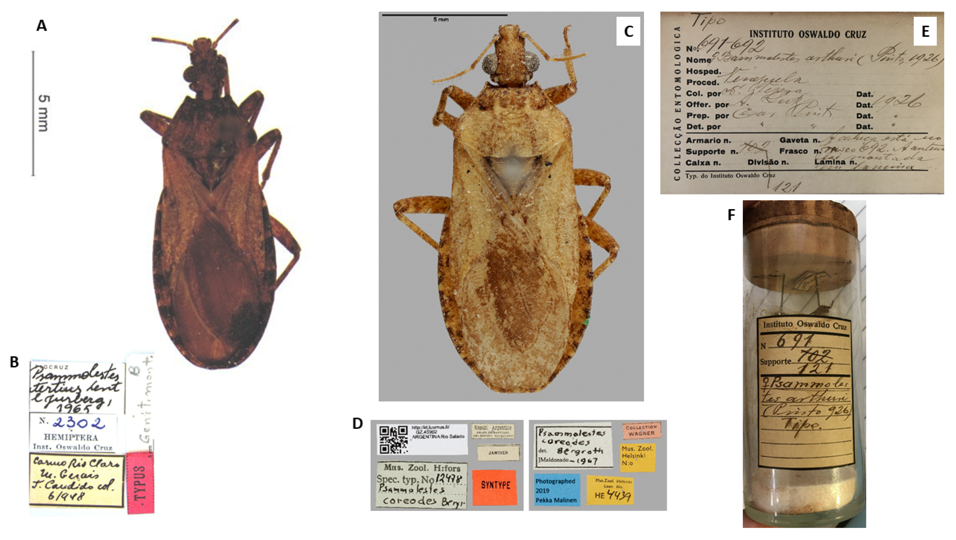
3.1. Triatomine Collections

In November of 2015, 172 specimens of P. tertius were collected in the municipality of Castro Alves, Bahia, Brazil, and 26 in Seabra, Bahia, Brazil. In December 2015, 137 specimens of P. coreodes were collected in the municipality Corumbá, Mato Grosso do Sul, Brazil. Lastly, 40 specimens of P. arthuri were collected in March 2017 at the Central University of Venezuela, Maracay, Aragua, Venezuela. All detailed information is organized in Table 1.

3.2. Morphological Characters

3.2.1. Male Genitalia (Redescription of the Male Genitalia)

Psammolestes arthuri

The phallosoma (Ph) presents a quadrangular shape, the anterior borders are rounded and the (PrPh) has a small, rounded curvature; the extension of the basal plate (Eplb) is medium in size but broad in shape (Figure 3A). A cylindrical and hollow gonopore (PrG) process with internal edges joined at the base and at the apex, in which each extension consists of two parts, was inserted into the basal bridge by a narrow rod (Figure 3A). The phallosoma (Ph) plate was convex in shape (Figure 3J). The parameres (Pa) are of medium length, with cylindrical conformation, and in the side view are a little arcuate. In the general profile, they are sinuous with a pointed apex projected out of a side flap; the outer face has many sensilla; the inner face has sensilla and the protruding edges, where the end has a chitinized tip of different coloration from the body and this coloring marks the insertion of these sensilla (Figure 3D). A median pygophore process (PrP) is pointed with a blunt apex, and the end of the plate where it is inserted is poorly chitinized, located inside the outer edge of the pygophore (Figure 3G).

Psammolestes coreodes

The morphology of the phallosoma (Ph) had an ovoid shape, the anterior borders are acuminate and the (PrPh) has a small curvature; the extension of the basal plate (Eplb) is short but broad (Figure 3B). A cylindrical and hollow gonopore (PrG) process with internal edges joined at the base and at the apex, where each extension consists of two parts, was inserted into the basal bridge by a short rod (Figure 3B). The phallosoma plate shows an equilateral triangle shape with a rounded apex (Figure 3K). The parameres (Pa) are short, with cylindrical conformation through the lateral view, besides being slightly arcuate. In the general profile they are sinuous with a pointed apex projected coming out of a side flap; the outer face has many sensillae, the inner face has only a few and the tip is a different to the body (Figure 3E). The median process of the pygophore (PrP) is pointed with a very sharp apex, and the end of the plate where it is inserted has two strongly chitinized striae located inside the outer border of the pygophore (Figure 3H).

Psammolestes tertius

The phallosoma (Ph) has a rounded shape, the anterior edges are rounded and the (PrPh) has a small irregular curvature, and one has a long size (Figure 3C). (PrG) has inner edges joined at the base and at the apex, where each extension consists of two parts, and are inserted in the basal bridge by a narrow rod (Figure 3C). A has a rounded trapezoidal shape (Figure 3L). The parameres (Pa) are of medium length, with cylindrical conformation, and in the side view, a few are arched. In the general profile, they are sinuous with a pointed apex and projected coming out of a side flap; the outer face has many bristles, the inner face has fewer sensillae and the edges protrude to the point of having a chitinized tip of different coloring from the body (Figure 3F). The median process of the pygophore (PrP) is pointed with a sharp apex, and the end of the plate where it is inserted is little chitinized, situated inside the outer edge of the pygophore (Figure 3I).

3.3. Egg Morphometry

The mean, maximum and minimum deviation of the measurements of total length and eggshell length of populations of P. arthuri, P. coreodes and P. tertius are presented below in the form of a table (Table 2).
When performing the intraspecific comparison, regarding significance, it was observed that they did not present any significance in any of the parameters used in the study. When performing the interspecific comparison, the species present a significant difference in the diameter of the opercular opening, but the total length is very significant (Table 3).

3.4. Morphological Studies

3.4.1. Head

In P. arthuri (Figure 4A), the ocelli are separated and arranged in a straight line. In P. coreodes and P. tertius (Figure 4B,C), the ocellus form a symmetrical cylindrical shape and are larger in P.tertius when compared to P.coreodes and P. arthuri. The clypeus in P. arthuri is broad in the anterior portion and narrow in the posterior portion, with lateral lines transversely arranged (Figure 4A). In P. coreodes, it is rectangular in shape with straight lateral lines (Figure 4B) and in P. tertius it has a small narrowing in the anterior portion (Figure 4C). In P. arthuri, the anteclypeus is concave and narrow in the anterior portion and straight and wide in the posterior portion (Figure 4A). In P. coreodes, the anteclypeus is straight and broad in the anterior and posterior portions (Figsure 4B) and in P. tertius, the anteclypeus is concave and broad in the anterior portion and straight and wide in the posterior portion (Figure 4C).

3.4.2. Thorax

Anterolateral angle: In P. arthuri, the insertion was performed near the border of the ocelli. The insertions are pronounced and the posterior extremities are triangular in shape (Figure 4D). In P. coreodes, the insertion was below the dividing line between the neck and the protorax; they are short, triangular and not very pronounced (Figure 4E). In P. tertius, the insertion was made below the dividing line between the neck and the protorax and are short and the ends are rounded (Figure 4F). In P. arthuri, the carinas are elevated from the anterior to the posterior lobe (Figure 4D). In P. coreodes and P. tertius, they are present but distributed with little prominence in the posterior lobe (Figure 4E,F). In P. arthuri, P. coreodes and P. tertius, the discal tubercles are not present (Figure 4D–F). The glabrous areas of all three species are distributed regularly in the central region of the pronotum and irregularly on the sides of the anterior lobe; in P. arthuri you can view six glabrous areas, P. coreodes presents another layout pattern with eight glabrous areas and P. terius has a pattern with twelve glabrous areas. The posterior lobe of P. arthuri is rough (Figure 4A) and in P. coreodes and P. tertius it is striated (Figure 4B,C). In P. arthuri, P. coreodes and P. tertius, the apical processes are extremely short; however, the tips of the apical processes are distinct due to the differences in the triangular conformation (Figure 5A–C). The central depression in P. arthuri is clearly delimited by two areas: a small, fluted area and a larger, flat area (Figure 5A). In P. coreodes and P. tertius, the delimitations are not accentuated and have irregular striations distributed throughout the region (Figure 5B,C).
In P. arthuri, P. coreodes and P. tertius, the limiting line of the estridulatory sulcus is slightly curved in the anterior portion (Figure 5G–I). In P. arthuri, the groove is short and broad throughout the groove body and shows a narrowing in the posterior portion (Figure 5G). In P. coreodes, the groove is short and broad in the anterior portion and presents two straight limiting lines in the medial and posterior “V” (Figure 5H). In P. tertius, the border is wide initially and narrows in the median region and the posterior portion is re-sharpened at the end of the posterior V-shaped portion (Figure 5I).

3.4.3. Abdomen

In P. arthuri, the urotergite has a striated general appearance, but with vertical striae and ribs distributed throughout the upper and lower areas. Its apex is subtly rounded and does not extend beyond the central area (Figure 5D). P. coreodes presents a urotergite with a striated aspect vertically on the sides and lower region, but with discrete ridges and ribs in the upper region and marks in the lower region. Its apex is pointed and has a rounded marking in the central region (Figure 5E). Psammolestes tertius presents a urotergite with striations and discrete veins and its apex is pointed with a central depression (Figure 5F).
Female external genitalia: dorsal view: The line dividing the seventh and eighth segments is concave in P. Arthuri (Figure 6A), straight transverse in P. coreodes (Figure 6B) and convex in the central portion in P. tertius (Figure 6C). The line dividing the eighth of the ninth segment is convex in P. arthuri (Figure 6A) and straight in P. coreodes and P. tertius (Figure 6B,C). The third segment of P. arthuri (Figure 6A) laterally presents as a pair of symmetric curves, differing from those of P. coreodes (Figure 6B) and P. tertius (Figure 6C), which are straight and diagonally arranged.
Posterior view: In P. arthuri, P. coreodes and P. tertius (Figure 6D–F), the ninth segment is flattened frontally, and is sharper in P. tertius (Figure 6F). The tenth segment in P. arthuri, P. coreodes and P. tertius (Figure 6D–F) is in the form of a “U” with more closed or more open contours. Gonocoxite 8 is visible in P. arthuri and not visible in P. tertius (Figure 6D,F). It has a peniform format, which varies in size, and is markedly higher in P. arthuri (Figure 6D). Gonopophysics 8 is visible in P. arthuri, is subtly visible in P. tertius (Figure 6D,F) and is not visible in P. coreodes (Figure 6E).
Ventral view: the dividing line of segment VII with gonocoxites 8 is concave and similar in P. arthuri and P. coreodes (Figure 6G,H) and differs in the central part in P. tertius (Figure 6I). Segment IX is irregular and has an undulating shape throughout its length in all three species (Figure 6G–I). The gonocoxite 8 in P. arthuri are rounded (Figure 6G), in P. coreodes they are convex in the lateral and border with gonapophysics 8 and are rounded in all other areas (Figure 6H) and in P. tertius they have a trapezoidal shape, gonapophysis 8 (Figure 6I). Gonapophysics 8 in P. arthuri (Figure 6G) is short and curved, while in P. coreodes and P. tertius they are triangular and similar (Figure 6H,I).

3.5. Dichotomous Key for Species of the Genus Psammolestes, Based on Adults

Head and thorax highly polished; head not constricted before neck in lateral view (Figure 7); head has wide percurrent yellowish band occupying entire interocular space dorsally (Figure 7); long hairs on apex of second and third rostral segments (Figure 7); anterolateral angles of pronotum projecting forward to level of ocelli (Figure 7); male genitalia with large, fused basal plate struts…………………………………P. arthuri (Figure 7).
Head and thorax dull; head constricted before neck in side view; head light yellowish brown dorsally, speckled with darker coloration or dark and with narrow yellow percurrent line dorsally; long hairs on entire second and third rostral segments; shorter anterolateral angles, not attaining the level of ocelli; male genitalia with shorter, not fused basal plate struts ………………………………………………………………………………………2.
2—Head as long as or slightly shorter than wide across eyes (Figure 8); anteocular region not over twice as long as postocular region (Figure 8); head strongly declivous behind ocelli (Figure 8); anterolateral angles of pronotum acuminate (Figure 8); male genitalia with very oval-shaped basal plate struts. Length of male: 11–13 mm and length of female: 12–14 mm. Width of pronotum of male: 3.0–3.5 and width of pronotum of female: 3.2–4.0 mm. Width of abdomen of male: 4–5 mm and width of abdomen of female: 4.5–5.5 mm. …………………………………………………………………………P. coreodes (Figure 8).
Head slightly longer than wide across eyes (Figure 9); anteocular region from two to two and one-half times as long as postocular (Figure 9); head moderately declivous behind ocelli (Figure 9); very short anterolateral angles of pronotum (Figure 9); male genitalia with broadly S-shaped basal plate struts; dorsal sclerotization of phallosoma is broadly rounded apically ……………………………………………………………P. tertius(Figure 9).

3.6. Morphological Description of Eggs

The eggs of Psammolestes spp. were studied. The eggs of this genus are strongly attached to the substrate and are of medium and small size.

3.6.1. Psammolestes arthuri

The coloration ranged from off-white to brown, then approaching light brown when about to hatch. Eggs with continuous coloring have a small stripe on the chorion border lighter than the body of the egg. They do not have a “collar”, they have a long, narrow “collar”, often presenting a “lateral flattening” of coloration equal to the body of the egg. The operculum is light in color, in the same shade of the corial border which is a tone lighter than the body of the egg, with translucent and whitish edges. Its shape is circular with elevated, irregular filamentous projections and the opercular borders are narrow and of a light color; in some cases, there is a slight inclination of the operculum in relation to the “lateral flattening”. The eggs were significantly cylindrical, at an average of 1.54 mm in length (Table 2). The general aspect of the exocorial is not uniform, but the body is organized by people who are regular and the patients do not have access to the exocoriais cells. The pattern of the exocoriais cells is irregular and is not of a standardized smooth appearance. In optical microscopy (OM), by transparency, only the small central holes at the entrance of the tubes are evident. In SEM, the “boundary lines” (LL) are high and well visible in the presence of granulations (Figure 10A–E).

3.6.2. Psammolestes coreodes

The coloration varies from whitish to brown, reaching light brown when about to hatch. They have a pronounced brown pigmentation pattern, with a specular appearance. Eggs have a double coloration, with one color clearly just below the “colo” and corial boards. They do not have a “collar”; they have a long, narrow “collar” and often have a “lateral flattening” of coloration equal to the body of the egg. The operculum is light in color, in the same shade as the corial border in a tone lighter than the body of the egg, with translucent and whitish edges. Its shape is circular with a subtle central projection and has no obvious opercular edges. There is a more evident pattern of pores in the central region. The eggs, markedly oval, have a mean diameter of 1.34 mm (Table 2). The general appearance of the exocore is uniform, in both the egg body and the operculum, and the polygonal areas vary from pentagonal to hexagonal, most of which are hexagonal. In OM, by transparency, only the small central holes at the entrance of the tubes are evident. In SEM, the “limiting lines” (LL), due to the presence of granulations, have a slightly rough appearance. By transparency, they are refringent and, consequently, not very evident. The granulations that cover the tegument are irregular in size, agglutinated and distributed throughout the area of the exocorial cell (Figure 11A–F).

3.6.3. Psammolestes tertius

The egg color has four patterns: one whitish with small spots of light brown color (Figure 12D), one with a gray color with larger brown stains (Figure 12A), one full brown (Figure 12B) and one light brown with a white collar and a corial lip (Figure 12C). A marginal pigmentation pattern determined a spotted appearance. They are “collared” and have a long, narrow “collar”, often showing a “lateral flattening” of coloration equal to the body of the egg. The operculum is light in color, in the same shade as the corial border in a tone lighter than the body of the egg, with translucent and whitish edges. Its shape is circular with a subtle central projection and has no obvious opercular edges. There is a more evident pattern of pores in the central region. The eggs, markedly oval, have an average diameter of 1.40 mm (Table 2). The general appearance of the exocoria is uniform, both in the egg body and in the operculum (the non-polygonal areas). In OM, by transparency, only the small central holes at the entrance of the tubes are evident. In SEM, the “boundary lines” (LL) are not present and cells have a slightly rough appearance. The granulations that cover the integument are of irregular size, agglutinated and distributed throughout the area of the exocorial cell (Figure 12A–G).

3.7. Dichotomous Key for Species of the Genus Psammolestes, Based on Eggs

           1. Long cylindrical shaped eggs with lateral flattening……                P. arthuri
                              2. Oval and circular shaped eggs……                                         3
          3a. Eggs of oval shape without the presence of the collar……            P. coreodes
         3b. Eggs in circular format with the presence of the collar……            P. tertius

4. Discussion

Although the integrative taxonomy shows a trend towards the taxonomy of Triatominae [5], the classical taxonomy based on morphological and morphometric characteristics is of great importance for the correct classification of species (mainly because the diagnosis of practically all triatomine species is based on morphological data) [5]. This methodology was used to describe the species of Psammolestes [8,9,11], and was the process used to characterize pygophore described by Lent and Jurberg [11]. They used the marked character to separate the three species.
In 1965, Lent and Jurberg [11] conducted a review of the genus Psammolestes, in which they described the species P. tertius. In this work, a brief redescription of the three species was given, describing the following characteristics: median process of pygophore, parameres and median extension of the basal plaque and phallosoma. Furthermore, the redescription of male internal genitalia was performed, with the following details: phallosoma, gonopore process, phallosoma plate, parameres and median process of the pygophore.
Based on this information, Lent and Wygodzisky [16] proposed a dichotomous key for the genus Psammolestes. Although alternative keys have been recently proposed to assist in the correct identification of triatomines (such as the keys proposed from cytogenetic data [35,36,37,38]), the dichotomous keys based on morphological characteristics are the most commonly used from a practical scientific point of view. Thus, we enriched the Lent and Wygodzisky [16] key with new characters to assist in the correct identification of the species within this genus.
In 1976, Carcavallo and Tonn [39] developed a dichotomous key to differentiate some triatomine genera from egg character; however, they did not include Psammolestes spp. Subsequently, Barata [40] and Santos et al. [41] presented specific keys for the genus Rhodnius. In 1975, Carcavallo et al. [42] published the first characteristics of Psammolestes eggs, with an emphasis on P. arthuri. Barata [43] succinctly described the eggs of the genus Psammolestes. Thus, the key proposed in the present study for Psammolestes, together with the keys of Barata (1981) and Santos et al. (2009), allow species of the Rhodniini tribe to be identified from eggs.
Recent phylogenetic studies demonstrate that Psammolestes species are phylogenetically related, with P. tertius and P. coreodes being sister species [25]. In addition, one of the models tested for species delimitation (the ‘deep divergence and large population size’ model) delimited only two species: (i) P. arthuri and (ii) P. coreodes + P. tertius [25]. However, reproductive barriers confirm the specific status of P. coreodes and P. tertius based on the biological species concept [44]. Furthermore, the morphological study conducted by SEM allowed to differentiate the three species of Psammolestes; in the dorsal portion of the thorax, differences were noted in the pronotum, anterior lateral angles, the sub median carinas, glabrous areas and the scutellum and in the ventral portion of the thorax, differences in the shape of the stridulatory sulcus were observed. The female external genitalia were examined and differences were observed when examined by the dorsal, posterior and ventral view.
Hypsa et al. [30] and Filée et al. [31] emphasize that within the Rhodniini, it is evident that Psammolestes spp. represent species of Rhodnius. However, the characteristics of the head described by SEM (arrangement and size of the ocelli and the shape of the clypeus and the anteclypeus) when compared to Rhodnius do not confirm the inclusion of Psammolestes in the genus Rhodnius. These results based on the phenetic species concept are in agreement with the recent confirmation of generic status proposed based on the biological species concept [32].

5. Conclusions

Based on this, it was possible to differentiate between three species of Psammolestes and confirm that this genus should not be classified in the genus Rhodnius, contributing to Rhodniini’s taxonomy. Overall, our morphological data will be clear and helpful for a field survey of Chagas disease in the near future.

Author Contributions

Conceptualization, J.d.O., K.C.C.A. and J.A.d.R.; methodology, J.d.O., K.C.C.A., C.E.A., N.O., G.L.C., C.G., H.M.H., F.M.S. and J.A.d.R.; formal analysis, J.d.O. and K.C.C.A.; investigation J.d.O., K.C.C.A., C.E.A., N.O., G.L.C., C.G., H.M.H., F.M.S. and J.A.d.R.; resources, J.d.O. and J.A.d.R.; writing—original draft preparation, J.d.O. and K.C.C.A.; writing—review and editing, J.d.O., K.C.C.A., C.E.A., N.O., G.L.C., C.G., H.M.H., F.M.S. and J.A.d.R.; supervision, J.A.d.R. and C.E.A.; project administration, J.d.O. and J.A.d.R.; funding acquisition, J.d.O., C.G. and J.A.d.R. All authors have read and agreed to the published version of the manuscript.

Funding

This research was funded by Fundação de Amparo à Pesquisa do Estado de São Paulo (FAPESP), Conselho Nacional de Desenvolvimento Científico e Tecnológico (CNPq) and Carlos Chagas Filho Research Foundation of the State of Rio de Janeiro (FAPERJ) process number E-26/203.837/2022; JAR, Research Productivity Scholarships, CNPq process 317 358/2021-9.

Institutional Review Board Statement

Not applicable.

Informed Consent Statement

Not applicable.

Data Availability Statement

All relevant data are within the manuscript.

Acknowledgments

We would like to express our gratitude to Wilma Savini and Jose Manuel Ayala for the support provided in Venezuela, and to the Central Laboratory of Public Health Professor Gonçalo Moniz (LACEN—BA) for their logistics and mobility support in Bahia. Additionally, we would like to extend a special thank you to everyone who assisted us in the field in Bahia, including Eduardo Oyama Lins Fonseca, Carlos Gustavo Silva dos Santos, Orlando Marcos Farias de Sousa and Jucimar Souza Vaz. I would like to acknowledge the curators of the Coleção Entomológica do Instituto Oswaldo Cruz (CEIOC), Márcio Felix and Claudia Leal Rodrigues, for allowing me access to the type of Psammolestes arthuri.. We also thank the curators of the Triatomines Collection of the Oswaldo Cruz Institute, Rio de Janeiro, Brazil, for providing images of the P. tertius type. We would like to express our gratitude to Heidi Viljanen, who provided images of the P. coreodes type (deposited at the Zoological Museum, Helsinki, Finland; http://id.luomus.fi/GZ.45902).

Conflicts of Interest

The authors declare no conflict of interest.

References

  1. World Health Organization. Chagas Disease (American Trypanosomiasis). Available online: http://www.who.int/news-room/fact-sheets/detail/chagas-disease-(american-trypanosomiasis) (accessed on 27 July 2022).
  2. Pan American Health Organization. Chagas in the Americas. Available online: https://www3.paho.org/hq/index.php?option=com_content&view=article&id=13566:chagas-in-americas&Itemid=40721&lang=en (accessed on 27 July 2022).
  3. Chagas, C. Nova tripanossomíase humana. Estudos sobre a morfologia e o ciclo evolutivo de Schizotrypanum cruz n. sp., agente etiológico de nova entidade mórbida do homem. Mem. Inst. Oswaldo Cruz 1909, 1, 159–218. [Google Scholar] [CrossRef]
  4. Shikanai-Yasuda, M.A.; Carvalho, N.B. Oral transmission of Chagas disease. Clin. Infect. Dis. 2012, 54, 845–852. [Google Scholar] [CrossRef] [PubMed]
  5. Alevi, K.C.C.; Oliveira, J.; Rocha, D.S.; Galvão, C. Trends in taxonomy of Chagas disease vectors (Hemiptera, Reduviidae, Triatominae): From Linnaean to integrative taxonomy. Pathogens 2021, 10, 1627. [Google Scholar] [CrossRef]
  6. Correia, J.P.S.O.; Gil-Santana, H.R.; Dale, C.; Galvão, C. Triatoma guazu Lent and Wygodzinsky Is a Junior Synonym of Triatoma williami Galvão, Souza and Lima. Insects 2022, 13, 591. [Google Scholar] [CrossRef] [PubMed]
  7. Téllez-Rendón, J.; Esteban, L.; Rengifo-Correa, L.; Díaz-Albiter, H.; Huerta, H.; Dale, C. Triatoma yelapensis sp. nov. (Hemiptera: Reduviidae) from Mexico, with a Key of Triatoma Species Recorded in Mexico. Insects 2023, 14, 331. [Google Scholar] [CrossRef]
  8. Bergroth, E. A new genus of Reduviidae. Psyche 1911, 18, 144–145. [Google Scholar] [CrossRef]
  9. Pinto, C. Triatomideos da Venezuela, com a descripção de uma nova espécie do gênero Eutriatoma. Ann. Fac. Med. Sao Paulo 1926, 1, 85–87. [Google Scholar]
  10. Del Ponte, E. Catalogo descriptivo de los géneros Triatoma Lap, Rhodnius Stal y Eratyrus Stal. Rev. Inst. Bacteriol. 1930, 5, 12. [Google Scholar]
  11. Lent, H.; Jurberg, J. O gênero Psammolestes Bergroth, 1911, com um estudo sobre a genitalia das espécies (Hemiptera, Reduviidae, Triatominae). Rev. Bras. Biol. 1965, 25, 27. [Google Scholar]
  12. Galvão, C.; Carvalho, R.U.; Rocha, D.S.; Juberg, J. A check-list of the current valid species of the subfamily Triatominae Jeannel, 1919 (Hemiptera, Reduviidae) and their geographical distribution, with nomenclatural and taxonomic notes. Zootaxa 2003, 2002, 1–36. [Google Scholar] [CrossRef]
  13. Santos, F.M.; Jansen, A.M.; Mourão, G.M.; Jurberg, J.; Nunes, A.P.; Herrera, H.M. Triatominae (Hemiptera, reduviidae) in the pantanal region: Association with Trypanosoma cruzi, different habitats and vertebrate hosts. Rev. Soc. Bras. Med. Trop. 2015, 48, 532–538. [Google Scholar] [CrossRef] [PubMed]
  14. Cabrera, R. Notas breves sobre Psammolestes tertius Bergroth, 1911 (Reduviidae: Hemiptera): Un triatomino silvestre. An. Fac. Med. Lima 2006, 67, 345–365. [Google Scholar] [CrossRef]
  15. Silva, A.N.B.; Diotaiuti, L.; Galvão, L.M.C.; Chiari, E.; Oliveira, P.I.C.; CâMARA, A.C.J.; Souza, R.C.M. First report of Psammolestes tertius Lent & Jurberg, 1965 (Hemiptera, Reduviidae, Triatominae) in Rio Grande do Norte state, Brazil. Check List 2018, 14, 1109–1113. [Google Scholar] [CrossRef]
  16. Lent, H.; Wygodzinsky, P. Revision of the Triatominae (Hemiptera: Reduviidae) and their significance as vector of Chagas’s disease. Bull. Am. Mus. Nat. Hist. 1979, 163, 123–520. [Google Scholar]
  17. Pinto, C.; Lent, H. Sobre as especies do genero Psammolestes Bergroth, 1911 (Hemiptera, Triatomidae). An. Acad. Bras. Ciênc. 1935, 7, 5. [Google Scholar]
  18. Pifano, F. Anotaciones acerca del Psammolestes arthuri (Pinto, 1926) (Hemiptera, Heteroptera, Triatomidae) reduvideo hematofago encontrado en nidos de “cucarachero de monte (probablemente, Dendrocolaptidae) en un sector de los valles de Yaracuy”. Gac. Med. Caracas 1938, 45, 5. [Google Scholar]
  19. Sherlock, I.A.; Guitton, N. Fauna Triatominae do estado da Bahia—Brasil III—Notas sobre ecótopos silvestres e o gênero Psammolestes. Mem. Inst. Oswaldo Cruz 1973, 1, 11. [Google Scholar] [CrossRef]
  20. Cazorla-Perfetti, D. Psammolestes arthuri naturally infected with Trypanosoma cruzi found in sympatry with Rhodnius prolixus and Triatoma maculata on bird nests in Anzoátegui state, Venezuela. Saber 2015, 27, 324–327. [Google Scholar]
  21. Oliveira, J.; Alevi, K.C.C.; Fonseca, E.O.L.; Souza, O.M.F.; Santos, C.G.S.; Azeredo-Oliveira, M.T.V.; Da Rosa, J.A. New record and cytogenetic analysis of Psammolestes tertius Lent & Jurberg, 1965 (Hemiptera, Reduviidae, Triatominae) from Bahia state, Brazil. Genet. Mol. Res. 2016, 15, 2–6. [Google Scholar] [CrossRef]
  22. Paiva, V.F.; Rosa, J.A.; Ceretti Junior, W.; Marrelli, M.T.; Oliveira, J. Confirmation of the first report of Psammolestes tertius Lent and Jurberg, 1965 (Hemiptera, Reduviidae, Triatominae) in Paraná State, Brazil. Rev. Soc. Bras. Med. Trop. 2021, 54, e0485-2020. [Google Scholar] [CrossRef]
  23. Oliveira, J.; Rosa, J.A.; Fontes, F.M.; Andrade, D.C.; Madi, R.R.; Melo, C.M. Psammolestes tertius Lent & Jurberg, 1965 (Hemiptera, Reduviidae, Triatominae): First report in Sergipe State, Brazil. Rev. Soc. Bras. Med. Trop. 2021, 54, e0708-2020. [Google Scholar] [CrossRef] [PubMed]
  24. Oliveira, J.; Alevi, K.C.C.; Ravazi, A.; Herrera, H.M.; Santos, F.M.; Azeredo-Oliveira, M.T.V.; Rosa, J.A. New evidence of the monophyletic relationship of the genus Psammolestes Bergroth, 1911 (Hemiptera, Reduviidae, Triatominae). Am. J. Trop. Med. Hyg. 2018, 99, 1485–1488. [Google Scholar] [CrossRef] [PubMed]
  25. Hernández, C.; Alvarado, M.; Salgado-Roa, F.C.; Ballesteros, N.; Rueda, -M.N.; Oliveira, J.; Alevi, K.C.C.; Rosa, J.A.; Urbano, P.; Salazar, C.; et al. Phylogenetic relationships and evolutionary patterns of the genus Psammolestes Bergroth, 1911 (Hemiptera: Reduviidae: Triatominae). BMC Ecol. Evol. 2022, 22, 30. [Google Scholar] [CrossRef] [PubMed]
  26. Pita, S.; Lorite, P.; Cuadrado, A.; Panzera, Y.; De Oliveira, J.; Alevi, K.C.C.; Rosa, J.A.; Freitas, S.P.C.; Gómez-Palacio, A.; Solari, A.; et al. High chromosomal mobility of rDNA clusters in holocentric chromosomes of Triatominae, vectors of Chagas disease (Hemiptera-Reduviidae). Med. Vet. Entomol. 2022, 36, 66–80. [Google Scholar] [CrossRef]
  27. Hernández, C.; Rosa, J.A.; Vallejo, G.A.; Guhl, F.; Ramirez, J.D. Taxonomy, Evolution and Biogeography of the Rhodniini Tribe (Hemiptera: Reduviidae). Diversity 2020, 2, 12–97. [Google Scholar] [CrossRef]
  28. Carcavallo, R. Aspects of the epidemiology of Chagas disease in Venezuela and Argentina. American Trypanosomiasis Research. PAHO Sci. Publ. 1976, 318, 347–358. [Google Scholar]
  29. Justi, S.; Galvão, C. The evolutionary origin of diversity in Chagas disease vectors. Trends Parasitol. 2017, 33, 42–52. [Google Scholar] [CrossRef]
  30. Hypsa, V.; Tietz, D.; Zrzavy, J.; Rego, R.O.; Galvão, C.; Jurberg, J. Phylogeny and biogeography of Triatominae (Hemiptera, Reduviidae): Molecular evidence of a New World origin of the asiatic clade. Mol. Phylogenet. Evol. 2012, 23, 447–457. [Google Scholar] [CrossRef]
  31. Filée, J.; Merle, M.; Bastide, H.; Mougel, F.; Bérenger, J.M.; Folly-Ramos, E.; Almeida, C.E.; Harry, M. Phylogenomics for Chagas Disease Vectors of the Rhodnius Genus (Hemiptera, Triatominae): What We Learn from Mito-Nuclear Conflicts and Recommendations. Front. Ecol. Evol. 2022, 9, 750317. [Google Scholar] [CrossRef]
  32. Ravazi, A.; Oliveira, J.d.; Madeira, F.F.; Reis, Y.V.d.; Oliveira, A.B.B.d.; Galvão, C.; Azeredo-Oliveira, M.T.V.d.; Rosa, J.A.d.; Alevi, K.C.C. Trends in Taxonomy of the Rhodniini Tribe (Hemiptera, Triatominae): Reproductive Incompatibility between Rhodnius neglectus Lent, 1954 and Psammolestes spp. Confirms the Generic Status of Psammolestes Bergroth, 1911. Diversity 2022, 14, 761. [Google Scholar] [CrossRef]
  33. Rosa, J.A.; Mendonça, V.J.; Rocha, C.S.; Gardim, S.; Cilense, M. Characterization of the external female genitalia of six species of Triatominae (Hemiptera: Reduviidade) by scanning electron microscopy. Mem. Inst. Oswaldo Cruz 2010, 105, 286–292. [Google Scholar] [CrossRef] [PubMed]
  34. Weirauch, C. From four- to three-segmented labium in Reduviidae (Hemiptera: Heteroptera). Acta Entomol. Musei Natl. Pragae 2008, 48, 331–344. [Google Scholar]
  35. Borsatto, K.C.; Azeredo-Oliveira, M.T.V.; Alevi, K.C.C. Identification Key for the Chagas Disease Vectors of Five Brazilian States, Based on Cytogenetic Data. Am. J. Trop. Med. Hyg. 2019, 100, 303–305. [Google Scholar] [CrossRef]
  36. Borsatto, K.C.; Reis, Y.V.; Garcia, A.C.C.; Sousa, P.S.; Azeredo-Oliveira, M.T.V.; Alevi, K.C.C. CytoKey: Identification Key for the Chagas Disease Vectors of the Largest Brazilian Urban Center (Sao Paulo State), Based on Cytogenetic Data. Am. J. Trop. Med. Hyg. 2019, 101, 113–115. [Google Scholar] [CrossRef]
  37. Gonzalez-Britz, N.E.G.; Alevi, K.C.C.; Caris-Garcia, A.C.; Martinez-Purroy, C.E.; Galvão, C.; Carrasco, H.J. Chagas disease vectors of Paraguay: Entomoepidemiological aspects of Triatoma sordida (Stal, 1859) and development of an identification key for Paraguayan triatomines based on cytogenetics data. Am. J. Trop. Med. Hyg. 2021, 105, 130–133. [Google Scholar] [CrossRef]
  38. Oliveira, J.; Rosa, J.A.; Alevi, K.C.C. Chagas Disease Vectors of Espirito Santo, Brazil: First Report of Triatoma infestans (Klug, 1834) (Hemiptera, Triatominae) in the Brazilian State and Development of an Identification Key Based on Cytogenetic Data. Am. J. Trop. Med. Hyg. 2021, 104, 653–655. [Google Scholar] [CrossRef] [PubMed]
  39. Carcavallo, R.; Tonn, R.J. Clave grafica de Reduviidae (Hemiptera) hematofagos de Venezuela. Bol. Dir. Malarioligia Saneam. Ambient. 1976, 16, 244–265. [Google Scholar]
  40. Barata, J.M.S. Aspectos morfologicos de ovos de Triatominae II—Caracteristicas macroscopicas e exocoriais de dez especies do genero Rhodnius Stal, 1859 (Hemiptera: Reduviidae). Rev. Saúde Publ. 1981, 15, 490–542. [Google Scholar] [CrossRef]
  41. Santos, C.M.; Jurberg, J.; Galvao, C.; Rosa, J.A.; Junior, W.C.; Barata, J.M.S.; Obara, M.T. Comparative descriptions of eggs from three species of Rhodnius (Hemiptera: Reduviidae: Triatominae). Mem. Inst. Oswaldo Cruz 2009, 104, 1012–1018. [Google Scholar] [CrossRef] [PubMed]
  42. Carcavallo, R.U.; Otero, M.A.; Martínez, A.; Tonn, R.J. Notas sobre la biología, ecología y distribución geográfica del Psammolestes arthuri (Pinto) 192(Hemiptera, Reduviidae). Descripción de los estádios preimagales. Bol. Dir. Malarioligia Saneam. Ambient. 1975, 15, 231–239. [Google Scholar]
  43. Barata, J.M.S. Macroscopic and exochorial structures of Triatominae eggs. Estruturas macroscópicas e exocoriais de ovos de Triatominae. In Atlas of Chagas’ Disease Vectors in the Américas; Carcavallo, R.U., Galíndez Girón, I., Jurberg, J., Lent, H., Eds.; Editora Fiocruz: Rio de Janeiro, Brazil, 1998; Volume II, pp. 409–448. [Google Scholar]
  44. Ravazi, A.; Oliveira, J.; Campos, F.F.; Madeira, F.F.; Reis, Y.V.; Oliveira, A.B.B.; Azeredo-Oliveira, M.T.V.; Rosa, J.A.; Galvão, C.; Alevi, K.C.C. Trends in evolution of the Rhodniini tribe (Hemiptera, Triatominae): Experimental crosses between Psammolestes tertius Lent & Jurberg, 1965 and P. coreodes Bergroth, 1911 and analysis of the reproductive isolating mechanisms. Parasit. Vectors 2021, 14, 350. [Google Scholar] [CrossRef] [PubMed]
Figure 1. Types of Psammolestes species: (A) P. tertius holotype, (B) labels in detail (C) P. coreodes syntype, (D) labels in detail, (E) form with information on the type of P. arthuri, (F) the type of P. arthuri stored and sealed inside a glass tube.
Life 13 01081 g001
Figure 2. Aspects of the fieldwork. (A) A nest of Phacellodomus sp. in Brazil. (B) Nest collection in Venezuela. (C) Nest collection in Brazil. (D) Eggs adhering to nest sticks. (E) Nest sorting in the field. (F) Phacellodomus sp. in Venezuela.
Life 13 01081 g002
Figure 3. Structures of the diaphanized male genitalia. Phallosoma: (A) P. arthuri, (B) P. coreodes and (C) P. tertius. Parameters: (D) P. arthuri, (E) P. coreodes and (F) P. tertius. Median process of pygophore: (G) P. arthuri, (H) P. coreodes and (I) P. tertius. Phallosome plaque: (J) P. arthuri, (K) P. coreodes and (L) P. tertius.
Life 13 01081 g003
Figure 4. Dorsal view of head: (A) P. arthuri, (B) P. coreodes and (C) P. tertius. Pronotum: (D) P. arthuri, (E) P. coreodes and (F) P. tertius.
Life 13 01081 g004
Figure 5. Scutellum: (A) P. arthuri, (B) P. coreodes and (C) P. tertius. Urotergite process I: (D) P. arthuri, (E) P. coreodes and (F) P. tertius. Stridulatory groove: (G). P. arthuri, (H). P. coreodes and (I). P. tertius.
Life 13 01081 g005
Figure 6. Female external genitalia using SEM. Dorsal view: (A) P. arthuri, (B) P. coreodes and (C) P. tertius. Posterior view: (D) P. arthuri, (E) P. coreodes and (F) P. tertius. Ventral view: (G) P. arthuri, (H) P. coreodes and (I) P. tertius.
Life 13 01081 g006
Figure 7. Adult male of P. arthuri. Dorsal view (A), ventral view (B), head dorsal view (C), head ventral view (D), thorax dorsal view (E).
Life 13 01081 g007
Figure 8. Adult female of P. coreodes. Dorsal view (A), ventral view (B), head dorsal view (C), head ventral view (D), thorax dorsal view (E).
Life 13 01081 g008
Figure 9. Adult male of P. tertius. Dorsal view (A), ventral view (B), head dorsal view (C), head ventral view (D), thorax dorsal view (E).
Life 13 01081 g009
Figure 10. Eggs of P. arthuri. (A) Eggs by means of optical microscopy; (B) eggs adhered together in a substrate; (C) eggshells by means of scanning electron microscopy; (D) detail of the chorion border by means of SEM and (E) pattern of an exocorial cell by SEM.
Life 13 01081 g010
Figure 11. Eggs of P. coreodes. (AC) Chromatic variations of eggs by means of light microscopy. (D) Eggshell by means of scanning electron microscopy. (E) Detail of the central border in SEM and (F) pattern of exocorial cells in SEM.
Life 13 01081 g011
Figure 12. Eggs of P. tertius. (AD) Chromatic patterns of eggs by means of optical microscopy; (E) eggshells by means of scanning electron microscopy; (F) detail of the chorion border by means of SEM and (G) pattern of exocorial cell in SEM.
Life 13 01081 g012
Table 1. Collection data of Psammolestes spp.
Species CountryStateMunicipalityLocality
P. tertiusBrazilBahiaSeabraAgreste
P. tertiusBrazilBahiaCastro AlvesMelancia II
P. coreodesBrazilMato Grosso do SulCorumbáAccess road to “Que Qué”
P. coreodesBrazilMato Grosso do SulCorumbáAccess road to Paraguai River
P. tertiusBrazilMato Grosso do SulCorumbáAccess road to Alegria farm
P. arthuriVenezuelaAraguaMaracayCentral University of Venezuela
Table 2. Results of the measurements and respective statistical analyses of the opercular opening and the total length of 50 eggshells of populations of three species of Psammolestes.
Opercular OpeningTotal Length
PopulationMinimumMaximumAverageStandard DeviationMinimumMaximumAverageStandard Deviation
P. tertius POP10.300.370.340.0171.271.471.360.052
P. tertius POP20.300.380.350.021.321.51.40.046
P. coreodes POP30.290.350.320.0161.251.431.340.05
P. coreodes POP40.290.370.320.0181.261.431.340.046
P. coreodes POP50.260.360.310.0221.21.461.330.051
P. coreodes POP60.280.340.310.0151.251.441.360.051
P. arthuri POP70.390.470.430.021.341.711.540.085
POP1: Castro Alves—BA; POP2: Santa Therezinha—BA; POP3: (Que Que) Corumbá—MS; POP4: (Alegria farm access) Corumbá—MS; POP5: (Paraguai River acess) Corumbá—MS; POP6: (Access to morro of Urucum) Corumbá—MS and POP7: Maracay, Aragua–Venezuela.
Table 3. Statistical analysis using unpaired t-test with Welch’s correction for measurements of the opercular opening and total length of 50 eggshells of the genus Psammolestes populations.
Opercular OpeningTotal Length
Populationsp-ValueSignificancep-ValueSignificance
POP1 vs. POP2p > 0.05NSp > 0.05NS
POP1 vs. POP3p > 0.05NSp > 0.05NS
POP1 vs. POP4p > 0.05NSp > 0.05NS
POP1 vs. POP5p > 0.05NSp > 0.05NS
POP1 vs. POP6p > 0.05NSp > 0.05NS
POP2 vs. POP3p > 0.05NSp < 0.05*
POP2 vs. POP4p > 0.05NSp < 0.05*
POP2 vs. POP5p > 0.05NSp < 0.05*
POP2 vs. POP6p > 0.05NSp > 0.05NS
POP3 vs. POP4p > 0.05NSp > 0.05NS
POP3 vs. POP5p > 0.05NSp > 0.05NS
POP3 vs. POP6p > 0.05NSp > 0.05NS
POP7 vs. POP1p > 0.05NSp < 0.01**
POP7 vs. POP2p > 0.05NSp < 0.01**
POP7 vs. POP3p < 0.05*p < 0.01**
POP7 vs. POP4p < 0.05*p < 0.01**
POP7 vs. POP5p < 0.05*p < 0.01**
POP7 vs. POP6p < 0.05*p < 0.01**
POP1: P. tertius: Castro Alves—BA; POP2: P. tertius: Santa Therezinha—BA; POP3: P. coreodes (Que Que) Corumbá—MS; POP4: P. coreodes (Alegria farm acess) Corumbá—MS; POP5: P. coreodes (Paraguai River acess) Corumbá—MS; POP6: P. coreodes (Acess to morro do Urucum) Corumbá—MS and POP7: P. arthuri Maracay, Aragua—Venezuela. NS = not significant (p > 0.05); * significant (p < 0.05); ** very significant (p < 0.01).
Disclaimer/Publisher’s Note: The statements, opinions and data contained in all publications are solely those of the individual author(s) and contributor(s) and not of MDPI and/or the editor(s). MDPI and/or the editor(s) disclaim responsibility for any injury to people or property resulting from any ideas, methods, instructions or products referred to in the content.

