
Duodenal Gangliocytic Paragangliomas—Case Series and Literature Review

 , , , , , and
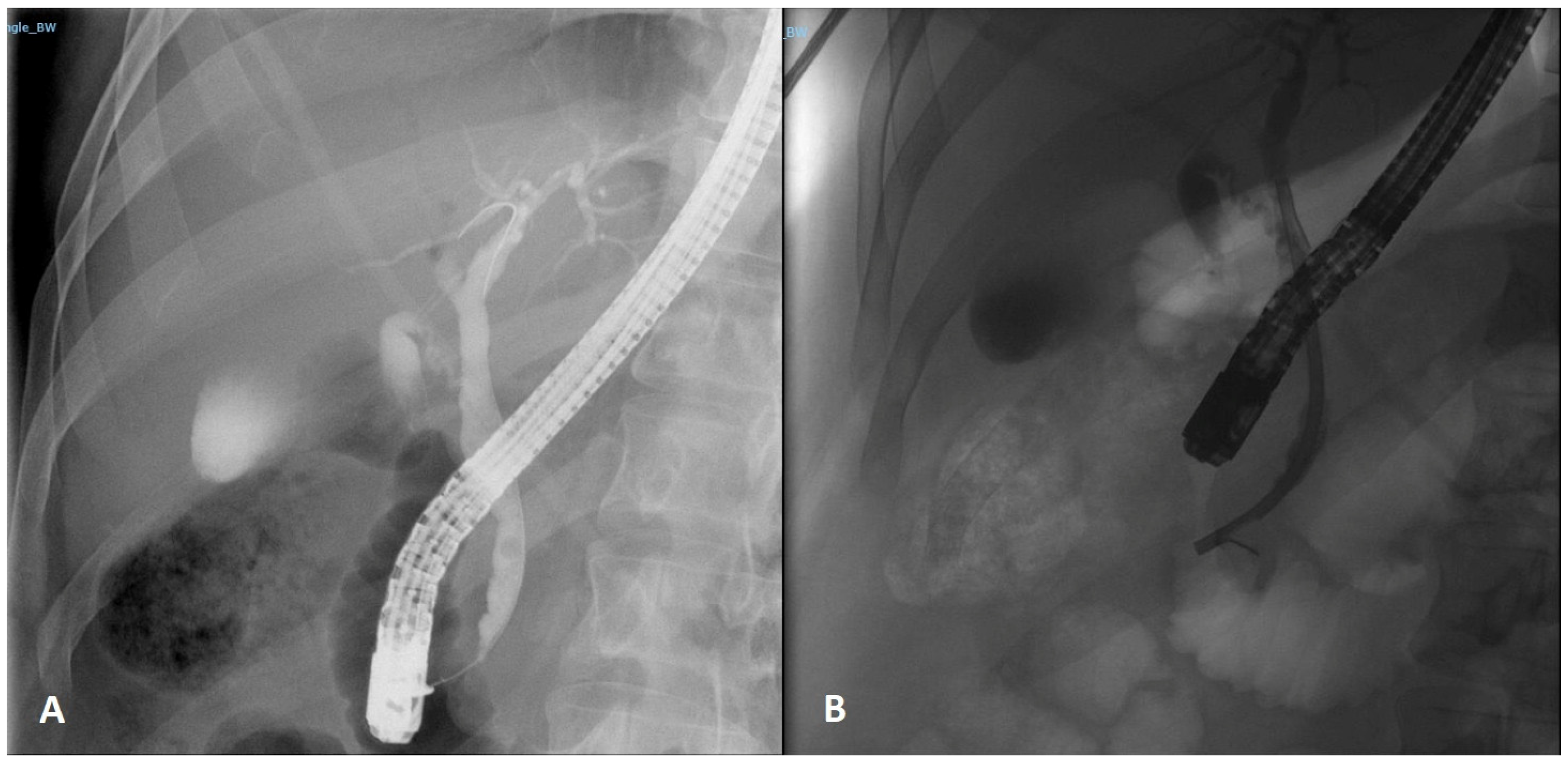
Abstract
:1. Introduction
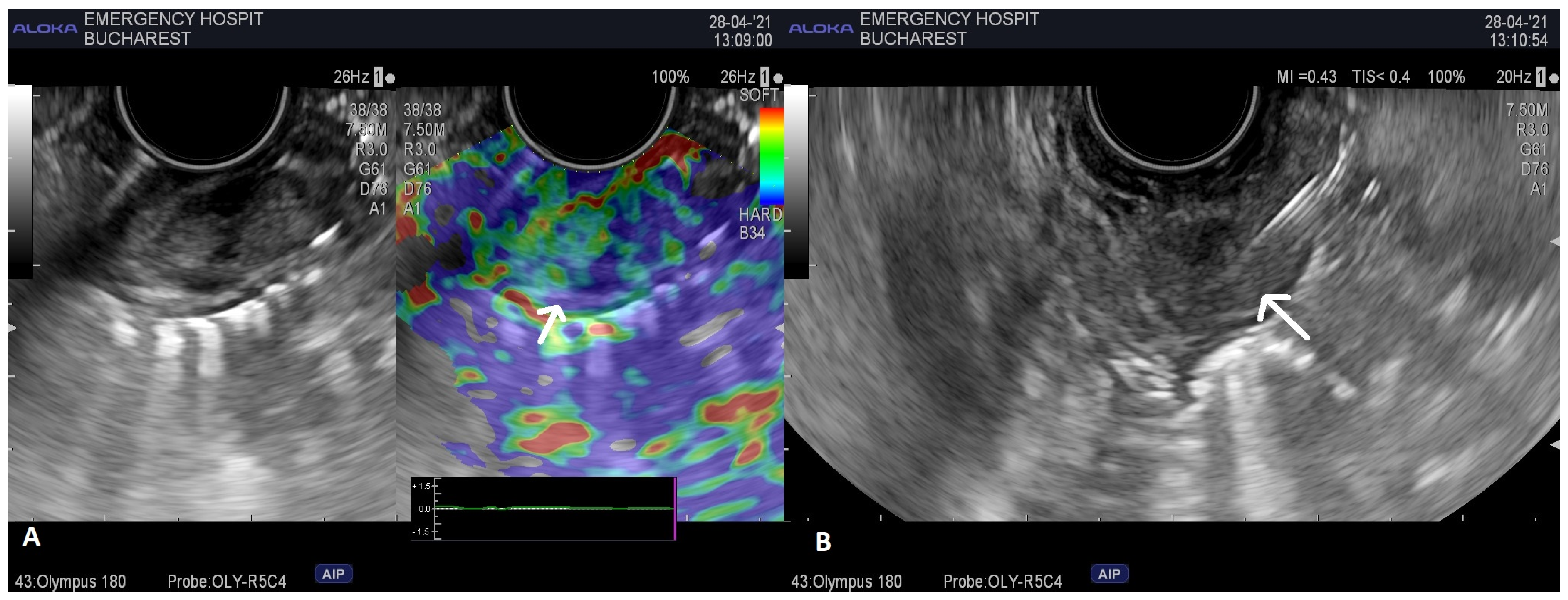
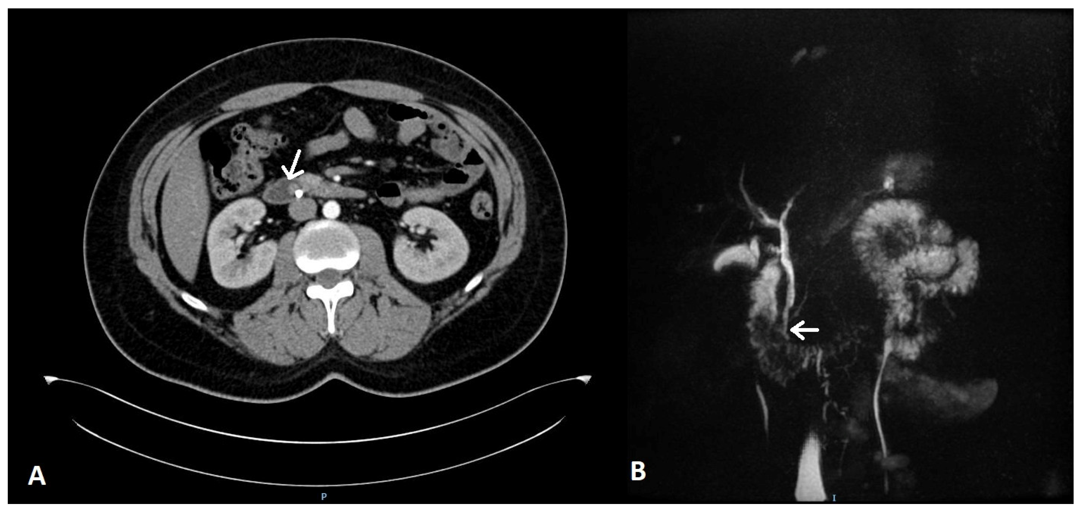
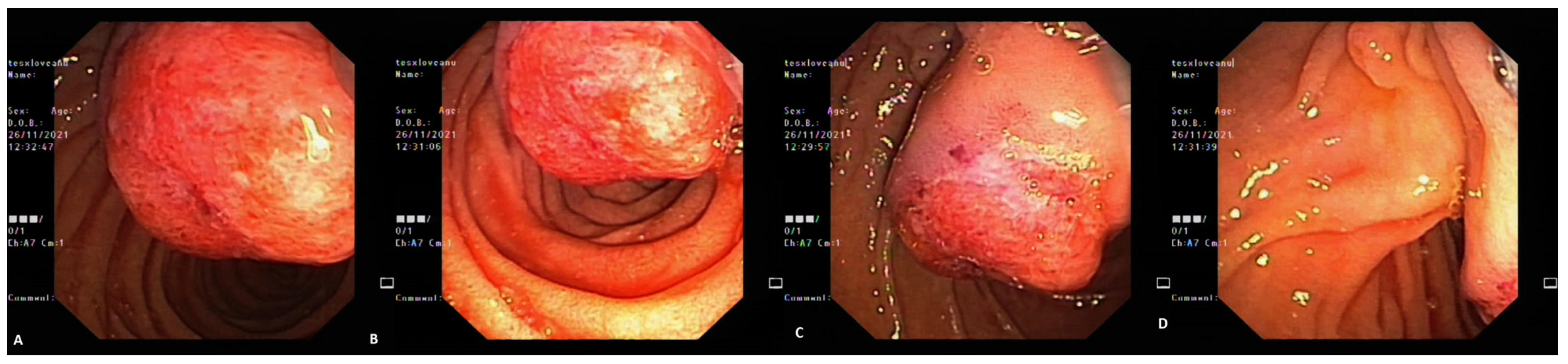
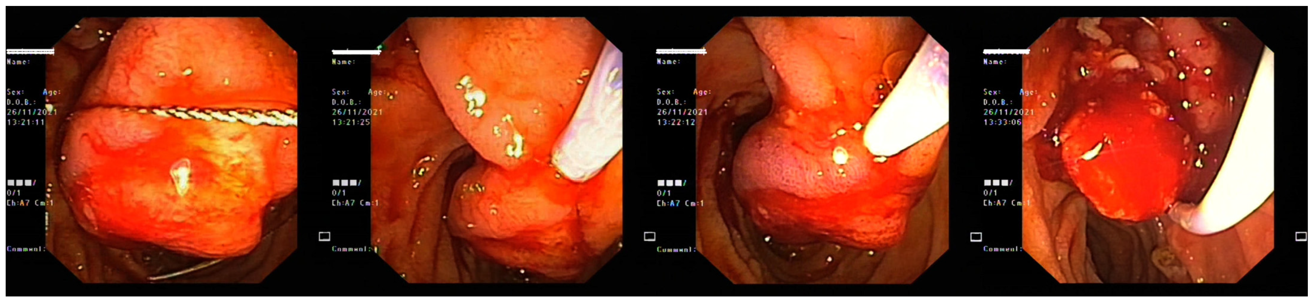
2. Case Report 1
3. Case Report 2
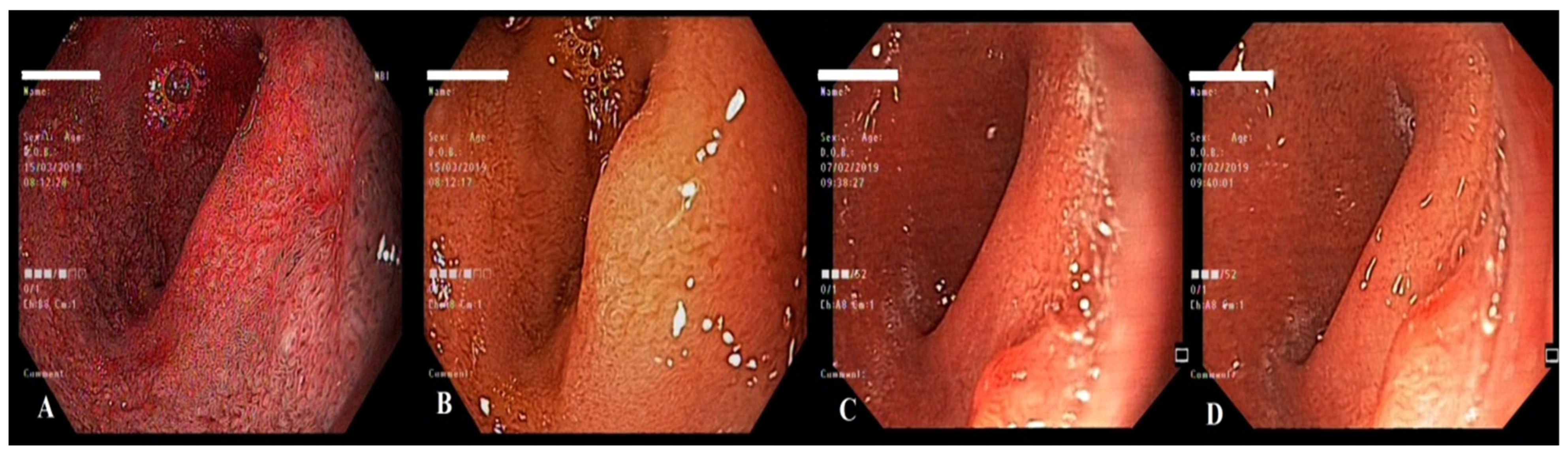
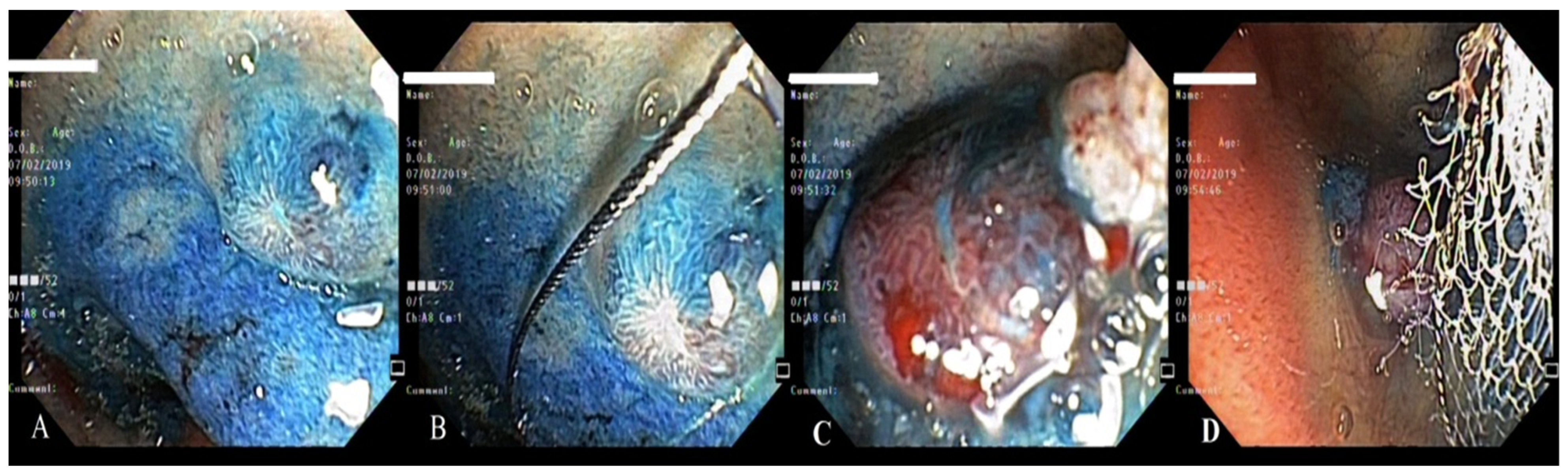
4. Case Report 3
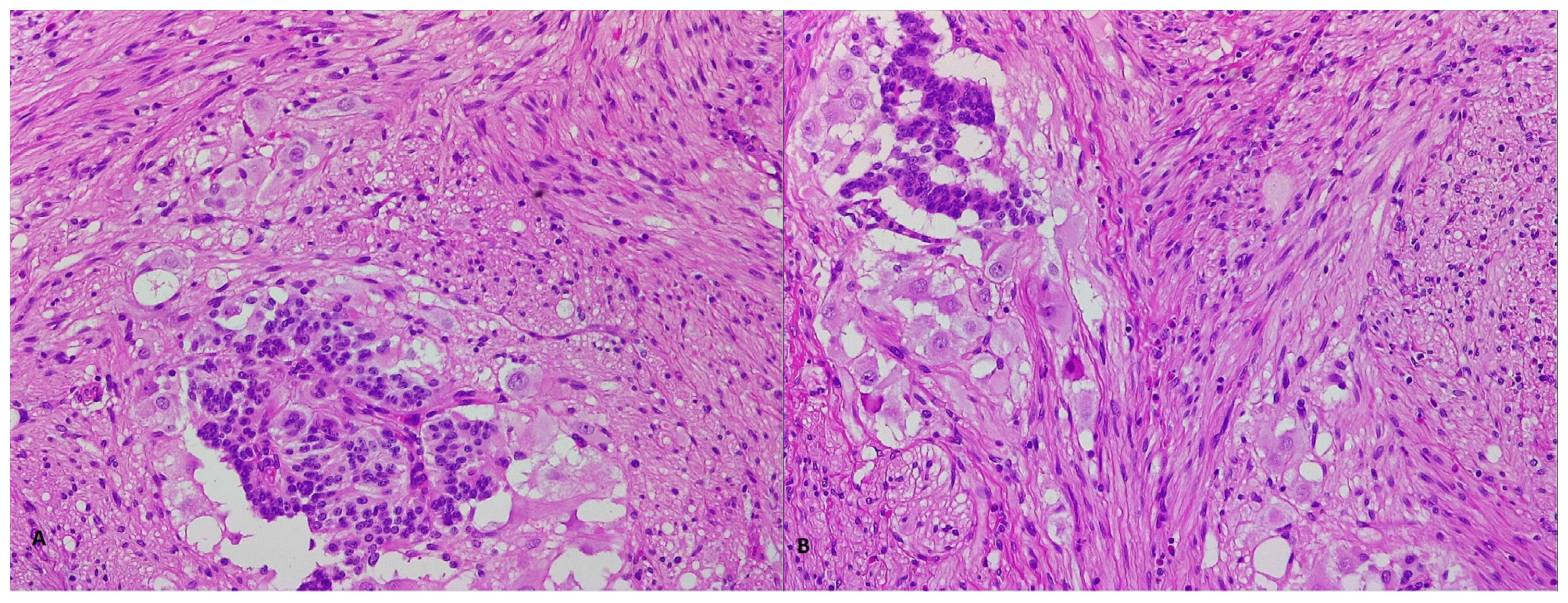
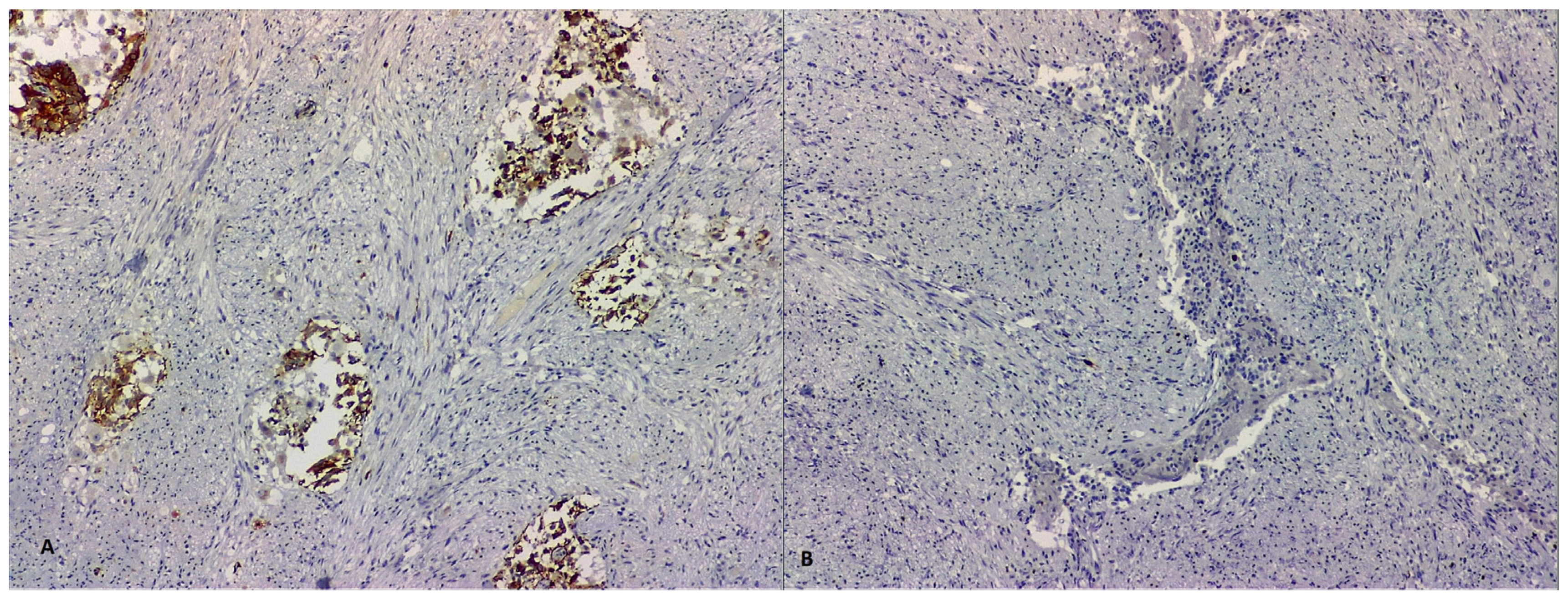
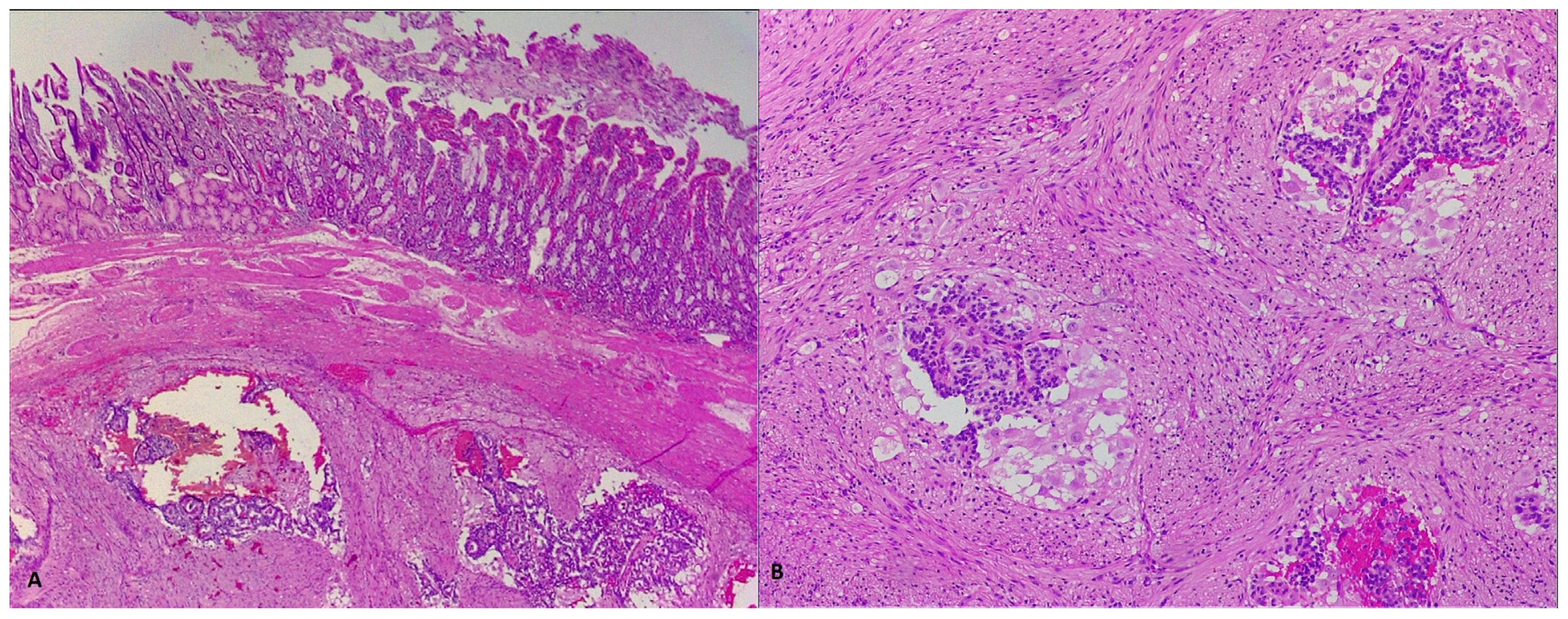
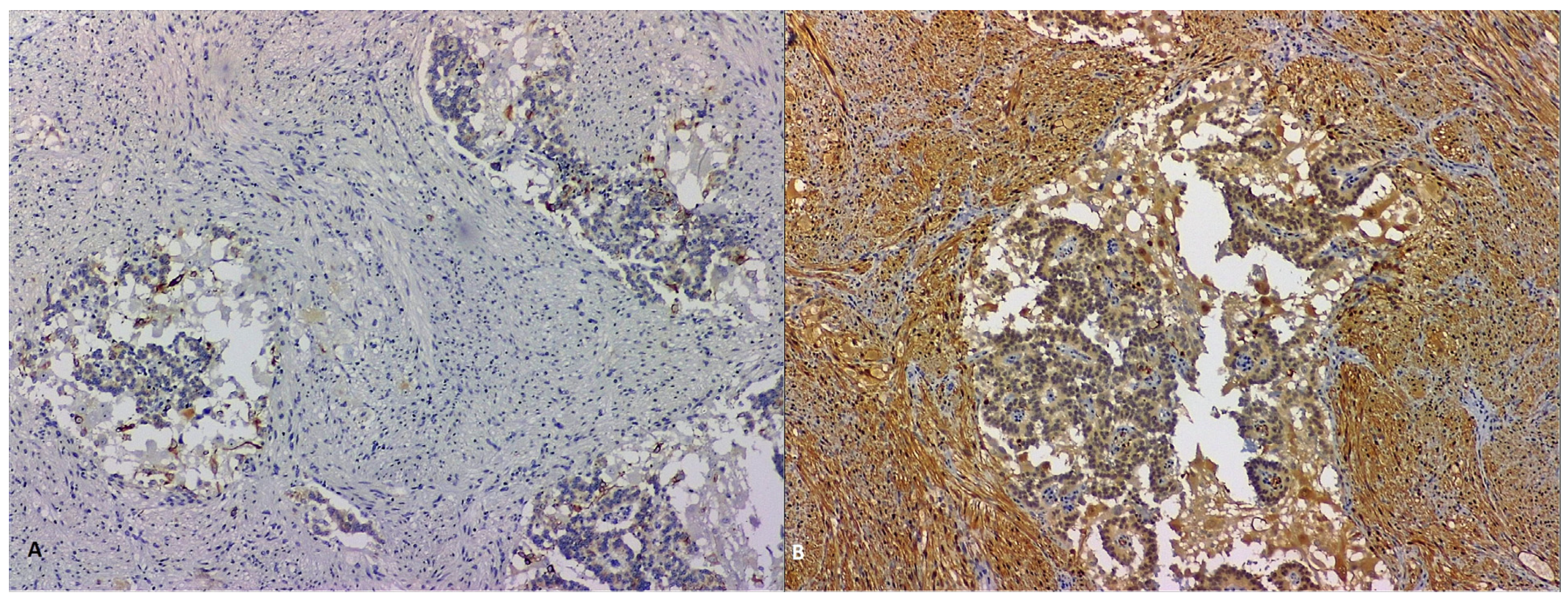
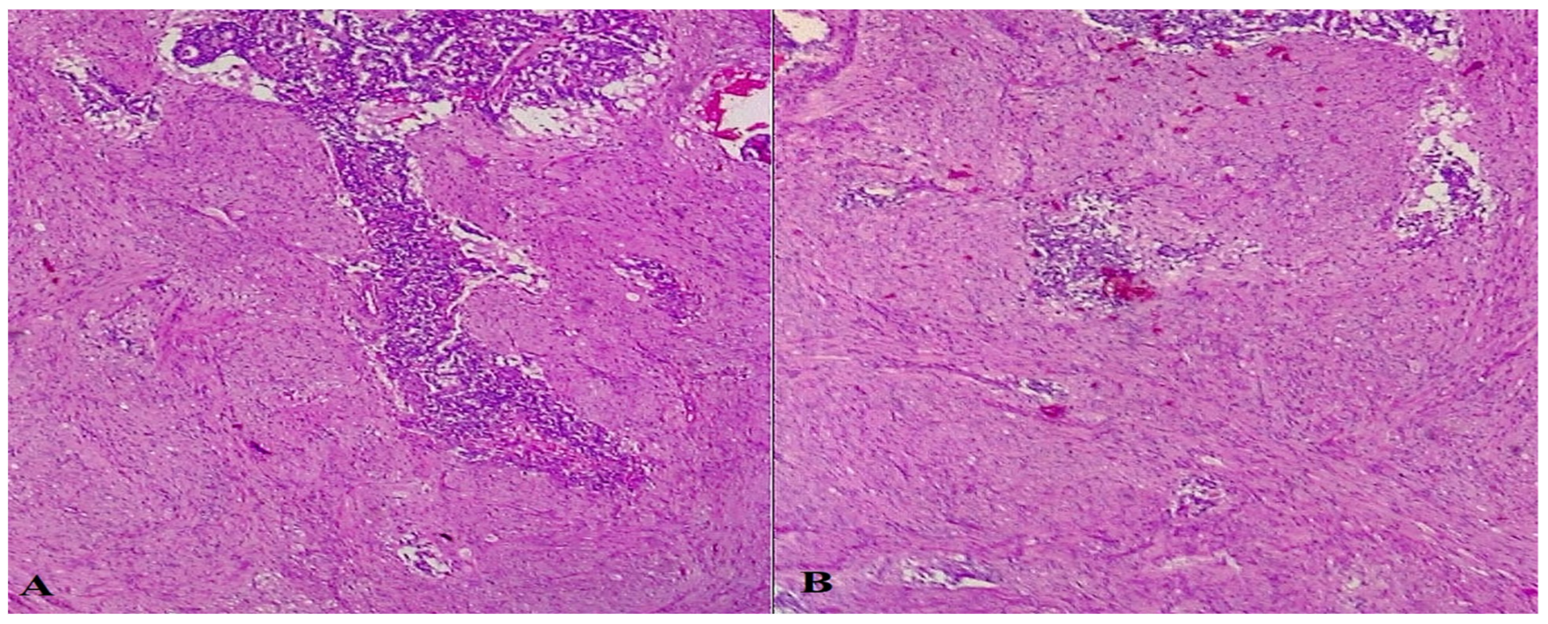
5. Discussions and Literature Review
6. Conclusions
Author Contributions
Funding
Institutional Review Board Statement
Informed Consent Statement
Data Availability Statement
Conflicts of Interest
References
- Bakshi, N.; Rao, S.; Dhawan, S.; Singla, V. Duodenal gangliocytic paraganglioma: A rare cause for gastrointestinal polyp. Indian J. Pathol. Microbiol. 2018, 61, 580–582. [Google Scholar] [CrossRef]
- Okubo, Y.; Yokose, T.; Motohashi, O.; Miyagi, Y.; Yoshioka, E.; Suzuki, M.; Washimi, K.; Kawachi, K.; Nito, M.; Nemoto, T.; et al. Duodenal Rare Neuroendocrine Tumor: Clinicopathological Characteristics of Patients with Gangliocytic Paraganglioma. Gastroenterol. Res. Pract. 2016, 2016, 5257312. [Google Scholar] [CrossRef] [PubMed] [Green Version]
- Sharma, S.; Gaspar, B.L.; Kumar, P.; Yadav, T.D.; Vasishta, R.K. Gangliocytic paraganglioma with atypical immunohistochemical features presenting as extrahepatic biliary obstruction. Int. J. Surg. Pathol. 2015, 23, 561–566. [Google Scholar] [CrossRef] [PubMed]
- Park, H.K.; Han, H.S. Duodenal Gangliocytic Paraganglioma With Lymph Node Metastasis. Arch. Pathol. Lab. Med. 2016, 140, 94–98. [Google Scholar] [CrossRef] [Green Version]
- Riley, D.S.; Barber, M.S.; Kienle, G.S.; Aronson, J.K.; von Schoen-Angerer, T.; Tugwell, P.; Kiene, H.; Helfand, M.; Altman, D.G.; Sox, H.; et al. CARE guidelines for case reports: Explanation and elaboration document. J. Clin. Epi. 2017, 89, 218–235. [Google Scholar] [CrossRef]
- Asa, S.L.; Ezzat, S.; Mete, O. The Diagnosis and Clinical Significance of Paragangliomas in Unusual Locations. J. Clin. Med. 2018, 7, 280. [Google Scholar] [CrossRef] [PubMed] [Green Version]
- Welander, J.; Andreasson, A.; Juhlin, C.C.; Wiseman, R.W.; Bäckdahl, M.; Höög, A.; Larsson, C.; Gimm, O.; Söderkvist, P. Rare germline mutations identified by targeted next-generation sequencing of susceptibility genes in pheochromocytoma and paraganglioma. J. Clin. Endocrinol. Metab. 2014, 99, E1352–E1360. [Google Scholar] [CrossRef] [PubMed]
- Milione, M.; Parente, P.; Grillo, F.; Zamboni, G.; Mastracci, L.; Capella, C.; Fassan, M.; Vanoli, A. Neuroendocrine neoplasms of the ampullary region. In WHO Classification of Tumors of the Digestive System; IARC Press: Lyon, France, 2010; pp. 92–94. [Google Scholar]
- Kepes, J.J.; Zacharias, D.L. Gangliocytic paragangliomas of the duodenum. A report of two cases with light and electron microscopic examination. Cancer 1971, 27, 61–67. [Google Scholar] [CrossRef] [PubMed]
- Loftus, T.J.; Kresak, J.L.; Gonzalo, D.H.; Sarosi, G.A.; Behrns, K.E. Duodenal gangliocytic paraganglioma: A case report and literature review. Int. J. Surg. Case Rep. 2015, 8C, 5–8. [Google Scholar] [CrossRef] [Green Version]
- Taylor, H.B.; Helwig, E.B. Benign nonchromaffin paragangliomas of the duodenum. Virchows. Arch. Pathol. Anat. Physiol. Klin. Med. 1962, 335, 356–366. [Google Scholar] [CrossRef]
- Dahl, E.V.; Waugh, J.M.; Dahlin, D.C. Gastrointestinal ganglioneuromas: Brief review with report of a duodenal ganglioneuroma. Am. J. Patho. 1957, 33, 953–965. [Google Scholar]
- Burke, A.P.; Helwig, E.B. Gangliocytic paraganglioma. Am. J. Clin. Pathol. 1989, 92, 1–9. [Google Scholar] [CrossRef] [PubMed]
- Evans, J.D.; Wilson, P.G.; Barber, P.C.; Neoptolemos, J.P. Duodenal gangliocytic paraganglioma presenting as an ampullary tumor. Int. J. Pancreatol. 1996, 20, 131–134. [Google Scholar] [CrossRef] [PubMed]
- Altavilla, G.; Chiarelli, S.; Fassina, A. Duodenal periampullary gangliocytic paraganglioma: Report of two cases with immunohistochemical and ultrastructural study. Ultrastruct. Pathol. 2001, 25, 137–145. [Google Scholar] [CrossRef]
- Hernández, A.G.; Lanuza, E.D.; Matias, A.C.; Huertas, R.P.; Rodriguez, K.M.; Perez, P.G.; Mompean, F.O. Large gangliocytic paraganglioma of the duodenum: A rare entity. World J. Gastrointest. Surg. 2015, 7, 170–173. [Google Scholar] [CrossRef]
- Hashimoto, S.; Kawasaki, S.; Matsuzawa, K.; Harada, H.; Makuuchi, M. Gangliocytic paraganglioma of the papilla of Vater with regional lymph node metastasis. Am. J. Gastroenterol. 1992, 87, 1216–1218. [Google Scholar]
- Nagai, T.; Torishima, R.; Nakashima, H.; Tanahashi, J.; Iwata, M.; Ookawara, H.; Yokoyama, S.; Yada, K.; Sato, R.; Murakami, K.; et al. Duodenal gangliocytic paraganglioma treated with endoscopic hemostasis and resection. J. Gastroenterol. 2004, 39, 277–283. [Google Scholar] [CrossRef]
- Scheithauer, B.W.; Nora, F.E.; LeChago, J.; Wick, M.R.; Crawford, B.G.; Weiland, L.H.; Carney, J.A. Duodenal gangliocytic paraganglioma. Clinicopathologic and immunocytochemical study of 11 cases. Am. J. Clin. Pathol. 1986, 86, 559–565. [Google Scholar] [CrossRef]
- Sardar, H.A.; Ayad, A.M.; Rafil, T.Y. Duodenal gangliocytic paraganglioma: A very rare cause for upper gastrointestinal bleeding: Case report with review of literature. Int. J. Surg. Case Rep. 2020, 75, 408–412. [Google Scholar] [CrossRef]
- Reis, D.; Damião, F.; Noronha, F.C.; Cruz, R.; Vitorino, E.; Carrilho, R.L.; Tato, M.R. Duodenal Gangliocytic Paraganglioma: A Unique Cause of Abdominal Pain. ACG Case Rep. J. 2020, 7, e00272. [Google Scholar] [CrossRef]
- Wong, A.; Miller, A.; Metter, J.; Thomas, C., Jr. Locally advanced duodenal gangliocytic paraganglioma treated with adjuvant radiation therapy: Case report and review of the literature. World J. Surg. Onc. 2005, 3, 15. [Google Scholar] [CrossRef] [PubMed] [Green Version]
- Adams, L.; Friedman, T.M.; Shaver, T.R.; Younan, G. Duodenal Gangliocytic Paraganglioma Requiring a Pancreaticoduodenectomy: A Case Report and Review of the Literature. Case Rep. Surg. 2018, 2018, 6292789. [Google Scholar] [CrossRef] [Green Version]
- Castoldi, L.; De Rai, P.; Marini, A.; Ferrero, S.; De Luca, V.; Tiberio, G. Neurofibromatosis-1 and ampullary gangliocytic paraganglioma causing biliary and pancreatic obstruction. Int. J. Gastrointest. Cancer. 2001, 29, 93–98. [Google Scholar] [CrossRef] [PubMed]
- Hoffmann, K.M.; Furukawa, M.; Jensen, R.T. Duodenal neuroendocrine tumors: Classification, functional syndromes, diagnosis and medical treatment. Best Pract. Res. Clin. Gastroenterol. 2005, 19, 675–697. [Google Scholar] [CrossRef] [PubMed]
- Sundararajan, V.; Robinson-Smith, T.M.; Lowy, A.M. Duodenal gangliocytic paraganglioma with lymph node metastasis: A case report and review of the literature. Arch. Pathol. Lab. Med. 2003, 127, 139–141. [Google Scholar] [CrossRef] [PubMed]
- Nakamura, T.; Ozawa, T.; Kitagawa, M.; Takehira, Y.; Yamada, M.; Yasumi, K.; Tamakoshi, K.; Kobayashi, Y.; Nakamura, H. Endoscopic resection of gangliocytic paraganglioma of the minor duodenal papilla: Case report and review of the literature. Gastrointest. Endosc. 2002, 55, 270–273. [Google Scholar] [CrossRef] [PubMed]
- Li, B.; Li, Y.; Tian, X.Y.; Luo, B.N.; Li, Z. Malignant gangliocytic paraganglioma of the duodenum with distant metastases and a lethal course. World J. Gastroenterol. 2014, 20, 15454–15461. [Google Scholar] [CrossRef] [PubMed]
- Ogata, S.; Horio, T.; Sugiura, Y.; Aiko, S.; Aida, S. Duodenal gangliocytic paraganglioma with regional lymph node metastasis and a glandular component. Pathol. Int. 2011, 61, 104–107. [Google Scholar] [CrossRef] [PubMed]
- Ghassemi, K.A.; Cortina, G.; Reber, H.A.; Farrell, J.J. Complete resection of ampullary paragangliomas confined to the submucosa on endoscopic ultrasound may be best achieved by radical surgical resection. Case Rep. Gastroenterol. 2009, 3, 169–174. [Google Scholar] [CrossRef]
- Okubo, Y.; Wakayama, M.; Nemoto, T.; Kitahara, K.; Nakayama, H.; Shibuya, K.; Yokose, T.; Yamada, M.; Shimodaira, K.; Sasai, D.; et al. Literature survey on epidemiology and pathology of gangliocytic paraganglioma. BMC Cancer 2011, 11, 187. [Google Scholar] [CrossRef] [Green Version]
- Dookhan, D.B.; Meittinen, M.; Finkel, G. Recurrent duodenal gangliocytic paraganglioma with lymph node metastasis. Histopathology 1993, 22, 399–401. [Google Scholar] [CrossRef] [PubMed]
- Ting, Y.; Singh, S.; Nghiemphu, P. Antiangiogenic and immunotherapeutic strategies in the treatment of paraganglioma. J. Neuro-Oncol. 2021, 150, 291–299. [Google Scholar] [CrossRef]


Disclaimer/Publisher’s Note: The statements, opinions and data contained in all publications are solely those of the individual author(s) and contributor(s) and not of MDPI and/or the editor(s). MDPI and/or the editor(s) disclaim responsibility for any injury to people or property resulting from any ideas, methods, instructions or products referred to in the content. |
© 2023 by the authors. Licensee MDPI, Basel, Switzerland. This article is an open access article distributed under the terms and conditions of the Creative Commons Attribution (CC BY) license (https://creativecommons.org/licenses/by/4.0/).
Share and Cite
Stan-Ilie, M.; Şandru, V.; Plotogea, O.-M.; Rînja, E.; Pavel, C.; Constantinescu, G.; Negreanu, L.; Paduraru, D.N.; Bolocan, A.; Andronic, O.; et al. Duodenal Gangliocytic Paragangliomas—Case Series and Literature Review. Life 2023, 13, 597. https://doi.org/10.3390/life13030597
Stan-Ilie M, Şandru V, Plotogea O-M, Rînja E, Pavel C, Constantinescu G, Negreanu L, Paduraru DN, Bolocan A, Andronic O, et al. Duodenal Gangliocytic Paragangliomas—Case Series and Literature Review. Life. 2023; 13(3):597. https://doi.org/10.3390/life13030597
Chicago/Turabian StyleStan-Ilie, Madalina, Vasile Şandru, Oana-Mihaela Plotogea, Ecaterina Rînja, Christopher Pavel, Gabriel Constantinescu, Lucian Negreanu, Dan Nicolae Paduraru, Alexandra Bolocan, Octavian Andronic, and et al. 2023. "Duodenal Gangliocytic Paragangliomas—Case Series and Literature Review" Life 13, no. 3: 597. https://doi.org/10.3390/life13030597