1. Introduction
Third molar extraction is one of the most common surgeries in any dental practice. The most common postoperative sequelae include pain and swelling [
1]. The primary reason for these signs and symptoms is the acute inflammation that occurs due to the surgical handling of soft and hard tissues [
2]. Such postoperative symptoms can be predicted by several factors, including peri-operative infection, pericoronitis, the difficulty of impaction, duration of the surgery, the technique of removal, perioperative use of antibiotics, etc. [
3,
4]. Given these wide ranges of variables, from the surgical technique employed to the optimal use of antibiotics, researchers have used specific markers of postoperative morbidity to compare and identify a better methodology. C-reactive protein (CRP) is one such marker. CRP is stimulated due to surgery-induced inflammation or any associated trauma/infection. The physiological value of CRP is <10 mg/L in healthy individuals. This value increased by double after 8 h of surgery and returned to the baseline value in a week [
5,
6]. CRP quantification is often used as a biomarker of systemic inflammatory status [
7,
8,
9].
Osteotomy, the technique used for the 3rd molar removal, is technique-sensitive, especially in the field of maxillo-facial surgery. It involves drilling or cutting by osteotomes, which are relatively rough instruments, especially those that are rotatory based. Due to the exceedingly high temperatures produced during osseous drilling, osteotomies could result in marginal osteonecrosis and hamper bone regeneration. Piezosurgery, or piezoelectric bone surgery, is a relatively precise system for hard tissue removal. The piezo-associated ultrasound frequency does not damage the associated soft tissues. Catuna et al. (1953) published their work regarding the effects of ultrasonic waves on cutting hard tissues of dental origin [
10]. Since then, the use of ultrasonics in dentistry, especially in periodontology and endodontics, has risen exponentially. The application of ultrasound methods in oral surgery was first introduced by Horton et al. at the end of the 1970s [
11]. Due to the piezoelectric effect of ultrasonic bone-cutting instruments, micro-vibrations are produced in the bone. This fact was first established and described in 1880 by Marie and Jean Curie. A more advanced device working on the principle of ultrasonic vibrations was established in 1988 by Tomaso Vercellotti, an Italian maxillofacial surgeon, to overcome the limitations of conventional instrumentation in oral and maxillofacial hard tissue surgery [
12]. This technique is not only clinically effective but also shows a more favorable tissue response in terms of the histological indications of wound healing and osteogenesis than other conventional bone removal techniques [
13]. In addition, the shock waves produced in the fluid medium aid in diminishing the bacterial load at the site of operation [
14]. The literature has been published on the use of piezo technology in dentistry for maxillary sinus lift procedures [
15], bone harvesting [
16], alveolar crest expansion, implantology [
17], periodontal surgery, orthognathic, maxillofacial surgery [
18], and dental extractions [
19].
Given the increasing demand to employ piezo-surgery, the present study was undertaken to assess if the postoperative morbidity associated with 3rd molar surgery was lower in piezo-surgery compared to the rotatory bur technique. The CRP values in the blood and the visual analog scale for pain were assessed between the two surgical techniques at varying time points. The C-reactive protein was used for quantitative analysis to assess the changes for the first time and to determine the effects of the two different techniques for the removal of bone.
2. Materials and Methods
This original prospective study was conducted on patients requiring bilateral third molar extraction between April 2015 and April 2018. Written informed consent was obtained from the patients for the study. Dr. D. Y. Patil Vidyapeeth’s institutional ethics committee approval (DPU/Research/159(7)/2013, research proposal no—DR-1—2012–2013) was obtained on 30-03-2013.
Inclusion criteria: Healthy patients in the age range of 20–30 years requiring surgical removal of a bilateral mesioangular impacted lower third molar of the same difficulty index were selected. As a pre-operative elevated CRP value would interfere with the study results, only patients with a negative baseline (pre-operative) CRP qualitative test [RHELAX CRP; Tulip Diagnostics] were included in the study. For the CRP qualitative assessment, 2 mL of the patient’s venous blood was collected before the 3rd molar extraction. An elevated CRP concentration (above 6 mg/L) leads to visible agglutination, demonstrating a positive test.
Exclusion criteria: Patients with any history of neurological disturbance and trauma, history of surgical procedures in the area of the third molar, pre-existing inflammation, or recent history of anti-inflammatory drugs or steroids intake, preoperatively (baseline) positive CRP qualitative test.
Group A—25 sites surgical removal by Piezosurgery
Group B—25 sites surgical removal with rotatory osteotomy.
The split-mouth study design was adapted 25 patients were divided into 2 groups.
Group A = 25 (one side) mesioangular impacted mandibular teeth removed by piezosurgery
Group B = 25 (mesioangular impacted mandibular tooth removed by Rotatory osteotomy
No. of sides total n = 50
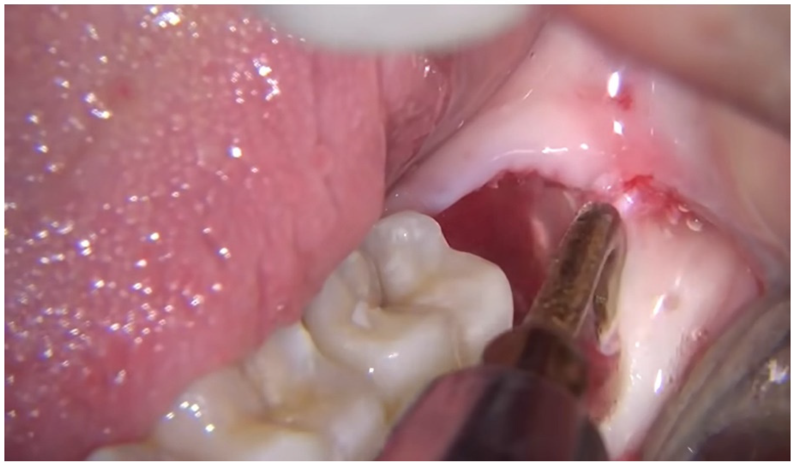
The patients were allocated by simple random sampling using the SNOSE technique. Surgical removal with piezosurgery or rotary was randomly allocated with double-blinding of both the investigator and evaluator. The period between the surgeries in the same patient was 21 days. Their case history was recorded pre-operatively, and the blood was withdrawn for CRP level (baseline) and assessed using enzyme-linked immunosorbent assay (ELISA) [Spin React, Tulip Diagnostics]. These pre-operative quantitative values were considered baseline values. The 25 patients underwent surgical removal of the third molar by rotatory bur on one side and piezo on the other side, with a gap of 21 days between the surgeries. Using local anesthesia, the third molar (
Figure 1) was surgically removed using the above-mentioned bone-cutting methods. Piezosurgery was conducted with a DMETEC surgystar with EXO1, EXO2, and EXO3. The rotatory osteotomy technique was done using a No. 10 bur and 703 SS white bur. Immediately post-surgery, a second blood sample was obtained. The CRP levels of these samples were estimated by ELISA. The third and fourth blood samples were collected at 24 and 72 h postoperatively using ELISA for the quantitative analysis of CRP. The Visual Analog Scale (VAS), which was a subjective evaluation, was used to assess postoperative pain at 24 h and 72 h. The patients were given the same anti-inflammatory drug (ibuprofen 400 mg) in both procedures for 3 days (once every 8 h) after the surgical procedure. The patients were followed for postoperative follow-up to check for postoperative inflammation in both groups. The statistical analysis was done with a paired
t-test and ANOVA.
Statistical analysis: The obtained data were compiled on an MS Office Excel Sheet (v 2010, Microsoft Office, Washington, DC, USA) and subjected to statistical analysis using a Statistical Package for the Social Sciences (SPSS v 21.0, IBM, Chicago, IL, USA). Intragroup comparison of VAS across various time intervals was assessed using a paired t-test, while CRP was assessed using repeated-measures ANOVA. An intergroup comparison of VAS and CRP between both groups was done using a t-test. For all the statistical tests, p < 0.05 was considered statistically significant, keeping α error at 5% and β error at 20%, thus designating 80% power to the study.
3. Results
The average durations of the piezo and rotary bur techniques were 50 min and 25 min, respectively.
Intra-group comparison: CRP value within the piezo group showed a difference from immediate post-op to 24 h, and 72 h post-op, although the difference was not statistically significant (
p > 0.05). The CRP value within the rotatory group showed a statistically significant difference (
p < 0.01) from immediate post-op to 24 h, and 72 h post-op (
Table 1). The VAS values within the piezo and rotatory groups showed a statistically significant difference (
p < 0.01) at postoperative 24 h and 72 h (
Table 2).
Inter-group comparison: CRP value in the piezo group was lower than in the rotary group, although the difference was not statistically significant (
p > 0.05). The VAS value in the piezo group was significantly lower (
p < 0.01) than in the rotatory group both post-op 24 h and 72 h (
Table 3).
4. Discussion
The past decade has seen a surge in the number of innovative surgical tools and technology being introduced to decrease the surgical difficulty and postoperative morbidity associated with third molar surgery. An example of such an innovation is piezosurgery or the use of ultrasonic piezoelectric vibrations for relatively safer and precise osteotomies [
20]. This method helps to perform precise osteotomy in areas even close to vital and important facial structures, including vessels and nerves [
21]. The present study employed a split-mouth research design. The advantage of this research design is that it eliminates the patient’s compliance bias from the evaluated treatment effect, as elicited by Zhu et al. [
22]. To standardize the results, the authors selected patients who had mandibular third molar impacted in a mesioangular class II position B (Pell and Gregory classification) [
23]. The impaction selection made in the present study was based on the relatively higher frequency of the mesioangular class II position and the fact that there is published literature using a similar research design [
20,
24], which, in turn, would allow reliable comparisons. The statistics indicate that the value of CRP post-surgery is always more than the preoperative value, which is on par with Chander et al. [
25], Desai et al. [
26], and Hao Shen et al. [
27]. This postoperative increase in the blood CRP (acute-phase protein) is a direct result of the local manipulation of the soft tissue and flap during the surgical extraction of impacted mandibular third molar procedures.
Maria et al. [
28] and Sanchis et al. [
29] observed that the duration of the surgical procedure was directly related to postoperative tissue reactions and hence increased CRP. This is contrary to the present study results wherein the surgical technique with a longer time duration (piezo-average 50 min) showed lower CRP levels than the technique with a relatively short time duration (rotary-average 25 min). The conflicting data obtained could be explained by Ohzato et al. [
30], who attributed the stimulation of inflammatory mediators to the degree of tissue destruction and the type of tissue. Thus, the higher CRP values obtained in the rotatory group could be a direct reflection of the amount of tissue destruction and the resulting influx of inflammatory mediators rather than surgical time. Calvo AM et al. and Larsen MK et al. also reported that CRP levels were not dependent on the duration of surgery. Smaller incisions, healing by primary intention, and the use of anti-inflammatory drugs were reported to be the major factors associated with low levels of CRP, while surgery-induced infection/aseptic traumatic inflammation was associated with increased CRP levels [
31,
32,
33,
34]. Bulut et al. [
32] and El-Sharrawy [
35] provided further evidence for the role of postoperative inflammation in increasing CRP levels, with the latter reporting a strong association between CRP levels and treatment response. Given the close association between CRP and postoperative morbidity, Lizuka and Lindqvist assessed the potential use of CRP as an early indicator of infection/aseptic inflammation. They reported that CRP was a relatively better indicator of inflammation than the erythrocyte sedimentation rate. Based on the abovementioned studies, CRP levels could be a vital tool for monitoring potential postoperative infection/aseptic inflammation following third molar surgery [
32,
35,
36]. Also, a higher pre-operative CRP level could indicate a risk for prolonged severe postoperative inflammation. Thus, CRP recorded at the baseline (pretreatment) could be used to stratify the patient as high or low risk for potential postoperative morbidity. Prophylactic anti-inflammatory regimens could be prescribed according to CRP-based risk stratification [
37].
The present study also evaluated postoperative pain. Published studies comparing post-surgical pain in piezosurgery and conventional rotatory surgery for mandibular third molar removal reported a relatively better outcome in the former [
20,
38,
39,
40,
41]. Although piezoelectric surgery increased the treatment duration, it had a positive impact on the patients’ operative experience and postoperative clinical sequelae. The time taken for the piezosurgery group was more than for the rotatory group. The postoperative inflammation assessed clinically with swelling was less on the fifth postoperative day compared to the rotatory group. The trismus also showed a similar pattern in presentation in comparison. The time taken, however, was greater in the piezo group compared to the rotary group. There was a difference of 30 min more in the piezo group compared to the rotary group. This is on par with the present study, wherein postoperative pain (as recorded in the VAS) was higher in the rotary group than in the piezo group. This is the only landmark study that has worked on the quantitative analysis of C-reactive protein in patients undergoing two different surgical techniques to make a basis for scientific quantification. The inference of the study was that quantitative analysis of Crp was an excellent parameter for evaluating the use of piezosurgery in comparison to rotatory osteotomy. The CRP values, which were lower in Group A, were suggestive that it has a role in reducing inflammation when the surgery was done with piezosurgery. The ELISA technique of evaluation is a single quantitative method for grading the amount of inflammation when we are comparing two different methods of surgical removal of impacted third molars.
5. Conclusions
The results of the present study showed an increase in the blood CRP levels compared to the pre-operative values, although the rise dropped immediately at 24 h for the piezo group. In contrast, the CRP levels remained high even at 24 h in the rotary group. The rise in the C-reactive protein can be largely attributed to surgery-induced postoperative inflammation. The ability of piezo-based surgical interventions to spare the surrounding soft tissue could be the reason for the relatively lower CRP and VAS levels compared to the rotary group. Further multi-center studies with a larger sample size and longer follow-up duration are required to validate the findings of the present study.
Author Contributions
Conceptualization, L.S. and K.G.; methodology, U.L.; software, S.B. (Shilpa Bhandi); validation, M.M.H.B., S.B. (Swati Bharadwaj), and A.A.; formal analysis, A.T.R.; investigation, R.R. and K.G.; resources, S.B. (Swati Bharadwaj); data curation, S.B. (Shilpa Bhandi); writing—original draft preparation, L.S., R.R. and K.G.; writing—review and editing, L.T., R.R. and S.P.; visualization, L.T.; supervision, L.T.; project administration, S.P. All authors have read and agreed to the published version of the manuscript.
Funding
This research received no external funding.
Institutional Review Board Statement
Dr. D. Y. Patil Vidyapeeth’s institutional ethics committee approval (DPU/Research/159(7)/2013) was obtained.
Informed Consent Statement
Informed consent was obtained from all individual participants included in the study.
Data Availability Statement
No data was used to support this study.
Acknowledgments
The study was supported by Dr. D. Y. Patil Dental College and Hospital (research proposal no—DR-1—2012–2013), D. Y. Patil Vidyapeeth, Pune, India.
Conflicts of Interest
The authors declare no conflict of interest.
References
- Nørholt, S.E. Treatment of acute pain following removal of mandibular third molars: Use of the dental pain model in pharmacological research and development of a comparable animal model. Int. J. Oral Maxillofac. Surg. 1998, 27, 1–41. [Google Scholar] [CrossRef]
- Esen, E.; Taşar, F.; Akhan, O. Determination of the anti-inflammatory effects of methylprednisolone on the sequelae of third molar surgery. J. Oral Maxillofac. Surg. 1999, 57, 1201–1206. [Google Scholar] [CrossRef]
- Blondeau, F.; Daniel, N.G. Extraction of impacted mandibular third molars: Postoperative complications and their risk factors. J. Can. Dent. Assoc. 2007, 73, 325. [Google Scholar] [PubMed]
- Bouloux, G.F.; Steed, M.B.; Perciaccante, V.J. Complications of Third Molar Surgery. Oral Maxillofac. Surg. Clin. 2007, 19, 117–128. [Google Scholar] [CrossRef]
- Stahl, W.M. Acute phase protein response to tissue injury. Crit. Care Med. 1987, 15, 545–550. [Google Scholar] [CrossRef]
- Giannoudis, P.V.; Smith, M.R.; Evans, R.T.; Bellamy, M.C.; Guillou, P.J. Serum CRP and IL-6 levels after trauma: Not predictive of septic complications in 31 patients. Acta Orthop. Scand. 1998, 69, 184–188. [Google Scholar] [CrossRef] [Green Version]
- Macy, E.M.; Hayes, T.E.; Tracy, R.P. Variability in the measurement of C-reactive protein in healthy subjects: Implications for reference intervals and epidemiological applications. Clin. Chem. 1997, 43, 52–58. [Google Scholar] [CrossRef] [Green Version]
- Black, S.; Kushner, I.; Samols, D. C-reactive protein. J. Biol. Chem. 2004, 279, 48487–48490. [Google Scholar] [CrossRef] [Green Version]
- Stürmer, T.; Raum, E.; Buchner, M.; Gebhardt, K.; Schiltenwolf, M.; Richter, W.; Brenner, H. Pain and high sensitivity C reactive protein in patients with chronic low back pain and acute sciatic pain. Ann. Rheum. Dis. 2005, 64, 921–925. [Google Scholar] [CrossRef] [Green Version]
- Catuna, M.C. Sonic energy: A possible dental application. Preliminary report of an ultrasonic cutting method. Ann. Dent. 1953, 12, 256–260. [Google Scholar]
- Horton, J.E.; Tarpley, T.M.; Jacoway, J.R. Clinical applications of ultrasonic instrumentation in the surgical removal of bone. Oral Surg Oral Med. Oral Pathol. 1981, 51, 236–242. [Google Scholar] [CrossRef]
- Vercellotti, T.; Nevins, M.L.; Kim, D.M.; Nevins, M.; Wada, K.; Schenk, R.K.; Fiorellini, J.P. Osseous response following resective therapy with piezosurgery. Int. J. Periodontics Restor. Dent. 2005, 25, 543–549. [Google Scholar]
- Chiriac, G.; Herten, M.; Schwarz, F.; Rothamel, D.; Becker, J. Autogenous bone chips: Influence of a new piezoelectric device (Piezosurgery®) on chip morphology, cell viability and differentiation. J. Clin. Periodontol. 2005, 32, 994–999. [Google Scholar] [CrossRef] [PubMed]
- Walsh, B.L.J. Piezosurgery: An increasing role in dental hard tissue surgery. Australas. Dent. Pract. 2007, 18, 52–56. [Google Scholar]
- Wallace, S.S.; Mazor, Z.; Froum, S.J.; Cho, S.-C.; Tarnow, D.P. Schneiderian membrane perforation rate during sinus elevation using piezosurgery: Clinical results of 100 consecutive cases. Int. J. Periodontics Restor. Dent. 2007, 27, 413–419. [Google Scholar]
- Happe, A. Use of a piezoelectric surgical device to harvest bone grafts from the mandibular ramus: Report of 40 cases. Int. J. Periodontics Restor. Dent. 2007, 27, 241–249. [Google Scholar]
- Vercellotti, T. Piezoelectric surgery in implantology: A case report--a new piezoelectric ridge expansion technique. Int. J. Periodontics Restor. Dent. 2000, 20, 358–365. [Google Scholar]
- Robiony, M.; Polini, F.; Costa, F.; Zerman, N.; Politi, M. Ultrasonic bone cutting for surgically assisted rapid maxillary expansion (SARME) under local anaesthesia. Int. J. Oral Maxillofac. Surg. 2007, 36, 267–269. [Google Scholar] [CrossRef]
- Sortino, F.; Pedullà, E.; Masoli, V. The Piezoelectric and Rotatory Osteotomy Technique in Impacted Third Molar Surgery: Comparison of Postoperative Recovery. J. Oral Maxillofac. Surg. 2008, 66, 2444–2448. [Google Scholar] [CrossRef]
- Piersanti, L.; Dilorenzo, M.; Monaco, G.; Marchetti, C. Piezosurgery or conventional rotatory instruments for inferior third molar extractions? J. Oral Maxillofac. Surg. 2014, 72, 1647–1652. [Google Scholar] [CrossRef]
- Crosetti, E.; Battiston, B.; Succo, G. Piezosurgery in head and neck oncological and reconstructive surgery: Personal experience on 127 cases. Acta Otorhinolaryngol. Ital. 2009, 29, 1–9. [Google Scholar] [PubMed]
- Zhu, H.; Zhang, S.; Sample, C. Sample size considerations for split-mouth design. Stat. Methods Med. Res. 2017, 26, 2543–2551. [Google Scholar] [CrossRef] [PubMed]
- Pell, G.J.; Gregory, G.T. Impacted mandibular third molars: Classification and modified technique for removal. Dent. Dig. 1933, 39, 330–338. [Google Scholar]
- Goyal, M.; Marya, K.; Jhamb, A.; Chawla, S.; Sonoo, P.R.; Singh, V.; Aggarwal, A. Comparative evaluation of surgical outcome after removal of impacted mandibular third molars using a Piezotome or a conventional handpiece: A prospective study. Br. J. Oral Maxillofac. Surg. 2012, 50, 556–561. [Google Scholar] [CrossRef]
- Chander, P.M.; Ali, F.M.; Aher, V. C-reactive protein a better indicator of inflammation after third molar extraction. Niger. J. Clin. Pract. 2013, 16, 297–301. [Google Scholar] [PubMed]
- Kiran, D.N.; Desai, R. Estimation of C-reactive Protein Associated with Mandibular Fracture. J. Maxillofac. Oral Surg. 2012, 11, 67–71. [Google Scholar] [CrossRef] [PubMed] [Green Version]
- Shen, H.; Zhang, N.; Zhang, X.; Ji, W. C-reactive protein levels after 4 types of arthroplasty. Acta Orthop. 2009, 80, 330–333. [Google Scholar] [CrossRef] [Green Version]
- Maria, A.; Malik, M.; Virang, P. Comparison of Primary and Secondary Closure of the Surgical Wound After Removal of Impacted Mandibular Third Molars. J. Maxillofac. Oral Surg. 2012, 11, 276–283. [Google Scholar] [CrossRef] [Green Version]
- Bielsa, J.M.S.; Bazán, S.H.; Diago, M.P. Flap repositioning versus conventional suturing in third molar surgery. Med. Oral Patol. Oral Cir. Bucal 2008, 13, 138–142. [Google Scholar]
- Ohzato, H.; Yoshizaki, K.; Nishimoto, N.; Ogata, A.; Tagoh, H.; Monden, M.; Gotoh, M.; Kishimoto, T.; Mori, T. Interleukin-6 as a new indicator of inflammatory status: Detection of serum levels of interleukin-6 and C-reactive protein after surgery. Surgery 1992, 111, 201–209. [Google Scholar]
- Guarnieri, R.; Miccoli, G.; Reda, R.; Mazzoni, A.; Di Nardo, D.; Testarelli, L. Sulcus fluid volume, IL-6, and Il-1b concentrations in periodontal and peri-implant tissues comparing machined and laser-microtextured collar/abutment surfaces during 12 weeks of healing: A split-mouth RCT. Clin. Oral Implant. Res. 2022, 33, 94–104. [Google Scholar] [CrossRef] [PubMed]
- Bulut, E.; Bulut, S.; Etikan, I.; Koseoglu, O. The value of routine antibiotic prophylaxis in mandibular third molar surgery: Acute-phase protein levels as indicators of infection. J. Oral Sci. 2001, 43, 117–122. [Google Scholar] [CrossRef] [PubMed] [Green Version]
- Calvo, A.M.; Brozoski, D.T.; Giglio, F.P.M.; Gonçalves, P.Z.; Sant’Ana, E.; Dionísio, T.J.; Lauris, J.R.P.; Santos, C.F. Are antibiotics necessary after lower third molar removal? Oral Surg. Oral Med. Oral Pathol. Oral Radiol. 2012, 114 (Suppl. S5), 199–208. [Google Scholar] [CrossRef] [PubMed]
- Larsen, M.K.; Kofod, T.; Duch, K.; Starch-Jensen, T. Short-term Haematological Parameters Following Surgical Removal of Mandibular Third Molars with Different Doses of Methylprednisolone Compared with Placebo: A Randomized Controlled Trial. J. Oral Maxillofac. Res. 2020, 11, 3–5. [Google Scholar] [CrossRef] [PubMed]
- El-Sharrawy, E.A.; El-Hakim, I.E.; Sameeh, E. Attenuation of C-reactive protein increases after exodontia by tramadol and ibuprofen. Anesth. Prog. 2006, 53, 78–82. [Google Scholar] [CrossRef]
- Iizuka, T.; Lindqvist, C. Changes in C-reactive protein associated with surgical treatment of mandibular fractures. J. Oral Maxillofac. Surg. 1991, 49, 464–467. [Google Scholar] [CrossRef]
- Hashish, I.; Harvey, W.; Harris, M. Anti-inflammatory effects of ultrasound therapy: Evidence for a major placebo effect. Rheumatology 1986, 25, 77–81. [Google Scholar] [CrossRef]
- Mantovani, E.; Arduino, P.G.; Schierano, G.; Ferrero, L.; Gallesio, G.; Mozzati, M.; Russo, A.; Scully, C.; Carossa, S. A split-mouth randomized clinical trial to evaluate the performance of Piezosurgery compared with traditional technique in lower wisdom tooth removal. J. Oral Maxillofac. Surg. 2014, 72, 1890–1897. [Google Scholar] [CrossRef]
- Pappalardo, S.; Guarnieri, R. Randomized clinical study comparing piezosurgery and conventional rotatory surgery in mandibular cyst enucleation. J. Cranio-Maxillofac. Surg. 2014, 42, e80–e85. [Google Scholar] [CrossRef]
- Mozzati, M.; Gallesio, G.; Russo, A.; Staiti, G.; Mortellaro, C. Third-molar extraction with ultrasound bone surgery: A case-control study. J. Craniofacial Surg. 2014, 25, 856–859. [Google Scholar] [CrossRef]
- Jiang, Q.; Qiu, Y.; Yang, C.; Yang, J.; Chen, M.; Zhang, Z. Piezoelectric versus conventional rotary techniques for impacted third molar extraction: A meta-analysis of randomized controlled trials. Medicine 2015, 94, e1685. [Google Scholar] [CrossRef] [PubMed]
| Publisher’s Note: MDPI stays neutral with regard to jurisdictional claims in published maps and institutional affiliations. |
© 2022 by the authors. Licensee MDPI, Basel, Switzerland. This article is an open access article distributed under the terms and conditions of the Creative Commons Attribution (CC BY) license (https://creativecommons.org/licenses/by/4.0/).


 ,
, 


