
Sperm Selection and Embryo Development: A Comparison of the Density Gradient Centrifugation and Microfluidic Chip Sperm Preparation Methods in Patients with Astheno-Teratozoospermia

 ,
,
Abstract
:1. Introduction
2. Materials and Methods
2.1. Patient Selection
2.2. Ovarian Stimulation
2.3. Semen Analysis
2.4. Sperm Preparation Using the Density Gradient Centrifugation Method
2.5. Sperm Preparation Using a Microfluidic Sorting Chip
2.6. Embryo Development
2.7. Statistics
3. Results
4. Discussion
5. Conclusions
Supplementary Materials
Author Contributions
Funding
Institutional Review Board Statement
Informed Consent Statement
Data Availability Statement
Acknowledgments
Conflicts of Interest
References
- Agarwal, A.; Baskaran, S.; Parekh, N.; Cho, C.-L.; Henkel, R.; Vij, S.; Arafa, M.; Panner Selvam, M.K.; Shah, R. Male infertility. Lancet 2021, 397, 319–333. [Google Scholar] [CrossRef]
- Neri, Q.V.; Lee, B.; Rosenwaks, Z.; Machaca, K.; Palermo, G.D. Understanding fertilization through intracytoplasmic sperm injection (ICSI). Cell Calcium 2014, 55, 24–37. [Google Scholar] [CrossRef] [Green Version]
- Cardona Barberán, A.; Boel, A.; Vanden Meerschaut, F.; Stoop, D.; Heindryckx, B. Diagnosis and Treatment of Male Infertility-Related Fertilization Failure. J. Clin. Med. 2020, 9, 3899. [Google Scholar] [CrossRef]
- Simopoulou, M.; Gkoles, L.; Bakas, P.; Giannelou, P.; Kalampokas, T.; Pantos, K.; Koutsilieris, M. Improving ICSI: A review from the spermatozoon perspective. Syst. Biol. Reprod. Med. 2016, 62, 359–371. [Google Scholar] [CrossRef] [Green Version]
- Parinaud, J.; Mieusset, R.; Vieitez, G.; Labal, B.; Richoilley, G. Influence of sperm parameters on embryo quality. Fertil. Steril. 1993, 60, 888–892. [Google Scholar] [CrossRef]
- Piccolomini, M.M.; Bonetti, T.C.; Motta, E.L.; Serafini, P.C.; Alegretti, J.R. How general semen quality influences the blastocyst formation rate: Analysis of 4205 IVF cycles. JBRA Assist. Reprod. 2018, 22, 89–94. [Google Scholar] [CrossRef] [PubMed]
- Colaco, S.; Sakkas, D. Paternal factors contributing to embryo quality. J. Assist. Reprod. Genet. 2018, 35, 1953–1968. [Google Scholar] [CrossRef]
- Oseguera-López, I.; Ruiz-Díaz, S.; Ramos-Ibeas, P.; Pérez-Cerezales, S. Novel Techniques of Sperm Selection for Improving IVF and ICSI Outcomes. Front. Cell Dev. Biol. 2019, 7, 298. [Google Scholar] [CrossRef]
- Pousette, A.; Akerlöf, E.; Rosenborg, L.; Fredricsson, B. Increase in progressive motility and improved morphology of human spermatozoa following their migration through Percoll gradients. Int. J. Androl. 1986, 9. [Google Scholar] [CrossRef]
- Jeyendran, R.S.; Caroppo, E.; Rouen, A.; Anderson, A.; Puscheck, E. Selecting the most competent sperm for assisted reproductive technologies. Fertil. Steril. 2019, 111, 851–863. [Google Scholar] [CrossRef] [PubMed]
- Aitken, R.J.; Finnie, J.M.; Muscio, L.; Whiting, S.; Connaughton, H.S.; Kuczera, L.; Rothkirch, T.B.; De Iuliis, G.N. Potential importance of transition metals in the induction of DNA damage by sperm preparation media. Hum. Reprod. 2014, 29, 2136–2147. [Google Scholar] [CrossRef] [PubMed]
- Muratori, M.; Tarozzi, N.; Carpentiero, F.; Danti, S.; Perrone, F.M.; Cambi, M.; Casini, A.; Azzari, C.; Boni, L.; Maggi, M.; et al. Sperm selection with density gradient centrifugation and swim up: Effect on DNA fragmentation in viable spermatozoa. Sci. Rep. 2019, 9, 7492. [Google Scholar] [CrossRef]
- Zini, A.; Finelli, A.; Phang, D.; Jarvi, K. Influence of semen processing technique on human sperm DNA integrity. Urology 2000, 56, 1081–1084. [Google Scholar] [CrossRef]
- Malvezzi, H.; Sharma, R.; Agarwal, A.; Abuzenadah, A.M.; Abu-Elmagd, M. Sperm quality after density gradient centrifugation with three commercially available media: A controlled trial. Reprod. Biol. Endocrinol. 2014, 12, 121. [Google Scholar] [CrossRef] [Green Version]
- Simon, L.; Zini, A.; Dyachenko, A.; Ciampi, A.; Carrell, D.T. A systematic review and meta-analysis to determine the effect of sperm DNA damage on in vitro fertilization and intracytoplasmic sperm injection outcome. Asian J. Androl. 2017, 19, 80–90. [Google Scholar] [CrossRef]
- Zini, A.; Boman, J.M.; Belzile, E.; Ciampi, A. Sperm DNA damage is associated with an increased risk of pregnancy loss after IVF and ICSI: Systematic review and meta-analysis. Hum. Reprod. 2008, 23, 2663–2668. [Google Scholar] [CrossRef] [PubMed] [Green Version]
- Zhao, J.; Zhang, Q.; Wang, Y.; Li, Y. Whether sperm deoxyribonucleic acid fragmentation has an effect on pregnancy and miscarriage after in vitro fertilization/intracytoplasmic sperm injection: A systematic review and meta-analysis. Fertil. Steril. 2014, 102, 998–1005.e1008. [Google Scholar] [CrossRef] [PubMed]
- Weng, L. IVF-on-a-Chip: Recent Advances in Microfluidics Technology for In Vitro Fertilization. SLAS Technol. 2019, 24, 373–385. [Google Scholar] [CrossRef]
- Kashaninejad, N.; Shiddiky, M.J.A.; Nguyen, N.-T. Advances in Microfluidics-Based Assisted Reproductive Technology: From Sperm Sorter to Reproductive System-on-a-Chip. Adv. Biosyst. 2018, 2, 1700197. [Google Scholar] [CrossRef]
- Alias, A.B.; Huang, H.-Y.; Yao, D.-J. A Review on Microfluidics: An Aid to Assisted Reproductive Technology. Molecules 2021, 26, 4354. [Google Scholar] [CrossRef]
- Chinnasamy, T.; Kingsley, J.L.; Inci, F.; Turek, P.J.; Rosen, M.P.; Behr, B.; Tüzel, E.; Demirci, U. Guidance and Self-Sorting of Active Swimmers: 3D Periodic Arrays Increase Persistence Length of Human Sperm Selecting for the Fittest. Adv. Sci. (Weinh) 2017, 5, 1700531. [Google Scholar] [CrossRef]
- Cho, B.S.; Schuster, T.G.; Zhu, X.; Chang, D.; Smith, G.D.; Takayama, S. Passively Driven Integrated Microfluidic System for Separation of Motile Sperm. Anal. Chem. 2003, 75, 1671–1675. [Google Scholar] [CrossRef] [PubMed]
- Nosrati, R.; Vollmer, M.; Eamer, L.; San Gabriel, M.C.; Zeidan, K.; Zini, A.; Sinton, D. Rapid selection of sperm with high DNA integrity. Lab. Chip 2014, 14, 1142–1150. [Google Scholar] [CrossRef] [PubMed]
- Phiphattanaphiphop, C.; Leksakul, K.; Phatthanakun, R.; Khamlor, T. A novel microfluidic chip-based sperm-sorting device constructed using design of experiment method. Sci. Rep. 2020, 10, 17143. [Google Scholar] [CrossRef]
- Schuster, T.G.; Cho, B.; Keller, L.M.; Takayama, S.; Smith, G.D. Isolation of motile spermatozoa from semen samples using microfluidics. Reprod. Biomed. Online 2003, 7, 75–81. [Google Scholar] [CrossRef]
- Zhang, Y.; Xiao, R.R.; Yin, T.; Zou, W.; Tang, Y.; Ding, J.; Yang, J. Generation of Gradients on a Microfluidic Device: Toward a High-Throughput Investigation of Spermatozoa Chemotaxis. PLoS ONE 2015, 10, e0142555. [Google Scholar] [CrossRef] [PubMed]
- Tasoglu, S.; Safaee, H.; Zhang, X.; Kingsley, J.L.; Catalano, P.N.; Gurkan, U.A.; Nureddin, A.; Kayaalp, E.; Anchan, R.M.; Maas, R.L.; et al. Exhaustion of racing sperm in nature-mimicking microfluidic channels during sorting. Small 2013, 9, 3374–3384. [Google Scholar] [CrossRef] [PubMed] [Green Version]
- Quinn, M.M.; Jalalian, L.; Ribeiro, S.; Ona, K.; Demirci, U.; Cedars, M.I.; Rosen, M.P. Microfluidic sorting selects sperm for clinical use with reduced DNA damage compared to density gradient centrifugation with swim-up in split semen samples. Hum. Reprod. 2018, 33, 1388–1393. [Google Scholar] [CrossRef] [PubMed] [Green Version]
- Asghar, W.; Velasco, V.; Kingsley, J.L.; Shoukat, M.S.; Shafiee, H.; Anchan, R.M.; Mutter, G.L.; Tüzel, E.; Demirci, U. Selection of functional human sperm with higher DNA integrity and fewer reactive oxygen species. Adv. Healthc. Mater. 2014, 3, 1671–1679. [Google Scholar] [CrossRef] [PubMed]
- Pujol, A.; García-Peiró, A.; Ribas-Maynou, J.; Lafuente, R.; Mataró, D.; Vassena, R. A microfluidic sperm-sorting device reduces the proportion of sperm with double-stranded DNA fragmentation. Zygote FirstView 2021, 27. [Google Scholar] [CrossRef]
- Ogata, K.; Nagata, M.P.B.; Nishizono, H.; Yamanouchi, T.; Matsuda, H.; Ogata, Y.; Takeda, K.; Hashiyada, Y.; Yamashita, K. In vitro survival kinetics of microfluidic-sorted bovine spermatozoa. Andrology 2021, 9, 977–988. [Google Scholar] [CrossRef]
- Anbari, F.; Khalili, M.A.; Sultan Ahamed, A.M.; Mangoli, E.; Nabi, A.; Dehghanpour, F.; Sabour, M. Microfluidic sperm selection yields higher sperm quality compared to conventional method in ICSI program: A pilot study. Syst. Biol. Reprod. Med. 2021, 67. [Google Scholar] [CrossRef] [PubMed]
- Veeck, L.; Zaninovic, N. An Atlas of Human Blastocysts; Informa Healthcare: New York, NY, USA; London, UK, 2003. [Google Scholar]
- Gardner, D.K.; Schoolcraft, W.B. Culture and transfer of human blastocysts. Curr. Opin. Obstet. Gynecol. 1999, 11, 307–311. [Google Scholar] [CrossRef] [PubMed]
- Nikshad, A.; Aghlmandi, A.; Safaralizadeh, R.; Aghebati-Maleki, L.; Warkiani, M.E.; Khiavi, F.M.; Yousefi, M. Advances of microfluidic technology in reproductive biology. Life Sci. 2021, 265, 118767. [Google Scholar] [CrossRef] [PubMed]
- Gode, F.; Bodur, T.; Gunturkun, F.; Gurbuz, A.S.; Tamer, B.; Pala, I.; Isik, A.Z. Comparison of microfluid sperm sorting chip and density gradient methods for use in intrauterine insemination cycles. Fertil. Steril. 2019, 112, 842–848. [Google Scholar] [CrossRef]
- Gode, F.; Gürbüz, A.S.; Tamer, B.; Pala, I.; Isik, A.Z. The Effects of Microfluidic Sperm Sorting, Density Gradient and Swim-up Methods on Semen Oxidation Reduction Potential. Urol. J. 2020, 17, 397–401. [Google Scholar] [CrossRef] [PubMed]
- Shirota, K.; Yotsumoto, F.; Itoh, H.; Obama, H.; Hidaka, N.; Nakajima, K.; Miyamoto, S. Separation efficiency of a microfluidic sperm sorter to minimize sperm DNA damage. Fertil. Steril. 2016, 105, 315–321.e311. [Google Scholar] [CrossRef] [Green Version]
- Miller, J.E.; Smith, T.T. The effect of intracytoplasmic sperm injection and semen parameters on blastocyst development in vitro. Hum. Reprod. 2001, 16, 918–924. [Google Scholar] [CrossRef] [PubMed] [Green Version]
- Chapuis, A.; Gala, A.; Ferrières-Hoa, A.; Mullet, T.; Bringer-Deutsch, S.; Vintejoux, E.; Torre, A.; Hamamah, S. Sperm quality and paternal age: Effect on blastocyst formation and pregnancy rates. Basic Clin. Androl. 2017, 27, 2. [Google Scholar] [CrossRef] [Green Version]
- Bartolacci, A.; Pagliardini, L.; Makieva, S.; Salonia, A.; Papaleo, E.; Viganò, P. Abnormal sperm concentration and motility as well as advanced paternal age compromise early embryonic development but not pregnancy outcomes: A retrospective study of 1266 ICSI cycles. J. Assist. Reprod. Genet. 2018, 35, 1897–1903. [Google Scholar] [CrossRef]
- Borges, E., Jr.; Setti, A.S.; Braga, D.P.; Figueira, R.C.; Iaconelli, A., Jr. Total motile sperm count has a superior predictive value over the WHO 2010 cut-off values for the outcomes of intracytoplasmic sperm injection cycles. Andrology 2016, 4, 880–886. [Google Scholar] [CrossRef]
- Hardarson, T.; Van Landuyt, L.; Jones, G. The blastocyst. Hum. Reprod. 2012, 27, i72–i91. [Google Scholar] [CrossRef] [Green Version]
- Gardner, D.K.; Balaban, B. Assessment of human embryo development using morphological criteria in an era of time-lapse, algorithms and ‘OMICS’: Is looking good still important? Mol. Hum. Reprod. 2016, 22, 704–718. [Google Scholar] [CrossRef]
- Yildiz, K.; Yuksel, S. Use of microfluidic sperm extraction chips as an alternative method in patients with recurrent in vitro fertilisation failure. J. Assist. Reprod. Genet. 2019, 36, 1423–1429. [Google Scholar] [CrossRef] [PubMed]
- Avendaño, C.; Oehninger, S. DNA fragmentation in morphologically normal spermatozoa: How much should we be concerned in the ICSI era? J. Androl. 2011, 32, 356–363. [Google Scholar] [CrossRef] [PubMed]
- Yalcinkaya Kalyan, E.; Can Celik, S.; Okan, O.; Akdeniz, G.; Karabulut, S.; Caliskan, E. Does a microfluidic chip for sperm sorting have a positive add-on effect on laboratory and clinical outcomes of intracytoplasmic sperm injection cycles? A sibling oocyte study. Andrologia 2019, 51, e13403. [Google Scholar] [CrossRef]
- Ozcan, P.; Takmaz, T.; Yazici, M.G.K.; Alagoz, O.A.; Yesiladali, M.; Sevket, O.; Ficicioglu, C. Does the use of microfluidic sperm sorting for the sperm selection improve in vitro fertilization success rates in male factor infertility? J. Obstet. Gynaecol. Res. 2021, 47, 382–388. [Google Scholar] [CrossRef] [PubMed]
- Smith, G.D.; Cantatore, C.; Ohl, D.A. Microfluidic Systems for Isolation of Spermatozoa from Testicular Specimens of Non-Obstructive Azoospermic Men: Does/Can It Improve Sperm Yield? J. Clin. Med. 2021, 10, 3667. [Google Scholar] [CrossRef] [PubMed]
- Neculai-Valeanu, A.S.; Ariton, A.M. Game-Changing Approaches in Sperm Sex-Sorting: Microfluidics and Nanotechnology. Animal 2021, 11, 1182. [Google Scholar] [CrossRef] [PubMed]


| Basal | |
|---|---|
| Semen volume (mL) | 3.18 ± 1.48 |
| Sperm concentration (106/mL) | 40.63 ± 42.00 |
| Total motility (%) | 33.14 ± 14.47 |
| Progressive motility (%) | 12.59 ± 8.33 |
| Morphology | 1.50 ± 0.67 |
| Microchip Group (n: 22) | Gradient Group (n: 22) | z/t | p Value | |||
|---|---|---|---|---|---|---|
| Mean ± SD | Median (Q1–Q3) | Mean ± SD | Median (Q1–Q3) | |||
| Volume of recovered sperm (mL) | 1.07 ± 0.15 | 1.00 (1.00–1.05) | 0.73 ± 0.08 | 0.70 (0.70–0.80) | −5.914 z | p < 0.0001 * |
| Sperm concentration (106/mL) | 4.37 ± 6.05 | 2.15 (0.70–5.25) | 19.80 ± 19.90 | 14.50 (6.25–28.00) | −3.911 z | p < 0.0001 * |
| Total motility (%) | 79.50 ± 17.19 | 81.00 (70.00–92.25) | 53.27 ± 24.32 | 55.00 (33.00–73.50) | −3.547 z | p < 0.0004 * |
| Progressive motility (%) | 68.41 ± 24.57 | 72.00 (54.00–90.00) | 31.73 ± 19.90 | 30.50 (17.25–42.75) | 5.441 t | p < 0.0001 * |
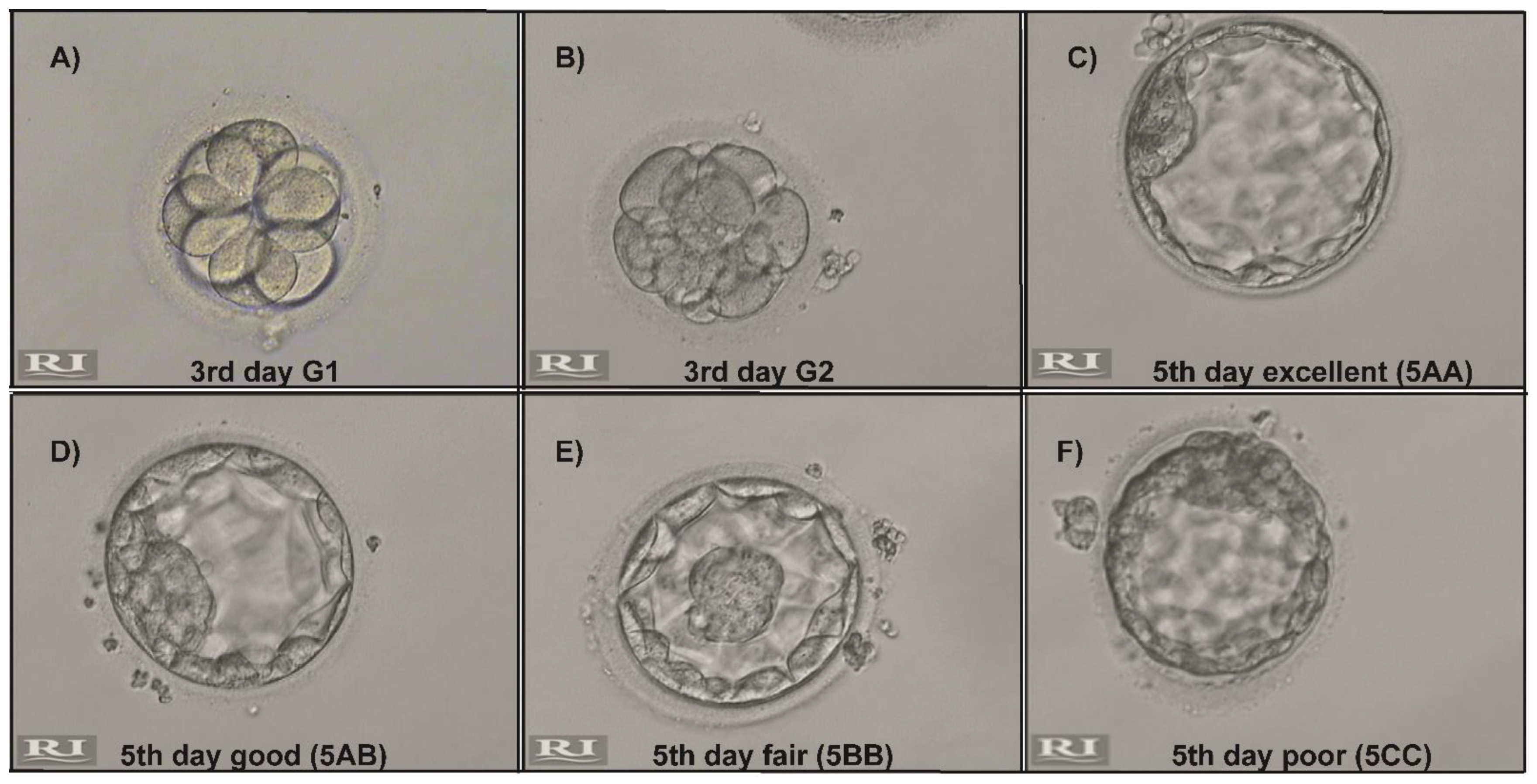
| Embryo Development | Microchip Group | Gradient Group | z | p-Value |
|---|---|---|---|---|
| 1st day fertilized (n: 186) | 0.89 ± 0.23 (n: 96) | 0.91 ± 0.16 (n: 90) | −0.214 | 0.966 |
| 3rd day total (n: 170) | 0.85 ± 0.26 (n: 89) | 0.83 ± 0.25 (n: 81) | −0.761 | 0.570 |
| 3rd day G1 (n: 144) | 0.83 ± 0.25 (n: 77) | 0.76 ± 0.31 (n: 67) | −0.235 | 0.625 |
| 3rd day G2 (n: 26) | 0.12 ± 0.23 (n: 12) | 0.13 ± 0.21 (n: 14) | −2.452 | 0.933 |
| 5th day total (n: 109) | 0.60 ± 0.31 (n: 62) | 0.47 ± 0.30 (n: 47) | −0.585 | 0.050 |
| 5th day excellent (n: 63) | 0.42 ± 0.28 (n: 41) | 0.23 ± 0.23 (n: 22) | −1.037 | 0.029 * |
| 5th day good (n: 18) | 0.10 ± 0.14 (n: 10) | 0.07 ± 0.12 (n: 8) | −1.497 | 0.471 |
| 5th day fair (n: 20) | 0.02 ± 0.06 (n: 8) | 0.01 ± 0.04 (n: 12) | −0.214 | 0.311 |
| 5th day poor (n: 8) | 0.07 ± 0.11 (n: 3) | 0.16 ± 0.19 (n: 5) | −0.761 | 0.104 |
Publisher’s Note: MDPI stays neutral with regard to jurisdictional claims in published maps and institutional affiliations. |
© 2021 by the authors. Licensee MDPI, Basel, Switzerland. This article is an open access article distributed under the terms and conditions of the Creative Commons Attribution (CC BY) license (https://creativecommons.org/licenses/by/4.0/).
Share and Cite
Guler, C.; Melil, S.; Ozekici, U.; Donmez Cakil, Y.; Selam, B.; Cincik, M. Sperm Selection and Embryo Development: A Comparison of the Density Gradient Centrifugation and Microfluidic Chip Sperm Preparation Methods in Patients with Astheno-Teratozoospermia. Life 2021, 11, 933. https://doi.org/10.3390/life11090933
Guler C, Melil S, Ozekici U, Donmez Cakil Y, Selam B, Cincik M. Sperm Selection and Embryo Development: A Comparison of the Density Gradient Centrifugation and Microfluidic Chip Sperm Preparation Methods in Patients with Astheno-Teratozoospermia. Life. 2021; 11(9):933. https://doi.org/10.3390/life11090933
Chicago/Turabian StyleGuler, Cagla, Sureyya Melil, Umit Ozekici, Yaprak Donmez Cakil, Belgin Selam, and Mehmet Cincik. 2021. "Sperm Selection and Embryo Development: A Comparison of the Density Gradient Centrifugation and Microfluidic Chip Sperm Preparation Methods in Patients with Astheno-Teratozoospermia" Life 11, no. 9: 933. https://doi.org/10.3390/life11090933