
Cytoskeletal Tensegrity in Microgravity

Independent Researcher, Glebe 2037, Australia
Life 2021, 11(10), 1091; https://doi.org/10.3390/life11101091
Submission received: 28 September 2021
/
Revised: 9 October 2021
/
Accepted: 14 October 2021
/
Published: 15 October 2021
(This article belongs to the Special Issue Space Flight Factors and Cytoskeleton Organization)
Abstract
:In order for Man to venture further into Space he will have to adapt to its conditions, including microgravity. Life as we know it has evolved on Earth with a substantial gravitational field. If they spend considerable time away from Earth, astronauts experience physiological, mental, and anatomical changes. It is not clear if these are pathological or adaptations. However, it is true that they experience difficulties on their return to stronger gravity. The cytoskeleton is a key site for the detection of gravitational force within the body, due to its tensegrity architecture. In order to understand what happens to living beings in space, we will need to unravel the role cytoskeletal tensegrity architecture plays in the building and function of cells, organs, the body, and mind.
“If we’re going to go to the moon again and we’re going to go to Mars and beyond, then we’ve got to get a little outside of our comfort zone” (Jared Isaacman in The Guardian 16th September 2021).
1. Definition
2. Tensegrity and the Universe including Life
Buckminster Fuller, the mathematician, first used the term “tensegrity” [2]. It should be noted that his student, the renowned artist Kenneth Snelson, preferred the term “floating compression”. Fuller thought that stars’ and planets’ compression elements were held in place by prestress tensional gravity. He also saw this—discontinuous compression, continuous tension—in atoms, electrons orbiting the nucleus and it all held together by subatomic attraction and repulsion. Thus, he saw that the Universe is a tensegrity fractal, with self-similarity at different scales. This enables a hierarchical nesting of living beings’ structure. Thus, the gravitational force acts at the whole organ or tissue level and is sensed at cellular and molecular levels. Indeed, gravity can lead to changes in many cellular functions, including proliferation, differentiation, signalling, and expression. Conversely, changes at the subatomic and molecular level can be translated into more macro-occurrences [3,4]. Just as gravity “prunes” all that is not part of a constructed building, biotensegrity “prunes” all that is not part of the great fractal continuum from micro to macro and vice versa, thus, acting as an ultimate proof-reading algorithm [5].
3. Tensegrity and the Body
Tensegrity lends itself to describing biomechanical joints like the elbow, whichare stable during shape changes due to “prestress”. Connective tissues integrate components into a single unit [6]. Indeed, the entire human frame with its many tensile components pulling on rigid bones and stabilising them against gravity, is tensegrity [1].
Furthermore, even human tensegrity architecture is part of this continuum. Richard Dawkins [7] makes the point that the phenotype of an organism is not limited to the actual body of that organism but extends to the ways in which the organism modifies its surrounding environment. Thus, a bird’s nest is the extended phenotype of a bird. A beaver’s dam is the extended phenotype of a beaver, and tensegrity-based architecture is the extended phenotype of humans. So, the human mind’s functioning is somehow based in tensegrity. “Embodiment” in robotics means that the robot body and its controller are not considered separate but that the interaction of the body with its environment is taken into account. This applies to tensegrity structures. Thus, the human brain may compute without need to invoke any special “soul” or atman because it is a tensegrity structure [8]. Interestingly, the nervous system can be seen as fractal, with hierarchical structure progressing from the micro to the macro and vice versa [9]. The brain is a tensegrity structure of its grey and white matters and fascia. These in turn are constructed of tensegrity assemblages of cells, then actin and MT cytoskeletons and attractive (tensional) and repulsive (compressive) forces between individual amino acids and so forth.
“The conception of a haptic medium as a nested tensegrity structure has been proposed to express the obtained information realized by myofascia deformation, by its invariants and transformations. The tensegrity proposal rationalizes the relative indifference of dynamic touch to the site of mechanical contact (hand, foot, torso or probe) and the overtness of exploratory activity. It also provides a framework for dynamic touching’s fractal nature, and the finding that its degree of fractality may matter to its accomplishments” [10]. Indeed.
4. Qualities of Tensegrity Structures: The Cytoskeleton
Tensegrity structures with discontinuous members are used both in terrestrial, outer-space, and morphing devices [11]. Composed of discontinuous struts and cables, these systems are only structurally stable when prestress is induced; otherwise, they lose their geometrical configuration (while keeping its topology) and, thus, can be tightly packed. This enables gigantic shape change, making such structures highly versatile and resilient [11]. These qualities also make nested hierarchies of tensegrity ideal for biological systems to create relatively large structures with a minimum of protein subunits. This includes cytoskeletal tensegrities.
Due to internal prestress in the cytoskeleton, cells take on a spherical shape when in suspension. However, if a cell is anchored to a resisting substrate, it spreads out taking on a flattened form. When cells are attached to a flexible substrate, it is pulled into compression wrinkles [12]. Both cell–cell and cell–extracellular matrix (ECM) adhesions have unique components, but both are linked to the cell’s actin cytoskeleton. Thus, mechanical and/or chemical stimuli are transferred and integrated to induce or control cell proliferation, migration, and differentiation [13]. Focal adhesions have hundreds of clustered integrins and other cytoplasmic proteins connecting with hundreds of actin and myosin (motor protein) proteins. This enables the force at a single focal adhesion to be tens of nN and the force can propagate along the cytoskeleton distances up to tens of µm. The size of a cell. The cytoskeleton mediating long distance force transmission in the cytoplasm has been shown in several cell types [14].
Actin is involved in mechanosensing through other pathways. In a process known as cytoskeletal strengthening, integrin-mediated cell–ECM adhesion is enhanced through interactions with cytoskeletal proteins, among which vinculin is important in stimulating actin polymerisation and recruiting actin remodelling proteins [15]. Cadherins are a large superfamily of calcium-dependent transmembrane adhesive proteins that are responsible for cell–cell adhesion in all soft tissues [16]. Extracellular regions of cadherin mediate the adhesion between neighbouring cells, while the cytoplasmic tails of cadherins interact with effector proteins, which connect them to the actin cytoskeleton [17].
The microtubule (MT) cytoskeleton can transmit mechanical signals and resist compression, thus, fulfilling its role as the compression-resistant component of subcellular cytoskeletal tensegrity. It is, along with the actin cytoskeleton, central to the perception of external forces by cells [18]. In contracting cardiomyocytes detyrosination of MTs (a post-translational modification of MTs) promoted buckling, stiffened the myocytes, and correlated with impaired function in cardiomyopathy. Thus, detyrosinated MTs represent crucial compression-resistant cytoskeletal components [19]. Importantly, MT post-translational modifications affect the binding of partner proteins, such as the motor proteins kinesins to the MT. MTs adhered to a two-dimensional substrate by MT-associated protein kinesin buckle when subjected to compression stress. The density of kinesin is found to play the key role in determining the buckling mode of MTs [20]. In fact, the cardiac MT network may act as a cytoskeletal shock-absorber [21].
5. Tensegrity and the Nervous System
The cytoskeletons of the brain and nervous systems may function as tensegrity systems. Nerve axons typically possess bundled MTs and the dendritic spines polymerised actin filaments. If MTs in the nerve-axon are in compression as in tensegrity, then pulling on the axon could reduce the compression. Normally, this is done by a growth cone but this could also be done by pulling. In either case, a decrease in the compression of the assembled MTs should favour MT assembly, and this is what is observed [22]. Various other models of the brain suggest the brain and nervous systems as tensegrity structures [23]. A fascial layer (prestressed structure) covers brain, spinal cord, and many nerves. The fascia mechanically connects neural tissue (compression-resistant structures) to the rest of the body [24]. Indeed, the brain even pulses, with the spinal cord acting as a piston [25], the force possibly dissipated through the brain’s cytoskeletal tensegrity network as heat.
6. Graviception and the Cytoskeleton
Cells probably sense changes in gravitational force through alterations in forces between their adhesions and the cytoskeleton or through force acting directly on the cytoskeleton, rather than through a “gravioreceptor” molecule. The entire cell or even entire tissue is a gravity sensor [26]. Local bending of the cytoskeleton is common to all cellular mechanotransduction. Mechanosensory cells experience gravitational force through dense organelles (e.g., otoliths, stereocilia). Other cells also feel gravitational force as cytoskeletal distortion, perhaps compression resistant elements (MT) tugging on prestressed tensional elements (actin filaments). Indeed, in human primary endothelial cells, there are alterations to the actin cytoskeleton under both hypogravity and hypergravity [27]. Another study found a collapse of the actin cytoskeleton of osteoblasts after four days of microgravity [28].
When organisms experience microgravity, they experience an acute decrease in pre-stress on the macro (tissue, organ) level, which, due to the hierarchical organisation of life, trickles down to produce corresponding changes in form and function at the cellular and molecular levels including an upregulation of cytoskeletal genes in cancer cells [29]. Cells derived from the nervous systems are affected. Under simulated microgravity, the adhesion of primary cells from human brain nervous tissue was decreased, accompanied by highly disorganized β-tubulin structures in a circular pattern around the nucleus [30]. In human SH-SY5Y neuroblastoma cells, weightlessness or changing gravity resulted in microtubular bending and loop formation [31].
The third class of cytoskeletal proteins, intermediate filaments, is also involved in the tensegrity architecture of human cells forming tensile cables that link the cell nucleus to its cortex [32]. There is evidence that intermediate filaments play a role in cells’ response to gravity (or lack thereof) [33].
7. Changes to Tissues in Microgravity
Simulated microgravity alters morphology, cytoskeletal organisation, and cellular motility in primary neurons as well as cytoskeleton protein expression in rat hypothalamus and mouse hippocampus and hypothalamus [34]. Neurons are in functionally involved in human adaptation to microgravity. After short exposures to simulated microgravity, neurons changed morphology followed by fast recovery on return to Earth surface gravity. Long exposure to microgravity resulted in possible adaptation of single neurons as well as neuronal networks. The latter was coupled to an increase of apoptosis. However, neurons and neuronal networks exposed for long-term to microgravity required longer recovery time to re-adapt to Earth surface gravity [35]. One possible irreversible change to the nervous system under microgravity is the hyperbranching of motor neurons [36].
Another change seen under microgravity is muscular atrophy. As mentioned above, muscles act as tensional elements in the musculoskeletal body. Thus, microgravity leads to the breakdown of this hierarchy of the body fractal. The muscle consists in a large part of actin filaments, which act in conjunction with myosin to provide the force of muscle contraction. Under microgravity, the expression of actin and myosin are altered [37] so changes in one iteration of the body fractal (cytoskeletal level) appear to have consequences at higher hierarchies. Bone is affected. Primary cilia act as microgravity sensors, depolymerising microtubules to inhibit osteolast differentiation and mineralisation [38].
Interestingly, one study found neither quantitative nor structural changes in the actin cytoskeleton of macrophages under microgravity [39]. The cells of the immune system are not directly involved in the cytoskeleton/tissue/organ hierarchies of fractal hierarchy. Thus, they may be spared the potentially negative effects that microgravity has upon these tensegrity-based systems.
8. Microgravity May Promote Mental Illness
Three of the most prevalent mental illnesses are depression, bipolar syndrome, and schizophrenia. Mental illnesses are not quite the same as neurological disorders, although there is certainly some overlap. Whereas with many neurological diseases it is possible to say THIS disorder is caused by THIS mutation and THIS process; in general mental (psychiatric) illnesses have a much more nebulous aetiology. Of course, there are exceptions, and I am sure many neurologists and psychiatrists would disagree! Machado-Joseph disease, despite its prevalence remains unsolved [40]; a gene for schizophrenia in a Scottish lineage, DISC1, has been identified [41].
In a previous paper [42], I suggested that depression, bipolar syndrome, and schizophrenia (and other conditions besides) may be malfunctions of the nervous system cytoskeletal tensegrity framework, particularly in the brain. Indeed, a tensegral understanding is beginning to be developed for treatments in allied health including osteopathy [43] and in oncological diagnosis [44]. In depression, the nervous system cytoskeleton may be too “floppy”, there might not be enough prestress, and in schizophrenia too “taut” with too much prestress. Bipolar syndrome is a condition that fluctuates between these extremes. There is certainly abundant evidence that the cytoskeleton plays a key role in mental health [45,46].
A number of psychiatric and psychological hurdles have arisen for astronauts [47]. In particular, there seems to be a tendency for them to develop anxiety- and depression-spectrum disorders [48]. These have typically been seen as psychological manifestations of loneliness, isolation, interpersonal stress. However, as I have laid out above, microgravity’s effect on the nervous system, in particular the nervous system cytoskeleton, may contribute here. Indeed, it seems to make some sense. Reduction in gravitational force leads to a reduction in the prestress of the actin network within neurons, which leads to depression and indeed space flight reduces the staining of actin fibres in human monocytes [49], perhaps similarly to neurons? Indeed, it is of interest that there seems to be few examples of astronauts suffering schizophrenia-spectrum problems during or following missions.
9. The Situation in Plants
Far less work has been done on tensegrity and the cytoskeleton in plants than in animals, let alone with gravity factored in. Plants are certainly affected in microgravity [50] with reduced yield. However, in some species that can withstand hurricanes, cellulose microfibrils that make up the walls of the cable-fibres are oriented parallel to the long axis of the fibres and make the cables strong under tension. Dorsal and ventral leaf surfaces and partitions contain lignified fibre bundles and vascular strands that are strong under compression [51]. Possibly importantly, the microtubule cytoskeleton is intimately involved in the deposition of cellulose in the cell wall.
A model envisaging the cytoplasm as pervaded by an actin-based cytoskeletal network that is denser in the ER-devoid central region than in the ER-rich cell cortex and is linked to stretch receptors in the plasma membrane has been proposed for plants. Sedimenting statoliths may produce a directional signal by locally disrupting the network, thus, altering the balance of forces acting on receptors in different plasma membrane regions [52]. This may turn out to be vitally important if we are to explore space further. Plants provide food and produce valuable oxygen gas.
10. Concluding Remarks
As, in the future, longer and more ambitious extra-terrestrial missions are planned, the health of astronauts in microgravity will be paramount. I have demonstrated a possible role for the nervous system cytoskeletal tensegrity network in the mental health of astronauts. Life has arisen through eons on a planet with a considerable gravitational field [53]. It would be strange if there were not consequences for departing from it for considerable lengths of time [28]. Interestingly, it has been observed that astronauts grow in outer space by a solid inch or two, and their bodies deteriorate. This syndrome has similarities to the genetic disease acromegaly. While acromegaly may not directly involve the cytoskeleton, it is possible that cellular tensegrity is affected at least in the skin [54].
Are physical and mental changes experienced by astronauts pathogenic or adaptive? [55] They may assist existence in space but be crippling upon a return to a region of relatively high gravity. I am reminded of the navigators in Frank Herbert’s Dune Sci-Fi series who morph into something other in order to guide spaceships to the stars. If it will take generations to travel so far, there will be children conceived, born, and matured in microgravity [56]. What will the effect of microgravity on cytoskeletal proteins mean for them? Brave new world.
Funding
This research received no external funding.
Institutional Review Board Statement
Not applicable.
Informed Consent Statement
Not applicable.
Data Availability Statement
Not applicable.
Acknowledgments
The author thanks John Harper of Charles Sturt University for critically reading the manuscript.
Conflicts of Interest
The author declares no conflict of interest.
References
- Ingber, D.E.; Landau, M. Tensegrity. Scholarpedia 2012, 7, 8344. [Google Scholar]
- Fuller, R.B. Synergetics: Explorations in the Geometry of Thinking; Macmillan: London, UK, 1975. [Google Scholar]
- Ingber, D.E. How cells (might) sense microgravity. FASEB J. 1999, 13, 3–15. [Google Scholar] [CrossRef] [Green Version]
- Ingber, D.E. Cellular mechanotransduction: Putting all the pieces together again. FASEB J. 2006, 220, 811–827. [Google Scholar] [CrossRef]
- Levin, S.M. The Biotensegrity Algorithm: Biotensegrity: The Structural Basis of Life, 2nd ed.; Handspring: Pencaitland, Scotland, 2018. [Google Scholar]
- Scarr, G. A consideration of the elbow as a tensegrity structure. Int. J. Osteopath. Med. 2012, 15, 171–180. [Google Scholar] [CrossRef]
- Dawkins, R. The Extended Phenotype; Oxford University Press: Oxford, UK, 1982. [Google Scholar]
- Caluwaerts, K.; D’Haene, M.; Verstraeten, D.; Schrauwen, B. Locomotion without a brain: Physical reservoir computing in tensegrity structures. Life 2012, 19, 35–66. [Google Scholar] [CrossRef] [PubMed]
- Gardiner, J.; Overall, R.; Marc, J. The fractal nature of the brain: EEG data suggests that the brain functions as a “quantum computer” in 5-8 dimensions. Neuroquantology 2010, 8, 137–141. [Google Scholar] [CrossRef]
- Turvey, M.T.; Carello, C. Obtaining information by dynamic (effortful), touching. Biochim. Biophys. Acta 2011, 366, 3123–3132. [Google Scholar] [CrossRef]
- Liu, K.; Wu, J.; Paulino, G.H.; Qi, H.J. Programmable Deployment of Tensegrity Structures by Stimulus-Responsive Polymers. PLoS ONE 2017, 7, e3511. [Google Scholar] [CrossRef] [Green Version]
- Zhu, C.; Bao, G.; Wang, N. Cell mechanics: Mechanical response, cell adhesion, and molecular deformation. Ann. Rev. Biomed. Eng. 2000, 2, 189–226. [Google Scholar] [CrossRef]
- Martino, F.; Perestrelo, A.R.; Vinarsky, V.; Pagliari, S.; Forte, G. Cellular Mechanotransduction: From Tension to Function. Front. Physiol. 2018, 9, 824. [Google Scholar] [CrossRef]
- Case, L.B.; Waterman, C.M. Integration of actin dynamics and cell adhesion by a three-dimensional, mechanosensitive molecular clutch. Nat. Cell Biol. 2015, 7, 955–963. [Google Scholar] [CrossRef] [PubMed]
- Bachir, A.I.; Horwitz, A.R.; Nelson, W.J.; Bianchini, J.M. Actin-Based Adhesion Modules Mediate Cell Interactions with the Extracellular Matrix and Neighboring Cells. Cold Spring Harb. Perspect. Biol. 2017, 9, a023234. [Google Scholar] [CrossRef] [PubMed] [Green Version]
- Halbleib, J.M.; Nelson, W.J. Cadherins in development: Cell adhesion, sorting, and tissue morphogenesis. Genes Dev. 2006, 20, 3199–3214. [Google Scholar] [CrossRef] [PubMed] [Green Version]
- Shapiro, L.; Weis, W.I. Structure and Biochemistry of Cadherins and Catenins. Cold Spring Harb Perspect. Biol. 2009, 1, a003053. [Google Scholar] [CrossRef]
- Ogneva, I.V. Cell mechanosensitivity: Mechanical properties and interaction with gravitational field. BioMed Res. Int. 2013. [Google Scholar] [CrossRef] [Green Version]
- Robison, P.; Caporizzo, M.A.; Ahmadzadeh, H.; Bogush, A.I.; Chen, C.Y.; Margulies, K.B.; Sheov, V.B.; Prosser, B.L. Detyrosinated microtubules buckle and bear load in contracting cardiomyocytes. Science 2016, 352, aaf0659. [Google Scholar] [CrossRef] [Green Version]
- Kabir, A.M.R.; Inoue, D.; Afrin, T.; Mayama, H.; Sada, K.; Kakugo, A. Buckling of Microtubules on a 2D Elastic Medium. Sci. Rep. 2015. [Google Scholar] [CrossRef] [PubMed] [Green Version]
- Caporizzo, M.; Chen, C.Y.; Salomon, A.K.; Margulies, K.B.; Prosser, B.L. Microtubules Provide a Viscoelastic Resistance to Myocyte Motion. Biophys. J. 2018, 115, 1796–1807. [Google Scholar] [CrossRef] [Green Version]
- Buxbaum, R.E. Nerves Are Tensegrity Structures and Grow When Pulled. 2014. Available online: https://www.rebresearch.com/blog/nerves-as-tensegrity-grow-when-pulled/ (accessed on 26 September 2021).
- Gardiner, J.; Marc, J.; Overall, R. Cytoskeletal thermal ratchets and cytoskeletal tensegrity: Determinants of brain asymmetry and symmetry? Front. Biosci. 2008, 13, 4649–4656. [Google Scholar] [CrossRef] [Green Version]
- Bordoni, B.; Mahabadi, N.; Varacallo, M. Anatomy, Fascia. StatPearls. Available online: ncbi.nlm.nih.gov/books/NBK493232/ (accessed on 22 July 2021).
- Greitz, D.; Wirestam, R.; Franck, A.; Nordell, B.; Thomsen, C.; Ståhlberg, F. Pulsatile brain movement and associated hydrodynamics studied by magnetic resonance phase imaging. The Monro-Kellie doctrine revisited. Neuroradiology 1992, 34, 370–380. [Google Scholar] [CrossRef]
- Najrana, T.; Sanchez-Esteban, J. Mechanotransduction as an adaptation to gravity. Front. Pediatr. 2016, 26, 140. [Google Scholar] [CrossRef] [Green Version]
- Versari, S.; Villa, A.; Bradamante, S.; Maier, J.A.M. Alterations of the actin cytoskeleton and increased nitric oxide synthesis are common features in human primary endothelial cell response to changes in gravity. Biochim. Biophys. Acta 2007, 1773, 1645–1652. [Google Scholar] [CrossRef] [Green Version]
- Hughes-Fulford, M. Function of the cytoskeleton in gravisensing during spaceflight. Adv. Space Res. 2003, 32, 1585–1593. [Google Scholar] [CrossRef]
- Corydon, T.J.; Kopp, S.; Wehland, M.; Braun, M.; Schütte, A.; Mayer, T.; Hülsing, T.; Oltmann, H.; Schmitz, B.; Hemmersbach, R.; et al. Alterations of the cytoskeleton in human cells in space proved by life-cell imaging. Sci. Rep. 2016, 6, 20043. [Google Scholar] [CrossRef] [Green Version]
- Wang, X.; Du, J.; Wang, D.; Zeng, F.; Wei, Y.; Wang, F.; Feng, C.; Li, N.; Dai, R.; Deng, Y.; et al. Effects of simulated microgravity on human brain nervous tissue. Neurosci. Lett. 2016, 627, 199–204. [Google Scholar] [CrossRef]
- Rosner, H.; Wassermann, T.; Moller, W.; Hanke, W. Effects of altered gravity on the actin and microtubule cytoskeleton of human SH-SY5Y neuroblastoma cells. Protoplasma 2006, 229, 225–234. [Google Scholar] [CrossRef]
- Wang, N.; Naruse, K.; Stamenović, D.D.; Fredberg, J.J.; Mijailovich, S.M.; Tolić-Nørrelykke, I.M.; Polte, T.; Mannix, R.; Ingber, D.E. Mechanical behavior in living cells consistent with the tensegrity model. Proc. Natl. Acad. Sci. USA 2001, 98, 7765–7770. [Google Scholar] [CrossRef] [PubMed] [Green Version]
- Vorselen, D.; Roos, W.H.; MacKintosh, F.C.; Wuite, G.J.L.; van Loon, J.J.W.L. The role of the cytoskeleton in sensing changes in gravity by nonspecialized cells. FASEB J. 2014, 28, 536–547. [Google Scholar] [CrossRef] [Green Version]
- Pani, G.; Verslegers, M.; Quintens, R.; Samari, N.; de Saint-Georges, L.; van Oostveldt, P.; Baatout, S.; Benotmane, M.A. Combined exposure to simulated microgravity and acute or chronic radiation reduces neuronal network integrity and survival. PLoS ONE 2016, 11, e0155260. [Google Scholar] [CrossRef]
- Pani, G.; Samari, N.; Quintens, R.; de Saint-Georges, L.; Meloni, M.; Baatout, S.; Van Oostveldt, P.; Adberrafi Benetmane, M. Morphological and physiological changes in mature in vitro neuronal networks towards exposure to short-, middle- or long-term simulated microgravity. PLoS ONE 2013, 8, e73857. [Google Scholar] [CrossRef] [PubMed] [Green Version]
- Laranjeiro, R.; Harinath, G.; Pollard, A.K.; Gaffney, C.J.; Deane, C.S.; Vanapalli, S.A.; Etheridge, T.; Sewczyk, N.J.; Driscoll, M. Spaceflight affects neuronal morphology and alters transcellular degradation of neuronal debris in adult Caenorhabditis elegans. Science 2021, 24, 102105. [Google Scholar] [CrossRef]
- Thomason, D.B.; Morrison, P.R.; Oganov, V.; Ilyina-Kakueva, E.; Booth, F.W.; Baldwin, K.M. Altered actin and myosin expression in muscle during exposure to microgravity. J. Appl. Physiol. 1985, 73, 90S–93S. [Google Scholar] [CrossRef] [PubMed]
- Shi, W.; Zhang, Y.; ChenHe, J.; Feng, X.; Wei, W.; Hua, J.; Wang, J. Primary cilia act as microgravity sensors by depolymerizing microtubules to inhibit osteoblastic differentiation and mineralization. Bone 2020, 136, 115346. [Google Scholar] [CrossRef]
- Tauber, S.; Lauber, B.A.; Paulsen, L.E.; Lemann, M.; Hauschild, S.; Shepherd, N.R.; Polzer, J.; Segerer, J.; Thiel, C.S.; Ullrich, O. Cytoskeletal stability and metabolic alterations in primary human macrophages in long-term microgravity. PLoS ONE 2017, 12, e0175599. [Google Scholar] [CrossRef] [PubMed] [Green Version]
- Bettencourt, C.; Lima, M. Macado-Joseph Disease: From first descriptions to new perspectives. Orphanet. J. Rare Dis. 2011, 6, 35. [Google Scholar] [CrossRef] [Green Version]
- Hennah, W.; Thomson, P.; Peltonen, L.; Porteous, D. Genes and Schizophrenia: Beyond Schizophrenia: The Role of DISC1 in Major Mental Illness. Schizophr. Bull. 2006, 32, 409–416. [Google Scholar] [CrossRef] [Green Version]
- Gardiner, J. The depression/schizophrenia continuum: Does cytoskeletal tensegrity play a role? Neuroquantology 2017, 15, 146–155. [Google Scholar] [CrossRef] [Green Version]
- Swanson, R.L. Biotensegrity: A unifying theory of biological architecture with applications to osteopathic practice, education, and research—A review and analysis. J. Am. Osteopath. Assoc. 2013, 113, 34–52. [Google Scholar] [CrossRef] [Green Version]
- Tadeo, I.; Berbegall, A.P.; Escudero, L.M.; Alvaro, T.; Noguera, R. Biotensegrity of the extracellular matrix: Physiology, dynamic mechanical balance, and implications in oncology and mechanotherapy. Front. Oncol. 2014, 4, 39. [Google Scholar] [CrossRef] [PubMed] [Green Version]
- Bianchi, M.; Hagan, J.J.; Heidbreder, C.A. Neuronal plasticity, stress and depression: Involvement of the cytoskeletal microtubular system? Curr. Drug. Targets CNS Neurol. Disord. 2005, 4, 597–611. [Google Scholar] [CrossRef]
- Yan, Z.; Kim, E.; Datta, D.; Lewis, D.A.; Soderling, S.H. Synaptic Actin Dysregulation, a Convergent Mechanism of Mental Disorders? J. Neurosci. 2016, 36, 11411–11417. [Google Scholar] [CrossRef] [Green Version]
- Kanas, N. Humans in Space: The Psychological Hurdles; Springer: Cham, Switzerland, 2015. [Google Scholar]
- Kanas, N. Psychiatric Issues in Space. Psychiatr. Times 2016, 33. Available online: https://www.psychiatrictimes.com/view/psychiatric-issues-space (accessed on 26 September 2021).
- Meloni, M.A.; Galleri, G.; Pani, G.; Saba, A.; Pippia, P.; Cogoli-Greuter, M. Space flight affects motility and cytoskeletal structures in human monocyte cell line J-111. Cytoskeleton 2011, 68, 125–137. [Google Scholar] [CrossRef]
- Kiss, J.Z.; Wolverton, C.; Wyatt, S.E.; Hasenstein, K.H.; van Loon, J.J.W.A. Comparison of microgravity analogs to spaceflight in studies of plant growth and development. Front. Plant Sci. 2019, 10, 1577. [Google Scholar] [CrossRef] [Green Version]
- Annals of Botany Office. Fibre Cables and Tensegrity in Typha Leaves. 2014. Available online: https://www.botany.one/2014/09/fibre-cables-and-tensegrity-in-typha-leaves/ (accessed on 26 September 2021).
- Yoder, T.L.; Zheng, H.Q.; Todd, P.; Staehelin, L.A. Amyloplast sedimentation dynamics in maize columella cells supports a new model for the gravity-sensing apparatus of roots. Plant Physiol. 2001, 125, 1045–1060. [Google Scholar] [CrossRef] [Green Version]
- Volkmann, D.; Baluska, F. Gravity: One of the driving forces for evolution. Protoplasma 2006, 229, 143–148. [Google Scholar] [CrossRef]
- Quatresooz, P.; Hermanns-Le, T.; Ciccarelli, A.; Beckers, A.; Pierard, G.E. Tensegrity and type 1 dermal dendrocytes in acromegaly. Eur. J. Clin. Investig. 2005, 35, 133–139. [Google Scholar] [CrossRef]
- Demontis, G.C.; Germani, M.M.; Caiani, E.G.; Barravecchia, I.; Passino, C.; Angeloni, D. Human Pathophysiological Adaptations to the Space Environment. Front. Physiol. 2017, 8, 547. [Google Scholar] [CrossRef]
- Crawford-Young, S.J. Effects of microgravity on cell cytoskeleton and embryogenesis. Int. J. Dev. Biol. 2006, 50, 183–191. [Google Scholar] [CrossRef]
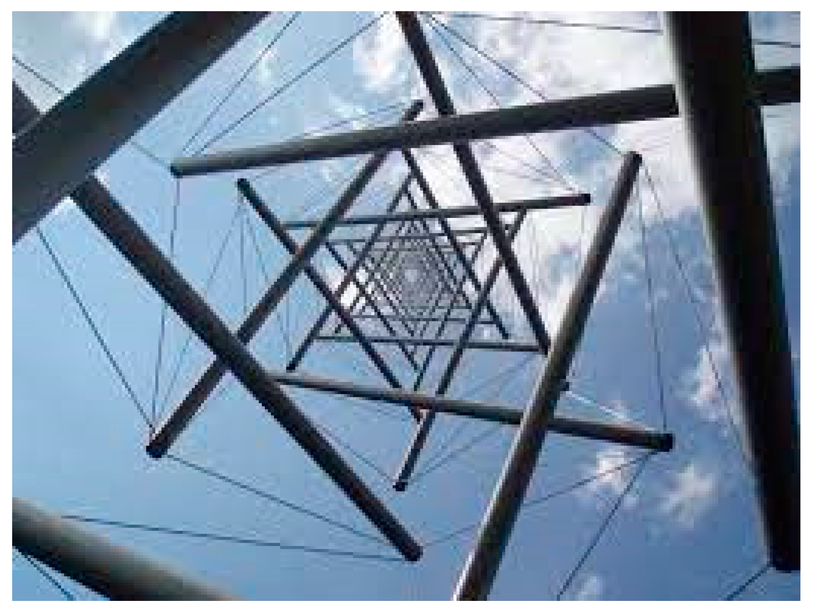
Figure 1.
“Needle Tower” by Kenneth Snelson demonstrating tensegrity architecture with compression-resistant and tensional elements.
Figure 1.
“Needle Tower” by Kenneth Snelson demonstrating tensegrity architecture with compression-resistant and tensional elements.

Publisher’s Note: MDPI stays neutral with regard to jurisdictional claims in published maps and institutional affiliations. |
© 2021 by the author. Licensee MDPI, Basel, Switzerland. This article is an open access article distributed under the terms and conditions of the Creative Commons Attribution (CC BY) license (https://creativecommons.org/licenses/by/4.0/).
Share and Cite
MDPI and ACS Style
Gardiner, J. Cytoskeletal Tensegrity in Microgravity. Life 2021, 11, 1091. https://doi.org/10.3390/life11101091
AMA Style
Gardiner J. Cytoskeletal Tensegrity in Microgravity. Life. 2021; 11(10):1091. https://doi.org/10.3390/life11101091
Chicago/Turabian StyleGardiner, John. 2021. "Cytoskeletal Tensegrity in Microgravity" Life 11, no. 10: 1091. https://doi.org/10.3390/life11101091
Note that from the first issue of 2016, this journal uses article numbers instead of page numbers. See further details here.