Article

Bioleaching of Chalcopyrite by a New Strain Leptospirillum ferrodiazotrophum Ksh-L Isolated from a Dump-Bioleaching System of Kashen Copper-Molybdenum Mine

1
Laboratory of Metals Bioleaching, Institute of Microbiology, SPC “Armbiotechnology” NAS of Armenia,14 Gyurjyan str., Yerevan 0056, Armenia
2
Faculty of Natural Sciences I, Martin-Luther-Universität Halle-Wittenberg, Hoher Weg 8, 06120 Halle, Germany
3
Department of Ecology, Faculty of Biology, Yerevan State University, 1 Alex Manoogian str., Yerevan 0025, Armenia
4
Key Laboratory of Marine Environmental Corrosion and Biofouling, Institute of Oceanology, Chinese Academy of Sciences, No. 7 Nanhai Road, Qingdao 266071, China
*
Authors to whom correspondence should be addressed.
Minerals 2024, 14(1), 26; https://doi.org/10.3390/min14010026
Submission received: 18 October 2023 / Revised: 14 December 2023 / Accepted: 22 December 2023 / Published: 25 December 2023
(This article belongs to the Special Issue Valuable Metals Recovery by Mineral Processing and Hydrometallurgy)

Abstract

:
A new strain of Leptospirillum sp. Ksh-L was isolated from a dump-bioleaching system of the Kashen copper-molybdenum mine (South Caucasus). Ksh-L is an obligate chemolithoautotroph, capable of oxidizing ferrous iron (Fe2+). Cells are Gram-negative and vibrio- or spirillum-shaped of a 0.5–3 µm size. The optimal conditions for the growth are 35 °C and pH 1.6–1.8. Cu2+ and Zn2+ have different effects on the oxidizing ability of the Leptospirillum sp. Ksh-L culture depending on the phase of growth and concentration of Fe2+. Under the conditions of gradually increasing the concentration of copper in the medium, during 4–5 successive subculturing experiments, it was possible to obtain an adapted culture of Leptospirillum sp. Ksh-L, capable of growing in the medium in the presence of up to 400 mM Cu2+. A bioleaching experiment indicates that Ksh-L can efficiently oxidize chalcopyrite. However, the bioleaching of copper from chalcopyrite by Leptospirillum ferrodiazotropum Ksh-L increased about 1.8 times in association with At. thiooxidans ATCC 19377. Phylogenetic analysis based on 16S rRNA gene sequences (GenBank ID ON226845) shows that strain Ksh-L forms a single cluster into Group III. The strain possesses 99.59%, 99.52%, and 96.60% sequence similarity with the strains YTW-96-06, YTW-66-06, and Leptospirillum ferrodiazotrophum 5C in Group III, respectively.

1. Introduction

Bioleaching is a technique that uses microorganisms to remove metals from ore where traditional extraction methods are not economically viable. This method is frequently applied to sulfide mineral ores, which are the source of many valuable and precious metals, including copper, gold, and silver. Traditional methods of metal extraction from sulfide minerals, such as pyrometallurgy, are costly, energy-intensive, and environmentally damaging [1,2]. Bioleaching, using microbial metabolisms to break down metal ores, provides a low-cost solution to this issue. Microbes produce energy during the bioleaching process by oxidizing iron and sulfur from sulfide minerals.
Sulfide ores can be dissolved by ferrous-oxidizing acidophiles to create ferric iron, which then attacks minerals to form sulfur or polysulfide on the ore surface [3,4]. The target metals are released as a result of the oxidants’ attack on the sulfide minerals [2]. Due to the intimate connections between functional oxidizers, environmental conditions, and bioleaching performances, many researchers have recently focused on microbial communities [5,6].
Approximately 70% of the copper reserves in the world come from chalcopyrite [7,8]. It is one of the most resistant ores in hydrometallurgical processing and the most common copper ore. Modern research on the possibility of increasing total recoveries of metal values from such mineral resources plays a significant role in the bio-hydrometallurgical processing of complex low-grade non-ferrous metal concentrates [9,10,11,12,13].
Mesoacidophiles are the most frequently used microorganisms in industrial low-grade chalcopyrite heap-bioleaching operations because of their ambient temperature optimum and ranges, which have been shown to have a significant impact on the rate of copper extraction from chalcopyrite [14,15,16].
Leptospirilla was discovered to be the predominant iron-oxidizing bacteria in gold-arsenopyrite and pyrite bio-oxidation reactors working at 40 °C, although Acidithiobacillus ferrooxidans had long been thought to be the most significant microorganism in the bioleaching of metals [17,18,19,20,21,22].
Leptospirillum spp. bacteria are vibrio- and spiral-shaped chemolithotrophic organisms. They have been formally recognized as coherent bacteria since they fix carbon utilizing the Benson–Calvin cycle, employing oxygen as their only electron acceptor and ferrous iron as their sole electron donor [20,23,24,25,26]. The genus Leptospirillum has been classified into three groups, I, II, and III based on the 16S rRNA gene phylogeny [27]. Leptospirillum ferrooxidans is a representative of group I [28], Leptospirillum ferriphilum and Leptospirillum rubrum are representatives of group II, and Leptospirillum ferrodiazotrophum is a representative of group III [29,30,31]. In addition, microbial community genomics has identified further species, “Leptospirillum sp. group IV UBA BS” [30,31].
Notably, proteomics research has revealed that L. ferrodiazotrophum (group III) carbohydrate metabolism is significantly better than that of Leptospirillum group II and Ferroplasma type II [25]. Also, because the nif genes were only found in this bacterium’s genome sequence, it is thought to be a minor member of the genus and the only one that is capable of fixing nitrogen [30,31,32,33]. Based on this, one representative was isolated in a nitrogen-free liquid medium from AMD biofilm in which Leptospirillum group III was abundant [31]. This Fe(II)-oxidizing, free-living diazotroph was tentatively named “L. ferrodiazotrophum”. Hence, despite its low quantity in the microbial community, L. ferrodiazotrophum played a central role in biofilm formation and nitrogen fixation.
Gene complement variations highlight significant physiological variances that may have played a crucial role in the Leptospirillum groups’ likely sympatric separation. Leptospirillum group II is more capable of producing potentially important polymers for the establishment of floating biofilms, such as cellulose, cellobiose, and starch/amylose, than Leptospirillum group III in dealing with the osmotic challenges brought on by the near-molar FeSO4 solutions. With the potential completion of the glycolysis and TCA pathways, Leptospirillum group III appears to be more suited for energy production and nitrogen fixation. These results are in line with the descriptions of Leptospirillum group II as an early colonist and Leptospirillum group III as a component of late-stage biofilms [34,35]. Interestingly, the complements of signal transduction and chemotaxis genes in Leptospirillum groups II and III are quite different, as are many regulatory genes, pointing to adaptation to various microenvironments (such as those with particular levels of oxygen, redox potential, and availability of fixed nitrogen). Signal transduction, motility, and chemotaxis appear to be more crucial in Leptospirillum group III than in group II, according to genomic and proteomic evidence [33,35]. Leptospirillum group III is identified by biofilm characterization investigations as distributed cells and microcolonies in interior biofilm zones [34], where geochemical gradients are predicted to be prominent. Combining this distribution with the presumed metabolic traits may suggest that Leptospirillum group III is a microaerophile that prefers to grow in nutrient-poor areas of biofilms, where its capacity to fix nitrogen may be essential. Where oxygen availability is certainly low and Leptospirillum group II is present, a few studies on anaerobic metabolism (for example, making use of a within-biofilm nitrogen cycle) are available [33].
Most metals are soluble in acidic pH, and extremely acidophilic microorganisms should be tolerant to high concentrations of metals. Acidophiles are metal-tolerant by both active and passive mechanisms. The passive metal tolerance mechanism is based on the internal positive membrane potential by creating a chemiosmotic gradient that the cations should travel against to enter the cytoplasm [36,37]. Active systems include the efflux of metals from the cytoplasm to the periplasmic space, carried out by ATPases located in the internal membrane of the bacteria [38]. Some microorganisms may pump metal from the cytoplasm directly to the extracellular space by systems of the RND (resistance nodulation cell division) family of carriers, the Cus system of Escherichia coli being the best known of this kind of detoxification organization [39]. The capacity of some species to bind the metal in the periplasmic space using metal chaperones has also been reported for copper [40].
It should be noted that bacterial growth in the form of biofilms significantly increases the resistance of bacteria to metals [41,42,43]. Acidophiles attached to surfaces, such as sulfide minerals, form biofilms that usually include extracellular polymeric substances (EPSs) that can sorb metals to provide a further degree of metal tolerance [44,45].
Several mechanisms of resistance to Mo (VI) have been identified in acidophiles, including the putative Mo (V) resistance mechanism in At. ferrooxidans strain Funis 2–1 [46]. Mo (VI) is chemically reduced by Fe(II), and Mo(V) forms binds to the plasma membrane, probably to the cytochrome-c oxidase (lowering its activity), inhibiting Fe(II) oxidation and consequently growth. Resistance is based on a combination of a cytochrome-c oxidase that is tolerant to higher concentrations of Mo (V) and on Mo (V)-oxidizing activity six-fold greater than that detected in the sensitive At. ferrooxidans strain AP19-3 [46].
With the development of sequencing technology, many genomic-analysis-based studies have investigated the mechanisms underlying heavy metal resistance under extremely acidic conditions [47,48,49,50]. Several genes associated with EPS formation have been identified thus far [51]. Genome/transcriptome analyses showed the presence of genes involved in biofilm formation in Leptospirillum spp. [51,52]. In Acidithiobacillus and Leptospirillum spp. a membrane efflux pump encoded by the czcCBA cluster is responsible for resistance to cadmium, zinc, and cobalt [48,52,53,54]. The resistance of bacteria to copper is very important from the point of view of their application in biotechnological processes, where the concentration of copper ions can vary in the range from 15 to 100 mM CuSO4 [55]. Copper resistance systems in some acidophiles include a copper P-Type transporter identified in L. ferriphilum ML-04 [48]. In addition, earlier descriptions of acidophile copper resistance and the potential role of inorganic polyphosphates in metal resistance are also available [56,57].
This study addressed the characterization and identification of Leptospirillum Ksh-L, isolated from an acid mining drainage of copper ore in Armenia, its ability to degrade sulfide minerals, and its tolerance to metal ions.

2. Materials and Methods

2.1. Culture Conditions and Isolation

To obtain an enrichment culture of iron-oxidizing bacteria, 9K medium [58] with ferrous iron as a source of energy was inoculated with a sample of ore material from a dump-bioleaching system of Kashen copper-molybdenum mine (South Caucasus) and incubated at 35 °C and 150 rpm. The serial dilution method was used to obtain a pure culture of Leptospirillum spp.

2.2. Morphology Studies

Gram staining was performed using the Huker method [59]. The morphology of cells was studied with Motic BA310 trinocular (×1000) microscope supplied by Moticam A16 Camera (Barcelona, Spain, MoticIncorporation Ltd.).

2.3. Optimal pH and Temperature for Growth

Studies of the influence of temperature and pH on the growth of strain Ksh-L were carried out in 100 mL flasks containing 50 mL of 9K medium with ferrous iron (Fe2+) and 10% inoculum. Cultivation was performed on the orbital shaker-incubator ES-20/60 (Biosan, Riga, Latvia) at 150 rpm. The growth ranges for temperature and pH were set as 25–50 °C and 1.6 to 3.0, respectively.

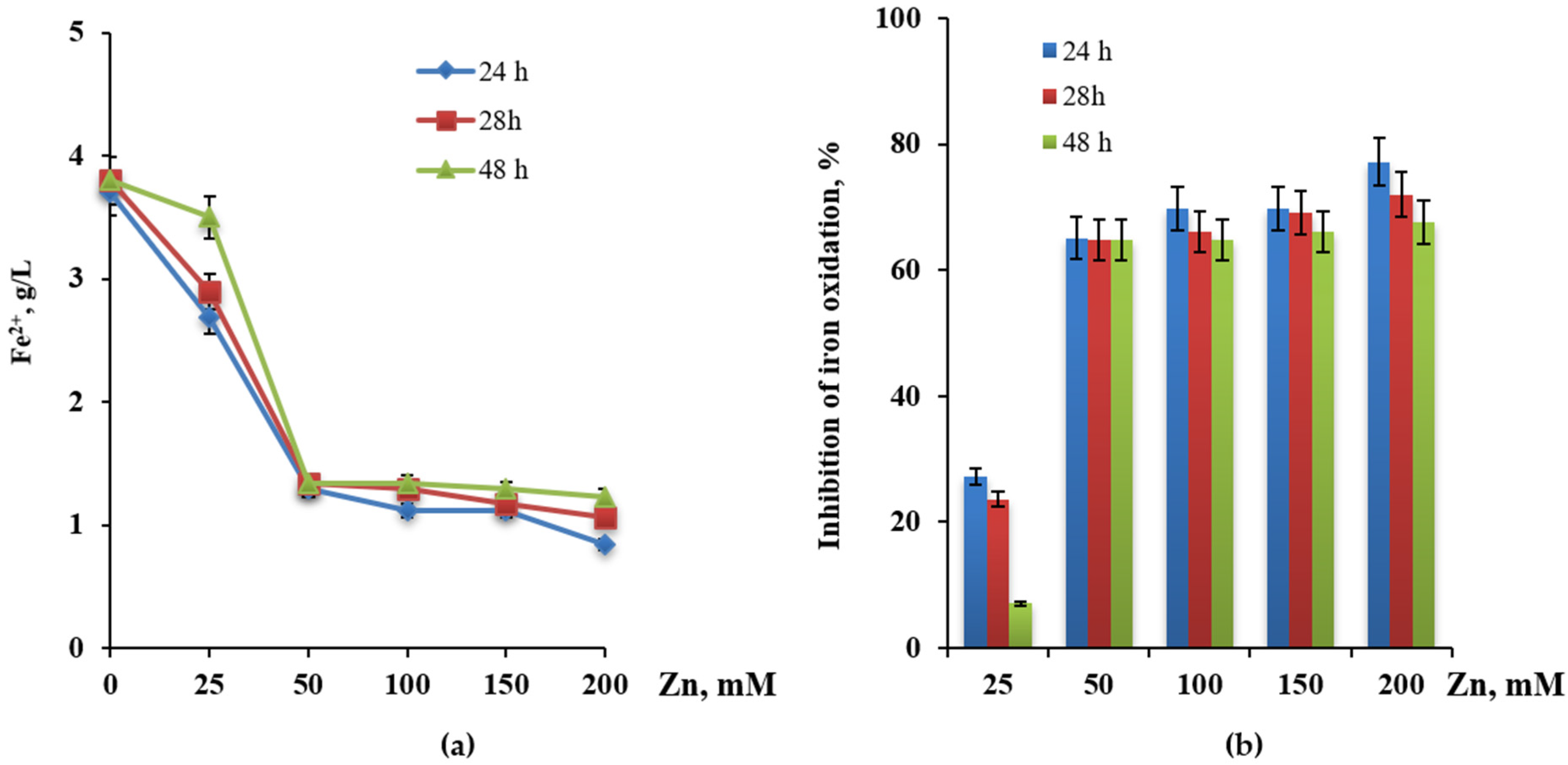
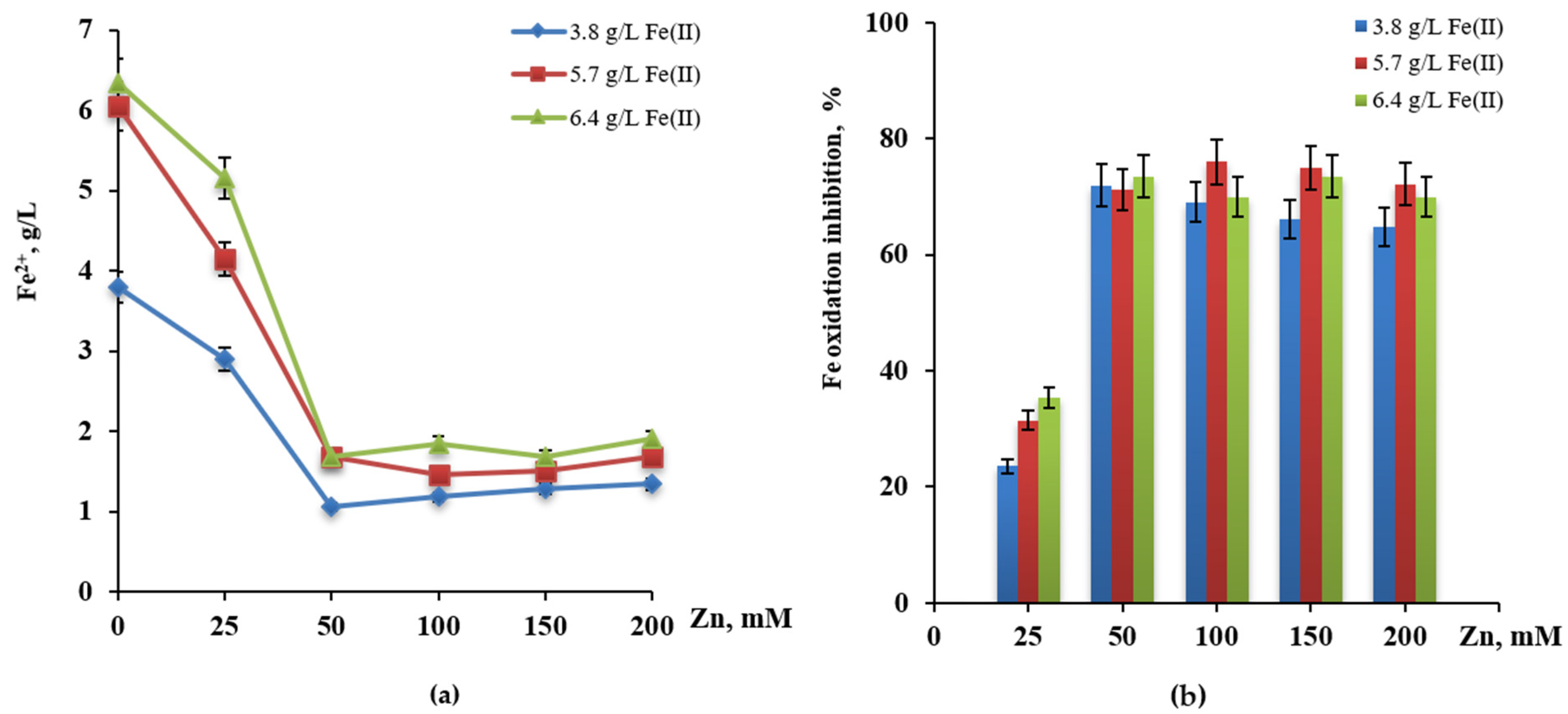
2.4. Influence of Copper and Zinc

The influence of copper and zinc ions on oxidation of Fe2+ by Leptospirillum sp. Ksh-L was studied in Mackintosh (MAC) medium [60] in the concentration range from 10 to 300 mM and at different concentrations of the source of energy (Fe2+). The data presented in the text are formed on the average from repeated experiments with ±2% variation of Fe2+.

2.5. EPS Analysis, Extraction, and Determination

2.5.1. EPS Extraction

For EPS extraction experiments, culture Leptospirillum Ksh-L was grown in 10 L MAC medium supplemented with FeSO4 × 7H2O as a source of energy at 35 °C with shaking at 160 rpm. In the stationary growth phase, cells were collected by centrifugation (10,000 rpm, 10 min) at 4 °C. To get rid of any remaining bacteria, the supernatant was collected and filtered through 0.2 m pore size filters under sterile conditions. Colloidal EPS was present in the fraction obtained. The pellet was re-suspended in 10 mL of 20 mM EDTA at pH 7 and centrifuged for 10 min at 7500 rpm (9900× g) and 4 °C to release the bound EPS. The washed fraction was applied to the supernatant. The fresh pellet was re-dissolved in 10 mL of 20 mM EDTA at pH 7, and the suspension was incubated at 4 °C for 1 h while being shaken. The remaining cells were then removed by centrifuging the bacterial suspension and extracting agent mixtures for 10 min at 7500 rpm (9900× g) and 4 °C [61].

2.5.2. Determination of EPS Composition

Total carbohydrate values were determined by spectrophotometry using the phenol-sulfuric acid method with D-glucose as the standard, as described by [62]. Total protein quantification was conducted by spectrophotometry using the Bradford procedure [63]. A calibration curve was developed using a series of bovine serum albumin (BSA) standards. Uronic acids were quantified using the protocol of Blumenkrantz and Asboe-Hansen [64]. Meta-hydroxydiphenyl solution in 0.5% NaOH, ortho-hydroxydiphenyl, H2SO4/sodium tetraborate solutions, and cetyltrimethylammonium bromide solution were used as a reaction mixture. Absorbance measurements were performed at 520 nm. The content of proteins, carbohydrates, and uronic acids in EPS was expressed in µg/mL culture medium.

2.6. DNA Extraction, PCR of 16s rRNA, Sequencing, and Phylogenetic Analysis

2.6.1. DNA Extraction

The identification of isolated strain Ksh-L was performed based on 16S rRNA gene nucleotide sequence analysis. DNA extraction was carried out according to the Macherey-NagelTM (NucleoSpinTM) protocol. Bacterial cultures in the logarithmic phase were centrifuged to obtain the bacterial biomass and frozen for DNA purification and taxonomic classification of the strain based on 16S rRNA gene sequences. For DNA extraction, the following reagents were used: BE buffer (Tris-HCl pH-8.0), proteinase K, MG-lysozyme solution, BS salt buffer, 96% ethanol, and distilled water.

2.6.2. Extraction and Purification of PCR Product

A 1.5% agarose gel Tris-HCl buffer solution was prepared. The agarose aqueous solution was heated in a microwave oven for 1–2 min before boiling and then cooled to 45–60 °C. A 1% 10 mM Tris-HCl, 1 mM EDTA aqueous solution (1 μL per 10 mL agarose solution) was added to the cooled agarose solution. A total of 5 μL of the test DNA solution was mixed with a 3 μL 6 μL 6X Thermo Scientific TriTrack DNA Loading Dye and 5 μL Thermo Scientific GeneRuler buffer containing 1 kb DNA Ladder markers. Electrophoresis was performed on GE Healthcare/Amersham Pharmacia EPS-601 Electrophoresis Power Supply for 25 min at a voltage of 104 V at 100 mA. The electropherograms and images were obtained using the GeneMarkerHID® Spectrum GeneMarkerHID® and Spectrum ProView™ Sequencing Software TM625 package (Promega Corporation 2800, Fitchburg, MA, USA).
The PCR amplification was carried out according to the Macherey-NagelTM (NucleoSpinTM) Extract II protocol. PCR amplification was carried out in 50 μL reaction mixture, which contained 10 mg of DNA sample, 1 μL universal bacterial PCR primers a-fD1 (27F) (AGAGTTTGATCCTGGCTCAG) and rP2 (ACGGCTACCTTGTTACGAG), b- 908 fwd and 796 rev, 5 μL Taq-DNA polymerase, 1 μL dNTPs, and 40 μL double distilled water. PCR temperature–time process is as follows: 1st cycle 95 °C × 5 min, further 30 cycles of denaturation −94 °C × 40 s, connection 50/53 °C × 55–60 s, synthesis 72 °C × 1.5 min, final cycle −72 °C × 3 min, and then standby at 8–10 °C. PCR products were tested by 1.5% agarose gel electrophoresis and sequenced with primers 908fwd (16Sfwd) (GTGCCAGCAGCCGCG) and 796rev (16Srev) (GGGTTGCGCTCGTTG) by Microsynth AG (Balgach, Switzerland). PCR product purification was performed using the QIAquick PCR purification kit following the manufacturer’s instructions. PCR fragments were sequenced by a 454 GS-FLX Titanium sequencer, using the Sanger method [65].

2.6.3. Construction of Phylogenetic Tree

Close relative and phylogenetic affiliation of the obtained 16S rRNA sequences were determined by submitting to the NCBI 16S ribosomal RNA GenBank database using NCBI blastn search analyses (www.ncbi.nlm.nih.gov) performed with Geneious prime 2022.0.2. (https://www.geneious.com) and the 16S Biodiversity tool (RDP tool version 2.12) [66,67]. The construction of phylogenetic trees was performed with MEGA 11 software using the neighbor-joining method [68,69].

2.7. Leaching Experiments

Chalcopyrite (CuFeS2) from Shamlugh ore deposit (Armenia) was tested in the bioleaching experiments. The chemical composition of minerals is presented in Table 1. Feed minerals were ground to a particle size ≤ 63 µm.
Bioleaching of chalcopyrite was performed using a pure culture of L. ferrodiazotropum Ksh-L as well as its associations with At. thiooxidans ATCC 19377 obtained from DSMZ. Bioleaching experiments were carried out in 250 mL Erlenmeyer flasks containing 100 mL of MAC medium without iron at 30 °C. Pulp density (PD) was 4% and pH 1.8. The inoculum of used cultures was 10%, and all experiments were carried out in triplicate. Chemical control with the same conditions and without inoculum was included. Copper, total iron, ferric (Fe(III)), and ferrous (Fe(II)) ions in leachate were analyzed for 30 days. pH was measured using Hi2211-01 Benchtop pH/mV Meter (Hanna Instruments, Vöhringen, Germany). The value of oxidation/reduction potential (ORP) was measured using a standard hydrogen electrode (SHE) in relation to an Ag/AgCl reference electrode (mV vs. Ag/AgCl). Copper and total iron were determined using atomic absorption spectrophotometer AAS SP-IAA1800H (Bioevopeak, Qingdao, China). Concentrations of ferric (Fe(III)) and ferrous (Fe(II)) ions were determined using the complexometric method with EDTA [70].
Consumption of Fe(II) was calculated as a difference between initial Fe(II) and concentration of Fe(II) determined at certain times of experiment (Equation (1)). Consumption is expressed by g/L and %.
[Fe(II)]c = [Fe(II)i−[Fe(II)]t/[Fe(II)]i × 100%
where [Fe(II)]c is the consumption of Fe(II); [Fe(II)i– is initial concentration of Fe(II) in the medium; and [Fe(II)]t—concentration of Fe(II) determined at certain point of time.
Inhibition of iron oxidation in the presence of metal ions was determined according to Equation (2).
Inhibition (%) = Fe(II)cm/Fe(II)c × 100
where Fe(II)cm is consumption of Fe in the presence of metal ions (g/L), and Fe(II)c is consumption of Fe in the absence of metal ions (g/L).

3. Results

3.1. Isolation of Strain Ksh-L

The selected sample from a dump-bioleaching system of the Kashen copper-molybdenum mine was transferred in a 9K liquid medium and incubated at 35 °C at 150 rpm for 5–7 days. As a result of the active growth of bacterial cells, the medium color changes from pale green to orange-red due to iron oxidation.

3.2. Morphology

Cells of Leptospirillum sp. Ksh-L are Gram-negative, motile, and vibrio- or spiral-shaped. Cells have a diameter of 0.5 µm and a length of 1.0–3.0 µm (Supplementary Figure S1). This is typical for the genus of Leptospirillum. The morphology results show that strain Ksh-L seems to be consistent with previously described Leptospirillum species [71].

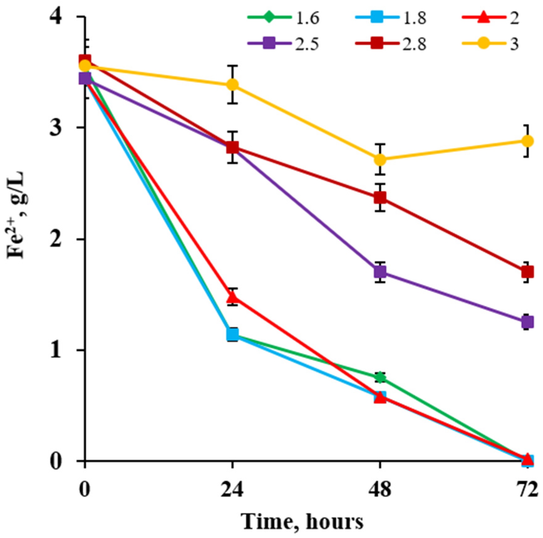
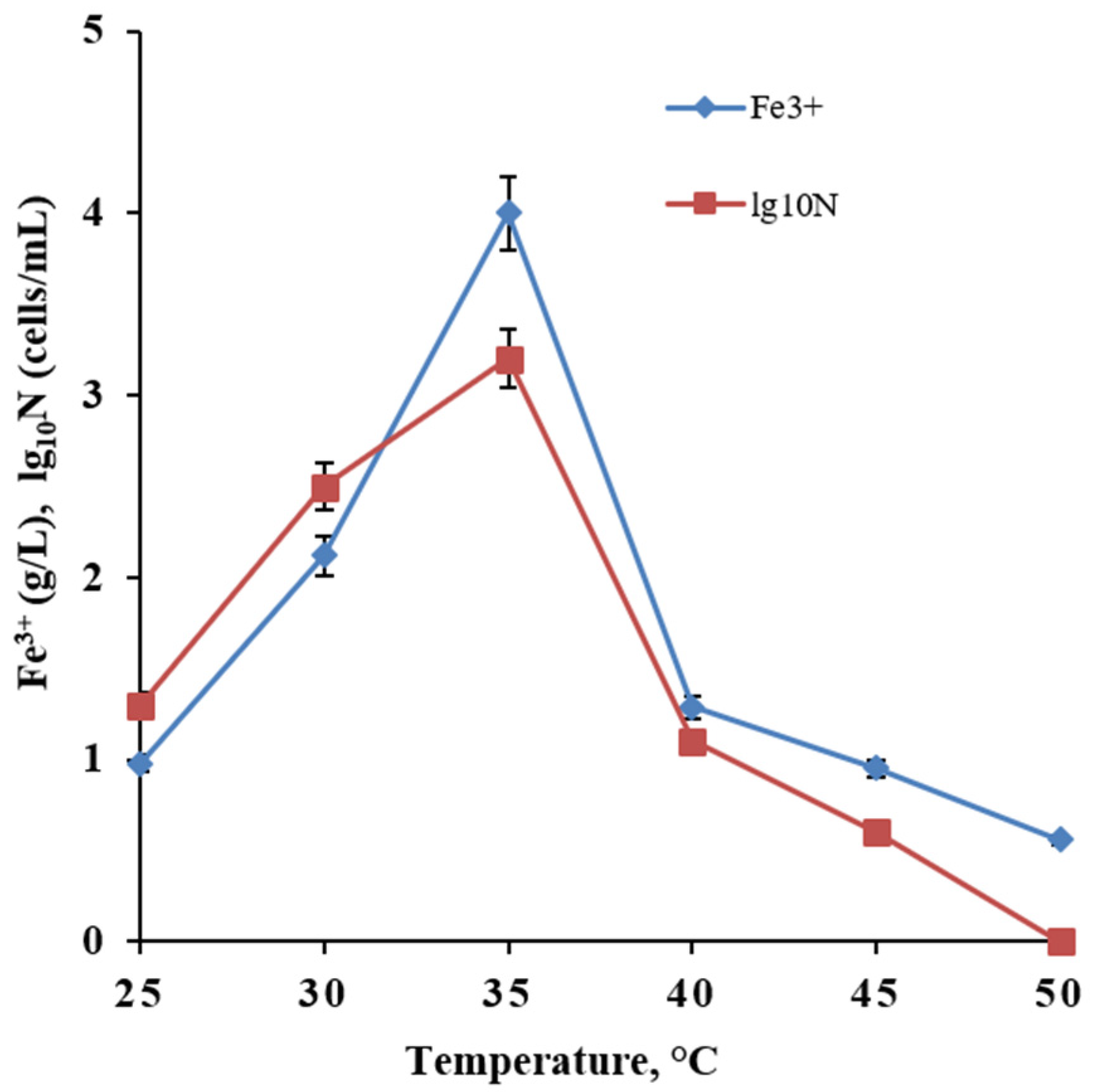
3.3. Optimal pH and Temperature for Growth

The growth of the newly isolated Khs-L strain was evaluated according to the increase in the number of cells and the results of their biological activity, increasing the amounts of Fe3+ in the medium. As shown in Figure 1, the growth curves of the Leptospirillum sp. Ksh-L strain in the initial pH range of 1.6–2.0 are S-shaped. The duration of the lag phase was about 48 h, but the growth of bacteria was more active at pH values of 1.6 and 1.8 (Figure 1).
The most intensive oxidation of Fe2+ was observed at pH values of 1.6 and 1.8. As can be seen from the presented data, at the initial pH values of 2.8 and 3.0, Fe2+ oxidation was sharply suppressed, and the duration of the lag phase was increased to 20 and 48 h, respectively (Figure 1). It can be explained that the pH variation affects enzyme activity because changes in ionization affect the system components. This indicates that the Ksh-L strain is sensitive to pH, and a much higher pH value will inhibit the activity of bacteria.
As can be seen from Figure 2, the optimum growth temperature of the Ksh-L strain was observed at 35 °C. Thus, optimal conditions for the growth of isolated strain Leptospirillum sp. were observed at 35 °C and a pH range from pH 1.6 to pH 2.0.

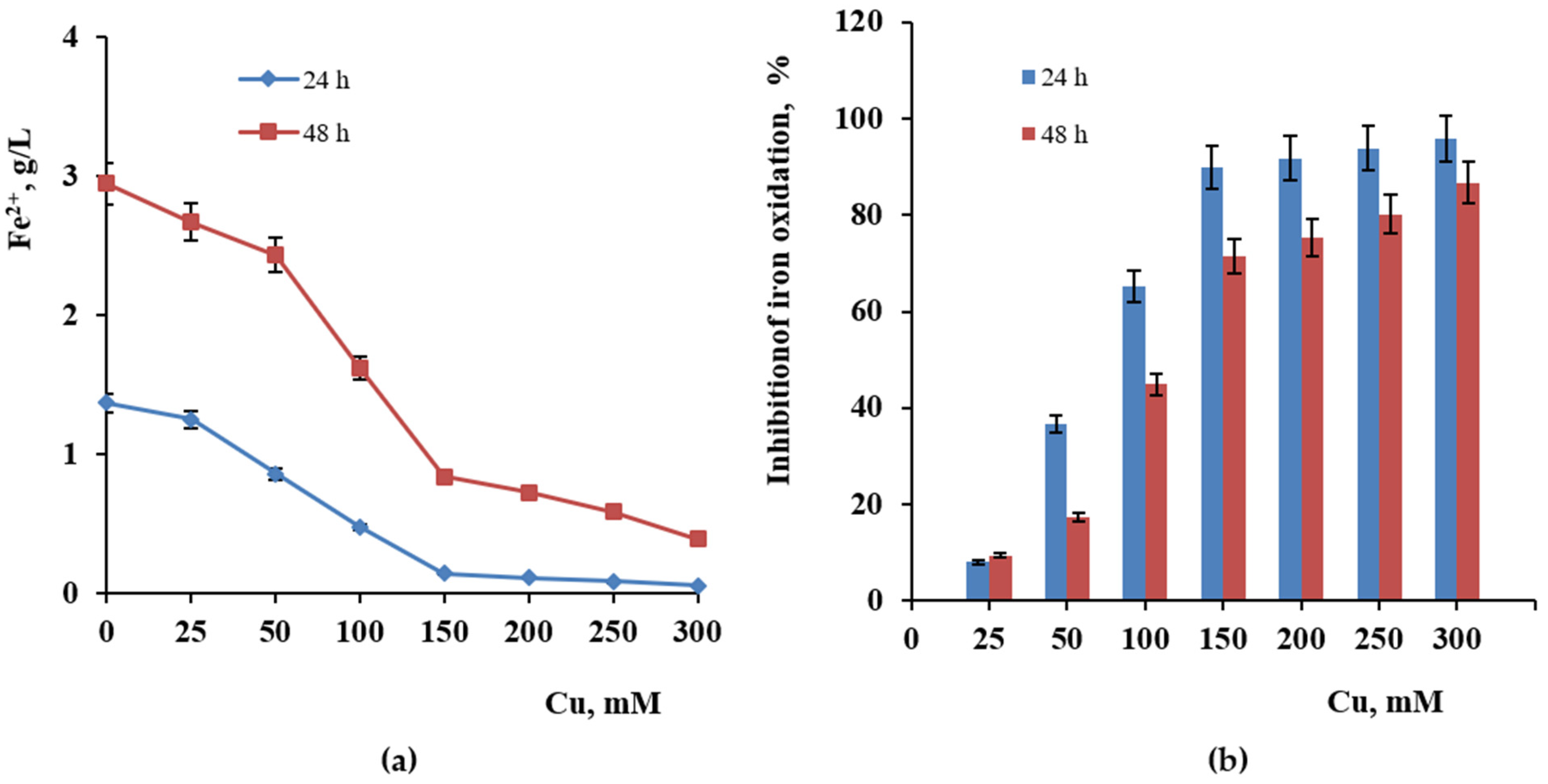
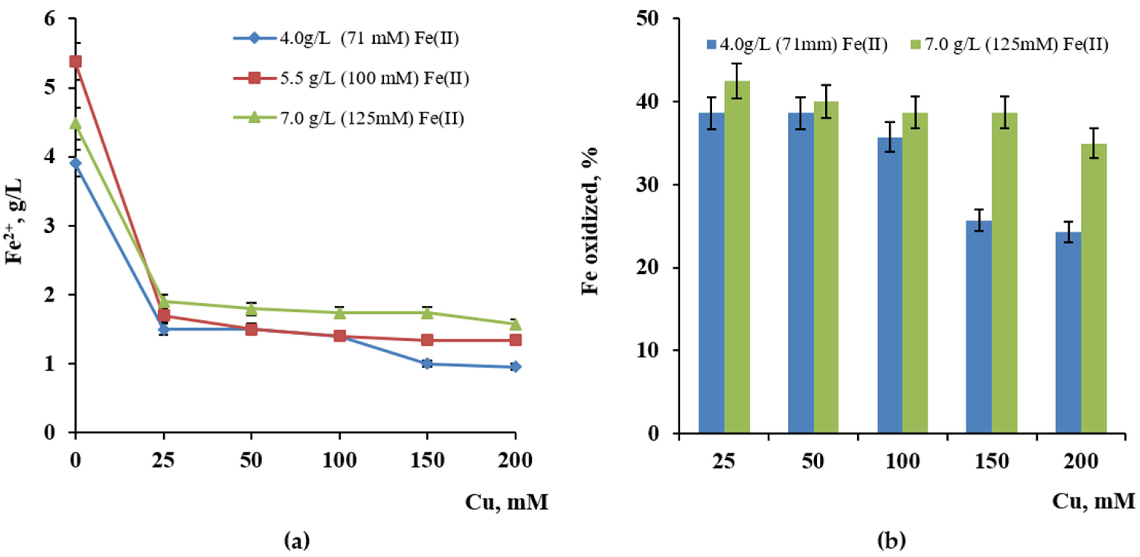
3.4. Influence of Copper and Zinc

Like most bioleaching microorganisms, strain Leptospirillum sp. Ksh-L has been isolated from an environment (dump-bioleaching system) that has unusually high concentrations of potentially toxic metals (e.g., copper and iron) as well as high concentrations of heavy metals (e.g., arsenic and silver). These metals can exert harmful effects on microorganisms. Their toxic effects include the blocking of biologically important functional groups and the denaturation of enzymes [72].
Cu (II) inhibition of growth and Fe(II) oxidation have also been demonstrated in Sulfobacillus thermosulfidooxidans subsp. asporogenes via competitive inhibition of Fe(II) oxidation [73]. The influence of Cu2+ and Zn2+ ions on the oxidation of Fe2+ by strain Ksh-L was studied in a concentration range from 25 to 200–300 mM. As can be seen from Figure 3a, copper in all tested concentrations inhibits the oxidation of Fe2+.
Furthermore, the higher the copper concentration, the higher the extent of inhibition of iron oxidation. Thus, iron oxidation by Leptospirillum sp. Ksh-L for 24 h was suppressed by 36.7, 65.3, and 89.8% at copper concentrations of 50, 100, and 150 mM, respectively, with a content of 3.2 g/L Fe2+ in the medium. Cu2+ in the concentration of 200–300 mM almost completely (90%–96%) inhibits iron oxidation by Leptospirillum sp. Ksh-L (Figure 3b). It can be noted that the extent of inhibition of iron oxidation by Cu2+ ion reduces for 48 h along with bacterial growth and is 17.4, 44.9, and 71.5%, respectively (Figure 3b).
The effect of copper on oxidation of Fe2+ Leptospirillum sp. Ksh-L was studied depending on substrate concentrations in the medium (Figure 4a,b). As shown in Figure 4, the degree of oxidation of Fe2+ by Leptospirillum sp. Ksh-L in the presence of tested copper concentrations was slightly higher when the content of Fe2+ in the medium increased from 4.0 g/L (71.4 mM) to 7.0 g/L (125 mM). Thus, the increase in the concentration of substrate from 4.0 to 7.0 g/L leads to the enhancement of the amount of oxidized iron by bacteria in the presence of the tested concentration of copper.
From the data presented, it can be seen that the oxidation of Fe2+ by Leptospirillum sp. Ksh-L is suppressed by about 25% at 25 mM of Zn2+ and is sharply inhibited (up to 65%–77%) at zinc concentrations in the range from 50 to 200 mM (Figure 5a,b).
As shown in Figure 6, an increase in the substrate concentration led to the enhancement of the amount of oxidized iron by Ksh-L in the presence of 25 mM Zn2+, while it did not facilitate iron oxidation by bacteria in concentrations of Zn above 100 mM. Moreover, no decrease in the inhibitory effect of zinc ions was observed along with the growth of bacteria, as shown in the case of copper (Figure 5).
Along with the growth of Leptospirillum sp. Ksh-L, the amount of oxidized iron in the presence of 50 mM Cu2+ increased. It is assumed that with the growth of bacteria, the cells form EPSs and create accordingly a less toxic and more favorable environment for the growth of cells in the presence of copper.

3.5. Adaptation of L. ferrodiazotrophum Ksh-L

Although bacteria do not react well to sudden and significant changes in heavy metal ion concentrations, they can be adapted to gradually increased concentrations over a while, to increase their tolerance to such metals. In the next series of the experiment under conditions of gradually increasing the concentration of copper in the medium, during 4–5 successive subculturing experiments, it was possible to obtain an adapted culture of Leptospirillum sp. Ksh-L capable of growing in the medium in the presence of up to 400 mM Cu2+ (Figure 7).

3.6. EPS Analysis

EPSs play an essential role in the formation of a biofilm, which mediates the adhesion of cells to the mineral surface and forms a cohesive three-dimensional polymer, interconnecting and immobilizing cells in the process of bioleaching by iron- and sulfur-oxidizing bacteria [74,75,76]. An important role of capsular polysaccharides as a fundamental structural element of the EPS, determining the mechanical stability of biofilm was disclosed. One of the objectives of the present study was to investigate the chemical composition of a colloidal polysaccharide of the newly isolated iron-oxidizing chemolithotrophic bacteria L. ferrodiazotrophum Ksh-L. The studies carried out showed that the total amounts of colloidal and capsular EPSs are 474.4 and 208.58 µg/mL, respectively.
As shown in Table 2, the amount of carbohydrates in the capsular EPS is considerably higher than that in the colloidal EPS. The amounts of protein in colloidal and capsular EPSs are approximately the same, 24.41 µg/mL and 27.33 µg/mL, respectively. Uronic acids were not detected in both EPSs. The obtained data are comparable and agree with the corresponding data of other species of the genus Leptospirillum, L. ferriphilum CC and L. ferrooxidans ZC studied by us previously [77,78].

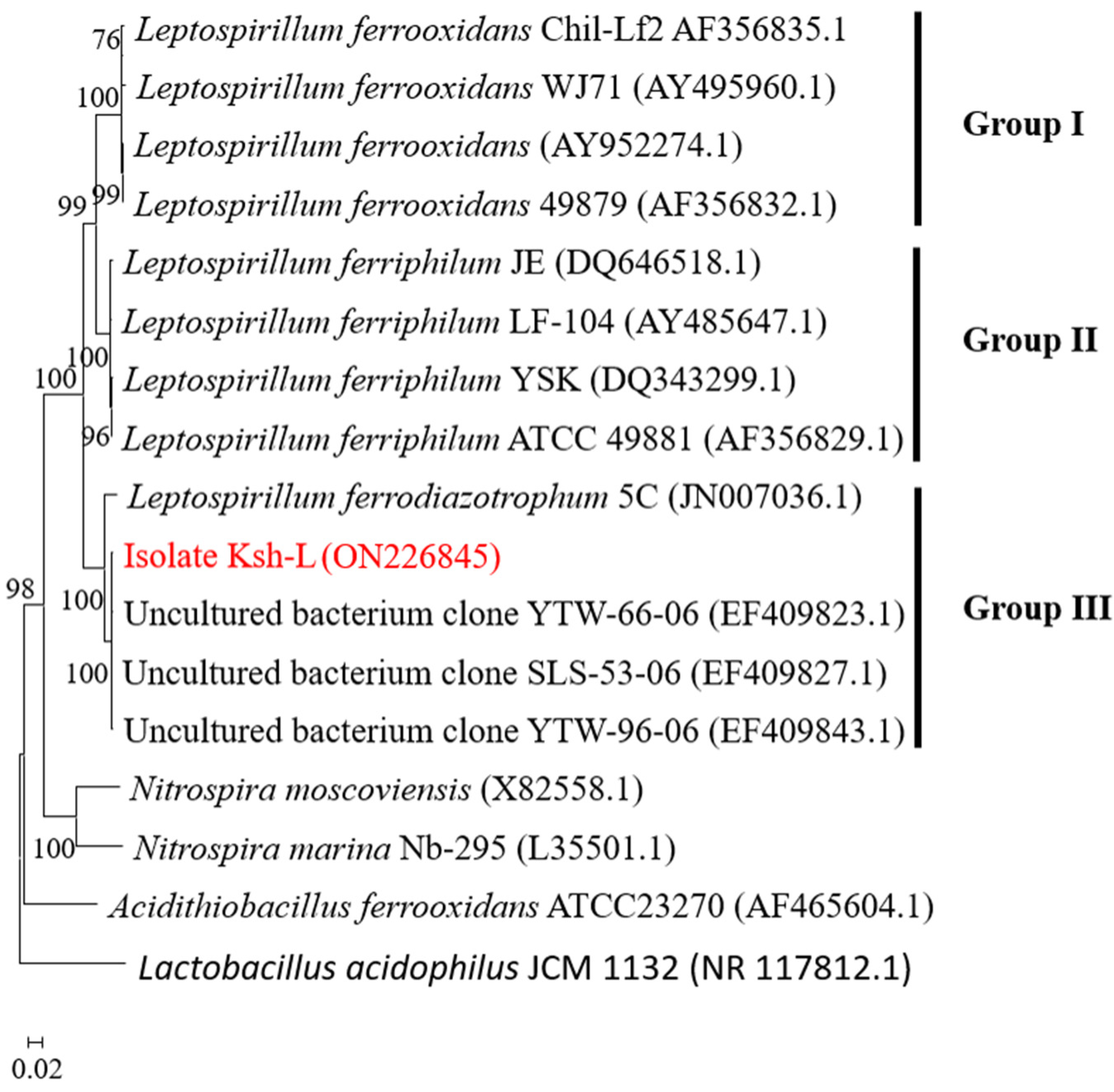
3.7. Phylogenetic Analysis of 16S rRNA

A PCR-amplified 16S rRNA product was detected by 1% agarose gel electrophoresis (Supplementary Figure S2). The length of the 16S rRNA of Leptospirillum sp. Ksh-L is about 1.5 kb and is sequenced sequentially. The sequence of the 16S rRNA of Leptospirillum sp. Ksh-L was submitted to GenBank, and the accession number ON226845 was obtained. The length of sequenced fragments of the gene encoding 16S rRNA is 1469 bp. The nucleotide sequence of the strain Leptospirillum sp. Ksh-L was phylogenetically compared with the Leptospirillum species (Figure 8).
Preliminary screening of the GenBank database was performed using BLAST (http:www.ncbi.nlm.nih.gov/blast). Based on the homology of 16S rRNA, the phylogenetic development tree was built as shown in Figure 8. The sequence was divided into three groups: Group I—L. ferrooxidans; Group II—L. ferriphilum; and Group III—Leptospirillum ferrodiazotrophum and uncultured clones. The isolated Leptospirillum sp. Ksh-L strain formed a single cluster into Group III and possessed 99.66% sequence similarity with uncultured bacterium clone SLS-53-06 (Table 3). As shown in Figure 8, the isolate Ksh-L compared to other strains in Group III with strains YTW-96-06, YTW-66-06, and Leptospirillum ferrodiazotrophum 5C and possessed 99.59%, 99.52%, and 96.60% sequence similarity, respectively (Table 3).
In Figure 8, the Lactobacillus acidophilus JCM 1132 strain is used as an out-group to root the tree, and the database accession numbers of the gene sequences used are given in parentheses.
Thus, the newly iron-oxidizing strain Ksh-L obtained from a dump-bioleaching system of the Kashen copper-molybdenum mine was identified as Leptospirillum ferroodiazotrophum. Type strain: Leptospirillum ferroodiazotrophum Ksh-L.

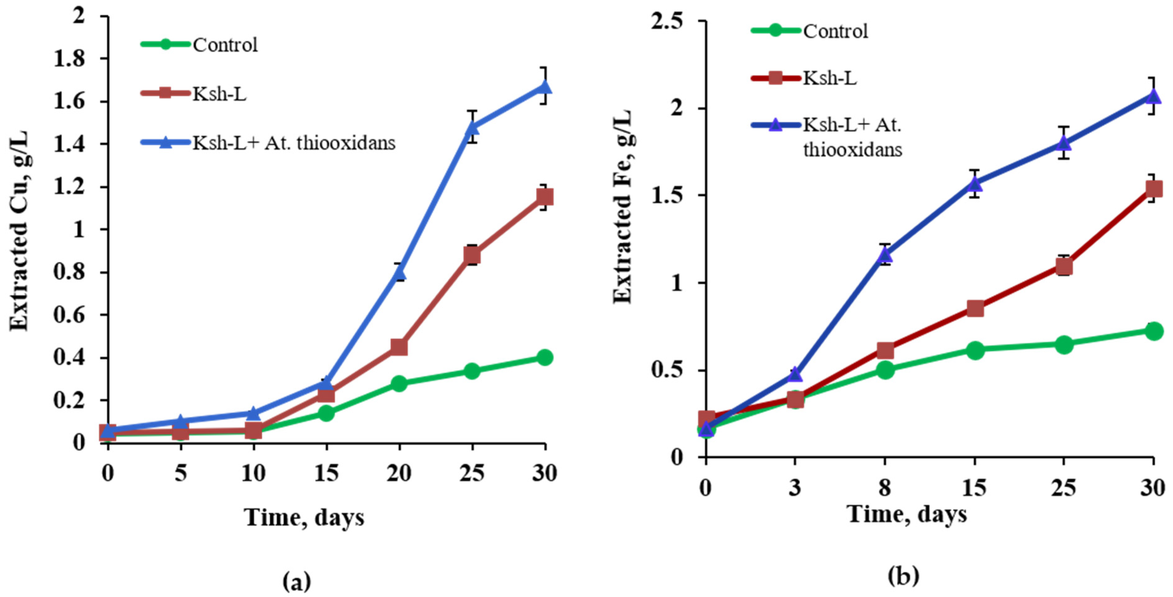
3.8. Bioleaching of Chalcopyrite

A comparative study was carried out on the bioleaching of chalcopyrite by a pure culture L. ferrodiazotrophum Ksh-L and its association with sulfur-oxidizing bacteria At. thiooxidans ATCC 19377 at 30 °C.
The data presented in Figure 9a,b show that compared to uninoculated control, isolated bacterium L. ferrodiazotropum Ksh-L stimulated the extraction of copper and iron by 1.7 and 2.3 times, respectively. However, in association with sulfur-oxidizing At. thiooxidans, L. ferrodiazotropum Ksh-L oxidizes chalcopyrite much more actively than in pure culture. This finding corresponds with the literature data from similar studies conducted using Leptospirillum ferooxidans with the association of At. thiooxidnas or At. caldus. According to some researchers, mixed cultures of mesophiles have been reported to oxidize sulfide minerals more efficiently than pure cultures [85,86,87].
Thus, in the presence of At. thiooxidans, the extraction of copper and iron from chalcopyrite by L. ferrodiazotropum Ksh-L for 30 days increases approximately 1.8 and 1.9 times, respectively (Figure 9a,b, Table 4).
The data presented in Table 1 show that chalcopyrite leaching is correlated with the pH and ORP of the solution. When L. ferrodiazotropum Ksh-L was used in monoculture, the final pH was 1.7, and the ORP was 600 mV, while in the L. ferrodiazotropum Ksh-L variant with At. thiooxidans ATCC 19377, the pH value was comparatively lower (1.5), and the ORP was significantly higher (720 mV) (Table 4).
Chalcopyrite is an acid-soluble sulfide mineral and is therefore subject to attack by both ferric iron (Fe3+) and protons (H+) (Equations (3) and (4)) [55,88].
CuFeS 2 + 2 Fe 2 SO 4 3 CuSO 4 + 5 FeSO 4 + 2 S 0
CuFeS 2 + 4 H + Fe 2 + + Cu 2 + + 2 H 2 S
0 . 125 S 8 + 1 . 5   O 2 + H 2 O A t . t h i o o x i d a n s   ATCC   19377   SO 4 2 + 2 H +
At. thiooxidans ATCC 19377 in a mixed culture oxidizes sulfide and sulfur to sulfuric acid and contributes to the decrease in pH (pH 1.5), thereby preventing the formation of jarosite and a hydrophobic layer of sulfur on the surface of chalcopyrite (Equation (5)), removes the effect of passivation of the mineral, and promotes intense oxidation of chalcopyrite. According to Christel and Dopson, 2016, sulfur-oxidizing A. caldus seemed to have a supporting role in the early stages of mineral dissolution [89]. The attached sulfur-oxidizing bacteria on the mineral surface utilized the reduced inorganic sulfur compounds (RISCs) released from the mineral by a direct mechanism [90]. In a study conducted by Tao et al. (2021), six artificial communities with varying functions or biodiversity were recreated using six common bioleaching species for chalcopyrite leaching. Communities with low diversity also performed somewhat poorly in bioleaching, and the absence of sulfur oxidizers greatly decreased copper extraction rates in those communities [91].

4. Conclusions

A new strain of iron-oxidizing strain Leptospirillum sp. Ksh-L was isolated from a dump-bioleaching system of the Kashen copper-molybdenum mine. The cells of the strain Ksh-L are Gram-negative, motile, and vibrio- or spiral-shaped, with a 0.5 μm width and a 1.0–3.0 μm length.
The optimal temperature for the growth of Leptospirillum sp. Ksh-L is 35 °C, and the optimal pH is 1.6–1.8. The current study showed how different concentrations of commercially important metals (Cu2+, Zn2+) have various effects on the oxidizing ability of the strain Leptospirillum sp. Ksh-L, depending on the phase of growth and concentration of ferrous iron as a source of energy.
Based on the homology of 16S rRNA, the isolated strain Leptospirillum sp. Ksh-L formed a single cluster into Group III and, compared to other strains in Group III, possessed 99.59%, 99.52%, and 96.60% sequence similarity with strains YTW-96-06, YTW-66-06, and Leptospirillum ferrodiazotrophum 5C, respectively. Thus, the newly iron-oxidizing strain Ksh-L obtained from a dump-bioleaching system of the Kashen copper-molybdenum mine was identified as Leptospirillum ferroodiazotrophum. Type strain: Leptospirillum ferroodiazotrophum Ksh-L.
It was also shown that the bioleaching of copper and iron from chalcopyrite by the association of L. ferrodiazotropum Ksh-L and At. thiooxidans ATCC 19377 in comparison with pure-culture L. ferrodiazotropum Ksh-L for 30 days increases about 1.8 and 1.9 times, respectively. Thus, it is supposed that the association of isolated L. ferrodiazotropum Ksh-L with sulfur-oxidizing At. thiooxidans ATCC 19377 can be successfully used to enhance the efficiency of copper extraction from chalcopyrite.

Supplementary Materials

The following supporting information can be downloaded at https://www.mdpi.com/article/10.3390/min14010026/s1, Figure S1: Microphotography of iron-oxidized bacteria (Motic BA 310 trinocular (×1000) microscope with Digital Camera Moticam A16 (×0.5)). Figure S2: Gel electrophoresis of PCR amplification of 16S rRNA amplified product of 16S rDNA for bacterial isolate Leptospirillum sp. Ksh-L.

Author Contributions

Conceptualization, A.K., N.V., S.W., G.S., R.Z., and A.V.; methodology, N.V., S.W., G.S., and R.Z.; software, A.K. and A.V.; validation, N.V., S.W., G.S., and R.Z.; formal analysis, A.K. and A.V.; investigation, A.K. and A.V.; resources, N.V., S.W., G.S., R.Z., and A.V.; data curation, A.V.; writing—original draft preparation, A.K. and A.V.; writing—review and editing, N.V., S.W., G.S., and R.Z.; visualization, A.V. and R.Z.; supervision, N.V., S.W., and R.Z.; project administration, N.V. and A.V.; funding acquisition, S.W., R.Z., and A.V. All authors have read and agreed to the published version of the manuscript.

Funding

This research was funded by the Higher Education Science Committee of the Ministry of Education, Science, Culture and Sports of Republic of the Armenia, grant number 22rl-031.

Data Availability Statement

Data are contained within the article.

Acknowledgments

All authors express gratitude to Martin Herzberg and Diana Galea from the Institute for Biology/Microbiology Martin-Luther University Halle-Wittenberg for molecular biology analysis.

Conflicts of Interest

The authors declare no conflict of interest. The funders had no role in the design of the study; in the collection, analyses, or interpretation of data; in the writing of the manuscript; or in the decision to publish the results.

References

  1. Zhao, H.; Zhang, Y.; Zhang, X.; Qian, L.; Sun, M.; Yang, Y.; Zhang, Y.; Wang, J.; Kim, H.; Qiu, G. The dissolution and passivation mechanism of chalcopyrite in bioleaching: An overview. Miner. Eng. 2019, 136, 140–154. [Google Scholar] [CrossRef]
  2. Jones, S.; Santini, J.M. Mechanisms of bioleaching: Iron and sulfur oxidation by acidophilic microorganisms. Essays Biochem. 2023, 67, 685–699. [Google Scholar] [CrossRef]
  3. Rohwerder, T.; Gehrke, T.; Kinzler, K.; Sand, W. Bioleaching review part A: Progress in bioleaching: Fundamentals and mechanisms of bacterial metal sulfide oxidation. Appl. Microbiol. Biotechnol. 2003, 63, 239–248. [Google Scholar] [CrossRef]
  4. Huang, C.; Qin, C.; Feng, X.; Liu, X.; Yin, H.; Jiang, L.; Liang, Y.; Liu, H.; Tao, J. Chalcopyrite bioleaching of an in situ leaching system by introducing different functional oxidizers. RSC Adv. 2018, 8, 37040. [Google Scholar] [CrossRef]
  5. Watling, H.R.; Watkin, E.L.; Ralphe, D.E. The resilience and versatility of acidophiles that contribute to the bio-assisted extraction of metals from mineral sulphides. Environ. Technol. 2010, 31, 915–933. [Google Scholar] [CrossRef]
  6. Spolaore, P.; Joulian, C.; Gouin, J.; Morin, D.; D’Hugues, P. Relationship between bioleaching performance, bacterial community structure and mineralogy in the bioleaching of a copper concentrate in stirred-tank reactors. Appl. Microb. Biotechnol. 2011, 89, 441–448. [Google Scholar] [CrossRef]
  7. Ghorbani, Y.; Franzidis, J.P.; Petersen, J. Heap leaching technology—Current State, innovations, and future directions: A review. Miner. Process. Extr. Metall. Rev. 2016, 37, 73–119. [Google Scholar] [CrossRef]
  8. Veloso, T.C.; Peixoto, J.J.M.; Pereira, M.S.; Leao, V.A. Kinetics of chalcopyrite leaching in either ferric sulphate or cupric sulphate media in the presence of NaCl. Int. J. Miner. Process 2016, 148, 147–154. [Google Scholar] [CrossRef]
  9. Rawlings, D.E.; Johnson, D.B. The microbiology of biomining: Development and optimization of mineral-oxidizing microbial consortia. Microbiology 2007, 153, 315–324. [Google Scholar] [CrossRef] [PubMed]
  10. Qin, W.Q.; Li, W.Z.; Lan, Z.Y.; Qiu, G.Z. Simulated small-scale pilot heap leaching of low-grade oxide zinc ore with integrated selective extraction of zinc. Miner. Eng. 2007, 20, 694–700. [Google Scholar] [CrossRef]
  11. Giaveno, A.; Lavalle, L.; Chiacchiarini, P.; Donati, E. Bioleaching of zinc from low-grade complex sulphide ores in an airlift by isolated Leptospirillum ferrooxidans. Hydrometallurgy 2007, 89, 117–126. [Google Scholar] [CrossRef]
  12. Gericke, M.; Muller, H.H.; van Staden, P.J.; Pinches, A. Development of a tank bioleaching process for the treatment of complex Cu-polymetallic concentrates. Hydrometallurgy 2008, 94, 23–28. [Google Scholar] [CrossRef]
  13. Patel, B.C.; Tippe, D.R.; Dave, S.R. Optimization of copper and bioregeneration under metallic stress. Hydrometallurgy 2012, 117–118, 18–23. [Google Scholar] [CrossRef]
  14. Pradhan, N.; Nathsarma, K.C.; Srinivasa Rao, K.; Sukla, L.B.; Mishra, B.K. Heap bioleaching of chalcopyrite: A review. Miner. Eng. 2008, 21, 355–365. [Google Scholar] [CrossRef]
  15. Abhilash; Mehta, K.D.; Pandey, B.D. Bacterial Leaching Kinetics for Copper Dissolution from a Low- Grade Indian Chalcopyrite Ore. Rev. Esc. Minas 2013, 66, 245–250. [Google Scholar] [CrossRef]
  16. Panda, S.; Akcil, A.; Pradhan, N.; Deveci, H. Current scenario of chalcopyrite bioleaching: A review on the recent advances to its heap-leach technology. Bioresour. Technol. 2015, 196, 694–706. [Google Scholar] [CrossRef]
  17. Lawson, E.N. The composition of mixed populations of leaching bacteria active in gold and nickel recovery from sulphide ores. In ternational Biohydrometallurgy Symposium. In Biomine 97; Australian Mineral Foundation: Glenside, Australia, 1997; pp. QP4.1–QP4.10. [Google Scholar]
  18. Rawlings, D.E.; Tributsch, H.; Hansford, G.S. Reasons why ‘Leptospirillum’-like species rather than Thiobacillus ferrooxidans are the dominant iron-oxidizing bacteria in many commercial processes for the biooxidation of pyrite and related ores. Microbiol. Read. 1999, 145, 5–13. [Google Scholar] [CrossRef]
  19. Rawlings, D.E.; Coram, N.J.; Gardner, M.N.; Deane, S.M. Thiobacillus caldus and Leptospirillum ferrooxians are widely distributed in continuous-flow biooxidation tanks used to treat a variety of metalcontaining ores and concentrates. In Biohydrometrallurgy and the Environment toward the Mining of the 21st Century, Part A; Amils, R., Balleste, A., Eds.; Elsevier Press: Amsterdam, The Netherlands, 1999; pp. 773–778. [Google Scholar]
  20. Coram, N.J.; Rawlings, D.E. Molecular relationship between two groups of the genus Leptospirillum and the finding that Leptospirillum ferriphilum sp. nov. dominates in South African commercial bio-oxidation tanks that operate at 40 °C. App Environ. Microbiol. 2002, 68, 838–845. [Google Scholar] [CrossRef]
  21. Schippers, A. Microorganisms involved in bioleaching and nucleic acid-based molecular methods for their identification and quantification. In Microbial Processing of Metal Sulfides; Donati, R.E., Sand, W., Eds.; Springer: New York, NY, USA, 2007; pp. 3–33. [Google Scholar]
  22. Dave, S.R. Selection of Leptospirillum ferrooxidans SRPCBL and development for enhanced ferric regeneration in stirred tank and airlift column reactor. Bioresour. Technol. 2008, 99, 7803–7806. [Google Scholar] [CrossRef]
  23. Markosyan, G.E. New iron oxidizing bacteria Leptospirillum ferrooxidans nov. gen. nov. sp. Biol. J. Armen. 1972, 25, 26–29. (In Russian) [Google Scholar]
  24. Vardanyan, N.; Akopyan, V. Leptospirillum-like bacteria and evaluation of their role in pyrite oxidation. Microbiology 2003, 72, 438–442. [Google Scholar] [CrossRef]
  25. Ram, R.J.; Verberkmoes, N.C.; Thelen, M.P.; Tyson, G.W.; Baker, B.J.; Blake, R.C., II; Shah, M.; Hettich, R.L.; Banfield, J.F. Community proteomics of a natural microbial biofilm. Science 2005, 308, 1915–1920. [Google Scholar] [CrossRef] [PubMed]
  26. Liu, J.S.; Xie, X.H.; Xiao, S.M.; Wang, X.M.; Zhao, W.J.; Tian, Z.L. Isolation of Leptospirillum ferriphilum by single-layered solid medium. J. Centr South. Univ. Technol. 2007, 14, 467–473. [Google Scholar] [CrossRef]
  27. Bond, P.L.; Banfield, J.F. Design and performance of rRNA targeted oligonucleotide probes for in situ detection and phylogenetic identification of microorganisms inhabiting acid mine drainage environments. Microb. Ecol. 2001, 41, 149–161. [Google Scholar] [CrossRef] [PubMed]
  28. Hippe, H. Leptospirillium gen. nov (ex Markoysan 1972), nom. rev., including Leptospirillium ferrooxidans sp. nov. (ex Markoysan 1972), nom. rev. and Leptospirillium thermoferrooxidans sp. nov. (Golovacheva et al. 1992). Int. J. Syst. Evol. Microbiol. 2000, 50, 501–503. [Google Scholar] [CrossRef] [PubMed]
  29. Bond, P.L.; Smriga, S.P.; Banfield, J.F. Phylogeny of microorganisms populating a thick, subaerial predominantly lithitrophic biofilm at an extreme acid mine drainage site. Appl. Environ. Microbiol. 2000, 66, 3842–3849. [Google Scholar] [CrossRef]
  30. Tyson, G.W.; Chapman, J.; Hugenholtz, P.; Allen, E.E.; Ram, R.J.; Richardson, P.M.; Solovyev, V.V.; Rubin, E.M.; Rokhsar, D.S.; Banfield, J.F. Community structure and metabolism through reconstruction of microbial genomes from the environment. Nature 2004, 428, 37–43. [Google Scholar] [CrossRef]
  31. Tyson, G.W.; Lo, I.; Baker, B.J.; Allen, E.E.; Hugenholtz, P.; Banfield, J.F. Genome-directed isolation of the key nitrogen fixer Leptospirillum ferrodiazotrophum sp. nov. from an acidophilic microbial community. Appl. Environ. Microbiol. 2005, 71, 6319–6324. [Google Scholar] [CrossRef]
  32. Allen, E.E.; Tyson, G.W.; Whitaker, R.J.; Detter, J.C.; Richardson, P.M.; Banfield, J.F. Genome dynamics in a natural archaeal population. Proc. Natl. Acad. Sci. USA 2007, 104, 1883–1888. [Google Scholar] [CrossRef]
  33. Goltsman, D.S.; Denef, V.J.; Singer, S.W.; VerBerkmoes, N.C.; Lefsrud, M.; Mueller, R.S.; Dick, G.J.; Sun, C.L.; Wheeler, K.E.; Zemla, A.; et al. Community genomic and proteomic analyses of chemoautotrophic iron-oxidizing “Leptospirillum rubarum” (group II) and “Leptospirillum ferrodiazotrophum” (group III) bacteria in acid mine drainage biofilms. Appl. Environ. Microbiol. 2009, 75, 4599–4615. [Google Scholar] [CrossRef]
  34. Wilmes, P.; Remis, J.P.; Hwang, M.; Auer, M.; Thelen, M.P.; Banfield, J.F. Natural acidophilic biofilm communities reflect distinct organismal and functional organization. ISME J. 2009, 3, 266–270. [Google Scholar] [CrossRef] [PubMed]
  35. Goltsman, A.D.S.; Dasari, M.; Thomas, B.C.; Shah, M.B.; VerBerkmoes, N.C.; Hettich, R.L.; Banfield, J.F. New group in the Leptospirillum clade: Cultivation-independent community genomics, proteomics, and transcriptomics of the new species “Leptospirillum group IV UBA BS”. Appl. Environ. Microbiol. 2013, 79, 5384–5393. [Google Scholar] [CrossRef] [PubMed]
  36. Baker-Austin, C.; Dopson, M. Life in acid: pH homeostasis in acidophiles. Trends Microbiol. 2007, 15, 165–171. [Google Scholar] [CrossRef] [PubMed]
  37. Slonczewski, J.L.; Fujisawa, M.; Dopson, M.; Krulwich, T.A. Cytoplasmic pH measurement and homeostasis in bacteria and archaea. Adv. Microb. Physiol. 2009, 55, 1–79. [Google Scholar] [PubMed]
  38. Rensing, C.; Grass, G. Escherichia coli mechanism of copper homeostasis in a changing environment. FEMS Microbiol. Rev. 2003, 27, 197–213. [Google Scholar] [CrossRef] [PubMed]
  39. Outten, F.W.; Huffman, D.L.; Hale, J.A.; Ó Halloran, T.V. The independent cue and cus system confer copper tolerance during aerobic and anaerobic growth in Escherichia coli. J. Biol. Chem. 2001, 276, 30670–30677. [Google Scholar] [CrossRef]
  40. Puig, S.; Thiele, D.J. Molecular mechanisms of copper uptake and distribution. Curr. Opin. Chem. Biol. 2002, 6, 171–180. [Google Scholar] [CrossRef]
  41. Costerton, J.W.; Lewandowski, Z.; Caldwell, D.E.; Korber, D.R.; Lappin-Scott, H.M. Microbial biofilms. Ann. Rev. Microbiol. 1995, 49, 711–745. [Google Scholar] [CrossRef]
  42. Sutherland, I.W. The biofilm matrix--an immobilized but dynamic microbial environment. Trends Microbiol. 2001, 9, 222–227. [Google Scholar] [CrossRef]
  43. Harneit, K.; Goksel, A.; Kock, D.; Klock, J.H.; Gehrke, T.; Sand, W. Adhesion to metal sulfide surfaces by cells of Acidithiobacillus ferrooxidans, Acidithiobacillus thiooxidans and Leptospirillum ferrooxidans. Hydrometallurgy 2006, 83, 245–254. [Google Scholar] [CrossRef]
  44. Teitzel, G.M.; Parsek, M.R. Heavy metal resistance of biofilm and planktonic Pseudomonas aeruginosa. Appl. Environ. Microbiol. 2003, 69, 2313–2320. [Google Scholar] [CrossRef] [PubMed]
  45. Harrison, J.J.; Ceri, H.; Turner, R.J. Multimetal resistance and tolerance in microbial biofilms. Nat. Rev. Microbiol. 2007, 5, 928–938. [Google Scholar] [CrossRef] [PubMed]
  46. Yong, N.K.; Oshima, M.; Blake, R.C.; Sugio, T. Isolation and some properties of an iron-oxidizing bacterium Thiobacillus ferrooxidans resistant to molybdenum ion. Biosci. Biotechnol. Biochem. 1997, 61, 1523–1526. [Google Scholar] [CrossRef]
  47. Valdés, J.; Pedroso, I.; Quatrini, R.; Dodson, R.J.; Tettelin, H.; Blake, R., II; Eisen, J.A.; Holmes, D.S. Acidithiobacillus ferrooxidans metabolism: From genome sequence to industrial applications. BMC Genom. 2008, 9, 597. [Google Scholar]
  48. Mi, S.; Song, J.; Lin, J.; Che, Y.; Zheng, H.; Lin, J. Complete genome of Leptospirillum ferriphilum ML-04 provides insight into its physiology and environmental adaptation. J. Microbiol. 2011, 49, 890–901. [Google Scholar] [CrossRef] [PubMed]
  49. Cárdenas, J.P.; Lazcano, M.; Ossandon, F.J.; Corbett, M.; Holmes, D.S.; Watkin, E. Draft genome sequence of the iron-oxidizing acidophile Leptospirillum ferriphilum type strain DSM 14647. Genome Announc 2014, 2, e01153-14. [Google Scholar] [CrossRef] [PubMed]
  50. Jiang, H.; Liang, Y.; Yin, H.; Xiao, Y.; Guo, X.; Xu, Y.; Hu, Q.; Liu, H.; Liu, X. Effects of arsenite resistance on the growth and functional gene expression of Leptospirillum ferriphilum and Acidithiobacillus thiooxidans in pure culture and coculture. Biomed. Res. Int. 2015, 2015, 203197. [Google Scholar] [CrossRef] [PubMed]
  51. Valenzuela, L.; Chi, A.; Beard, S.; Orell, A.; Guiliani, N.; Shabanowitz, J.; Hunt, D.F.; Jerez, C.A. Genomics, metagenomics and proteomics in biomining microorganisms. Biotechnol. Adv. 2006, 24, 197–211. [Google Scholar] [CrossRef]
  52. Moreno-Paz, M.; Gómez, M.J.; Arcas, A.; Parro, V. Environmental transcriptome analysis reveals physiological differences between biofilm and planktonic modes of life of the iron oxidizing bacteria Leptospirillum spp. in their natural microbial community. BMC Genom. 2010, 11, 404. [Google Scholar] [CrossRef]
  53. Mangold, S.; Potrykus, J.; Björn, E.; Lövgren, L.; Dopson, M. Extreme zinc tolerance in acidophilic microorganisms from the bacterial and archaeal domains. Extremophiles 2013, 17, 75–85. [Google Scholar] [CrossRef]
  54. Zhang, Y.; Wu, X.; Liu, D.; Duan, H.; Fan, H. Sequencing and bioinformatics analysis of the metal-related genes in Acidithiobacillus ferrooxidans strain DC. Folia Microbiol. 2013, 58, 551–560. [Google Scholar] [CrossRef] [PubMed]
  55. Watling, H.R. The bioleaching of sulphide minerals with emphasis on copper sulphides—A review. Hydrometallurgy 2006, 84, 81–108. [Google Scholar] [CrossRef]
  56. Orell, A.; Navarro, C.A.; Arancibia, R.; Mobarec, J.C.; Jerez, C.A. Life in blue: Copper resistance mechanisms of bacteria and Archaea used in industrial biomining of minerals. Biotechnol. Adv. 2010, 28, 839–848. [Google Scholar] [CrossRef] [PubMed]
  57. Orell, A.; Navarro, C.A.; Rivero, M.; Aguilar, J.S.; Jerez, C.A. Inorganic polyphosphates in extremophiles and their possible functions. Extremophiles 2012, 16, 573–583. [Google Scholar] [CrossRef] [PubMed]
  58. Silverman, M.P.; Lundgren, D.G. Studies on the chemoautotrophic iron bacterium Ferrobacillus ferrooxidans. I. An improved medium and a harvesting procedure for securing high cell yields. J. Bacteriol. 1959, 77, 642–647. [Google Scholar] [CrossRef] [PubMed]
  59. Gerhardt, P.; Murrayn, R.G.E.; Costilow, R.N.; Nestar, E.W.; Wood, W.A.; Krieg, N.R.; Phillips, G.B. Manual of Methods for General Bacteriology. American Society of Microbiology: Washington, DC, USA, 1981. [Google Scholar]
  60. Mackintosh, M.E. Nitrogen fixation by Thiobacillus ferrooxidans. J. Gen. Microbiol. 1987, 105, 215–218. [Google Scholar] [CrossRef]
  61. Castro, L.; Zhang, R.; Munoz, J.A.; Gonzalez, F.; Blazquez, M.L.; Sand, W. Characterization of exopolymeric substances (EPS) produced by Aeromonas hydrophila under reducing conditions. Biofouling 2014, 30, 501–511. [Google Scholar] [CrossRef]
  62. Dubois, M.; Gilles, K.A.; Hamilton, J.K.; Rebers, P.; Smith, F. Colorimetric method for determination of sugars and related substances. Anal. Chem. 1996, 28, 350–356. [Google Scholar] [CrossRef]
  63. Bradford, M.M. A rapid and sensitive method for the quantitation of microgram quantities of protein utilizing the principle of protein-dye binding. Anal. Biochem. 1976, 72, 248–254. [Google Scholar] [CrossRef]
  64. Blumenkrantz, N.; Asboe-Hansen, G. New method for quantitative determination of uronic acids. Anal. Biochem. 1973, 54, 484–489. [Google Scholar] [CrossRef]
  65. Sanger, F.; Nicklen, S.; Coulson, A.R. DNA sequencing with chain-terminating inhibitors. Proc. Natl. Acad. Sci. USA 1977, 84, 5463–5467. [Google Scholar] [CrossRef] [PubMed]
  66. Altschul, S.F.; Madden, T.L.; Schäffer, A.A.; Zhang, J.; Zhang, Z.; Miller, W.; Lipman, D.J. Gapped BLAST and PSI-BLAST: A new generation of protein database search programs. Nucleic Acids Res. 1997, 25, 3389–3402. [Google Scholar] [CrossRef] [PubMed]
  67. Kearse, M.; Moir, R.; Wilson, A.; Stones-Havas, S.; Cheung, M.; Sturrock, S.; Buxton, S.; Cooper, A.; Markowitz, S.; Duran, C.; et al. Geneious Basic: An integrated and extendable desktop software platform for the organization and analysis of sequence data. Bioinformatics 2012, 28, 1647–1649. [Google Scholar] [CrossRef]
  68. Tamura, K.; Nei, M.; Kumar, S. Prospects for inferring very large phylogenies by using the neighbor-joining method. Proc. Natl. Acad. Sci. USA 2004, 101, 11030–11035. [Google Scholar] [CrossRef] [PubMed]
  69. Kumar, S.; Stecher, G.; Li, M.; Knyaz, C.; Tamura, K. MEGA X: Molecular evolutionary genetics analysis across computing platforms. Mol. Biol. Evol. 2018, 35, 1547–1549. [Google Scholar] [CrossRef] [PubMed]
  70. Lucchesi, C.A.; Hirn, C.F. EDTA Titration of total Iron in Iron(II) and Iron(III) mixtures. Application to Iron driers. Anal. Chem. 1960, 32, 1191–1193. [Google Scholar] [CrossRef]
  71. Zhang, R.Y.; Xia, J.L.; Peng, J.H.; Zhang, Q.; Zhang, C.G.; Nie, Z.Y.; Qui, G.Z. A new strain Leptospirillum ferriphilum YTW315 for bioleaching of metal sulfides ores. Trans. Nonferrous Met. Soc. China 2010, 20, 135–141. [Google Scholar] [CrossRef]
  72. Valix, M.; Loon, L. Adaptive tolerance behaviour of fungi in heavy metals. Miner. Eng. 2003, 16, 193–198. [Google Scholar] [CrossRef]
  73. Vartanyan, N.S.; Karavaiko, G.I.; Pivovarova, T.A.; Dorofeev, A.G. Resistance of Sulfobacillus thermosulfidooxidans subspecies asporogenes to Cu2+, Zn2+ and Ni2+ ions. Microbiology 1990, 59, 399–404, (English translation of Mikrobiologiya). [Google Scholar]
  74. Sand, W.; Gehrke, T. Analysis and function of the EPS from the strong acidophile Thiobacillus ferrooxidans. In Microbial Extracellular Polymeric Substances; Wingender, J., Neu, T.R., Flemming, H.C., Eds.; Springer: Berlin/Heidelberg, Germany, 1999; pp. 127–140. [Google Scholar]
  75. Sheng, G.P.; Yu, H.Q.; Li, X.Y. Extracellular polymeric substances (EPS) of microbial aggregates in biological wastewater treatment systems: A review. Biotechnol. Adv. 2010, 28, 882–894. [Google Scholar] [CrossRef]
  76. Vardanyan, A.; Vardanyan, N.; Markosyan, L.; Sand, W.; Vera, M.; Zhang, R. Biofilm formation and extracellular polymeric substances (EPS) analysis by new isolates of Leptospirillum, Acidithiobacillus and Sulfobacillus from Armenia. Adv. Mater. Res. 2015, 1130, 153–156. [Google Scholar] [CrossRef]
  77. Vardanyan, A.; Vardanyan, N.; Khachatryan, A.; Zhang, R.; Sand, W. Adhesion to mineral surfaces by cells of Leptospirillum, Acidithiobacillus and Sulfobacillus from Armenian sulfide ores. Minerals 2019, 9, 69. [Google Scholar] [CrossRef]
  78. Vardanyan, N.; Badalyan, H.; Markosyan, L.; Vardanyan, A.; Zhang, R.; Sand, W. Newly isolated Acidithiobacillus sp. Ksh from Kashen copper ore: Peculiarities of EPS and colloidal exopolysaccharide. Front. Microbiol. 2020, 11, 1–11. [Google Scholar] [CrossRef] [PubMed]
  79. Saitou, N.; Nei, M. The neighbor-joining method: A new method for reconstructing phylogenetic trees. Mol. Biol. Evol. 1987, 4, 406–425. [Google Scholar] [PubMed]
  80. Felsenstein, J. Confidence limits on phylogenies: An approach using the bootstrap. Evolution 1985, 39, 783–791. [Google Scholar] [CrossRef] [PubMed]
  81. Yin, H.; Cao, L.; Qiu, G.; Wang, D.; Kellogg, L.; Zhou, J.; Liu, X.; Dai, Z.; Ding, J.; Liu, X. Molecular diversity of 16S rRNA and gyrB genes in copper mines. Arch. Microbiol. 2008, 189, 101–110. [Google Scholar] [CrossRef] [PubMed]
  82. Corbett, M.K. Leptospirillum: A Study of the Nitrogen Fixing Capabilities. Ph.D. Thesis, Philosophy of Curtin University, Perth, Australia, 2011; pp. 1–202. Available online: https://espace.curtin.edu.au/handle/20.500.11937/81 (accessed on 31 December 2011).
  83. Hallberg, K.B.; Johnson, D.B. Microbiology of a wetland ecosystem constructed to remediate mine drainage from a heavy metal mine. Sci. Total Environ. 2005, 338, 53–66. [Google Scholar] [CrossRef] [PubMed]
  84. Ehrich, S.; Behrens, D.; Lebedeva, E.; Ludwig, W.; Bock, E. A new obligately chemolithoautotrophic, nitrite-oxidizing bacterium, Nitrospira moscoviensis sp. nov. and its phylogenetic relationship. Arch. Microbiol. 1995, 164, 16–23. [Google Scholar] [CrossRef]
  85. Akcil, A.; Ciftci, H.; Deveci, H. Role and contribution of pure and mixed cultures of mesophiles in bioleaching of a pyritic chalcopyrite concentrate. Miner. Eng. 2007, 20, 310–318. [Google Scholar] [CrossRef]
  86. Fu, B.; Zhou, H.; Zhang, R.; Qiu, G. Bioleaching of chalcopyrite by pure and mixed cultures of Acidithiobacillus spp. and Leptospirillum ferriphilum. Int. Biodeterior. Biodegrad. 2008, 62, 109–115. [Google Scholar] [CrossRef]
  87. Wang, Y.; Zeng, W.; Qiu, G.; Chen, X.; Zhou, H. A moderately thermophilic mixed microbial culture for bioleaching of chalcopyrite concentrate at high pulp density. Appl. Environ. Microbiol. 2014, 80, 741–750. [Google Scholar] [CrossRef] [PubMed]
  88. Schippers, A.; Sand, W. Bacterial Leaching of Metal Sulfides Proceeds by Two Indirect Mechanisms via Thiosulfate or via Polysulfides and Sulfur. Appl. Env. Microbiol. 1999, 65, 319–321. [Google Scholar] [CrossRef] [PubMed]
  89. Christel, S.; Dopson, M. Less may be more: Improving chalcopyrite bioleaching kinetics via sequential inoculation of acidophilic model species. Book of abstracts of the 10th International Conference on Establishment of Cooperation between Companies and Institutions in the Nordic Countries, the Baltic Sea Region and the World. In Proceedings of the Linnaeus Eco-Tech, Kalmar, Sweden, 21–23 November 2016; p. 169. [Google Scholar]
  90. Watling, H.R.; Collinson, D.M.; Li, J.; Mutch, L.A.; Perrot, F.A.; Rea, S.M.; Reith, F.; Watkin, E.L.J. Bioleaching of a low-grade copper ore, linking leach chemistry and microbiology. Miner. Eng. 2014, 56, 35–44. [Google Scholar] [CrossRef]
  91. Tao, J.; Liu, X.; Luo, X.; Teng, T.; Jiang, C.; Drewniak, L.; Yang, Z.; Yin, H. An integrated insight into bioleaching performance of chalcopyrite mediated by microbial factors: Functional types and biodiversity. Bioresour. Technol. 2021, 319, 124219. [Google Scholar] [CrossRef]
Figure 1. Dynamics of ferrous iron oxidation by Leptospirillum sp. Ksh-L at different pH values (Fe2+—4 g/L, temperature 35 °C, 160 rpm).
Minerals 14 00026 g001
Figure 2. Effect of temperature on growth and iron oxidation by Leptospirillum sp. Ksh-L (pH 2.0, duration 48 h). N represents cell number (cells/mL).
Minerals 14 00026 g002
Figure 3. Oxidation of Fe2+ g/L (a) and inhibition of iron oxidation % (b) by Leptospirillum sp. Ksh-L at different concentrations of copper during 24 and 48 h of growth.
Minerals 14 00026 g003
Figure 4. Effect of Cu on oxidation of Fe2+ g/L (a) and % (b) by Leptospirillum sp. Ksh-L at different initial concentrations of Fe2+ in the medium (pH 1.95, temperature 35 °C, 180 rpm, duration—43 h).
Minerals 14 00026 g004
Figure 5. Influence of different concentrations of Zn2+ ions on oxidation of Fe2+ (a) and % (b) by Leptospirillum sp. Ksh-L depending on cultivation period (Fe2+—3.8 g/L, pH 1.95, temperature 35 °C, 180 rpm).
Minerals 14 00026 g005
Figure 6. Influence of Zn2+ on oxidation of Fe2+ g/L (a) and Fe oxidation inhibition % (b) by Leptospirillum sp. Ksh-L at different concentrations of substrate (Fe2+) (pH 1.95, temperature 35 °C, 180 rpm, 24 h).
Minerals 14 00026 g006
Figure 7. Oxidation of Fe2+ during an adaptation of Leptospirillum sp. Ksh-L under conditions of gradually increased concentrations of Cu in the medium (50–400 mM Cu2+).
Minerals 14 00026 g007
Figure 8. Phylogenetic position of strain Leptospirillum sp. Ksh-L. The evolutionary history was inferred using the neighbor-joining method [79]. The percentage of replicate trees in which the associated taxa clustered together in the bootstrap test (10,000 replicates) are shown next to the branches [80]. New isolate highlighted in red.
Minerals 14 00026 g008
Figure 9. Bioleaching of copper (a) and iron (b) from chalcopyrite by pure-culture L. ferrodiazotrophum Ksh-L and association with At. thiooxidans ATCC 19377 (CuFeS2—4%, pH 1.8, temperature 30 °C, 180 rpm).
Minerals 14 00026 g009
Table 1. Chemical composition of the analyzed chalcopyrite (wt%).
SampleFeCuS
Chalcopyrite29.730.233.8
Table 2. EPS composition of L.ferrodiazotrophum Ksh-L grown on ferrous iron (Fe (II)) as a source of energy (pH 1.95, temperature 35 °C, 180 rpm, cultivation 72 h).
EPS CompositionProtein (µg/L)Carbohydrates (µg/L)Uronic Acids
Capsular16.36.0BDL *
Colloidal8.33.9BDL *
* BDL: below the detection limit.
Table 3. Identity of 16S rRNA gene of isolated Leptospirillum sp. Ksh-L with other strains.
Isolated StrainStrain Name, GenBank (Accession Number)Identity, %Reference
L. ferrodiazotrophum Ksh-LUncultured bacterium clone YTW-66-06 (EF409823.1)99.52[81]
Uncultured bacterium clone SLS-53-06 (EF409827.1)99.66[81]
Uncultured bacterium clone YTW-96-06 (EF409843.1)99.59[81]
Leptospirillum ferrodiazotrophum 5C (JN007036.1)96.60[82]
Leptospirillum ferriphilum ATCC 49,881 (AF356829.1)91.02[20]
Leptospirillum ferrooxidans WJ71 (AY495960.1)89.60[83]
Nitrospira moscoviensis (X82558.1)81.22[84]
Table 4. Leaching of iron and copper from chalcopyrite by L. ferrodiazotropum Ksh-L and association with sulfur-oxidizing bacteria At. thiooxidans ATCC 19377.
BacteriaExtraction of Fe 30 DaysExtraction of CuFinal
g/L%g/L%pHORP, mV
Fe3+Fe2+Fe TotalFe Total
Control (uninoculated)00.6720.6725.60.524.11.8520
L. ferrodiazotropum Ksh-L1.0960.4481.54412.80.8846.91.7600
L. ferrodiazotropum Ksh-L + At. thiooxidans2.0160.6162.74423.01.6713.41.5720
Disclaimer/Publisher’s Note: The statements, opinions and data contained in all publications are solely those of the individual author(s) and contributor(s) and not of MDPI and/or the editor(s). MDPI and/or the editor(s) disclaim responsibility for any injury to people or property resulting from any ideas, methods, instructions or products referred to in the content.

