Article

New Features of Acidophilic Bacteria of the Genus Sulfobacillus: Polysaccharide Biosynthesis and Degradation Pathways

Winogradsky Institute of Microbiology, Research Centre “Fundamentals of Biotechnology”, Russian Academy of Sciences, Leninsky Ave., 33, Bld. 2, 119071 Moscow, Russia
*
Author to whom correspondence should be addressed.
Minerals 2023, 13(2), 255; https://doi.org/10.3390/min13020255
Submission received: 31 December 2022 / Revised: 4 February 2023 / Accepted: 9 February 2023 / Published: 11 February 2023

Abstract

:
Bacteria of the genus Sulfobacillus are predominant members of acidophilic microbial communities involved in the bioprocessing of sulfide raw materials. Genomic analysis of different Sulfobacillus species revealed a starch/glycogen GlgE-dependent biosynthesis pathway of α-glucans from trehalose in S. thermotolerans and S. thermosulfidooxidans. The key enzyme of this pathway, a fused maltose-trehalose/α-amylase protein, was not encoded in the genomes of other Sulfobacillus bacteria. At the same time, the presence of all genes encoding enzymes for α-glucan decomposition allowed the prediction of polysaccharide degradation pathways in these two species. Despite the optimum mixotrophic type of metabolism, a gradual adaptation of Sulfobacillus bacteria to polysaccharides resulted in their active organotrophic growth. Moreover, the enzyme assay determined the activities of the extracellular enzymes involved in glycogen and starch degradation. In acidophilic communities of natural and industrial habitats, an essential function of polysaccharides in the composition of extracellular polymeric substances of slime matrices is to promote the attachment of the microbial cells to solid surfaces, such as mineral particles. Polysaccharides can also be storage compounds used for energy and carbon metabolism under specific environmental conditions. Understanding the metabolic capabilities of Sulfobacillus bacteria in consuming and synthesizing α-glucans, which are provided in this study, is of fundamental importance in understanding acidophilic microbial communities and their application in practice.

1. Introduction

Acidophilic chemolithotrophic bacteria and archaea form a unique group of biomining microorganisms that are widely used to recover nonferrous metals from sulfide raw materials in biotechnological processes. Many bioleaching/biooxidation approaches have been developed, commercialized, and successfully used at an industrial scale [1,2], and several (bio)technologies are promising for future applications [1,3]. Biomining processes employ microbial consortia that are efficient under selected conditions of processing. Microbial communities vary in their composition, which depends on different factors, such as type of sulfide materials, temperature, pH, pulp density, and others [4]. Common members of acidophilic consortia belong to several genera of acidophilic bacteria (Acidithiobacillus, Leptospirillum, Sulfobacillus, Acidiphilium, Alicyclobacillus, Acidimicrobium, and some others) and archaea (such as Ferroplasma, Sulfolobus, Acidianus, and Metallosphaera) [4,5,6,7].
Bacteria of the genus Sulfobacillus are biotechnologically important members of metal-tolerant acidophilic microbial communities that promote the dissolution of sulfide minerals in natural and industrial environments. They efficiently oxidize elemental sulfur, reduced inorganic sulfur compounds, and ferrous iron [4,8,9]. These bacteria are key players in various acidophilic microbial communities participating in the bioleaching/biooxidation of metal sulfides. To date, six Sulfobacillus species have been reported, and several Sulfobacillus genomes have been sequenced and analyzed. These species are S. thermosulfidooxidans and S. thermosulfidooxidans subsp. asporogenes, S. acidophilus, S. sibiricus, S. thermotolerans, and S. benefaciens (summarized in [9]). One more species, S. harzensis, has been recently described [10]. The moderate thermophile S. thermosulfidooxidans (the optimum temperature of growth (Topt), 50 °C) and the thermotolerant species S. thermotolerans (Topt, 40 °C) are common members of microbial leaching consortia [4,5,6,7,8,9,11] because they are advantageous in metal-rich acidic environments. These features include versatile carbon and energy metabolism, high level of metal(loid) resistance, and tolerance to low pH values, as well as the spore-forming ability and cell polymorphism as a strategy for bacterial survival under stressful conditions [9,12,13,14,15,16,17,18,19].
Organic compounds have been shown to play a pivotal role in the metabolism of Sulfobacillus bacteria and for their functioning in microbial communities. Mixotrophy is one of the key features of Sulfobacillus bacteria, which are able to use both mineral and organic compounds as energy sources [12,15,16]. Bacteria of the genus Sulfobacillus are also capable of chemoorganoheterotrophic growth over several transfers [12,16]. It has also been shown that the serial subculturing of two S. thermotolerans and S. thermosulfidooxidans strains in a nutrient medium containing different organic substrates resulted in the continuous growth of these isolates under heterotrophic conditions, while the strain assigned to S. sibiricus possessed a significantly weaker capacity for organoheterotrophic growth [20].
In acidophilic microbial communities, the ability of a part of the microbial population to use organic substances in their constructive and/or energy metabolism is essential for the detoxification of the environment for obligately autotrophic microorganisms. The toxic effect of microbial lysis products on autotrophic microorganisms in acidophilic microbial communities has been previously shown [21,22]. Thus, the consumption of organic compounds is an important role of Sulfobacillus bacteria in microbial communities during effective bioleaching [11]. Due to the utilization of small amounts of different organic substrates, bacteria of the genus Sulfobacillus have certain advantages over obligately autotrophic members of microbial consortia in the presence of elevated concentrations of organic compounds in the medium of growth. The study of S. thermosulfidooxidans and S. thermotolerans metabolic capacities has indicated their organoheterotrophic growth using carbohydrates, organic acids, individual amino acids, and some complex organic compounds (yeast extract, peptone, tryptone, and casamino acids) as the sole energy sources [20,23]. However, little is known about their ability to degrade and synthesize polysaccharides. To date, Galliguillos et al. have shown that S. thermosulfidooxidans cells grown at elevated osmotic strength in liquid media contain trehalose as a compatible solute. In acidophiles, trehalose biosynthesis pathways may involve the synthesis of trehalose from glycogen [24]. These results correlate with the data on the genome of the strain S. thermosulfidooxidans DSM 9293T (=VKM B-1269T), which possesses genes for trehalose synthesis. In this species, trehalose synthesis occurs due to the combined action of trehalose-6-phosphate synthase and trehalose-6-phosphate phosphatase or via the second trehalose biosynthetic pathway that involves trehalose synthase [25].
To survive under temporarily occurring starvation conditions, many microorganisms accumulate carbon and energy reserves when organic compounds are in excess. Glycogen biosynthesis is the main strategy for such metabolic storage [26]. Guo et al. [27] have reported that S. thermosulfidooxidans and other Sulfobacillus genomes lack the genes encoding three key enzymes for the common glycogen biosynthesis pathway from glucose-1-phosphate, as well as the gene coding for glycogen phosphorylase that decomposes glycogen and, thus, regenerates glucose-1-phosphate from it. However, the ability to grow in nutrient media containing glycogen and/or starch has been reported for some acidophiles, including Alicyclobacillus spp., Sulfolobus spp., and the strain S. thermotolerans PCG-2 [28,29,30]. The goal of this study was to understand the ability of S. thermosulfidooxidans- and S. thermotolerans-type strains to degrade α-glucans (glycogen and starch). The aim was also to propose enzymatic pathways for α-glucan metabolism that may operate in these two Sulfobacillus species.

2. Materials and Methods

2.1. Objectives of Research, Culture Media, and Conditions of Cultivation

Two strains S. thermosulfidooxidans 1269T (VKM B-1269T = DSM 9293T) and S. thermotolerans Kr1T (VKM B-2339T = DSM 17362T) were obtained from the Collection of Microorganisms of Winogradsky Institute of Microbiology (Research Centre “Fundamentals of Biotechnology”, Moscow, Russia). The pure cultures were maintained in the modified 9K medium [31] of the following composition (g/L): (NH4)2SO4, 3.0; KCl, 0.1; KH2PO4, 0.5; MgSO4·7H2O, 0.5; Ca(NO3)2·4H2O, 0.01; FeSO4·7H2O (36 mM Fe2+); yeast extract (0.02%, w/v). To test the ability of these strains to grow in the media supplemented with polysaccharides, yeast extract was substituted for other organic substrates (0.02%, w/v): soluble starch or glycogen (Sigma-Aldrich, St. Louis, MO, USA). Under heterotrophic conditions, the iron-free medium contained α-glucans or trehalose (0.03%, w/v) as sole energy sources. The strains were subjected to serial transfers in the medium containing only organic energy substrates to adapt the strains to heterotrophic conditions. The solutions of H2SO4 (5 M) and NaHCO3 (20%, w/v) were used to adjust the initial pH value to 1.8 (mixotrophic conditions) or 2.2 (heterotrophic conditions).
Sulfobacillus strains were cultured in 250 mL Erlenmeyer flasks (100 mL of the medium). The amount of inoculum was 10% (v/v), or 1 × 10⁷ cells/mL. The flasks were incubated on Unimax-1010 rotor shakers (Heidolph Instruments, Schwabach, Germany; 200× rpm) in Inkubator-1000 thermostats (Heidolph Instruments, Schwabach, Germany) at 40 and 50 °C for the strain Kr1 and 1269, respectively.

2.2. Analytical Techniques

The pH values and concentrations of Fe3+ and Fe2+ were measured as previously described [30]. Quantitative and qualitative assessment of microbial cells was carried out by direct counts and by the method of serial terminal tenfold dilutions using an Olympus CX41 microscope (Olympus Corporation, Tokyo, Japan) equipped with a phase contrast device.

2.3. Enzyme Assays

To determine the activities of the extracellular enzymes degrading α-glucans (starch and glycogen), the strains 1269 and Kr1 were gradually adapted to heterotrophic media containing polysaccharides (0.03%, w/v) as sole energy substrates during five serial transfers. Enzyme activities were measured in the cell-free supernatants obtained after centrifugation (7000× g, 20 min) of the late-exponential S. thermosulfidooxidans 1269 (32 h of growth) and S. thermotolerans Kr1 (40 h of growth) cells by determining the reducing sugars using the Somogyi–Nelson colorimetric method [32,33] with modifications. The enzyme activities were routinely measured at 40 or 50 °C for the strains Kr1 or 1269, respectively, in the reaction mixtures (final pH 3.0) containing 0.2 M sodium phosphate buffer (2.5 mM CaCl2 at pH 7.0), soluble starch or glycogen (0.5%, w/v), and an equal amount of the cell-free supernatant (1:1, v/v). The following variants were used as controls: (i) a supernatant was substituted for acidified distilled water (pH 1.8) in the reaction mixture; (ii) the reaction mixture contained no organic substrate; and (iii) the reaction mixture contained a supernatant obtained after incubation of the acidified 9 K medium at 40 or 50 °C without inoculum (under the same experimental conditions for the growth of the strains Kr1 and 1269). Enzyme activities were measured at 600 nm using a PE-5400UV spectrophotometer (ECROS, St. Petersburg, Russia). The total enzymatic activity (µmol/(min·mg protein)) was calculated using maltose or glucose as a standard. One unit of the enzyme was defined as the amount of enzyme that produced 1 μmol per min of reducing sugars under the assay conditions. Protein content was measured by the Lowry method [34].

2.4. Protein Alignments

The protein sequences of S. thermotolerans Kr1 (NCBI accession number CP019454, https://www.ncbi.nlm.nih.gov/nuccore/CP019454.1?report=genbank), S. thermosulfidooxidans DSM 9293 (1269) (NCBI accession number NZ_FWWY01000001, https://www.ncbi.nlm.nih.gov/nuccore/NZ_FWWY01000001.1), and other Sulfobacillus strains (https://www.ncbi.nlm.nih.gov/genome/?term=Sulfobacillus) were aligned using the NCBI BlastP algorithm (https://blast.ncbi.nlm.nih.gov/Blast.cgi; accessed on 28 December 2022) and the MEGA 11 software package [35].

2.5. Protein Characterization and Pathway Mapping

Functional characterization of the proteins of interest, encoded by the S. thermotolerans Kr1 and S. thermosulfidooxidans 1269 genomes, and pathway mapping were carried out using KOALA (KEGG Orthology And Links Annotation) tools [36]. The final metabolic map was constructed using MetaCyc [37] and Pathway collages [38].

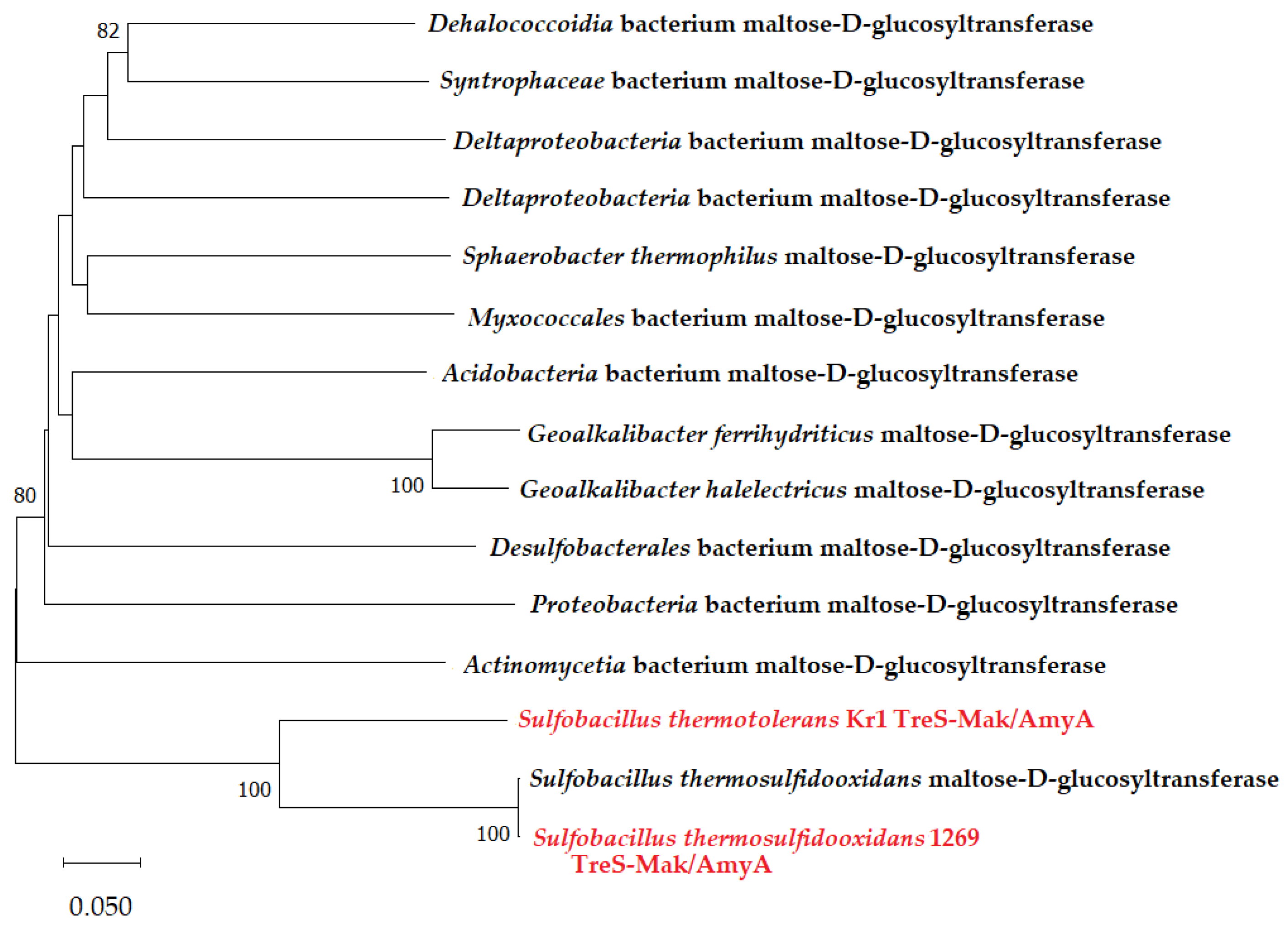
2.6. Phylogenetic Analysis of Proteins

A phylogenetic tree was constructed in the MEGA 11 software package [35]. The evolutionary history was inferred using the neighbor-joining method [39] and amino acid sequences of phylogenetically close microbial proteins. The bootstrap test (1000 replicates) was applied [40]. The evolutionary distances were computed using the Poisson correction method [41] and were in the units of the number of amino acid substitutions per site.
All microbiological experiments with flasks and enzyme measurements were carried out in duplicate. Statistical processing was performed using Microsoft Excel 2013. The standard deviation (SD) of the arithmetic mean was calculated, and the significance of the results was assessed using Student’s t-test at the significance level p ≤ 0.1.

3. Results and Discussion

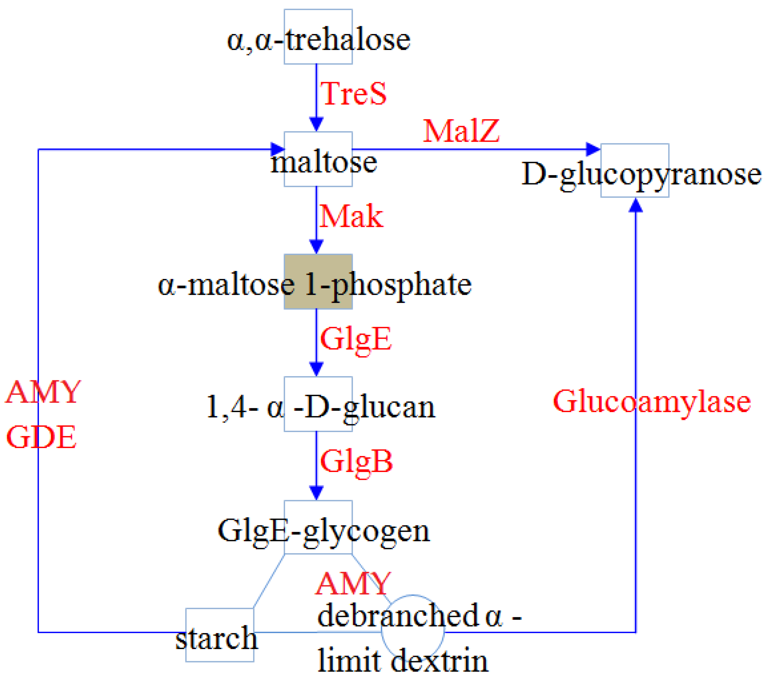
3.1. α-Glucan metabolism in Sulfobacillus Species: Biosynthesis and Degradation of Polysaccharides

In this work, we analyzed genomes of S. thermotolerans Kr1, S. thermosulfidooxidans 1269, and other species of the genus Sulfobacillus for the presence of the genes encoding proteins related to glycogen/starch metabolism pathways. Polysaccharides are the main storers of sugars used for the metabolic needs of diverse microorganisms. Biomining acidophilic microorganisms, such as bacteria of the genus Sulfobacillus, use sugars in catabolic and biosynthetic pathways, including biosynthesis of the polysaccharide capsule, which has several important functions in the microbe–microbe and microbe–mineral interactions [1,12,21].

3.1.1. Biosynthesis of α-Glucans in Sulfobacillus Species

Enzymes of the classical GlcA-GlcC pathway found in the majority of microorganisms were not identified in any genomes of Sulfobacillus bacteria available in the databases. At the same time, all key enzymes involved in the biosynthesis of α-glucans from trehalose were revealed in both S. thermotolerans Kr1 and S. thermosulfidooxidans 1269 (Figure 1, Table 1). These enzymes belonged to the GlgE-dependent four-step biosynthesis pathway previously discovered in mycobacteria [42,43].
According to the analysis of the Sulfobacillus genomes carried out in this study, the genome of S. thermotolerans Kr1 encoded an enzyme that was homologous to α-amylase/trehalose synthase of S. thermosulfidooxidans strains (TreS-Mak/Amy). A comparison to other Sulfobacillus genomes did not reveal any genes coding for this protein in other Sulfobacillus species. Phylogenetic analysis of S. thermotolerans Kr1 and S. thermosulfidooxidans 1269 TreS-Mak/Amy proteins showed that they occupied a separate branch in the phylogenetic tree, forming a common protein cluster for these two species (Figure 2). Their closest homologs belonged to microorganisms of distant phylogenetic groups that were not represented in acidophilic chemolithotrophic communities. Therefore, in the case of horizontal gene transfer to one of these Sulfobacillus species (and further to another one), the TreS-Mak/Amy gene was not acquired from the common members of acidophilic microbial communities but from other bacteria. Alternatively, and most probably, these genes were inherited from the ancestors of the phylum Firmicutes.
This enzyme is multifunctional. It contains an α-amylase catalytic domain (Amy) and a trehalose synthase-fused maltokinase domain (TreS-Mak), as in the case of the enzymes found in other bacterial phyla [43]. In this fused domain, the function of the trehalose synthase (TreS) is to interconvert maltose and trehalose. In turn, the maltokinase domain (Mak) catalyzes the second step of glycogen synthesis: maltose conversion into maltose 1-phosphate.
Genomes of the strains S. thermotolerans Kr1 and S. thermosulfidooxidans 1269 encoded a starch/glycogen synthase (SS, or GlgE), catalyzing the synthesis of linear α-1,4-glucans. The GlgE synthase found in these two Sulfobacillus species showed no similarity to any of the proteins encoded by S. acidophilus. However, it is encoded by S. benefaciens genome. The glgB gene encoding 1,4-α-glucan (glycogen) branching enzyme (BE, or GlgB) was identified in close proximity to the treS gene. In both S. thermotolerans and S. thermosulfidooxidans, the GlgE, TreS-Mak/Amy, and GlgB proteins were encoded by the gene clusters (ORF IDs BXT84_11955–11960 (strain Kr1) and SAMN00768000_3316–3318 (strain 1269) (Table 1). The 1,4-α-glucan (glycogen) BE was classified as a member of the glycoside hydrolase family 13 (GH13). BEs of the strains S. thermotolerans Kr1 and S. thermosulfidooxidans 1269 showed 68% a. a. similarity (99% coverage) (Table 1). The function of BE is to catalyze the intra- or intermolecular transglucosylation (by cleaving α-1,4-glucosidic bonds to form new α-1,6 branching points), and this enzyme plays a pivotal role in determining the branched structure of α-glucan [44]. A glycogen debranching enzyme (GDE), alternative to GlgB and essential for the final step of glycogen biosynthesis from α-1,4-glucans, was also identified in the genomes of S. thermotolerans and S. thermosulfidooxidans. GDEs of the two studied strains showed 53% a. a. similarity (Table 1). α-D-glucose is another product of the reaction catalyzed by the GDE. Thus, the glycogen debranching enzyme also promotes the destruction of the storage α-glucan (Section 3.1.2; Figure 1).

3.1.2. α-Glucan Degradation Pathways in Bacteria of the Genus Sulfobacillus

Branched α-glucans with α-1,6 linkages (glycogen and starch) can be degraded via reactions catalyzed by the GDE (amylo-α-1,6-glucosidase) mentioned above and two key enzymes identified in the genomes of the strains Kr1 and 1269 (Figure 1, Table 1). The first one is α-amylase possessing amylolytic activity (acting at) and decomposing glycogen/starch into α-limited dextrins and maltose, while maltase-glucoamylase (α-glucosidase, MalZ) converts maltose into glucose. The second key enzyme glucoamylase (GH15 family) degrades dextrins to α-D-glucose molecules. In addition to α-1,4 activity, glucoamylases also hydrolyze α-1,6 glucosidic linkages at the branching points of α-glucan molecules, although with lower activity than α-1,4 bonds (with rare exceptions, such as an enzyme of the fungus Amorphoteca (Hormoconis) resinae, showing an unusually high α-1,6 activity) [45]. Thus, glucoamylases can catalyze the hydrolysis of both α-1,4 and α-1,6 glucosidic linkages to release free D-glucose monomers from the non-reducing ends of starch and other branched α-glucans (including limited dextrins). S. thermotolerans Kr1 and S. thermosulfidooxidans 1269 genomes encoded glucoamylase homologs showing 70–84% (a. a.) similarity between them (Table 1).
Thus, glucoamylases, α-amylases, and debranching enzymes were the main enzymes identified in the genomes of the two Sulfobacillus species, responsible for the degradation of starch/glycogen, with soluble sugars as main products.

3.2. Microbial Growth and Polysaccharide Oxidation

Since the four-step biosynthesis pathway of α-glucans from trehalose was revealed in the genomes of two Sulfobacillus species in this study, the ability of Sulfobacillus strains to grow in the medium containing polysaccharides was tested. For this purpose, the strains S. thermotolerans Kr1 and S. thermosulfidooxidans 1269 were cultivated for several consecutive passages under heterotrophic conditions in the iron-free modified 9 K medium containing glycogen or soluble starch. In addition, the strains were also grown in the iron-free modified 9 K medium containing trehalose as the sole energy substrate.
In the first transfer of S. thermotolerans Kr1 and S. thermosulfidooxidans 1269 cells from mixotrophic to heterotrophic conditions, their cell yields were as follows. After 48 h of growth, the cell numbers were similar, reaching 5 (±0.5) × 107 cells/mL in the medium containing glycogen or starch and 0.9–1.0 × 10⁸ cells/mL in the trehalose-containing medium. Bacterial populations contained not only vegetative cells but also ~25–30% of endospores (dormant forms) and forespores (an intermediate state between an active cell and a dormant form). Thus, these conditions were not optimal for the growth of these strains. However, a gradual adaptation of the two strains to heterotrophic conditions during five sequential culture transfers resulted in an increase in the overall cell numbers up to 2.4–3.0 × 10⁸ cells/mL. At the same time, the proportion of spores and forespores decreased to 1.5–2%.
These results agree with the previously obtained data on the organoheterotrophic growth of S. thermosulfidooxidans 1269 and S. thermotolerans Kr1. These two species are capable of more active organoheterotrophic growth than other members of the genus Sulfobacillus [9,46,47]. High rates of utilization of carbohydrates and other organic compounds, as well as high activities of relevant enzymes, have been reported for the strains Kr1 and 1269 [46,47,48]. S. thermotolerans Kr1 sustained growth for more passages and had a higher intracellular ATP pool under heterotrophic conditions than other species of the genus Sulfobacillus [49]. Moreover, the genome of S. thermotolerans Kr1 has been previously shown to encode all components of the lactose–galactose catabolism pathway, which is not present in other species of this genus [9].
Previously, the ability of S. thermosulfidooxidans 1269 to grow with trehalose under heterotrophic conditions has been reported [12]. In this study, active growth in the medium containing trehalose as the sole energy source was also shown for the strain Kr1 after five transfers, although it has not previously been identified [23]. After the first passage (36 h of growth), the maximum cell yield of S. thermotolerans Kr1 was 1.1 (±0.2) × 108 cells/mL. In the fifth culture transfer, the cell number increased up to 2.9 (±0.3) × 108 cells/mL, indicating the successful adaptation of the strain to heterotrophic conditions of growth in the presence of trehalose.
Trehalose was found to be synthesized by S. thermosulfidooxidans as a compatible solute [24]. In the present work, the specific characteristic of S. thermotolerans Kr1 growth under heterotrophic conditions with trehalose was the formation of cell aggregates, along with single and dividing cells. We suggest that such aggregation might be associated with the presence of trehalose in the medium. These results are in agreement with the previous studies indicating that the size of the mucous polysaccharide capsule surrounding cells increased twice (from 0.12 to 0.25 μm) in the strains 1269 [46] and Kr1 [47] under heterotrophic conditions of growth with sugars, in comparison with the optimal mixotrophic conditions in the presence of inorganic and organic substrates. The encapsulation of cells may be of particular importance for bacteria of the genus Sulfobacillus, which are able to survive and proliferate at elevated temperatures, low pH values, high concentrations of metal ions, and under deficiency of energy and carbon substrates.
At the same time, the production and secretion of extracellular polymeric substances (EPS) by acidophilic microorganisms is accepted as a key mechanism promoting irreversible cell attachment to surfaces (including mineral surfaces) and biofilm development. The building blocks in EPS biosynthesis are a series of modified sugars (UDP-galactose, UDP-glucose, dTDP-rhamnose, and GDP-mannose), which are synthesized from the simple carbohydrates: glucose-1-phosphate (glucose-1-P) and glucose-6-phosphate (glucose-6-P) [50]. Potentially, due to the presence of all enzymes necessary for trehalose conversion to glucose monomers in S. thermotolerans and S. thermosulfidooxidans, EPS could be produced under these specific conditions, resulting in cell agglomeration.

3.3. Activities of Enzymes Degrading α-Glucans

Specific activities of extracellular enzymes degrading α-glucans (starch and glycogen) were detected in the cell-free supernatants obtained from the late-exponential cultures of S. thermotolerans Kr1 and S. thermosulfidooxidans 1269. This assay was carried out to confirm the results on the presence of α-glucan degradation pathways identified in the genomes of these two Sulfobacillus species. Measurements of reduced sugars in the cell-free supernatants detected activities of the α-glucan-degrading enzymes in both strains. The results are summarized in Table 2.
The data obtained indicated that the activities in both S. thermotolerans Kr1 and S. thermosulfidooxidans 1269 were rather low but comparable with the activities of other enzymes of carbon metabolism in these species, which also remain at low levels under different conditions of cultivation [16,47,48]. Thus, this is a common characteristic of bacteria of the genus Sulfobacillus. In the case of the strain S. thermosulfidooxidans 1269, all enzymes of the starch/glycogen degradation pathway in the supernatants showed only slightly higher values than those of S. thermotolerans Kr1. Bai et al. have reported a 2.3 U mL−1 α-amylase production in the heterotrophic acidophilic strain Alicyclobacillus sp. A4 in the presence of starch as a carbon source [51]. Nevertheless, literature data on the activities of these enzymes in acidophiles are scarce.

4. Conclusions

In the case of acidophilic chemolithotrophic microorganisms, the production of polysaccharides and other EPS can promote close attachment of the cells to solid surfaces, such as mineral particles. In turn, attachment to the surface may induce polysaccharide and EPS formation [50,52].
In the current study, we showed that two species of the genus Sulfobacillus possessed enzymes for the trehalose-dependent pathway of α-glucan biosynthesis. Moreover, the key enzyme, a fused multidomain TreS-Mak/Amy protein, was encoded only in the genomes of these two species and was not present in the genomes of other bacteria of the genus Sulfobacillus.
The ability to grow organoheterotrophically and consume trehalose, starch, and glycogen as sole organic substrates under heterotrophic conditions of growth was also shown. Although the optimum metabolic type of all Sulfobacillus species is a mixotrophic one, gradual adaptation to polysaccharides within several culture transfers resulted in sufficient growth under these conditions. Thus, we can conclude that S. thermosulfidooxidans and S. thermotolerans can use polysaccharide metabolism pathways not only for extracellular EPS production for the slime matrix and capsule but also for their growth processes. Active organoheterotrophic growth is beneficial for Sulfobacillus species in the presence of organic compounds formed as a result of microbial cell lysis in the dense pulp of industrial bioreactors. The property of some members of acidophilic microbial communities to use organic substances is also essential for the detoxification of the environment for obligately autotrophic microorganisms [11,21,22]. Under adverse conditions characterized by the lack of organic substrates in the medium of growth, studied bacteria of the genus Sulfobacillus can use storage polysaccharide material (glycogen) as a source of organic substrates.

Author Contributions

Conceptualization, A.P.; methodology, A.P. and M.M.; software, A.P. and M.M.; validation, A.P. and M.M.; formal analysis, A.P. and M.M.; investigation, A.P. and M.M.; resources, A.P. and M.M.; data curation, A.P. and M.M.; writing—original draft preparation, A.P.; writing—review and editing, A.P. and M.M.; visualization, A.P. and M.M.; supervision, A.P.; project administration, A.P.; funding acquisition, A.P. and M.M. All authors have read and agreed to the published version of the manuscript.

Funding

This research was funded by the Ministry of Science and Higher Education (Russian Federation).

Data Availability Statement

The data presented in this study are available on request from the corresponding author.

Conflicts of Interest

The authors declare no conflict of interest.

References

  1. Kaksonen, A.H.; Deng, X.; Bohu, T.; Zea, L.; Khaleque, H.N.; Gumulya, Y.; Boxall, N.J.; Morris, C.; Cheng, K.Y. Prospective directions for biohydrometallurgy. Hydrometallurgy 2020, 195, 105376. [Google Scholar] [CrossRef]
  2. Wills, B.A.; Finch, J.A. Wills’ Mineral Processing Technology: An Introduction to the Practical Aspects of Ore Treatment and Mineral Recovery, 8th ed.; Butterworth-Heinemann: Oxford, UK, 2015. [Google Scholar]
  3. Muravyov, M.; Panyushkina, A.; Fomchenko, N. Bulk flotation followed by selective leaching with biogenic ferric iron is a promising solution for eco-friendly processing of complex sulfidic ores. J. Environ. Manag. 2022, 318, 115587. [Google Scholar] [CrossRef]
  4. Kondrat’eva, T.F.; Pivovarova, T.A.; Tsaplina, I.A.; Fomchenko, N.V.; Zhuravleva, A.E.; Murav’ev, M.I.; Melamud, V.S.; Bulayev, A.G. Diversity of the communities of acidophilic chemolithotrophic microorganisms in natural and technogenic ecosystems. Microbiology 2012, 81, 1–24. [Google Scholar] [CrossRef]
  5. Hedrich, S.; Schippers, A. Distribution of acidophilic microorganisms in natural and man-made acidic environments. Curr. Issues Mol. Biol. 2021, 40, 25–48. [Google Scholar] [CrossRef]
  6. Panyushkina, A.; Bulaev, A.; Belyi, A.V. Unraveling the central role of sulfur-oxidizing Acidiphilium multivorum LMS in industrial bioprocessing of gold-bearing sulfide concentrates. Microorganisms 2021, 9, 984. [Google Scholar] [CrossRef] [PubMed]
  7. Panyushkina, A.; Muravyov, M.; Fomchenko, N. A Case of predominance of Alicyclobacillus tolerans in microbial community during bioleaching of pentlandite-chalcopyrite concentrate. Minerals 2022, 12, 396. [Google Scholar] [CrossRef]
  8. Rawlings, D.E.; Johnson, D.B. The microbiology of biomining: Development and optimization of mineral-oxidizing microbial consortia. Microbiology 2007, 153, 315–324. [Google Scholar] [CrossRef]
  9. Panyushkina, A.E.; Babenko, V.V.; Nikitina, A.S.; Selezneva, O.V.; Tsaplina, I.A.; Letarova, M.A.; Kostryukova, E.S.; Letarov, A.V. Sulfobacillus thermotolerans: New insights into resistance and metabolic capacities of acidophilic chemolithotrophs. Sci. Rep. 2019, 9, 15069. [Google Scholar] [CrossRef] [PubMed]
  10. Zhang, R.; Hedrich, S.; Jin, D.; Breuker, A.; Schippers, A. Sulfobacillus harzensis sp. nov., an acidophilic bacterium inhabiting mine tailings from a polymetallic mine. Int. J. Syst. Evol. Microbiol. 2021, 71, 004871. [Google Scholar] [CrossRef]
  11. Muravyov, M.; Panyushkina, A. Distinct roles of acidophiles in complete oxidation of high-sulfur ferric leach product of zinc sulfide concentrate. Microorganisms 2020, 8, 386. [Google Scholar] [CrossRef] [Green Version]
  12. Tsaplina, I.A.; Bogdanova, T.I.; Sayakin, D.D.; Karavaiko, G.I. Effects of organic substances on the growth of Sulfobacillus thermosulfidooxidans and pyrite oxidation. Microbiology 1992, 60, 686–692. [Google Scholar]
  13. Watling, H.R. The bioleaching of nickel-copper sulfides. Hydrometallurgy 2008, 91, 70–88. [Google Scholar] [CrossRef]
  14. Collinson, D.M.; Usher, K.M.; Nichols, P.D.; Watling, H.R. Habituation of Sulfobacillus thermosulfidooxidans to 4-nonylphenol in ferrous ion growth medium. Process Biochem. 2011, 46, 108–115. [Google Scholar] [CrossRef]
  15. Zhuravleva, A.E.; Ismailov, A.D.; Tsaplina, I.A. Electron donors at oxidative phosphorylation in bacteria of the genus Sulfobacillus. Microbiology 2009, 78, 811–814. [Google Scholar] [CrossRef]
  16. Tsaplina, I.A.; Krasil’nikova, E.N.; Zakharchuk, L.M.; Egorova, M.A.; Bogdanova, T.I.; Karavaiko, G.I. Carbon metabolism in Sulfobacillus thermosulfidooxidans subsp. asporogenes, strain 41. Microbiology 2000, 69, 271–276. [Google Scholar] [CrossRef]
  17. Tsaplina, I.A.; Zhuravleva, A.E.; Egorova, M.A.; Bogdanova, T.I.; Krasil’nikova, E.N.; Zakharchuk, L.M.; Kondrat’eva, T.F. Response to oxygen limitation in bacteria of the genus Sulfobacillus. Microbiology 2010, 79, 13–22. [Google Scholar] [CrossRef]
  18. Hedrich, S.; Johnson, D.B. Aerobic and anaerobic oxidation of hydrogen by acidophilic bacteria. FEMS Microbiol. Lett. 2013, 349, 40–45. [Google Scholar] [CrossRef]
  19. Panyushkina, A.; Matyushkina, D.; Pobeguts, O. Understanding stress response to high-arsenic gold-bearing sulfide concentrate in extremely metal-resistant acidophile Sulfobacillus thermotolerans. Microorganisms 2020, 8, 1076. [Google Scholar] [CrossRef] [PubMed]
  20. Zhuravleva, A.E.; Tsaplina, I.A.; Kondrat’eva, T.F. Specific characteristics of the strains isolated from a thermoacidophilic microbial community oxidizing antimony sulfide ore. Microbiology 2011, 80, 70–81. [Google Scholar] [CrossRef]
  21. Vardanyan, A.; Vyrides, I. Acidophilic bioleaching at high dissolved organic compounds: Inhibition and strategies to counteract this. Miner. Eng. 2019, 143, 105943. [Google Scholar] [CrossRef]
  22. Merino, M.P.; Andrews, B.A.; Parada, P.; Asenjo, J.A. Characterization of Ferroplasma acidiphilum growing in pure and mixed culture with Leptospirillum ferriphilum. Biotechnol. Prog. 2016, 32, 1390–1396. [Google Scholar] [CrossRef]
  23. Bogdanova, T.I.; Tsaplina, I.A.; Kondrat’eva, T.F.; Duda, V.I.; Suzina, N.E.; Melamud, V.S.; Tourova, T.P.; Karavaiko, G.I. Sulfobacillus thermotolerans sp. nov., a thermotolerant, chemolithotrophic bacterium. Int. J. Syst. Evol. Microbiol. 2006, 56, 1039–1042. [Google Scholar] [CrossRef] [PubMed]
  24. Galleguillos, P.A.; Grail, B.M.; Hallberg, K.B.; Demergasso, C.S.; Johnson, D.B. Identification of trehalose as a compatible solute in different species of acidophilic bacteria. J. Microbiol. 2018, 56, 727–733. [Google Scholar] [CrossRef]
  25. Justice, N.B.; Norman, A.; Brown, C.T.; Singh, A.; Thomas, B.C.; Banfield, J.F. Comparison of environmental and isolate Sulfobacillus genomes reveals diverse carbon, sulfur, nitrogen, and hydrogen metabolisms. BMC Genom. 2014, 15, 1107. [Google Scholar] [CrossRef] [PubMed]
  26. Wilson, W.A.; Roach, P.J.; Montero, M.; Baroja-Fernández, E.; Muñoz, F.J.; Eydallin, G.; Viale, A.M.; Pozueta-Romero, J. Regulation of glycogen metabolism in yeast and bacteria. FEMS Microbiol. Rev. 2010, 34, 952–985. [Google Scholar] [CrossRef]
  27. Guo, X.; Yin, H.; Liang, Y.; Hu, Q.; Zhou, X.; Xiao, Y.; Ma, L.; Zhang, X.; Qiu, G.; Liu, X. Comparative genome analysis reveals metabolic versatility and environmental adaptations of Sulfobacillus thermosulfidooxidans strain ST. PLoS ONE 2014, 9, e99417. [Google Scholar] [CrossRef]
  28. Matzke, J.; Schwermann, B.; Bakker, E.P. Acidostable and acidophilic proteins: The example of the α-amylase from Alicyclobacillus acidocaldarius. Comp. Biochem. Physiol. A Physiol. 1997, 118, 475–479. [Google Scholar] [CrossRef]
  29. Karavaiko, G.I.; Bogdanova, T.I.; Tourova, T.P.; Kondrat’eva, T.F.; Tsaplina, I.A.; Egorova, M.A.; Krasil’nikova, E.N.; Zakharchuk, L.M. Reclassification of ‘Sulfobacillus thermosulfidooxidans subsp. thermotolerans’ strain K1 as Alicyclobacillus tolerans sp. nov. and Sulfobacillus disulfidooxidans Dufresne et al. 1996 as Alicyclobacillus disulfidooxidans comb. nov., and emended description of the genus Alicyclobacillus. Int. J. Syst. Evol. Microbiol. 2005, 55, 941–947. [Google Scholar] [PubMed]
  30. Panyushkina, A.E.; Tsaplina, I.A.; Grigor’eva, N.V.; Kondrat’eva, T.F. Thermoacidophilic microbial community oxidizing the gold-bearing flotation concentrate of a pyrite-arsenopyrite ore. Microbiology 2014, 83, 539–549. [Google Scholar] [CrossRef]
  31. Silverman, M.P.; Lundgren, D.G. Studies on the chemoautotrophic iron bacterium Ferrobacillus ferrooxidans. I. An improved medium and a harvesting procedure for securing high cell yields. J. Bacteriol. 1959, 77, 642–647. [Google Scholar] [CrossRef]
  32. Nelson, N. A photometric adaptation of the Somogyi method for the determination of glucose. J. Biol. Chem. 1944, 153, 375–380. [Google Scholar] [CrossRef]
  33. Somogyi, M. Notes on sugar determination. J. Biol. Chem. 1952, 195, 19–23. [Google Scholar] [CrossRef]
  34. Lowry, O.H.; Rosebrough, N.J.; Farr, A.L.; Randal, R.J. Protein measurement with the Folin phenol reagent. J. Biol. Chem. 1951, 193, 265–275. [Google Scholar] [CrossRef]
  35. Tamura, K.; Stecher, G.; Kumar, S. MEGA 11: Molecular evolutionary genetics analysis version 11. Mol. Biol. Evol. 2021, 38, 3022–3027. [Google Scholar] [CrossRef]
  36. Kanehisa, M.; Sato, Y.; Kawashima, M.; Furumichi, M.; Tanabe, M. KEGG as a reference resource for gene and protein annotation. Nucleic Acids Res. 2016, 44, D457–D462. [Google Scholar] [CrossRef]
  37. Caspi, R.; Billington, R.; Ferrer, L.; Foerster, H.; Fulcher, C.A.; Keseler, I.M.; Kothari, A.; Krummenacker, M.; Latendresse, M.; Mueller, L.A.; et al. The MetaCyc database of metabolic pathways and enzymes and the BioCyc collection of pathway/genome databases. Nucleic Acids Res. 2016, 44, D471–D480. [Google Scholar] [CrossRef]
  38. Paley, S.; O’Maille, P.E.; Weaver, D.; Karp, P.D. Pathway collages: Personalized multi-pathway diagrams. BMC Bioinform. 2016, 17, 529. [Google Scholar] [CrossRef] [PubMed]
  39. Saitou, N.; Nei, M. The neighbor-joining method: A new method for reconstructing phylogenetic trees. Mol. Biol. Evol. 1987, 4, 406–425. [Google Scholar]
  40. Felsenstein, J. Confidence limits on phylogenies: An approach using the bootstrap. Evolution 1985, 39, 783–791. [Google Scholar] [CrossRef]
  41. Zuckerkandl, E.; Pauling, L. Evolutionary divergence and convergence in proteins. In Evolving Genes and Proteins; Bryson, V., Vogel, H.J., Eds.; Academic Press: New York, USA, 1965; pp. 97–166. [Google Scholar]
  42. Kalscheuer, R.; Syson, K.; Veeraraghavan, U.; Weinrick, B.; Biermann, K.E.; Liu, Z.; Sacchettini, J.C.; Besra, G.; Bornemann, S.; Jacobs, W.R., Jr. Self-poisoning of Mycobacterium tuberculosis by targeting GlgE in an alpha-glucan pathway. Nat. Chem. Biol. 2010, 6, 376–384. [Google Scholar] [CrossRef]
  43. Fraga, J.; Maranha, A.; Mendes, V.; Pereira, P.J.; Empadinhas, N.; Macedo-Ribeiro, S. Structure of mycobacterial maltokinase, the missing link in the essential GlgE-pathway. Sci. Rep. 2015, 5, 8026. [Google Scholar] [CrossRef]
  44. Suzuki, R.; Koide, K.; Hayashi, M.; Suzuki, T.; Sawada, T.; Ohdan, T.; Takahashi, H.; Nakamura, Y.; Fujita, N.; Suzuki, E. Functional characterization of three (GH13) branching enzymes involved in cyanobacterial starch biosynthesis from Cyanobacterium sp. NBRC 102756. Biochim. et Biophys. Acta (BBA) Proteins Proteom. 2015, 1854, 476–484. [Google Scholar] [CrossRef] [PubMed]
  45. Marín-Navarro, J.; Polaina, J. Glucoamylases: Structural and biotechnological aspects. Appl. Microbiol. Biotechnol. 2011, 89, 1267–1273. [Google Scholar] [CrossRef] [PubMed]
  46. Senyushkin, A.A.; Severina, L.O.; Mityushina, L.L. Capsule formation by Sulfobacillus thermosulfidooxidans cells growing under oligotrophic and mixotrophic conditions. Microbiology 1997, 66, 455–461. [Google Scholar]
  47. Tsaplina, I.A.; Krasil’nikova, E.N.; Zhuravleva, A.E.; Egorova, M.A.; Zakharchuk, L.M.; Suzina, N.E.; Duda, V.I.; Bogdanova, T.I.; Stadnichuk, I.N.; Kondrat’eva, T.F. Phenotypic properties of Sulfobacillus thermotolerans: Comparative aspects. Microbiology 2008, 77, 654–664. [Google Scholar] [CrossRef]
  48. Zakharchuk, L.M.; Tsaplina, I.A.; Krasil’nikova, E.N.; Bogdanova, T.I.; Karavaiko, G.I. Carbon Metabolism in Sulfobacillus thermosulfidooxidans. Microbiology 1994, 63, 573–580. [Google Scholar]
  49. Tsaplina, I.A.; Zhuravleva, A.E.; Ismailov, A.D.; Zakharchuk, L.M.; Krasil’nikova, E.N.; Bogdanova, T.I.; Karavaiko, G.I. The dependence of intracellular ATP level on the nutrition mode of the acidophilic bacteria Sulfobacillus thermotolerans and Alicyclobacillus tolerans. Microbiology 2007, 76, 654–662. [Google Scholar] [CrossRef]
  50. Donati, E.R.; Sand, W. (Eds.) Microbial Processing of Metal Sulfides; Springer: Dordrecht, The Netherlands, 2007. [Google Scholar]
  51. Bai, Y.; Huang, H.; Meng, K.; Shi, P.; Yang, P.; Luo, H.; Luo, C.; Feng, Y.; Zhang, W.; Yao, B. Identification of an acidic α-amylase from Alicyclobacillus sp. A4 and assessment of its application in the starch industry. Food Chem. 2012, 131, 1473–1478. [Google Scholar] [CrossRef]
  52. Li, Q.; Sand, W. Mechanical and chemical studies on eps from Sulfobacillus thermosulfidooxidans: From planktonic to biofilm cells. Colloids Surf. B Biointerfaces 2017, 153, 34–40. [Google Scholar] [CrossRef]
Figure 1. Predicted α-glucan metabolism pathways in S. thermotolerans Kr1 and S. thermosulfidooxidans 1269. Arrows indicate reactions catalyzed by the enzymes encoded by the genomes. TreS and Mak proteins compose a common trehalose synthase-fused maltokinase domain (TreS-Mak) of the multifunctional enzyme, which also contains an α-amylase catalytic domain (Amy). Abbreviations: TreS, trehalose synthase (EC 5.4.99.16); Mak, maltokinase (EC 2.7.1.175); GlgE (SS), starch/glycogen synthase (α-1,4-glucan:maltose-1-phosphate maltosyltransferase) (EC 2.4.99.16); GlgB (BE), 1,4-α-glucan (glycogen) branching enzyme (EC 2.4.1.18); GDE, glycogen debranching enzyme (amylo-α-1,6-glucosidase) (EC 3.2.1.133); AMY, α-amylase (EC 3.2.1.1); MalZ, maltase-glucoamylase (α-glucosidase) (EC 3.2.1.20); glucoamylase, glucan 1,4-α-glucosidase (EC 3.2.1.3) hydrolyzing glycogen, starch, or dextrins to glucose monomers. The metabolic map was constructed using MetaCyc [37] and Pathway collages [38].
Minerals 13 00255 g001
Figure 2. Phylogenetic position of Sulfobacillus α-amylase/trehalose synthase. The phylogenetic tree was constructed using MEGA 11 [35]. The evolutionary history was inferred using the neighbor-joining method [39]. The optimal tree is shown. The percentage of replicate trees in which the associated taxa clustered together in the bootstrap test (1000 replicates) are shown next to the branches (values lower than 80% are hidden) [40]. The tree is drawn to scale, with branch lengths in the same units as those of the evolutionary distances used to infer the phylogenetic tree. The evolutionary distances were computed using the Poisson correction method [41] and are in the units of the number of amino acid substitutions per site. This analysis involved 15 amino acid sequences. All ambiguous positions were removed. There were a total of 1125 positions in the final dataset.
Minerals 13 00255 g002
Table 1. Enzymes of the GlgE-dependent polysaccharide biosynthesis and degradation pathways in S. thermotolerans Kr1 and S. thermosulfidooxidans 1269.
Strain, ORF IDKO 1Protein Size (a. a.)Protein Similarity (Coverage), %Putative Homolog
Kr1, BXT84_1269,
SAMN00768000_
Kr11269
119553318K1614765566163 (98)GlgE, α-1,4-glucan:maltose-1-phosphate maltosyltransferase (EC 2.4.99.16)
119603317K053431082108173 (99)TreS-Mak/AmyA, fused trehalose synthase (EC 5.4.99.16)/maltokinase (EC 2.7.1.175)/α-amylase (EC 3.2.1.1)
119653316K0070063263068 (99)GlgB, 1,4-α-glucan branching enzyme (EC 2.4.1.18)
161001445-746102053 (72)GDE; amylo-α-1,6-glucosidase (glycogen debranching enzyme) (EC 3.2.1.133)
080050408K0117880178970 (98)Glucoamylase, maltase-glucoamylase, (glucan 1,4-α-glucosidase) (EC 3.2.1.3)
121952946-65265484 (99.7)Glucoamylase, glycoside hydrolase family 15 (EC 3.2.1.3)
161251977-60161444 (96)
135351977-60161474 (97)
051152343K1592279580653 (96)Maltase-glucoamylase, α-glucosidase (EC 3.2.1.20)
1 KO, KEGG Orthology identifiers.
Table 2. α-Glucan (starch and glycogen) degradation by extracellular enzymes of S. thermotolerans Kr1 and S. thermosulfidooxidans 1269.
SubstrateProductPutative Enzyme(s)Enzyme Activity, µmol/(min∙mg Protein) 1
Kr11269
StarchMaltoseα-Amylase (EC 3.2.1.1)0.54 ± 0.050.66 ± 0.05
GlycogenMaltoseα-Amylase (EC 3.2.1.1)0.62 ± 0.030.71± 0.04
StarchGlucoseγ-Amylase/maltase-glucoamylase (EC 3.2.1.3) + α-amylase (EC 3.2.1.1) +
α-glucosidase (EC 3.2.1.20)
1.31 ± 0.041.40 ± 0.05
GlycogenGlucoseγ-Amylase/maltase-glucoamylase (EC 3.2.1.3) + α-amylase (EC 3.2.1.1) +
α-glucosidase (EC 3.2.1.20)
1.87 ± 0.061.93 ± 0.06
1 Mean values ± SD (p ≤ 0.05) are shown.
Disclaimer/Publisher’s Note: The statements, opinions and data contained in all publications are solely those of the individual author(s) and contributor(s) and not of MDPI and/or the editor(s). MDPI and/or the editor(s) disclaim responsibility for any injury to people or property resulting from any ideas, methods, instructions or products referred to in the content.

