
Manganese Oxide Minerals from the Xiangtan Manganese Deposit in South China and Their Application in Formaldehyde Removal

 , ,
, ,
Abstract
:1. Introduction
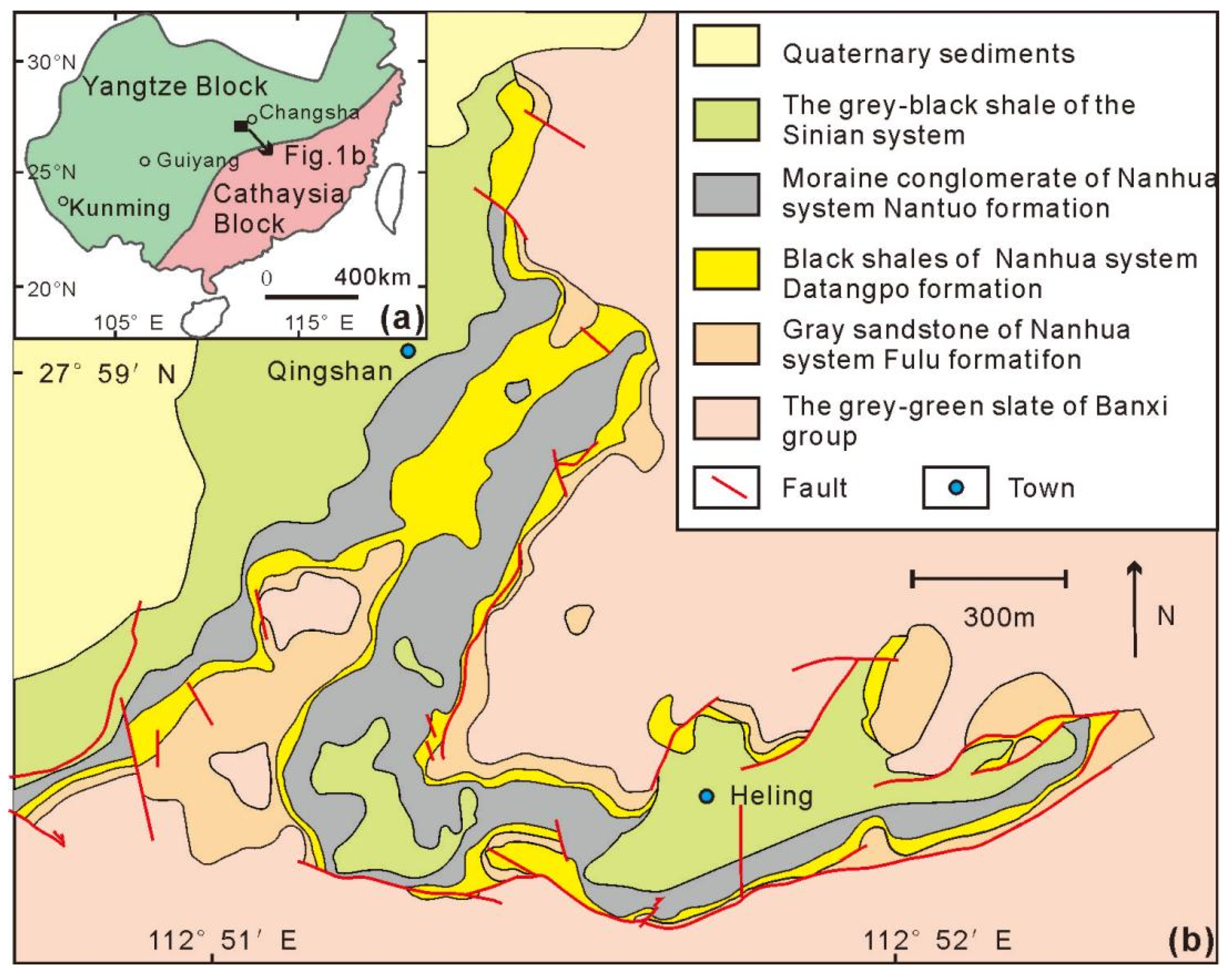
2. Geological Setting
3. Sampling and Methods
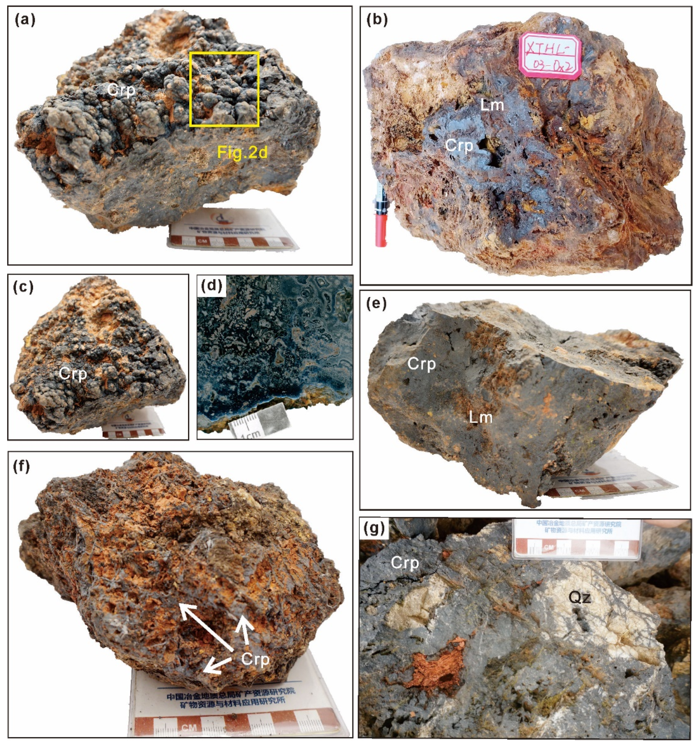
3.1. Sample Preparation
3.2. Mineralogical Analysis
3.3. Formaldehyde Removal Test
4. Results
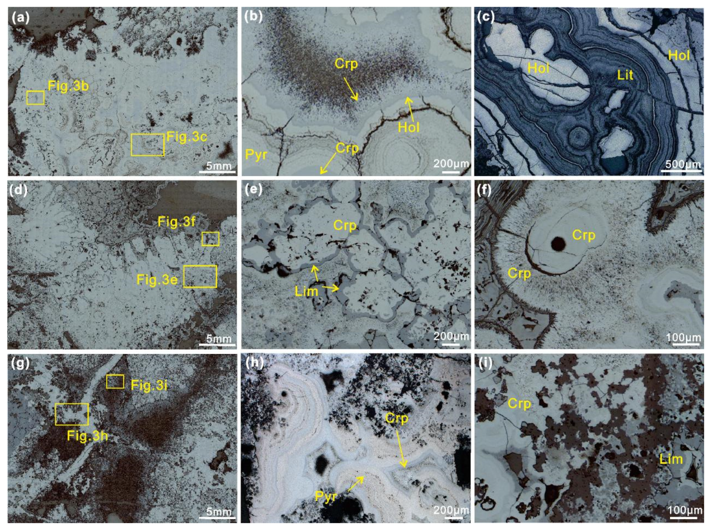
4.1. Optical Microscopy
4.2. EPMA Analysis
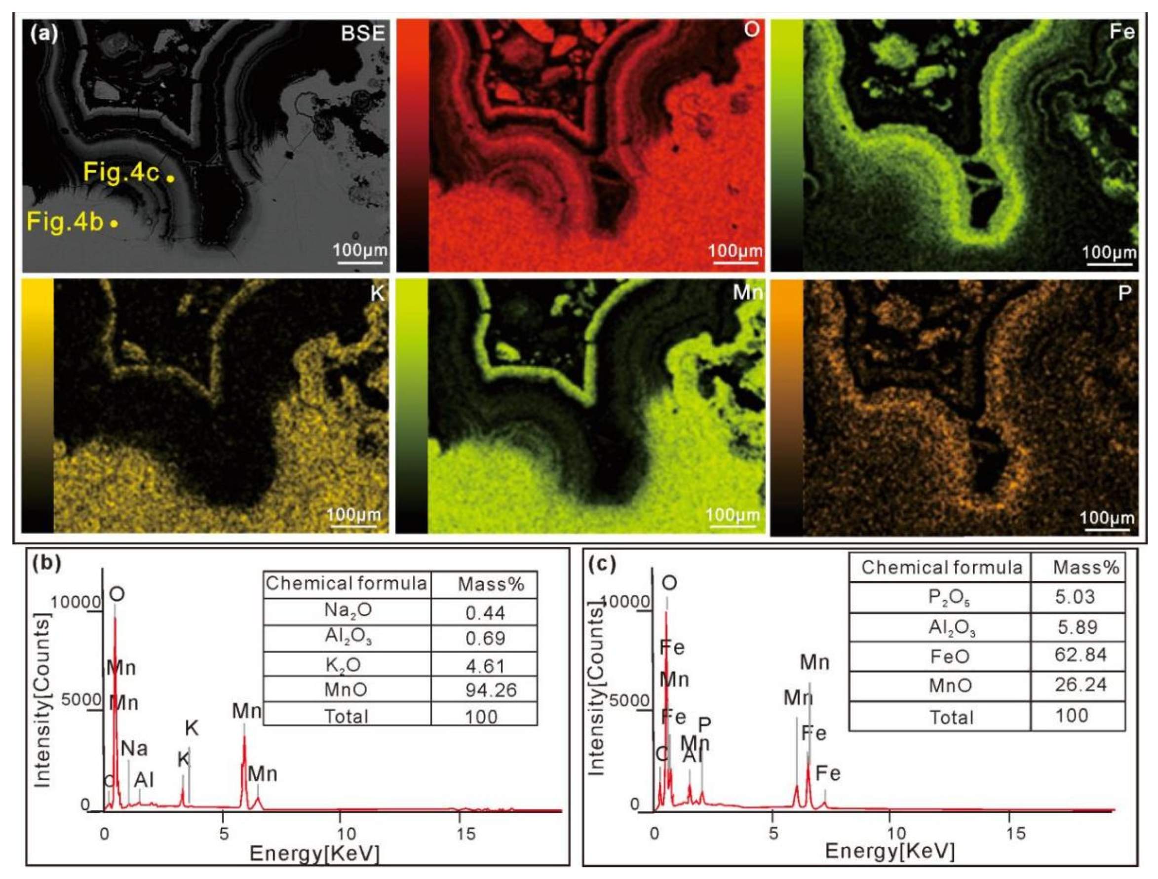
4.3. EDS Mappings
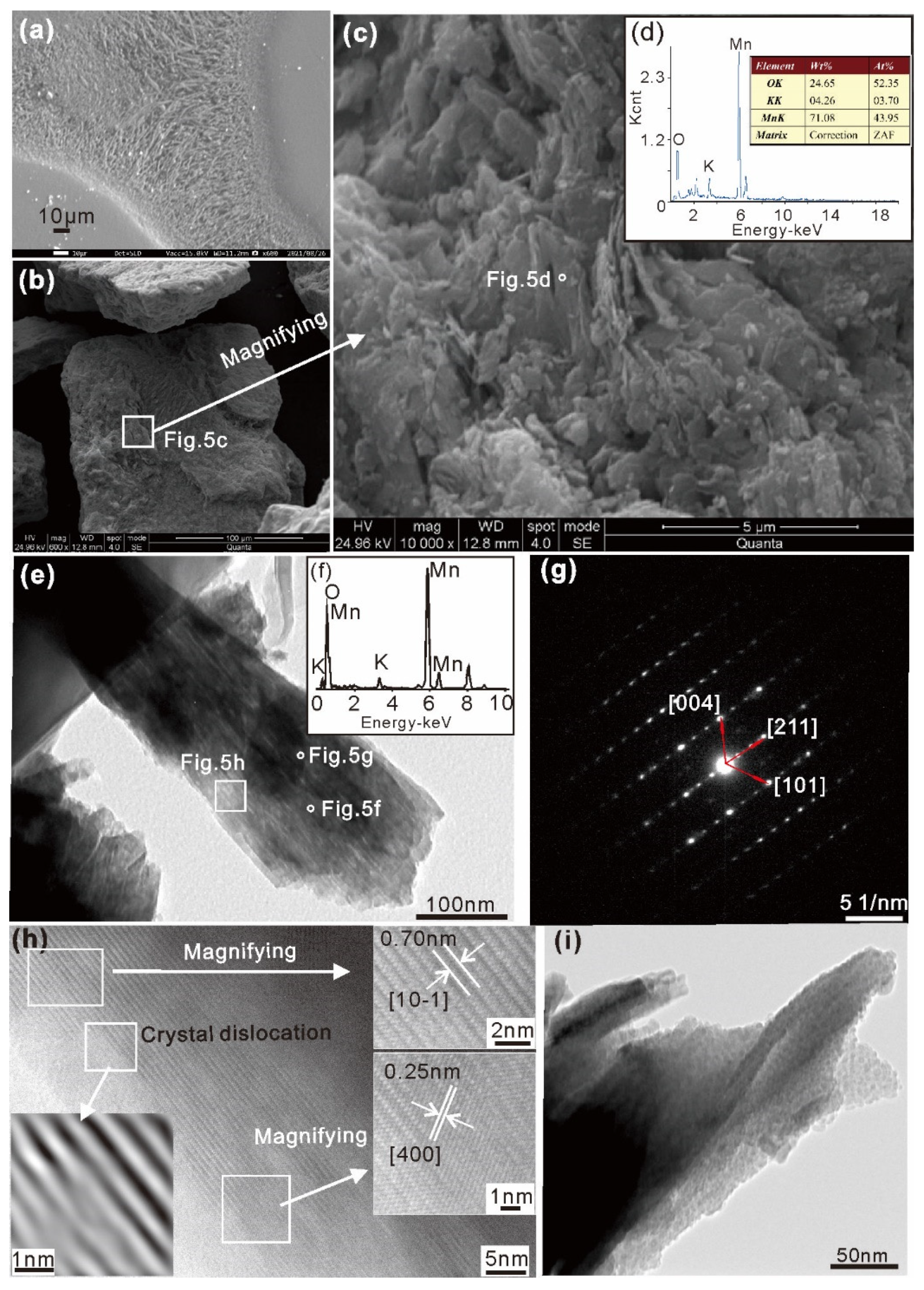
4.4. SEM and TEM Analysis
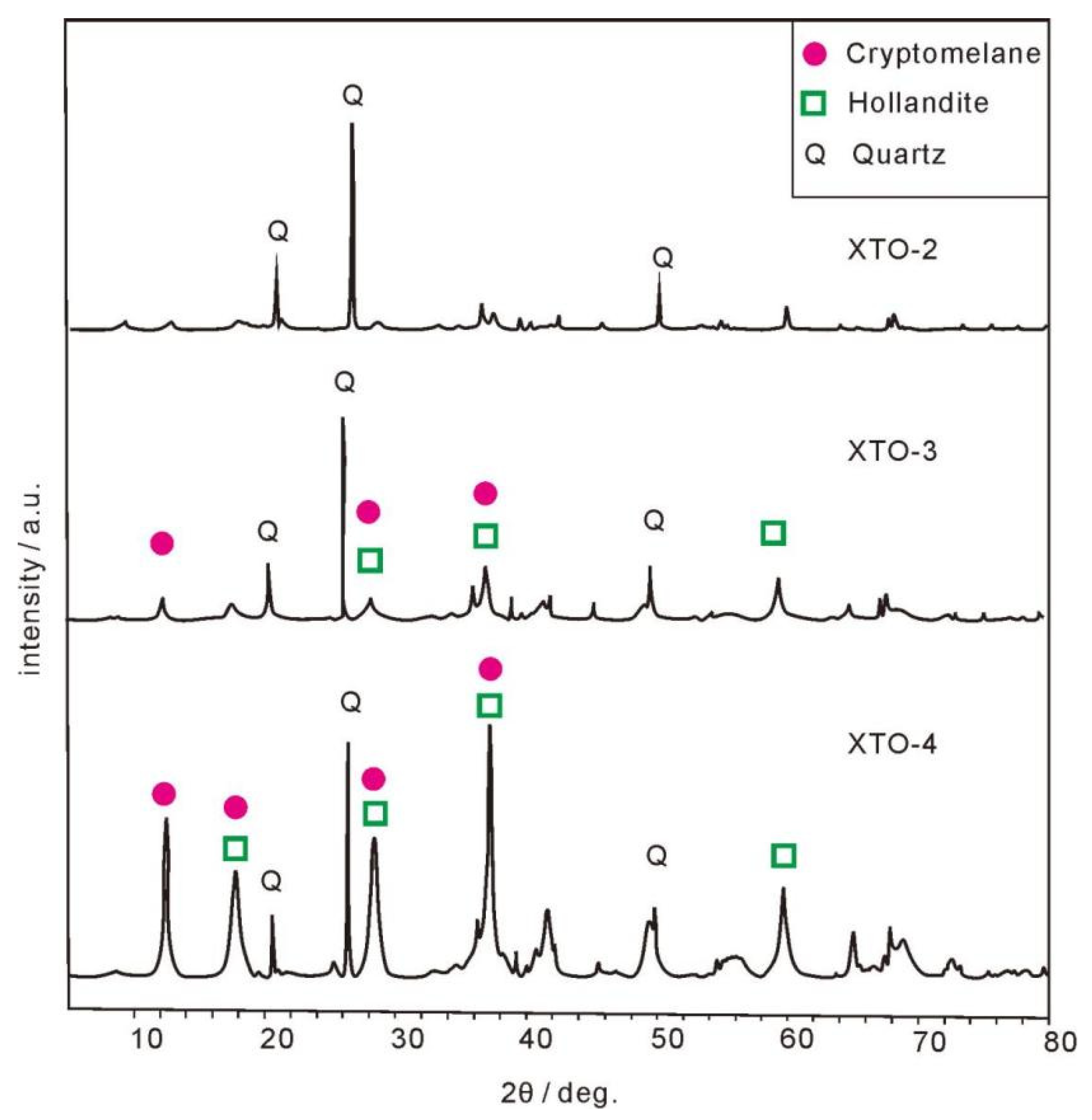
4.5. XRD Analysis
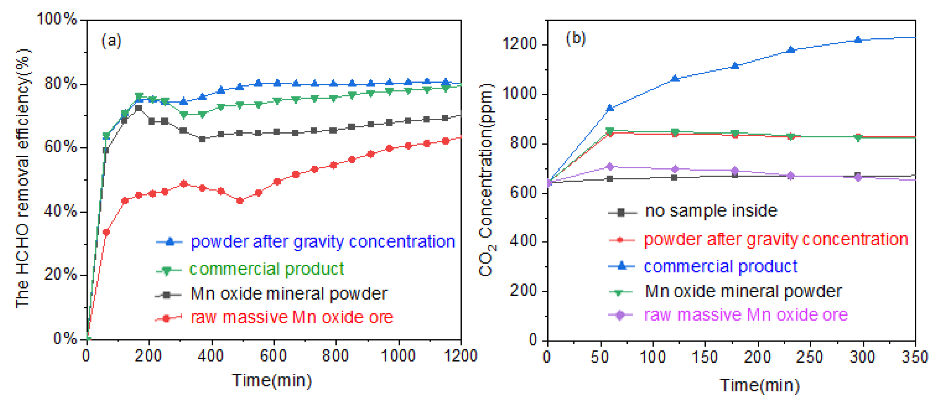
4.6. Evaluation of Formaldehyde Removal Activity
5. Discussion
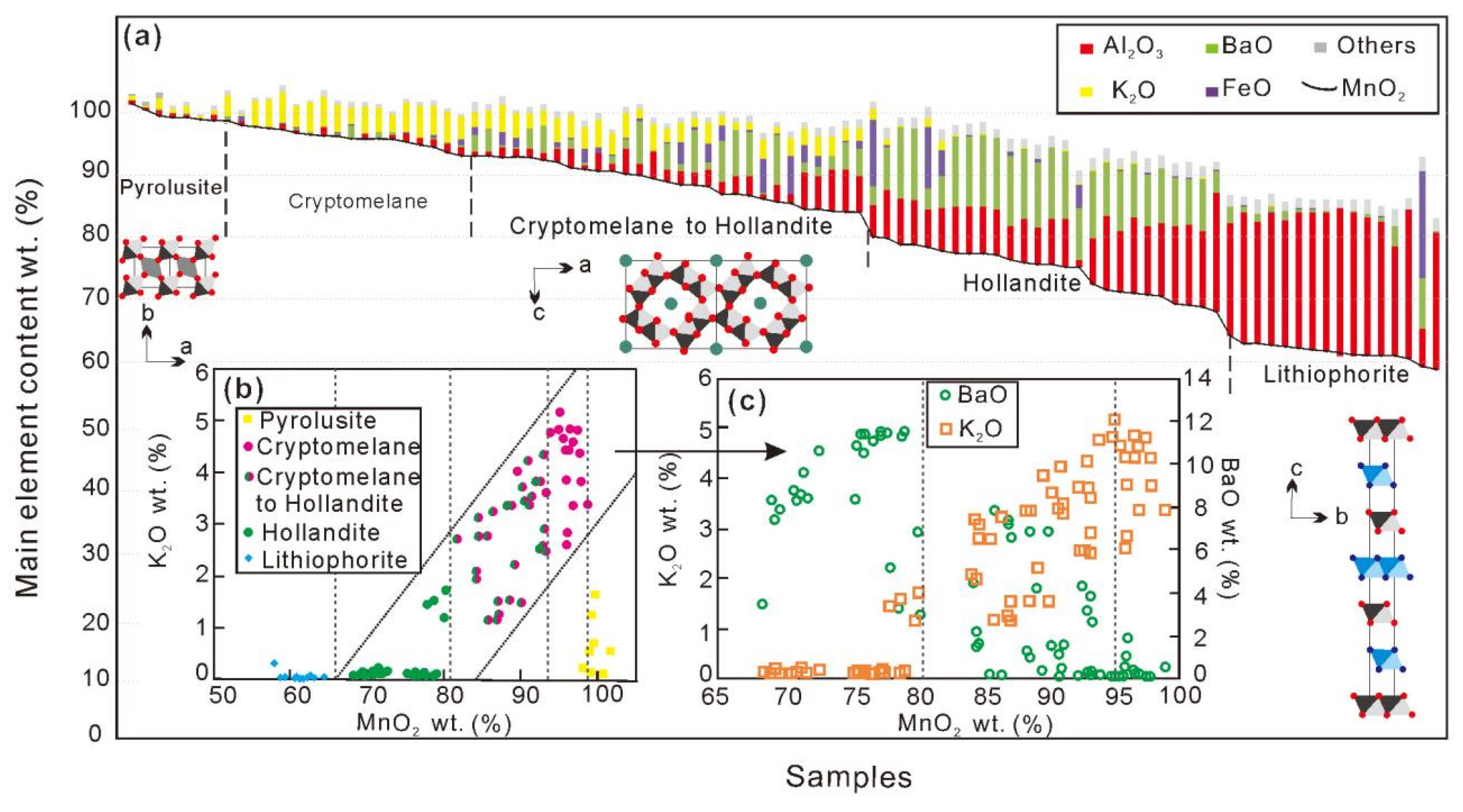
5.1. Characteristics of Main Manganese Oxide Minerals
5.2. Crystallochemical Characteristics of the Cryptomelane
5.3. Catalyst Activity of Formaldehyde by Natural Cryptomelane
6. Conclusions
7. Patents
Supplementary Materials
Author Contributions
Funding
Data Availability Statement
Acknowledgments
Conflicts of Interest
References
- Turekian, K.K.; Wedepohl, K.L. Distribution of the elements in some major units of the earth’s crust. Geol. Soc. Am. Bull. 1961, 72, 175–192. [Google Scholar] [CrossRef]
- Post, J.E. Manganese oxide minerals: Crystal structures and economic environmental significance. Proc. Natl. Acad. Sci. USA 1999, 96, 3447–3454. [Google Scholar] [CrossRef] [PubMed] [Green Version]
- Prasad, V.S.; Chaudhuri, M. Removal of bacteria turbidity from water by chemically treated manganese iron ores. Aqua 1995, 44, 80–82. [Google Scholar]
- Ghodbane, O.; Pascal, J.L.; Favier, F. Microstructural effects on Charge Storage Properties in MnO2 based Electrochemical Supercapacitors. ACS Appl. Mater. Interfaces 2009, 1, 1130. [Google Scholar] [CrossRef]
- Huang, M.; Li, F.; Dong, F.; Zhang, Y.; Zhang, L.-L. MnO2-based nanostructures for high-performance supercapacitors. J. Mater. Chem. A 2015, 3, 21380–21423. [Google Scholar] [CrossRef]
- Huang, H.; Xu, Y.; Feng, Q.; Leung, D.Y.C. Low temperature catalytic oxidation of volatile organic compounds: A review. Catal. Sci. Technol. 2015, 5, 2649–2669. [Google Scholar] [CrossRef]
- Nie, L.; Yu, J.; Jaroniec, M.; Tao, F.-F. Room-temperature catalytic oxidation of formaldehyde on catalysts. Catal. Sci. Technol. 2016, 6, 3649–3669. [Google Scholar] [CrossRef]
- Su, J.; Chen, C.; Guo, Y.; Xu, H.; Ke, Q. OMS-2-based catalysts with controllable hierarchical morphologies for highly efficient catalytic oxidation of formaldehyde. J. Hazard. Mater. 2019, 380, 120890. [Google Scholar] [CrossRef]
- Sekine, Y.; Nishimura, A. Removal of formaldehyde from indoor air by passive type air-cleaning materials. Atmos. Environ. 2001, 35, 2001–2007. [Google Scholar] [CrossRef]
- Tang, X.-F.; Li, Y.-G.; Huang, X.-M.; Xu, Y.-D.; Zhu, H.-Q.; Wang, J.-G.; Shen, W.-J. MnOx–CeO2 mixed oxide catalysts for complete oxidation of formaldehyde: Effect of preparation methodcalcination temperature. Appl. Catal. B Environ. 2006, 62, 265–273. [Google Scholar] [CrossRef]
- Tang, X.-F.; Chen, J.-L.; Huang, X.-M.; Xu, Y.-D.; Shen, W.-J. Pt/MnOx–CeO2 catalysts for the complete oxidation of formaldehyde at ambient temperature. Appl. Catal. B Environ. 2008, 81, 115–121. [Google Scholar] [CrossRef]
- Adelodun, A.A. Influence of Operation Conditions on the Performance of Non-thermal Plasma Technology for VOC Pollution Control. J. Ind. Eng. Chem. 2020, 92, 41–55. [Google Scholar] [CrossRef]
- Brown, G.E.; Calas, G. Mineral-aqueous solution interfaces and their impact on the environment. Geochem. Perspect. 2012, 1, 483–742. [Google Scholar] [CrossRef] [Green Version]
- Lu, A.H.; Li, Y.; Liu, F.-F.; Liu, Y.-W.; Ye, H.; Zhuang, Z.-Y.; Li, Y.-Z.; Ding, H.-R.; Wang, C.-Q. The photogeochemical cycle of Mn oxides on Earth’s surface. Mineral. Mag. 2021, 85, 22–38. [Google Scholar] [CrossRef]
- Wang, J.; Zhang, P.; Li, J.; Jiang, C.; Yunus, R.; Kim, J. Room-temperature oxidation of formaldehyde by layered manganese oxide: Effect of water. Environ. Sci. Technol. 2015, 49, 12372–12379. [Google Scholar] [CrossRef]
- Li, J.-G.; Zhang, P.-Y.; Wang, J.-L.; Wang, M.-X. Birnessite-type manganese oxide on granular activated carbon for formaldehyde removal at room temperature. J. Phys. Chem. C 2016, 120, 24121–24129. [Google Scholar] [CrossRef]
- Birkner, N.; Navrotsky, A. Thermodynamics of manganese oxides: Sodium, potassium, and calcium birnessite and cryptomelane. Proc. Natl. Acad. Sci. USA 2017, 114, E1046–E1053. [Google Scholar] [CrossRef] [Green Version]
- Potter, R.M.; Rossman, G.R. The tetravalent manganese oxides; identification, hydration, structural relationships by infrared spectroscopy. Am. Mineral. 1979, 64, 1199–1218. [Google Scholar]
- Mineral Commodity Summaries 2019; U.S. Geological Survey: Reston, VA, USA, 2019; 204p. [CrossRef]
- Gao, X.; Lu, A.-H.; Qin, S.; Zheng, Z. The refinement of crystal unit cell parameters of native cryptomelane in the supergene oxidation zone of the Xiangtan manganese deposit, Hunan Province. Acta Petrol. Mineral. 2003, 1, 77–79. (In Chinese) [Google Scholar]
- Gao, X.; Lu, A.-H.; Zheng, Z.; Jia, X.-X. Natural oxide octahedral molecular sieve OMS-2—The study of the characters of microstructure by for HRTEM cryptomelane of Xiangtan manganese deposit, Hunan province. J. Mineral. Petrol. 2005, 3, 52–57. (In Chinese) [Google Scholar]
- Zheng, J.-Y.; Zhao, W.-K.; Wang, X.; Zheng, Z.; Han, C.-B. Electric-enhanced hydrothermal synthesis of manganese dioxide for the synergistic catalytic of indoor low-concentration formaldehyde at room temperature. Chem. Eng. J. 2020, 401, 125790. [Google Scholar] [CrossRef]
- Liu, X.-F.; Wang, Q.-S.; Gao, X.-J. Mangenese Deposits of Guihzou, China; Guizhou People Press: Guiyang, China, 1989; pp. 1–194. (In Chinese) [Google Scholar]
- Fan, D.; Yang, P. Introduction to classification of manganese deposits of China. Ore Geol. Rev. 1999, 15, 1–13. [Google Scholar] [CrossRef]
- Pracejus, B.; Bolton, B.R.; Frakes, L.A. Nature and development of supergene manganese deposits, Groote Eylandt, Northern Territory, Australia. Ore Geol. Rev. 1988, 4, 71–98. [Google Scholar] [CrossRef]
- Lu, A.-H.; Gao, X.; Qin, S.; Wang, C.-Q. Cryptomelane KxMn8−xO16: Natural active octahedral molecular sieve OMS-2. Chin. Sci. Bull. 2003, 48, 920–923. [Google Scholar] [CrossRef]
- Richmond, W.E.; Fleischer, M. Cryptomelane, a new name for the commonest of the “psilomelane” minerals. Am. Miner. 1942, 27, 607–610. [Google Scholar]
- Sato, H.; Enoki, T.; Yamaura, J.I.; Yamamoto, N. Charge localization and successive magnetic phase transitions of mixed-valence manganese oxides K−1.5 (H3O) x Mn8O16 (0 < x < 0.5). Phys. Rev. B 1999, 59, 12836–12841. [Google Scholar]
- Wang, J.-L.; Jiang, C.-J.; Zhou, P.; Zhang, P.-Y.; Yu, J.-G. The effect of manganese vacancy in birnessite-type MnO2 on room-temperature oxidation of formaldehyde in air. Appl. Catal. B Environ. 2017, 204, 147–155. [Google Scholar] [CrossRef]
- Post, J.E.; Dreele, R.B.V.; Buseck, P.R. Symmetrycation displacements in hollandites: Structure refinements of hollandite, cryptomelane priderite. Acta Crystallogr. 1982, 38, 1056–1065. [Google Scholar] [CrossRef]
- Qi, Q.-P.; Zhang, W.-R.; Zhang, Y.-S.; Bai, G.-M.; Wang, S.-W.; Peng, L. Formaldehyde oxidation at room temperature over layered MnO2. Catal. Commun. 2021, 153, 106293. [Google Scholar] [CrossRef]
- Chen, T.; Dou, H.-Y.; Li, X.-L.; Tang, X.-F.; Li, L.-H.; Hao, J.-M. Tunnel structure effect of manganese oxides in complete oxidation of formaldehyde. Microporous Mesoporous Mater. 2009, 122, 270–274. [Google Scholar] [CrossRef]
- Espinal, L.; Wong-Ng, W.; Kaduk, J.A.; Allen, A.J.; Suib, S.L. Time–Dependent CO2 Sorption Hysteresis in a One-Dimensional Microporous Octahedral Molecular Sieve. J. Am. Chem. Soc. 2012, 134, 7944–7951. [Google Scholar] [CrossRef] [PubMed]
- Gao, T.; Norby, P. Frame stability of tunnel-structured cryptomelane nanofibers: The role of tunnel cations. Eur. J. Inorg. Chem. 2013, 28, 4948–4957. [Google Scholar] [CrossRef] [Green Version]
- De Guzman, R.N.; Shen, Y.F.; Neth, E.J.; Suib, S.L.; O’ Young, C.L.; Levine, S.; Newsam, J.M. Synthesis characterization of octahedral molecular sieves OMS-2 having the hollandite structure. Chem. Mater. 1994, 6, 815–821. [Google Scholar] [CrossRef]
- Vasconcelos, P.M.; Wenk, H.R.; Echer, C. In-situ study of the thermal behavior of cryptomelane by high-voltage analytical electron microscopy. Am. Mineral. 1994, 79, 80–90. [Google Scholar]
- Sekine, Y. Oxidative decomposition of formaldehyde by metal oxides at room temperature. Atmos. Environ. 2002, 36, 5543–5547. [Google Scholar] [CrossRef]
- Zhang, J.-H.; Li, Y.-B.; Wang, L.; Zhang, C.-B.; He, H. Catalytic oxidation of formaldehyde over manganese oxides with different crystal structures. Catal. Sci. Technol. 2015, 5, 2305–2313. [Google Scholar] [CrossRef]
- Wu, Y.; Lu, Y.; Song, C.-J.; Ma, Z.-C.; Xing, S.-T.; Gao, Y.-Z. A novel redox-precipitation method for the preparation of α-MnO2 with a high surface Mn4+ concentration and its activity toward complete catalytic oxidation of o-xylene. Catal. Today 2013, 201, 32–39. [Google Scholar] [CrossRef]
- Li, K.; Chen, C.; Zhang, H.-B.; Hu, X.-J.; Sun, T.-H.; Jia, J.-P. Effects of phase structure of MnO2 morphology of δ-MnO2 on toluene catalytic oxidation. Appl. Surf. Sci. 2019, 496, 143662. [Google Scholar] [CrossRef]
- Hou, J.-T.; Liu, L.-L.; Li, Y.-Z.; Mao, M.-Y.; Lv, H.-Q.; Zhao, X.-J. Tuning the K+ concentration in the tunnel of OMS-2 nanorods leads to a significant enhancement of the catalytic activity for benzene oxidation. Environ. Sci. Technol. 2013, 47, 13730–13736. [Google Scholar] [CrossRef]
- Xu, F.; Huang, Z.-W.; Hu, P.-P.; Chen, Y.-X.; Zheng, L.; Gao, J.-Y.; Tang, X.-F. The promotion effect of isolated potassium atoms with hybridized orbitals in catalytic oxidation. Chem. Commun. 2015, 51, 9888–9891. [Google Scholar] [CrossRef]
- Yusuf, A.; Sun, Y.; Snape, C.; He, J.; Wang, C.; Ren, Y.; Jia, H. Low-temperature formaldehyde oxidation over manganese oxide catalysts: Potassium mediated lattice oxygen mobility. Mol. Catal. 2020, 497, 111204. [Google Scholar] [CrossRef]
- Luo, J.; Zhang, Q.-H.; Huang, A.-M.; Suib, S.L. Total oxidation of volatile organic compounds with hydrophobic cryptomelane-type octahedral molecular sieves. Microporous Mesoporous Mater. 2000, 35–36, 209–217. [Google Scholar] [CrossRef]
- Bondi, A. van der Waals Volumes Radii. J. Phys. Chem. 1964, 68, 441–451. [Google Scholar] [CrossRef]
- Wang, Z.-M.; Tezuka, S.; Kanoh, H. Characterization of the structural surface properties of a synthesized hydrous hollandite by gaseous molecular adsorption. Chem. Mater. 2001, 13, 530–537. [Google Scholar] [CrossRef]
- Kijima, N.; Ikeda, T.; Oikawa, K.; Izumi, F.; Yoshimura, Y. Crystal structure of an open-tunnel oxide α-MnO2 analyzed by rietveld refinements MEM-based pattern fitting. J. Solid State Chem. 2004, 177, 1258–1267. [Google Scholar] [CrossRef]


| SiO2 | Al2O3 | CaO | Fe2O3 | K2O | MgO | MnO | Na2O | P2O5 | TiO2 | LOI | |
|---|---|---|---|---|---|---|---|---|---|---|---|
| Sample XTO-2 | 22.94 | 2.81 | 0.16 | 9.44 | 2.43 | 0.073 | 48.11 | 0.14 | 0.84 | 0.037 | 11.39 |
| Sample XTO-3 | 9.82 | 2.05 | 0.19 | 6.17 | 3.15 | 0.083 | 63.67 | 0.16 | 0.75 | 0.014 | 12.52 |
| Element | Standard | X-ray | Crystal | Method | S. D. (%) | D. L. (ppm) |
|---|---|---|---|---|---|---|
| Al | 02-rAl2SiO5-0315 | Ka | TAP | ZAF | 0.16 | 100 |
| Si | 03-SiO2-0224 | Ka | TAP | ZAF | 0.13 | 120 |
| Ca | 02-CaSiO3-0224 | Ka | PETL | ZAF | 0.14 | 80 |
| Ba | 21-BaSO4-0312 | La | PETL | ZAF | 0.17 | 210 |
| Mn | 14-MnTiO3-0224 | Ka | LIF | ZAF | 0.61 | 205 |
| Fe | 20-Fe3O4-0312 | Ka | LIF | ZAF | 0.33 | 320 |
| Mg | 1-MgSiO4-0312 | Ka | TAPH | ZAF | 0.17 | 160 |
| K | 17-KNbO3-0224 | Ka | PETH | ZAF | 0.21 | 70 |
| Cu | 23-Cu2O-0312 | Ka | LIF | ZAF | 0.29 | 440 |
| P | 7-CaPO4-0312 | Ka | PETL | ZAF | 0.34 | 220 |
| The Container Number | Test Samples | Characteristics | The Maximum Formaldehyde Removal Efficiency (%) | The Maximum Amount of CO2 (ppm) |
|---|---|---|---|---|
| A | with no sample inside | / | 0 | 0 |
| B | XTO-3, Mn oxide mineral powder after manual gravity concentration | 35 g, 200 mesh | 80.74 | 202 |
| C | XTO-1, the raw massive Mn oxide ore | The volume is approximately equal to 60 cm3 | 63.71 | 67 |
| D | XTO-2, Mn oxide mineral powder | 35 g, 200 mesh | 70.56 | 213 |
| E | commercial product | high purity synthetic birnessite loaded on felt, 16 g | 79.58 | 580 |
Publisher’s Note: MDPI stays neutral with regard to jurisdictional claims in published maps and institutional affiliations. |
© 2022 by the authors. Licensee MDPI, Basel, Switzerland. This article is an open access article distributed under the terms and conditions of the Creative Commons Attribution (CC BY) license (https://creativecommons.org/licenses/by/4.0/).
Share and Cite
Zhao, L.; Niu, S.; Niu, X.; Chen, T.; Wang, Y.; Li, L.; Huang, F.; Wu, H.; Mo, L.; Zhang, M. Manganese Oxide Minerals from the Xiangtan Manganese Deposit in South China and Their Application in Formaldehyde Removal. Minerals 2022, 12, 552. https://doi.org/10.3390/min12050552
Zhao L, Niu S, Niu X, Chen T, Wang Y, Li L, Huang F, Wu H, Mo L, Zhang M. Manganese Oxide Minerals from the Xiangtan Manganese Deposit in South China and Their Application in Formaldehyde Removal. Minerals. 2022; 12(5):552. https://doi.org/10.3390/min12050552
Chicago/Turabian StyleZhao, Liqun, Sida Niu, Xianglong Niu, Tong Chen, Yingchao Wang, Lei Li, Fei Huang, Huaying Wu, Lingchao Mo, and Min Zhang. 2022. "Manganese Oxide Minerals from the Xiangtan Manganese Deposit in South China and Their Application in Formaldehyde Removal" Minerals 12, no. 5: 552. https://doi.org/10.3390/min12050552