6.4. Chemical Compositions of Peridotitic Mineral Inclusions
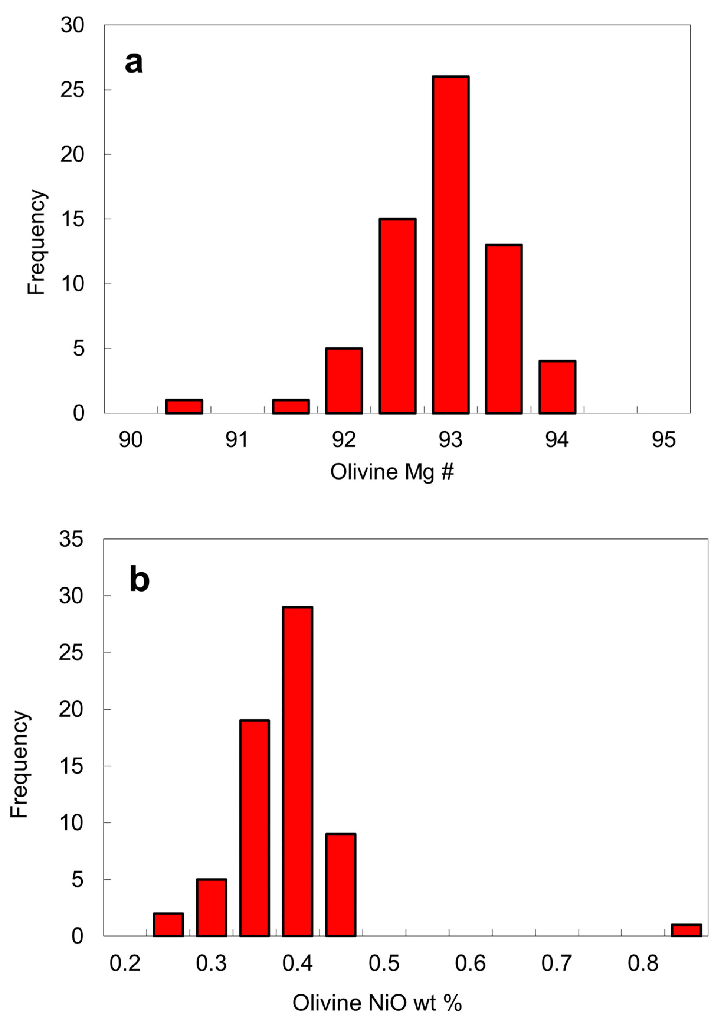
Olivine. Approximately 47 olivine inclusions were analyzed in this work (
Table 2) and additional EPMA analyses of olivine inclusions from the Fuxian kimberlites are also available in the literature [
3,
16,
17]. Olivine inclusions typically have a high Mg # of 92–94. Mg # is defined as 100Mg/(Mg + Fe
2+) by atom where all Fe is assumed to be Fe
2+ for olivine and Fe
3+ is calculated in the case of chromite. The peak position for the Mg # of olivine is 93, a value that is slightly lower than average Mg # of 94 for worldwide olivine inclusions [
1,
32,
33], but similar to those of olivine inclusions from the Akwatia diamonds [
24]. Only two olivine inclusions have a Mg # that is outside the range of 92–94 (
Figure 9a). The NiO contents of the olivine inclusions are usually in the range of 0.25 to 0.45 wt. % and are concentrated around 0.4 wt. % (
Table 2;
Figure 9b). One exceptional value is for the olivine from the diamond LN50D62, which has up to 0.80 wt. % NiO. Due to the wide NiO range (0.25–0.45 wt. %), the constant Ni content assumption for mantle olivine for the Ni-in-garnet thermometer may not always be valid [
34,
35].
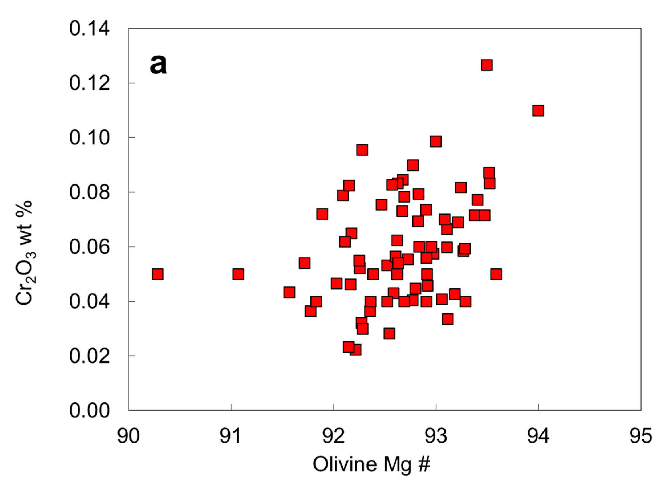
Chromium contents of the olivine inclusions (
Table 2;
Figure 10a) all fall within the normal reported range of olivine inclusions in diamonds [
24] and appear to increase with Mg #, likely owing to different degree of depletion in the mantle. If an olivine inclusion touches or is close to a chromite inclusion in diamond, an apparent high Cr
2O
3 content of the olivine may be caused by secondary X-ray fluorescence of Cr in chromite by Fe in olivine [
25,
26]. Therefore, a low accelerating voltage (10 kV) is preferred for analysis of olivine inclusions in olivine-chromite pairs in diamond. The unusually high Cr content of olivine inclusions in the Akwatia diamond [
24] should be checked for fluorescence effect. Nonetheless, high Cr contents in isolated olivine inclusions are likely be real. Chromium enrichment in olivine might result from specific P-T conditions, crystal-chemical factors, or reduction of Cr, which may enter olivine as Cr
2+ under reduced conditions [
36] or high crystallization temperatures [
37,
38]. Sutton et al. [
39] showed that Cr is predominantly divalent in lunar olivine.
Chromium contents of the olivine inclusions (
Table 2;
Figure 10a) all fall within the normal reported range of olivine inclusions in diamonds [
24] and appear to increase with Mg #, probably due to different degree of depletion in the mantle. If an olivine inclusion touches or is close to a chromite inclusion in diamond, an apparent high Cr
2O
3 content of the olivine may be caused by secondary X-ray fluorescence of Cr in chromite by Fe in olivine [
26]. Therefore, a low accelerating voltage (10 kV) is preferred for analysis of olivine inclusions in olivine-chromite pairs in diamond. The unusually high Cr content of olivine inclusions in the Akwatia diamond [
24] should be checked for fluorescence effect. Nonetheless, high Cr contents in isolated olivine inclusions are likely be real. Chromium enrichment in olivine might result from specific P-T conditions, crystal-chemical factors, or reduction in Cr, which may enter olivine as Cr
2+ under reduced conditions [
36] or high crystallization temperatures [
37,
38]. Sutton et al. [
39] showed that Cr is predominantly divalent in lunar olivine.
The Ca content of olivine is an indicator of pressure if it is in equilibrium with clinopyroxene. This is the basis of Ca exchange barometer between olivine and clinopyroxene [
40], although it is a system that depends much more on temperature than on pressure [
41]. The advantage of applying this system to olivine in diamond is that it is less likely to be reset during late stage processes. Very few olivine inclusions from North China contain CaO exceeding 0.06 wt. % (
Table 2;
Figure 10b), whereas olivine inclusions in diamond from other localities have CaO mainly in the range 0.00–0.13 and ≤0.23 wt. % [
24]. As indicated by the experimental data on Ca exchange between olivine and clinopyroxene [
40], a low Ca content in olivine (<0.08 wt. %) would require a high pressure, consistent with diamond stability field. The low Ca content of olivine inclusions might imply a Ca depletion of their source region (i.e., a harzburgite source) or crystallization at a lower temperature or a higher pressure than usual (if from lherzolite).
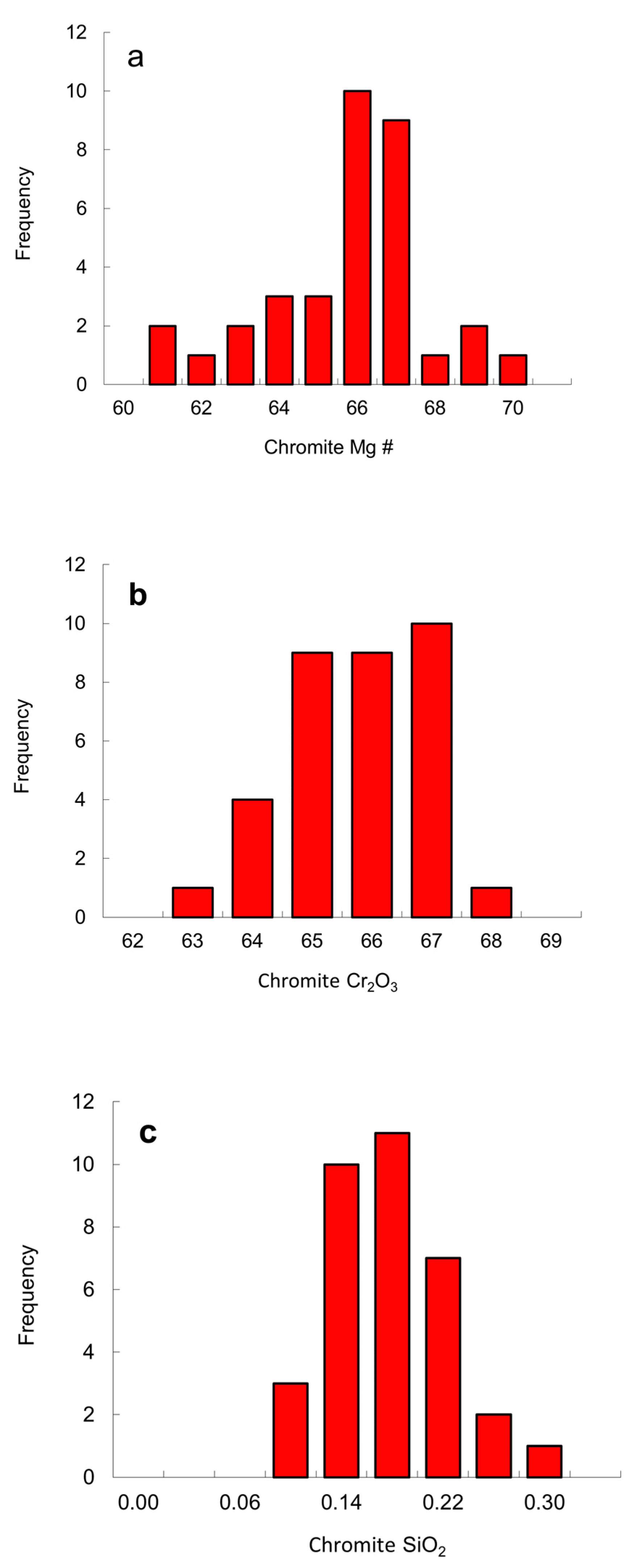
Chromite. Thirteen chromite inclusions were analyzed (
Table 3) and additional chromite analyses from the Fuxian pipes are available in the literature [
16,
17,
18]. Chromite occurs either as separate inclusions or with other minerals in a single diamond. Diamond commonly contains both chromite and olivine inclusions. The Mg # of the chromite inclusions varies from 60 to 70 with the peak position at a Mg # of 66 (
Figure 11), overlapping those of the Fe-rich part of the peridotitic worldwide range (Mg # of 60–80 [
24]). Recalculated 100Fe
2+/(Fe
3+ + Fe
2+) ratios are relatively high (76–95), probably indicating a relatively reduced environment. The Al
2O
3 ranges from 4.0 to 7.9 wt. %, within the worldwide range of chromite inclusions in diamond [
24]. A striking feature of the chromite inclusions in diamond is a high Cr content (63–67 wt. % Cr
2O
3 Table 3), which lie within the field defined by worldwide chromite inclusions in diamond [
1], but are centered around 66 wt. % Cr
2O
3 (
Figure 11).
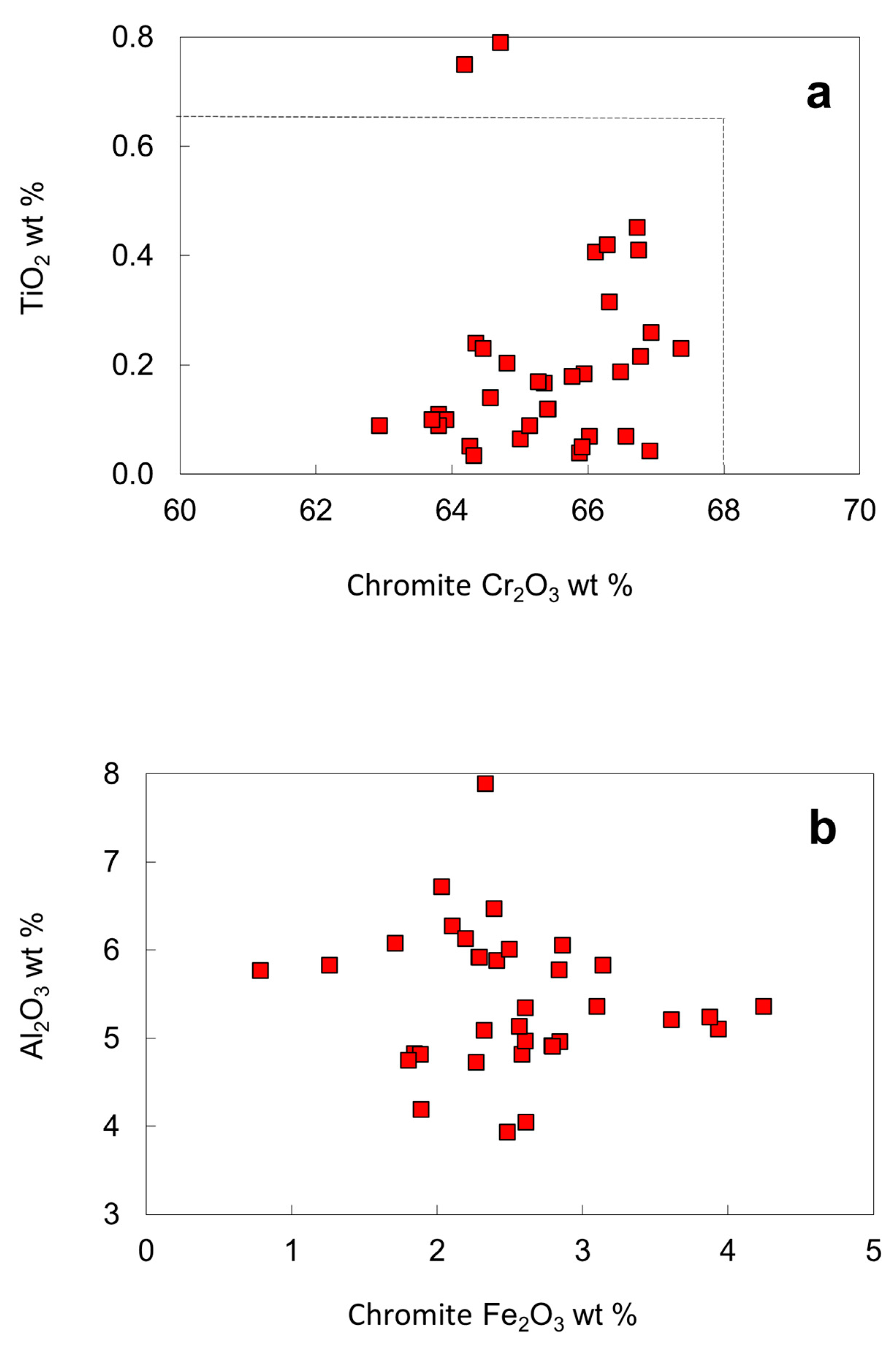
Except for two inclusions with 0.79 and 0.75 wt. % TiO
2, the TiO
2 contents (0.03–0.45 wt. %) of most chromites falls within the compositional range defined by worldwide chromites (maximum 0.65 wt. %), located in the "diamond inclusion field" for chromites (
Figure 12a). On a Fe
2O
3 vs. Al
2O
3 diagram, analyses of chromite inclusions fall within a limited compositional range (
Figure 12b). The recalculated Fe
2O
3 of the chromite inclusions is less than 4.5 wt. %. The SiO
2 contents of chromite inclusions are high, from 0.08 to 0.29 wt. %, with a pronounced peak around 0.18 wt. % (
Figure 11c). The solution of silicate-spinel component is favored by high pressure [
42], consistent with the high Cr
2O
3 contents that stabilize spinel towards higher pressures in lherzolites [
43].
Some diamonds contain more than one separated chromite inclusion (e.g., samples LN50D04, LN50D07 and LN50D20). Sample LN50D04 has 6 chromite inclusions, some at the edge of the diamond polished section, and others near the center of the diamond host; some are large and euhedral, while others are small and anhedral. There are no significant variations in composition for the multiple spinel grains in the diamond, although such inter-grain compositional variations have been recorded previously [
22,
23,
44,
45]. Multiple chromites in a single diamond have nearly the same compositions, probably indicating that the diamond formed in a very short time, or that all the chromite inclusions formed in the same environment, or that the composition of chromite did not evolve during growth of the diamond.
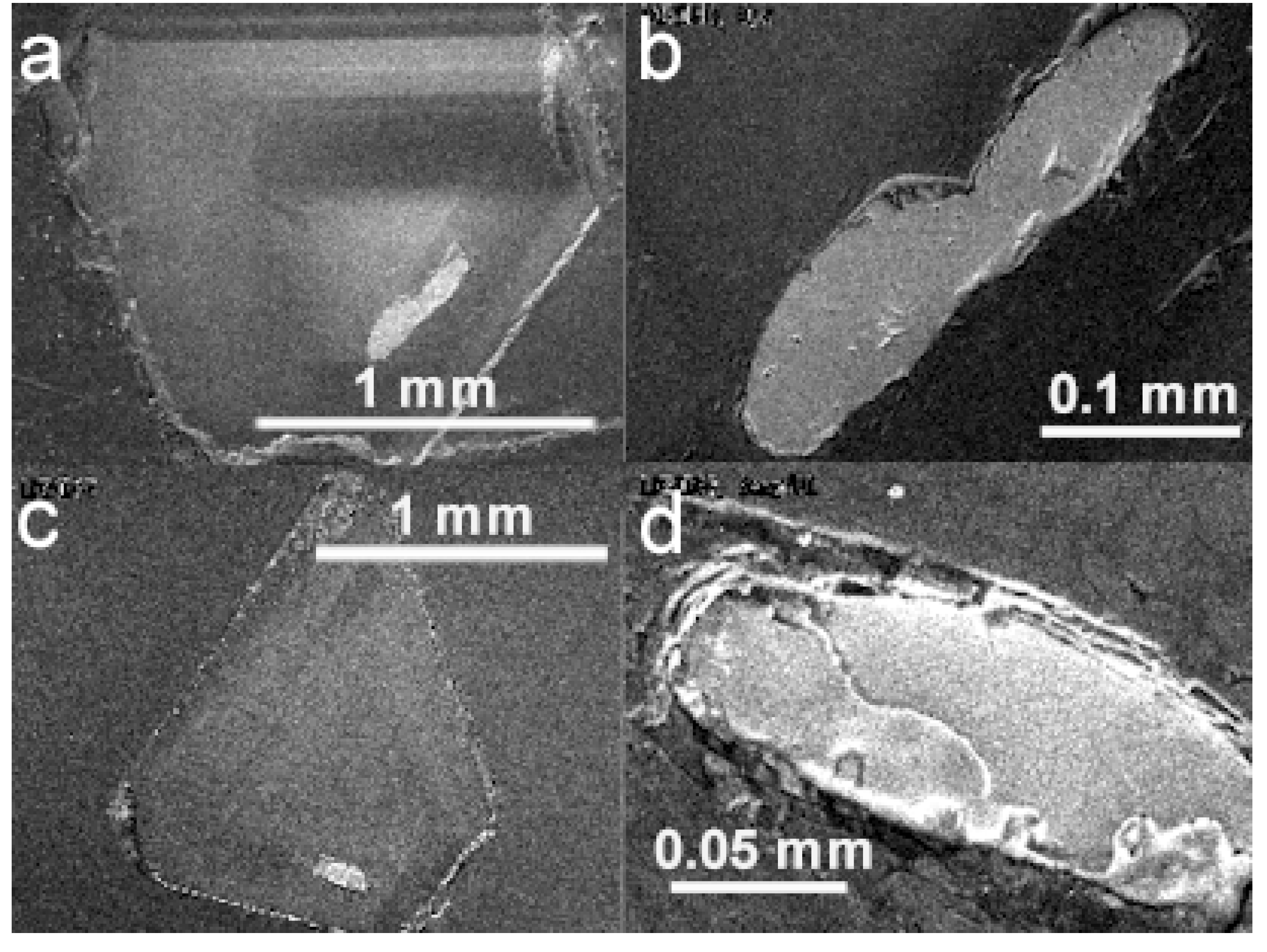
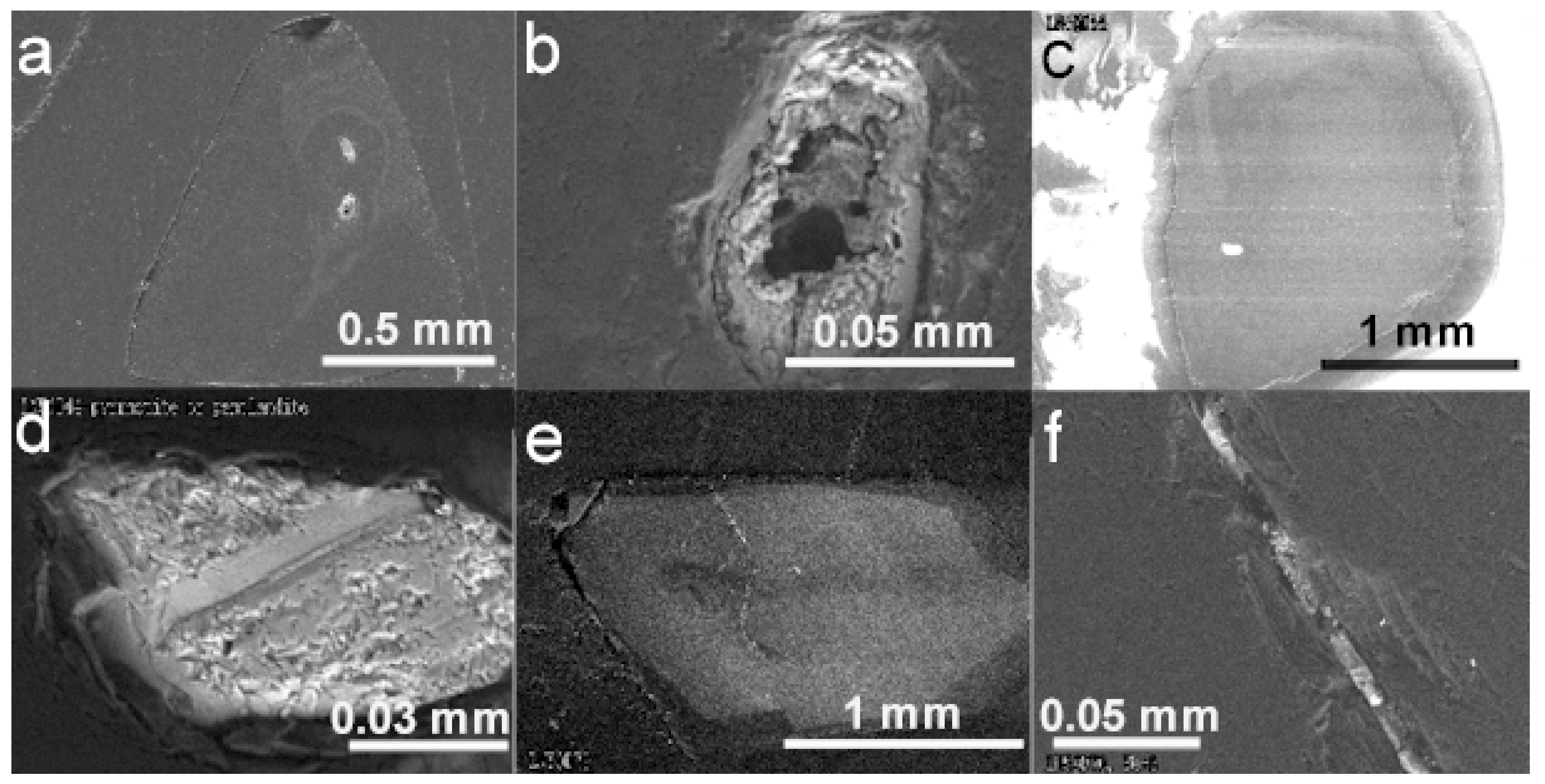
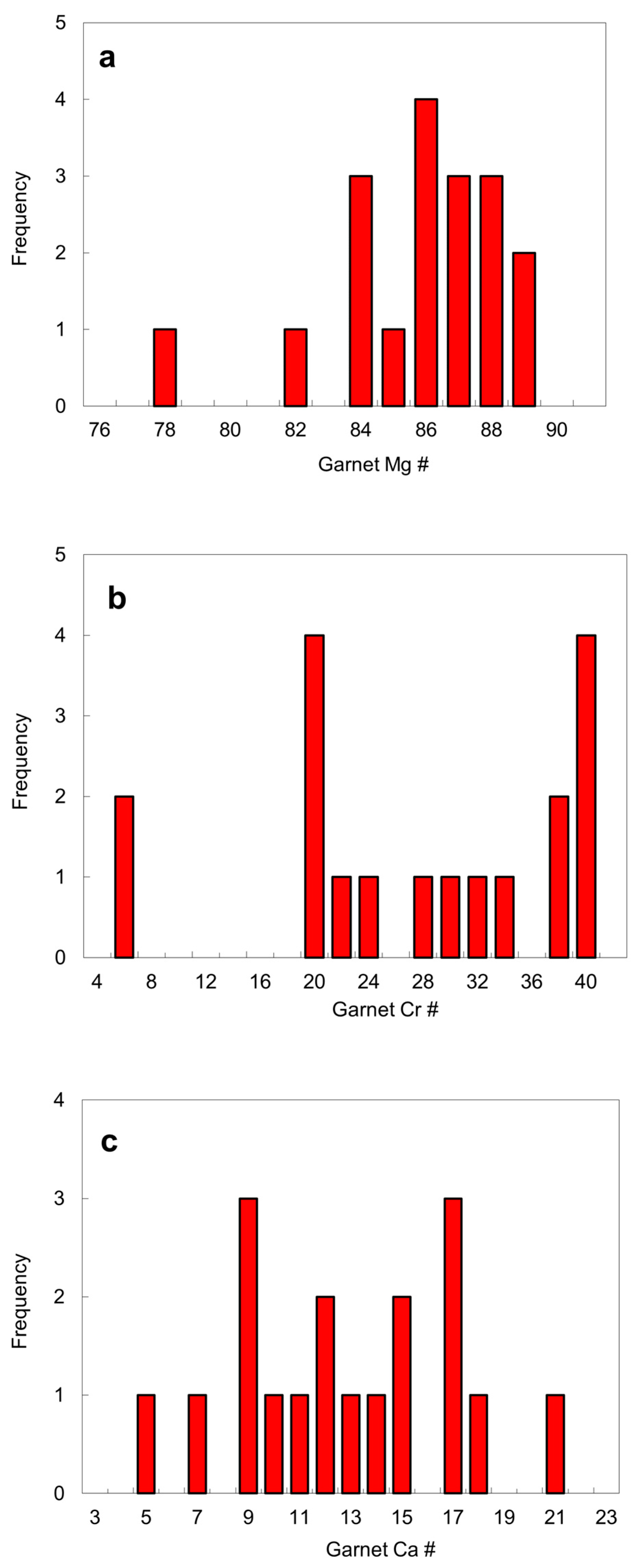
Garnet. Five garnet inclusions in diamond were exposed by polishing and analyzed (
Table 4). Garnet occurs either as a single inclusion or together with other mineral phases. Eclogitic garnet is extremely rare. No undisputed eclogitic inclusion was discovered in this study. A garnet numbered 52A in a garnet-orthopyroxene-clinopyroxene assemblage reported by Harris et al. [
16] is closest to eclogitic almandine-pyrope. Garnets in diamond from the No. 50 diatreme have the following: a Mg # of 81–88 (except for sample 52A that coexists with a low magnesian orthopyroxene and clinopyroxene); a Cr # of 19–39 except for samples 43A and 52A, where Cr # is defined as 100Cr/(Cr + Al) by atom; a Ca # of 5–21, where the Ca # is defined as 100Ca/(Ca + Mg) by atom (
Figure 13).
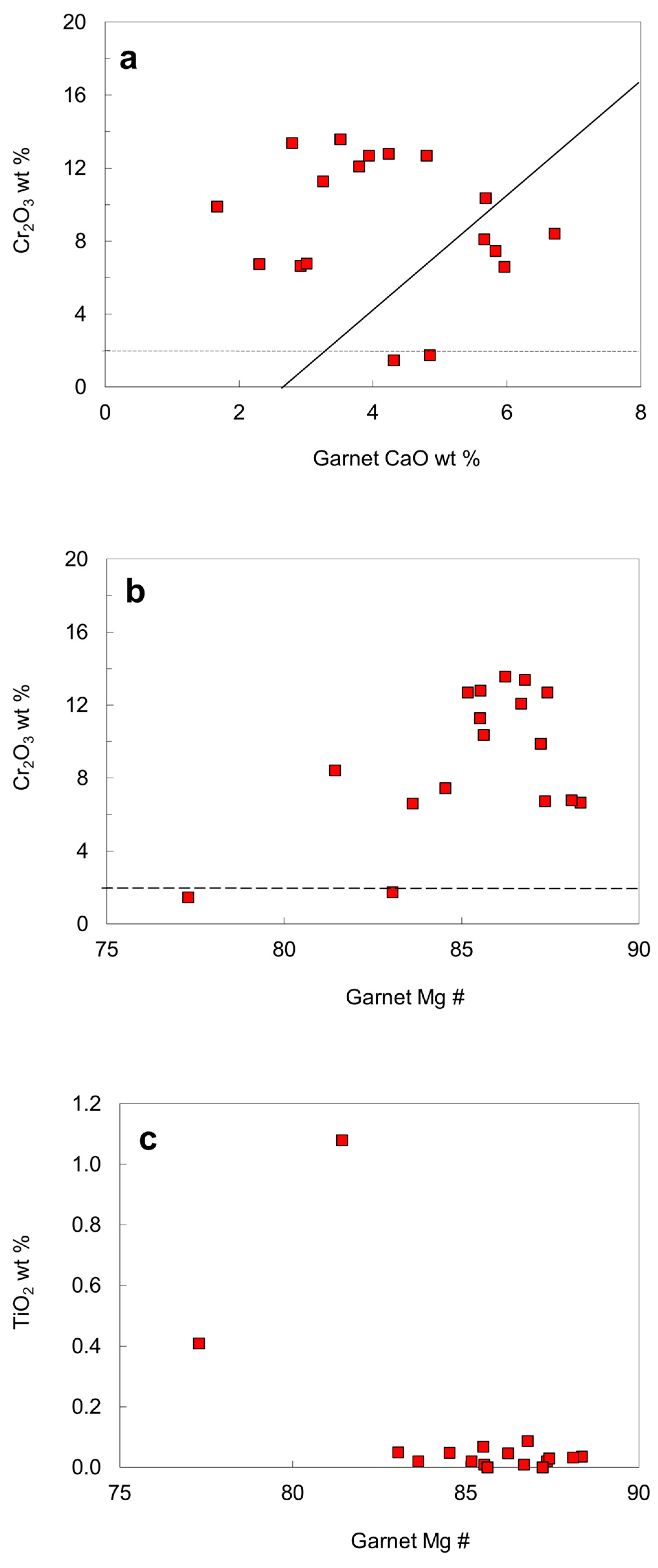
To distinguish diamondiferous from non-diamondiferous assemblages on the basis of the composition of peridotitic pyrope, curves on a CaO versus Cr
2O
3 diagram (
Figure 14) were derived by various authors [
27,
46,
47] to separate a high (lherzolitic) and a low (harzburgitic) calcium field. Most garnets in diamond from the No. 50 diatreme belong to the harzburgitic paragenesis, and only a few garnet-bearing diamond crystals are part of the lherzolitic association. Chemical compositions of two garnets (samples 43A and 52A, cf. [
16]) plot below the dashed line distinguishing peridotitic from non-peridotitic garnets [
47], indicating that they may belong to a different paragenesis. The garnet 52A has the lowest Mg # and it also coexists with low Mg # orthopyroxene and clinopyroxene, and therefore the assemblage garnet-orthopyroxene-clinopyroxene may belongs to a websterite or eclogitic paragenesis.
Figure 14c shows that only lherzolitic garnets with the lowest Mg # contain significant amounts of TiO
2 (0.41 and 1.08 wt. %), while the other garnets contain less than 0.09 wt. % TiO
2. The Al/Cr ratio in garnet was taken as a measure for the fertility of the source, as there is a positive correlation between Al/Cr ratio and Ti in lherzolitic garnets [
24]. However, there is no positive correlation between Al/Cr ratio and Ti content for the garnet inclusions from the No. 50 diamond association.
Most garnet inclusions have Si atoms per formula unit (apfu) close to the ideal value of 3 when normalized to 12 oxygens. A few garnets have Si slightly greater than 3, up to 3.03 apfu (
Table 4). The slight excess of Si in garnet may suggest the presence of majoritic component (Mg
3(MgSi)Si
3O
12) [
32] and further suggests a high-pressure condition if equilibrated with orthopyroxene [
48,
49,
50]. The diamond LN50D10 has two garnet inclusions. One is small (about 80 µm across) and has no excess Si. The other, exposed after polishing away the first garnet, has a small amount of excess Si (approximately 1%). These two garnets have almost identical compositions except for Si. The difference in Si might be caused by systematic errors between different EPMA sessions. Since one garnet was no longer available, it was not possible to examine systematic errors between different EPMA sessions by analyzing both garnets during a same EPMA session. Therefore, the garnet inclusion with excess Si was re-analyzed, together with a garnet xenocryst sample without excess Si (a Four Corner ultramafic garnet, PY15 [
51]). The average of the second garnet inclusion from the second EPMA session has less excess Si (3.003 apfu) relative to the first EPMA session. However, the average of garnet xenocrysts from the same EPMA session also has less Si (2.977 apfu) relative to early EPMA analyses (2.994 apfu). Although inconclusive, these measurements suggest that the small excess Si is real, probably indicating the existence of a very minor majoritic component.
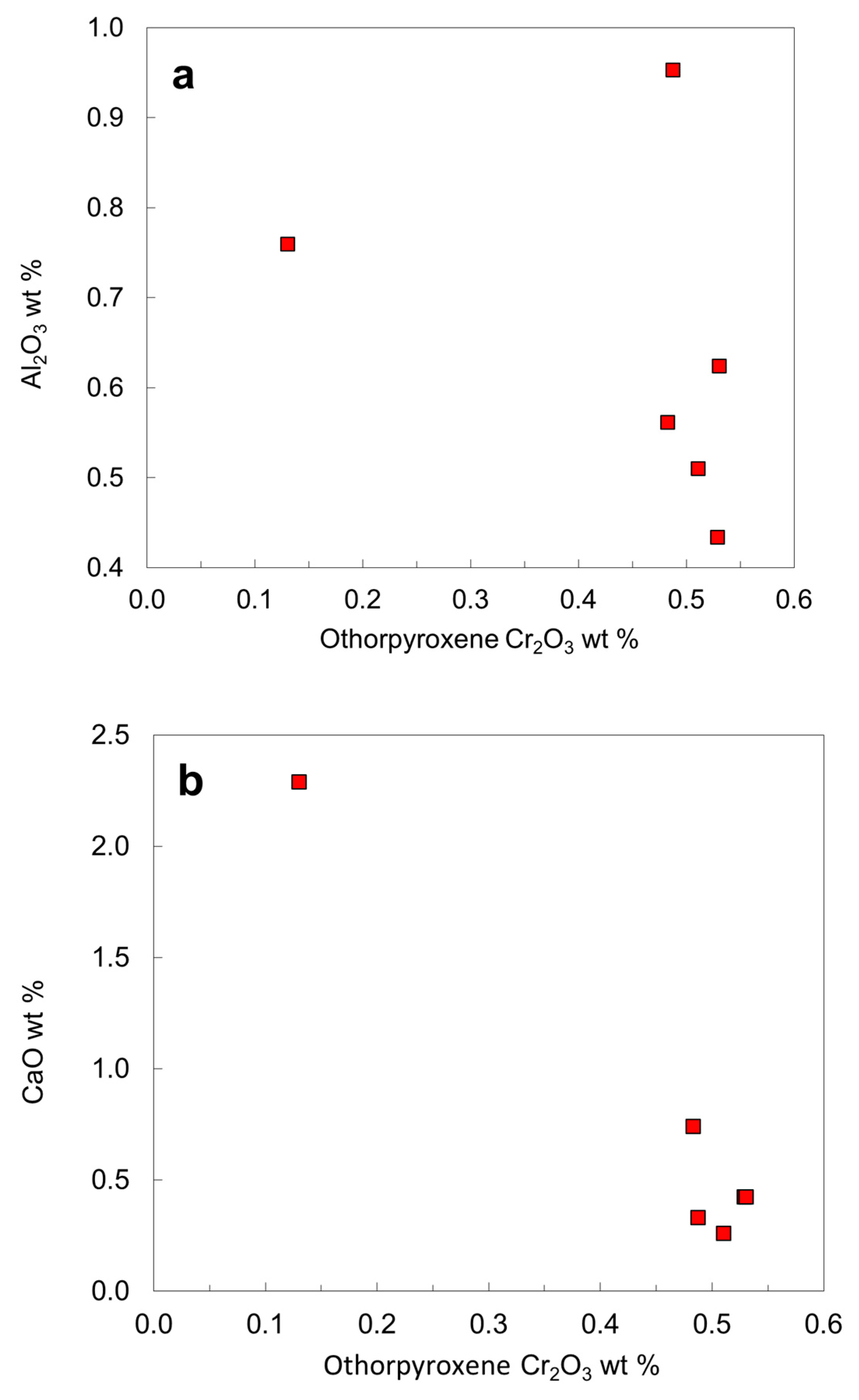
Pyroxene. Four orthopyroxene inclusions in three diamond hosts were recovered (
Table 5). In addition, Harris et al. [
16] reported two coexisting orthopyroxene inclusions and one websteritic orthopyroxene inclusion. Clinopyroxene was not identified in this work, although six clinopyroxene inclusions were recovered by Harris et al. [
16]. Two coexisting orthopyroxene grains in the diamond LN50D40 are homogeneous and have the same composition (
Table 5). The Mg # of the orthopyroxene is about 94, except for the websteritic orthopyroxene with a Mg # of 88 (sample 52B, cf. [
16]).
The orthopyroxene inclusions have low Ca contents (CaO < 0.50 wt. % except for the websteritic and the green orthopyroxene in LN50D65), probably indicating that some orthopyroxene might not be in equilibrium with clinopyroxene and might represent a harzburgitic origin [
52]. Compared to harzburgitic orthopyroxene, the websteritic orthopyroxene has higher Ti, Al, Fe, Ca and Na and lower Si, Cr and Mg (
Table 5;
Figure 15).
The olivine and orthopyroxene inclusions in the No 50 diamond are slightly enriched in iron compared with harzburgitic phases from worldwide sources [
1], probably suggesting a relatively fertile character of harzburgitic mantle. In agreement with the expected olivine–orthopyroxene partitioning relationship in peridotite xenoliths [
53], the peak of Mg # for the orthopyroxene is slightly higher than that of the olivine.
6.5. Chemical Compositions of Rare Mineral Inclusions
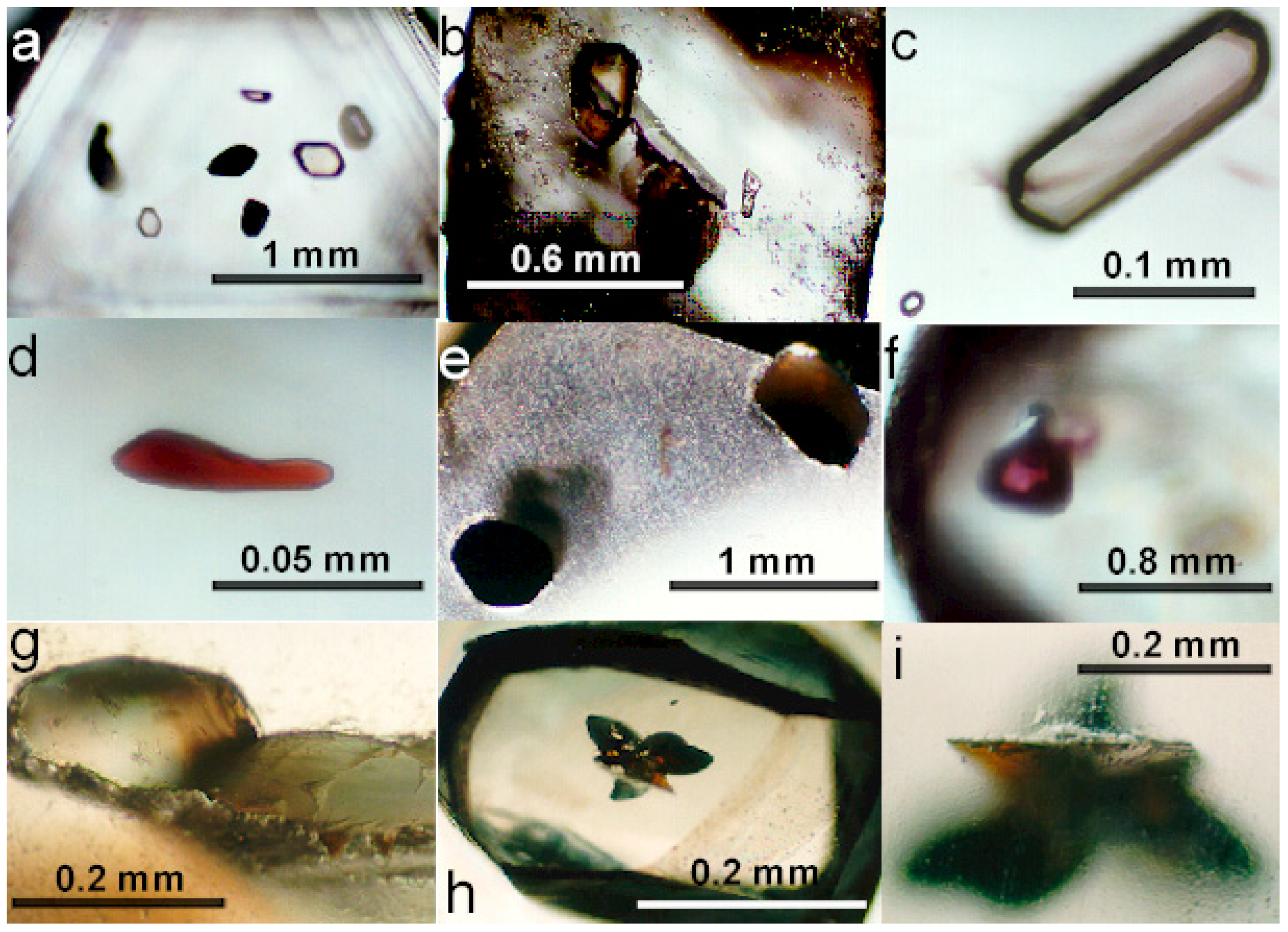
Carbonate. Carbonate inclusions in diamond are rare. The Ca-carbonate inclusion in sample LN50D11 (
Figure 6b) was initially described as a silicate under optical microscope. The analyses using a EPMA procedure for silicate showed that the Ca-carbonate inclusion is mainly composed of CaCO
3 with minor MgCO
3 and FeCO
3. The loss of the Ca-carbonate inclusion precludes further study of this interesting sample. A magnesite inclusion was found in a fracture of the diamond LN50D13 and coexists with a garnet grain. The coexistence of magnesite with garnet can be used to obtain an upper limit of oxygen fugacity from the reaction 6MgCO
3 + 22Al
2SiO
5 + 4SiO
2 = 2Mg
3Al
2Si
3O
12 + 6C + 9O
2 and a lower limit of oxygen fugacity in absence of periclase from the reaction MgCO
3 = MgO + C + O
2. Wang et al. [
54] reported a magnesite inclusion in a diamond from the No. 50 kimberlite. Carbonate inclusions in diamonds from other kimberlites include Ca carbonate, magnesite, and dolomite [
55,
56,
57,
58].
Sulfides. Four primary inclusions and one secondary sulfide inclusion in diamond were exposed and analyzed. Representative chemical compositions of the sulfide inclusions are given in
Table 6. Harris et al. [
16] reported pyrrhotite with less than 1.0 wt. % of Co, Ni, Cu and Zn and pentlandite with 34–35 wt. % Ni in diamonds from the No. 50 kimberlite, but did not provide complete analyses. Some sulfide inclusions from other localities are given in
Table 6 for comparison.
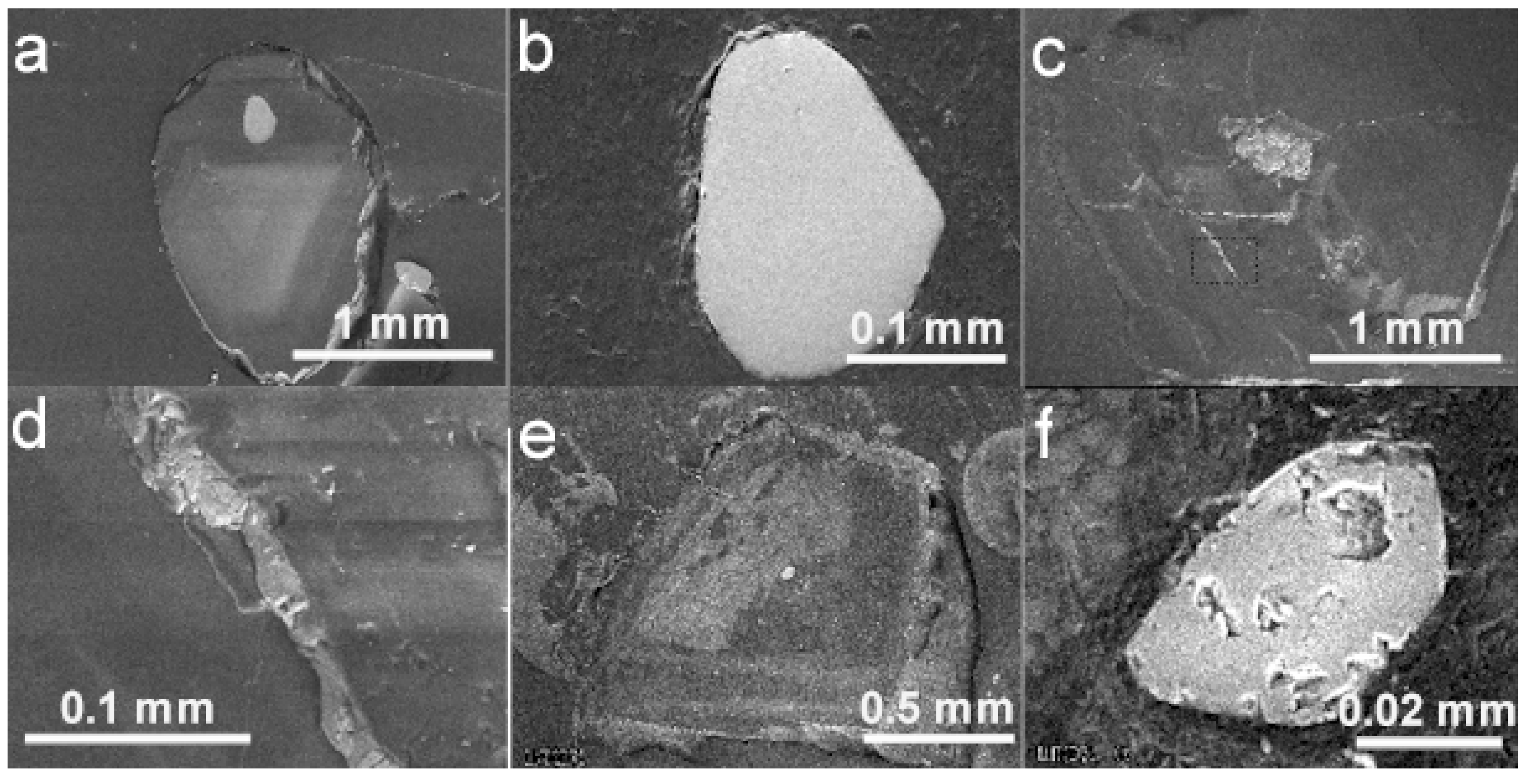
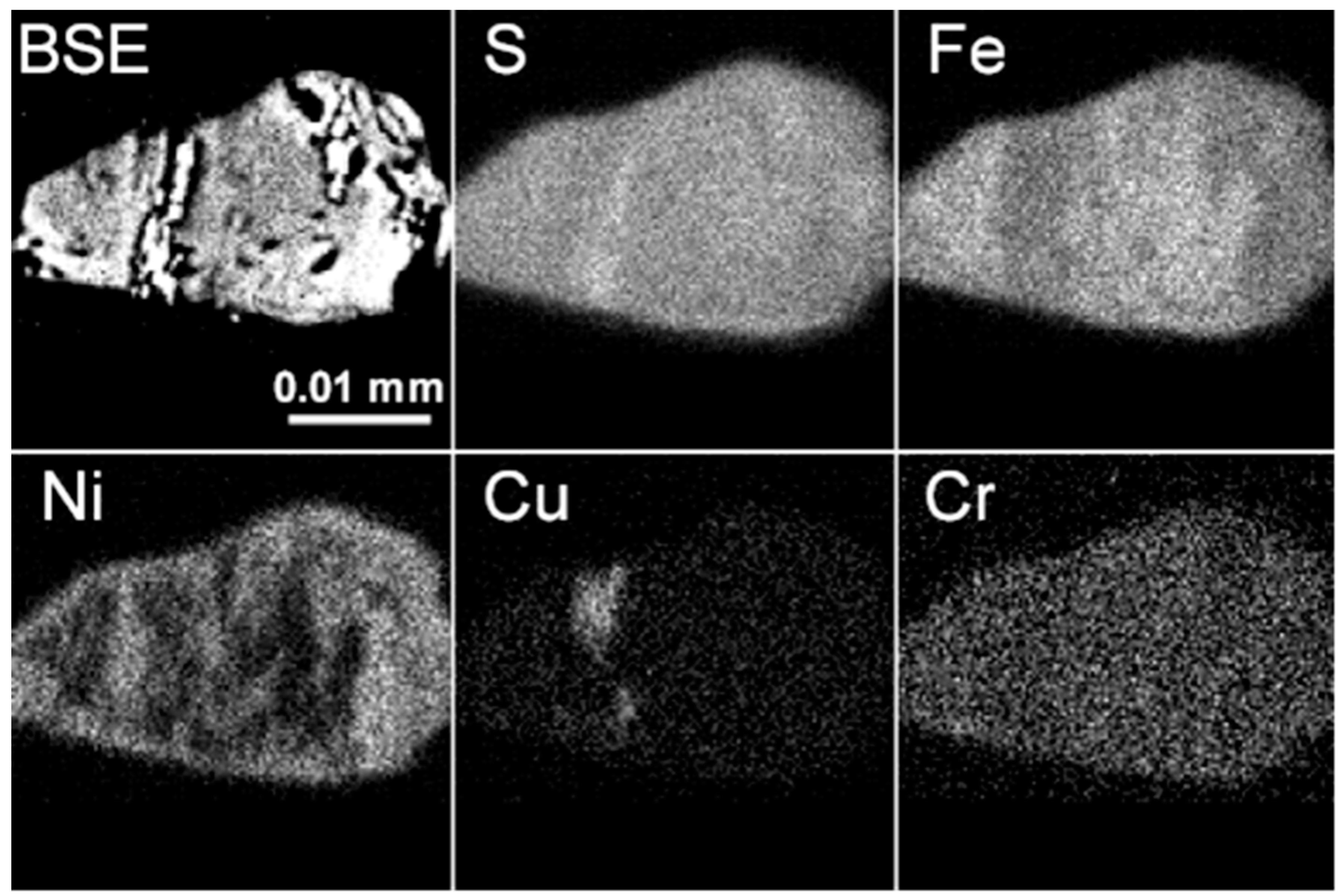
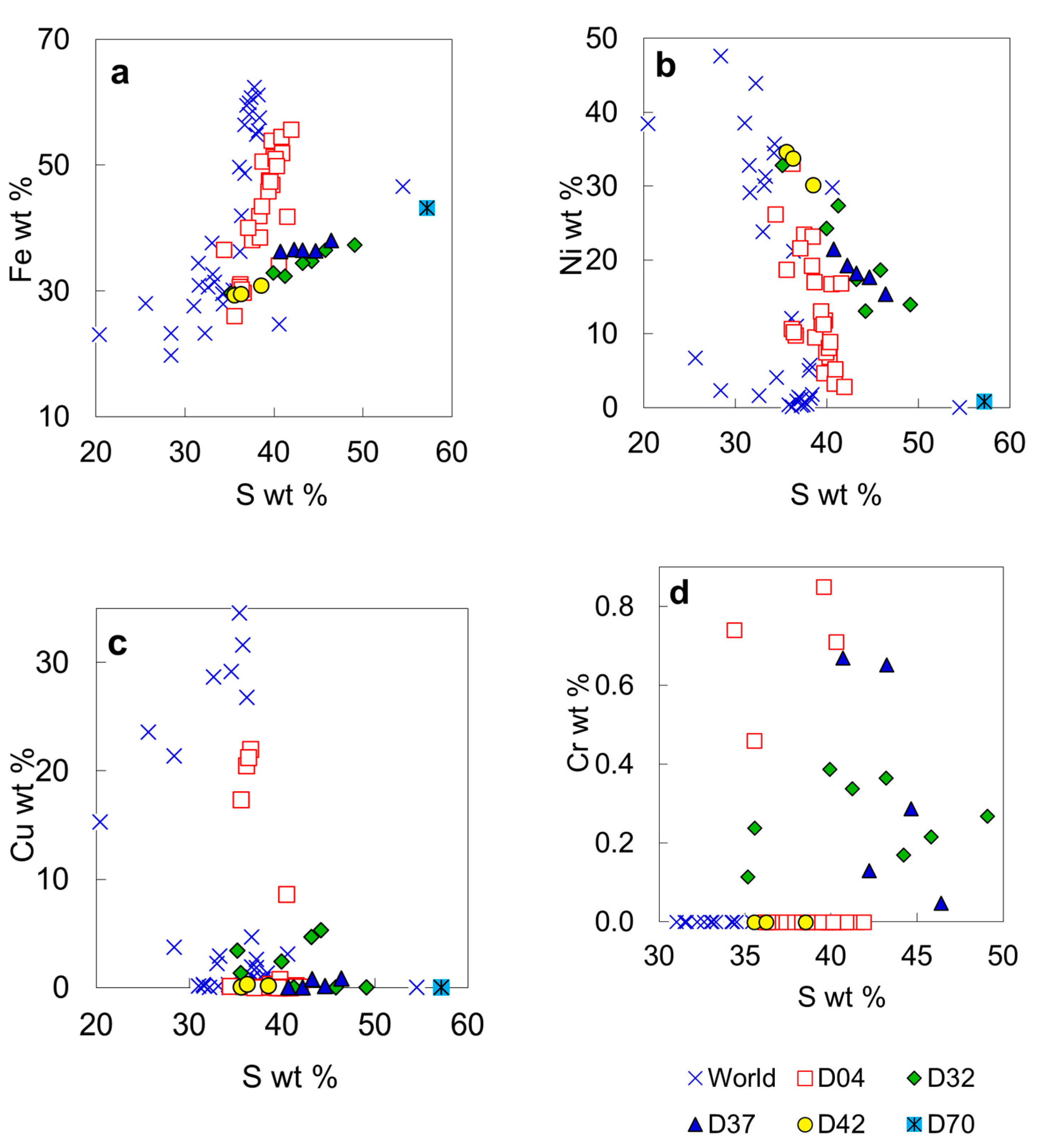
A sulfide inclusion in the diamond LN50D04 has a heterogeneous composition and shows Fe-rich, Ni-rich, and Cu-rich domains on X-ray map (
Figure 7). The content of Cu could be high as 22 wt. % in some areas. The ∑cation/sulfur ratio of the most sulfide analyses is <1 (down to 0.800), indicating that the sulfide is predominantly pyrrhotite (R
1–xS). One analysis with ∑cation/sulfur = 1 may be troilite and one with a ratio of 1.047 may be pentlandite. Normalized to four atoms, some analyses have ∑cations slightly higher than, although close to, four, suggesting the existence of R
3S
4 minerals in the linnaeite group that includes violarite FeNi
2S
4, daubreelite FeCr
2S
4, greigite Fe
3S
4, and carrollite Cu(Co,Ni)
2S
4. The sulfide inclusion in LN50D04 was initially analyzed at 20 kV and 10 nA. At these conditions, the Fe counts of Ni-rich domains could increase due to the fluorescence of Fe in the adjacent Fe-rich domain by Ni in the Ni-rich domain, thus producing excess cations relative to R
3S
4 phases. To examine this possibility, the inclusion was analyzed again at 10 kV and 20 nA condition. The effects of secondary fluorescence were not detected, and the calculated ∑cation/sulfur ratios remain similar. Chromium contents of the sulfide inclusion may be up to 0.85 wt. % (
Figure 16d); K, Mn, Co, As, Se, Sb, Te, Ba and Bi in the sulfide are low or below the EPMA detection limit.
Sulfides from LN50D32 and LN50D37 have formulae near NiFe
2S
4 (
Table 6). The sulfide in LN50D37 contains no Cu and its chemical variations are insignificant. This sulfide appears to be a solid solution between greigite (Fe
2+Fe
3+2S
4) with polydymite (Ni
2+Ni
3+2S
4), and/or violarite (Fe
2+Ni
3+2S
4). The exact identity depends on knowing the valence of Ni and Fe, which could vary during the P-T history of the sulfide. Moreover, the composition of the LN50D37 sulfide is likely an average of a submicroscopic intergrowth. Sulfide from the LN50D42 belongs to pyrrhotite or pentlandite groups. Sulfide from the LN50D70 is located in a fracture and is secondary in origin (
Table 6).
Bulanova et al. [
59] used a proton microprobe to analyze sulfide inclusions in diamond. However, sulfide inclusions are compositionally heterogeneous and their sizes are usually small (mostly 20–50 µm in this work) and similar to the typical size of the proton beam spot on the sample (30–50 µm). Therefore, the large volumes (essentially entire inclusions) analyzed by the proton microprobe will give information on the average bulk composition of the original sulfide or melt.
Unknown hydrous silicate phase. The unknown silicate phase in sample LN50D67 (
Figure 5c,d) is yellowish optically and has no fractures connected to the diamond surface. The inclusion has two compositional domains with one domain enriched in MgO and poor in SiO
2 relative to the other. The EPMA totals are low, from 85.9 to 90.2 wt. %, as is the case for serpentine, chlorite and humite (
Table 7). The inclusion contains ~10 wt. % Al
2O
3, higher than most serpentine and lower than most chlorite, common alteration products of olivine. If H
2O is the only other component, the inclusion is a hydrous silicate in the system MgO-FeO-Al
2O
3-SiO
2-H
2O. There is another hydrous silicate inclusion reported in diamond from the same locality (
Table 7). Hydrous dense silicate phases in system MgO-SiO
2-H
2O could be stable at very high pressure [
60,
61,
62]. The unknown hydrous silicate phase is a hydrous Mg-Fe aluminosilicate, similar to Phase F (1.2MgO·1.8SiO
2·1.2H
2O) in MgO and H
2O if Al substitutes for Si and Mg simultaneously, or close to Phase D (MgO·SiO
2·H
2O) or Phase E (2.3MgO·1.25SiO
2·1.2H
2O) in SiO
2 and H
2O. The sample was lost during additional polishing, and no additional characterization could be undertaken.
Table 7.
Compositions of unknown hydrous phases in diamonds from the No. 50 kimberlite diatreme.
Table 7.
Compositions of unknown hydrous phases in diamonds from the No. 50 kimberlite diatreme.
| | LN50D67
Domain 1 | LN50D67
Domain 2 | Miao et al. [10] | | Smyth & Kawamoto [61] | | Burnley and Navrotsky [60] | |
|---|
| Mineral | | | | | Wadsleyite II | | Phase D | Phase E | Phase F |
|---|
| Average | 3 Analyses | 3 Analyses | 4 Analyses | | 1 | 2 | | | |
|---|
| SiO2 | 47.27 | 41.29 | 65.81 | SiO2 | 40.04 | 40.01 | 50.74 | 39.65 | 60.71 |
| TiO2 | 0.01 | 0.01 | 0.06 | TiO2 | na | na | 0.00 | 0.00 | 0.00 |
| Al2O3 | 10.01 | 10.42 | 0.91 | Al2O3 | 0.41 | 0.34 | 0.00 | 0.00 | 0.00 |
| Cr2O3 | 0.08 | 0.10 | 0.11 | Cr2O3 | 0.19 | 0.22 | 0.00 | 0.00 | 0.00 |
| FeO | 8.19 | 11.90 | 0.22 | FeO | 8.80 | 10.60 | 0.00 | 0.00 | 0.00 |
| NiO | 0.13 | 0.21 | na | NiO | na | na | 0.00 | 0.00 | 0.00 |
| MnO | 0.01 | 0.02 | 0.02 | MnO | na | na | 0.00 | 0.00 | 0.00 |
| MgO | 20.37 | 24.39 | 29.36 | MgO | 47.57 | 44.37 | 34.04 | 48.94 | 27.15 |
| CaO | 0.87 | 0.38 | 0.11 | CaO | 0.00 | 0.00 | 0.00 | 0.00 | 0.00 |
| Σ | 86.95 | 88.72 | 96.60 | H2O | 2.99 | 4.46 | 15.21 | 11.41 | 12.14 |
| H2O | 12.90 | 12.68 | 4.84 | Σ | 100.00 | 100.00 | 100.00 | 100.00 | 100.00 |
| Σ | 99.85 | 101.40 | 101.44 | | | | | | |
| Si | 4.395 | 3.906 | 4.08 | Si | 0.975 | 0.980 | 1.000 | 1.250 | 1.800 |
| Al | 1.097 | 1.162 | 0.07 | Al | 0.012 | 0.010 | 0.000 | 0.000 | 0.000 |
| Ti | 0.001 | 0.000 | 0.00 | Ti | 0.000 | 0.000 | 0.000 | 0.000 | 0.000 |
| Cr | 0.006 | 0.008 | 0.01 | Cr | 0.004 | 0.004 | 0.000 | 0.000 | 0.000 |
| Fe | 0.637 | 0.942 | 0.01 | Fe | 0.179 | 0.217 | 0.000 | 0.000 | 0.000 |
| Ni | 0.008 | 0.013 | 0.00 | Ni | 0.000 | 0.000 | 0.000 | 0.000 | 0.000 |
| Mn | 0.000 | 0.001 | 0.00 | Mn | 0.000 | 0.000 | 0.000 | 0.000 | 0.000 |
| Mg | 2.822 | 3.439 | 2.71 | Mg | 1.726 | 1.620 | 1.000 | 2.300 | 1.200 |
| Ca | 0.087 | 0.039 | 0.01 | Ca | 0.000 | 0.000 | 0.000 | 0.000 | 0.000 |
| Σcation | 9.053 | 9.509 | 6.88 | H | 0.243 | 0.364 | 2.000 | 2.400 | 2.400 |
| ΣO | 14.000 | 14.000 | 11.00 | Σcation | 3.139 | 3.195 | 4.000 | 5.950 | 5.400 |
| Mg # | 81.6 | 78.5 | 99.58 | ΣO | 4.000 | 4.000 | 4.000 | 6.000 | 6.000 |
Fe-rich phase. An Fe-rich phase in the diamond LN50D09 was discovered and analyzed for Fe, Co, Ni, Cu, Mn, Ba, Al, Si, and Cr, and the total oxides are approximately 80 wt. %, indicating the presence of other components (
Table 8). Thus, the phase is neither native iron, wüstite, nor magnetite. Assuming the additional species is H
2O, the phase is likely to be a hydrous iron oxide, such as goethite, FeOOH (
Table 8). Other elements detected in the phase are Si (up to 3.8 wt. % SiO
2) and Al (up to 0.37 wt. % Al
2O
3) (
Table 8). The paragenesis of this Fe-rich inclusion is unknown. The diamond host of this inclusion is cloudy and of poor quality. Although there is no visible fracture observed in the diamond host, the possibility of penetration of external components into the inside of diamond cannot be completely excluded. The inclusion was probably originally trapped as native iron, wüstite, or magnetite, and was later altered or modified by an external fluid. A similar Fe-dominant inclusion in diamond was described by Miao et al. [
10] from the same locality (
Table 8). Significant SiO
2 (0.8 wt. %) was also reported in a Fe-phase by Stachel et al. [
57].
Table 8.
Compositions of an unknown Fe-rich phase in diamond from the No. 50 kimberlite diatreme.
Table 8.
Compositions of an unknown Fe-rich phase in diamond from the No. 50 kimberlite diatreme.
| | LN50D09 (Goethite or Limonite?) | | | | | Miao et al. [10] | |
|---|
| Analysis | a1 | a2 | b1 | b2 | b3 | b4 | Average | Goethite (?) | Ideal Goethite |
|---|
| Al2O3 | 0.11 | 0.37 | 0.26 | 0.10 | 0.35 | 0.18 | 0.25 | 0.54 | 0.00 |
| SiO2 | 3.77 | 3.53 | 3.66 | 1.39 | 3.64 | 3.63 | 3.27 | na | 0.00 |
| Cr2O3 | 0.02 | 0.03 | 0.12 | 0.00 | 0.04 | 0.09 | 0.05 | na | 0.00 |
| MnO | 0.00 | 0.00 | 0.00 | 0.00 | 0.00 | 0.00 | 0.00 | 0.09 | 0.00 |
| Fe2O3 | 80.68 | 81.75 | 80.54 | 78.86 | 82.04 | 79.92 | 80.63 | 85.30 | 87.98 |
| CoO | 0.00 | 0.00 | 0.00 | 0.00 | 0.00 | 0.00 | 0.00 | na | 0.00 |
| NiO | 0.10 | 0.02 | 0.08 | 0.11 | 0.04 | 0.09 | 0.07 | na | 0.00 |
| CuO | 0.00 | 0.00 | 0.06 | 0.06 | 0.08 | 0.33 | 0.10 | na | 0.00 |
| BaO | 0.14 | 0.08 | 0.14 | 0.02 | 0.07 | 0.10 | 0.09 | na | 0.00 |
| H2O | 15.19 | 14.21 | 15.14 | 19.47 | 13.74 | 15.66 | 15.53 | 14.07 | 12.02 |
| Σ | 100.00 | 100.00 | 100.00 | 100.00 | 100.00 | 100.00 | 100.00 | 100.00 | 100.00 |
| | Oxygen normalized to 3 | | | | | | | |
| Al | 0.004 | 0.013 | 0.009 | 0.004 | 0.012 | 0.007 | 0.009 | 0.020 | 0.000 |
| Si | 0.114 | 0.106 | 0.111 | 0.045 | 0.109 | 0.111 | 0.100 | 0.000 | 0.000 |
| Cr | 0.000 | 0.001 | 0.003 | 0.000 | 0.001 | 0.002 | 0.001 | 0.000 | 0.000 |
| Mn | 0.000 | 0.000 | 0.000 | 0.000 | 0.000 | 0.000 | 0.000 | 0.002 | 0.000 |
| Fe3+ | 1.841 | 1.844 | 1.837 | 1.933 | 1.840 | 1.836 | 1.853 | 1.979 | 2.000 |
| Co | 0.000 | 0.000 | 0.000 | 0.000 | 0.000 | 0.000 | 0.000 | 0.000 | 0.000 |
| Ni | 0.002 | 0.001 | 0.002 | 0.003 | 0.001 | 0.002 | 0.002 | 0.000 | 0.000 |
| Cu | 0.000 | 0.000 | 0.001 | 0.001 | 0.002 | 0.008 | 0.002 | 0.000 | 0.000 |
| Ba | 0.002 | 0.001 | 0.002 | 0.000 | 0.001 | 0.001 | 0.001 | 0.000 | 0.000 |
| Σcation | 1.963 | 1.965 | 1.965 | 1.986 | 1.965 | 1.967 | 1.968 | 2.001 | 2.000 |
| Σcharge | 6.000 | 6.000 | 6.000 | 6.000 | 6.000 | 6.000 | 6.000 | 6.000 | 6.000 |
| H2O molecule | 1.265 | 1.183 | 1.261 | 1.621 | 1.144 | 1.304 | 1.293 | 1.172 | 1.000 |
6.6. Origin of Sulfide Inclusions in Diamond
Sulfide inclusions in diamond are common [
1,
63,
64] and may occur as discrete crystals [
65]. Primary sulfide inclusions in diamonds have been shielded from the interaction with the outside environment. Therefore, such sulfide inclusions provide information on the primary compositions of mantle sulfide, the distribution and abundance of chalcophile elements in the mantle, and the formation environment of the host diamond [
66,
67]. Sulfide inclusions in diamonds are associated with either peridotitic or eclogitic assemblages [
59,
65]. On the base of the inclusion assemblage, the sulfide inclusion in the diamond LN50D04, which contains chromite and olivine, belongs to the peridotitic suite. In Siberian, peridotitic sulfides in diamond show significantly higher Ni and Cu contents than eclogitic sulfides [
59]. The boundary between peridotitic and eclogitic sulfide inclusions is 8 wt. % Ni [
65], or 12 wt. % Ni, or it is transitional [
59]. Experimental studies on Ni/Fe exchange between olivine and monosulfide solid solution [
68,
69,
70] indicate that, for peridotitic olivine with Fo88–Fo94 and 2500–3500 ppm Ni, the coexisting monosulfide solid solution will contain 30–55 mol % NiS (25–35 wt. % Ni). The sulfide inclusion in the diamond LN50D42 has a Ni content of ~30 wt. % and was probably in equilibrium with mantle olivine. If sulfide were the only inclusion in diamond, it may be difficult to determine which assemblage a sulfide inclusion might belong to based on Ni content. For example, the peridotitic sulfide in the diamond LN50D04 have Ni content from 6 to 34.6 wt. % (
Figure 16b), whereas an eclogitic sulfide in an omphacite- and coesite-bearing diamond contains >11 wt. % Ni [
59]. Deines and Harris [
71] demonstrated that assignment of Ni-rich monosulfide to the peridotitic paragenesis is ambiguous if there is no further evidence from coexisting silicate inclusions.
Iron–nickel–copper sulfides are the most abundant primary sulfide inclusions in diamond [
53,
59,
72]. Based on the stability of diamond (1500 K at 50 kbar [
73]) and silicate assemblages in diamond, sulfide was trapped at around 1000–1200 °C. At these temperatures, monosulfide solid solution and sulfide melt coexist over a wide range of compositions [
74,
75]. Therefore, iron–nickel–copper sulfides in diamonds might be trapped as droplets of primary, immiscible sulfide melt, such as those from silicate megacrysts in basalt [
76], or as crystals of monosulfide solid solution which then exsolved to different sulfide after subsolidus re-equilibration [
1,
2,
27,
63,
64,
77,
78,
79,
80] (. Element partitioning between sulfide melt and monosulfide crystal at mantle conditions is controlled by composition, temperature, and pressure. The effect of melt composition and temperature on element partitioning between sulfide melts and monosulfide crystals suggest that Cu and Ni are slightly concentrated in residual melt during fractional crystallization at 1000–1100 °C [
81]. Therefore, monosulfide solid solution crystallized from melts would contain low Ni and Cu, whereas the residual melts would be enriched in Ni and Cu. According to Bulanova et al. [
59], peridotitic sulfide inclusions with Cu > 3 wt. % and Ni > 17 wt. % may represent entrapped melts. The bulk composition of each sulfide inclusion in diamond from the No. 50 diatreme likely has low Cu content (<3 wt. %), although a few analyses of the sulfide inclusions have higher Cu contents (
Table 6). The Ni contents of the sulfide inclusions are widespread, from ~3 to >30 wt. % (
Table 6). The Cu and Ni contents appear to suggest that the most sulfide inclusions were trapped as monosulfide crystals and some may be trapped as melts.


