
Evaluation of Respiratory, Genotoxic and Cytotoxic Effects from Occupational Exposure to Typography Activities
 , ,
, ,
Abstract
:1. Introduction
2. Materials and Methods
2.1. Study Participants
2.2. Buccal Micronucleus Cytome Assay: DNA Damage
2.3. Spirometry Tests: Respiratory Effects
2.4. Statistical Analysis
3. Results
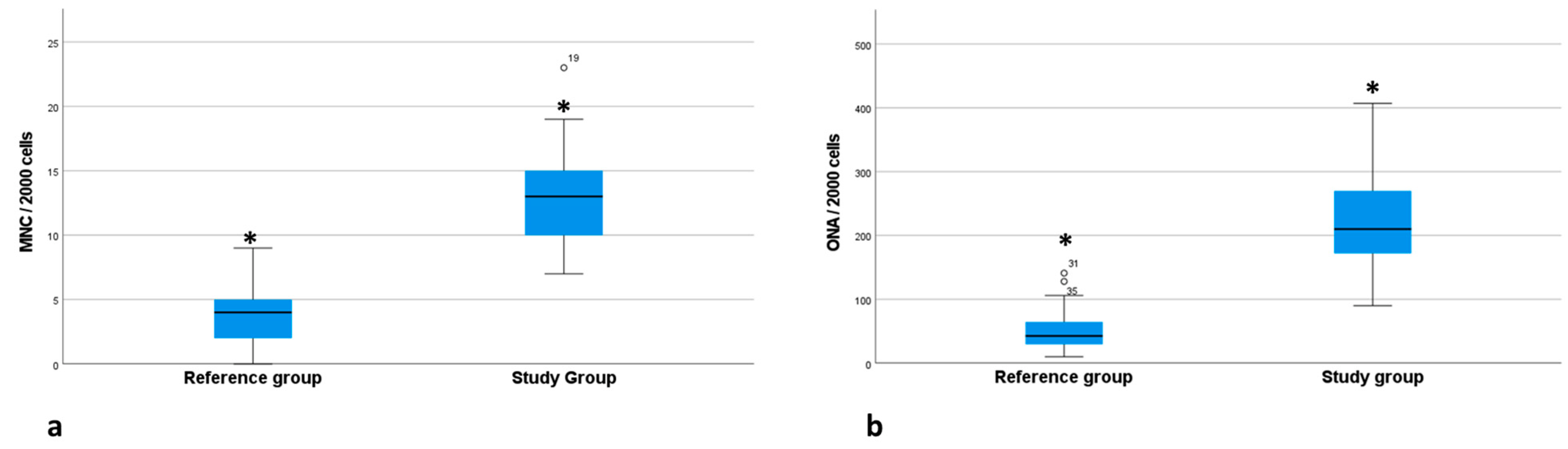
3.1. DNA Damage
3.2. Respiratory Effects
4. Discussion
5. Conclusions
Supplementary Materials
Author Contributions
Funding
Institutional Review Board Statement
Informed Consent Statement
Conflicts of Interest
References
- Goud, K.I.; Shankarapppa, K.; Vijayashree, B.; Prabhakar, R.K.; Ahuja, Y.R. AND Damage and Repair Studies in Individuals Working with Photocopying Machines. Int. J. Health Geogr. 2001, 1, 139–143. [Google Scholar] [CrossRef]
- WHO. Exposure to Hazardous Chemicals at Work and Resulting Health Impacts: A Global Review; International Labour Office—ILO: Geneva, Switzerland, 2021; ISBN 978-9-22-034219-0. [Google Scholar]
- National Institute for Occupational Safety and Health (NIOSH). Disease and Injury. 2014. Available online: https://www.cdc.gov/niosh/docs/96-115/diseas.html (accessed on 27 February 2023).
- Khatri, M.; Bello, D.; Gaines, P.; Martin, J.; Pal, A.K.; Gore, R.; Woskie, S. Nanoparticles from photocopiers induce oxidative stress and upper respiratory tract inflammation in healthy volunteers. Nanotoxicology 2013, 7, 1014–1027. [Google Scholar] [CrossRef]
- Gu, J.; Karrasch, S.; Salthammer, T. Review of the characteristics and possible health effects of particles emitted from laser printing devices. Indoor Air. 2020, 30, 396–421. [Google Scholar] [CrossRef]
- Prica, M.; Kecić, V.; Adamović, S.; Radonić, J.; Sekulić, M.T. Occupational Exposure to Hazardous Substances in Printing Industry Paper 14. In Proceedings of the 8th International Engineering Symposium at Bánki, Budapest, Hungary, 15 August 2016; ISBN 978-615-5460-95-1. [Google Scholar]
- Caselli, M.; Gennaro, G.; Saracino, M.S.; Tutino, M. Indoor contaminants from newspapers: VOCs emissions in newspaper stands. Environ. Res. 2009, 109, 149–157. [Google Scholar] [CrossRef] [PubMed]
- Guo, H.; Lee, S.C.; Chan, L.Y.; Li, W.M. Risk assessment of exposure to volatile organic compounds in different indoor environments. Environ. Res. 2004, 94, 57–66. [Google Scholar] [CrossRef]
- Al-Khulaifi, N.; Al-Mudhaf, H.; Alenezi, R.; Abu-Shady, R.; Selim, M. Seasonal and Temporal Variations in Volatile Organic Compounds in Indoor and Outdoor Air in Al-Jahra City, Kuwait. J. Environ. Prot. 2014, 5, 310–326. [Google Scholar] [CrossRef]
- Sutton, P.; Wolf, K.; Quin, J. Implementing Safer Alternatives to Lithographic Cleanup Solvents to Protect the Health of Workers and the Environment. J. Occup. Environ. Hyg. 2009, 6, 174–187. [Google Scholar] [CrossRef]
- Garcia, P.; Linhares, D.; Amaral, A.F.S.; Rodrigues, A.S. Exposure of thermoelectric power-plant workers to volatile organic compounds from fuel oil: Genotoxic and cytotoxic effects in buccal epithelial cells. Mutat. Res. Genet. Toxicol. Environ. Mutagen. 2012, 747, 197–201. [Google Scholar] [CrossRef] [PubMed]
- Sancini, A.; Caciari, T.; Chighine, A.; Gioffrè, P.A.; Andreozzi, G.; Sacchi, L.; RGiubilati, R.; Tomei, G.; Suppi, A.; Sacco, C.; et al. Workers of the printing industry and hepatic damage. Ann. Ig. 2014, 26, 321–329. [Google Scholar]
- Brina, K.R.; Carvalho, T.S.; Ardenghi, P.G.; Basso da Silva, L. Micronuclei and other nuclear anomalies in exfoliated buccal cells of urban solid waste collectors and recyclers in southern Brazil. Chemosphere 2018, 193, 1058–1062. [Google Scholar] [CrossRef]
- Fenech, M. The micronucleus assay determination of chromosomal level DNA damage. Methods Mol. Biol. 2008, 410, 185–216. [Google Scholar] [PubMed]
- Tolbert, P.E.; Shy, C.M.; Allen, J.W. Micronucleus and other nuclear anomalies in buccal smears: A field test in snuff users. Am. J. Epidemiol. 1991, 134, 840–850. [Google Scholar] [CrossRef] [PubMed]
- Thomas, P.; Holland, N.; Bolognesi, C.; Kirsch-Volders, M.; Bonassi, S.; Zeiger, E.; Knasmueller, S.; Fenech, M. Buccal micronucleus cytome assay. Nat. Protoc. 2009, 4, 825–837. [Google Scholar] [CrossRef] [PubMed]
- Lowry, J.B. A Guide to Spirometry for Primary Care Physicians; The College of Family Physicians: Mississauga, ON, Canada, 1998. [Google Scholar]
- Bolognesi, C.; Knasmueller, S.; Nersesyan, A.; Thomas, P.; Fenech, M. The HUMNxl scoring criteria for different cell types and nuclear anomalies in the buccal micronucleus cytome assay—An update and expanded photogallery. Mutat. Res. 2013, 753, 100–113. [Google Scholar] [CrossRef]
- Bolognesi, C.; Roggieri, P.; Ropolo, P.; Thomas, P.; Hor, M.; Fenech, M.; Nersesyan, A.; Knasmueller, S. Buccal micronucleus cytome assay: Results of an intra- and inter-laboratory scoring comparison. Mutagenesis 2015, 30, 545–555. [Google Scholar] [CrossRef] [PubMed]
- Çelik, A.; Yildirim, S.; Ekinci, S.Y.; Tasdelen, B. Biomonitoring for the genotoxic assessment in road construction workers as determined by the buccal micronucleus cytome assay. Ecotoxicol. Environ. Saf. 2013, 92, 265–270. [Google Scholar] [CrossRef] [PubMed]
- Bruschweiler, E.D.; Hopf, N.B.; Wild, P.; Huynh, C.K.; Fenech, M.; Thomas, P.; Hor, M.; Charriere, N.; Savova-Bianchi, D.; Danuser, B. Workers exposed to wood dust have an increased micronucleus frequency in nasal and buccal cells: Results from a pilot study. Mutagenesis 2014, 29, 201–207. [Google Scholar] [CrossRef] [PubMed]
- Tolbert, P.E.; Shy, C.M.; Allen, J.W. Micronucleus and other nuclear anomalies in buccal smears: Methods development. Mutat. Res. 1992, 271, 69–77. [Google Scholar] [CrossRef]
- Holland, N.; Bolognesi, C.; Kirsch-Volders, M.; Bonassi, S.; Zeiger, E.; Knasmueller, S.; Fenech, M. The micronucleus assay in human buccal cells as a tool for biomonitoring DNA damage: The HUMN project perspective on current status and knowledge gaps. Mutat. Res. Rev. Mutat. Res. 2008, 659, 93–108. [Google Scholar] [CrossRef]
- Miller, M.R.; Hankinson, J.; Brusasco, V.; Burgos, F.; Casaburi, R.; Coates, A.; Crapo, R.; Enright, P.; van der Grinten, C.P.M.; Gustafsson, P.; et al. Standardisation of spirometry. Eur. Respir. J. 2005, 26, 320–329. [Google Scholar] [CrossRef]
- American Thoracic Society. Standardization of spirometry. Am. J. Respir. Crit. Care Med. 1995, 152, 1107–1136. [Google Scholar]
- Johns, D.P.; Pierce, R. Spirometry: The Measurement and Interpretation of Ventilatory Function in Clinical Practice, 2nd ed.; McGraw-Hill: Sydney, Australia, 2007; 24p. [Google Scholar]
- Gold, W.M.; Koth, L.L. Pulmonary function testing. In Murray and Nadel’s Textbook of Respiratory Medicine, 6th ed.; Broaddus, V.C., Mason, R.J., Ernst, J.D., King, T.E., Jr., Lazarus, S.C., Murray, J.F., Nadel, J.A., Slutsky, A.S., Gotway, M.B., Eds.; Elsevier: Amsterdam, The Netherlands, 2015; Chapter 25; pp. 407–435. [Google Scholar]
- Saran, R.; Tiwari, R.K.; Reddy, P.P.; Ahuja, Y.R. Risk assessment of oral cancer in patients with pre-cancerous states of the oral cavity using micronucleus test and challenge assay. Oral Oncol. 2008, 44, 354–360. [Google Scholar] [CrossRef] [PubMed]
- Mahimkar, M.B.; Samant, T.A.; Kannan, S.; Patil, T. Influence of genetic polymorphisms on frequency of micronucleated buccal epithelial cells in leukoplakia patients. Oral Oncol. 2010, 46, 761–766. [Google Scholar] [CrossRef]
- Backman, H.; Lindberg, A.; Sovijärvi, A.; Larsson, K.; Lundbäck, B.; Rönmark, E. Evaluation of the global lung function initiative 2012 reference values for spirometry in a Swedish population sample. BMC Pulm. Med. 2015, 15, 26. [Google Scholar] [CrossRef]
- Shashikala, R.; Indira, A.P.; Manjunath, G.S.; Arathirao, K.; Akshatha, B.K. Role of micronucleus in oral exfoliative cytology. J. Pharm. Bioallied Sci. 2015, 7, 409–413. [Google Scholar]
- Bazyka, D.; Finch, S.C.; Ilienko, I.M.; Lyaskivska, O.; Dyagil, I.; Trotsiuk, N.; Gudzenko, N.; Chumak, V.V.; Walsh, K.M.; Wiemels, J.; et al. Buccal mucosa micronuclei counts in relation to exposure to low dose-rate radiation from the Chornobyl nuclear accident and other medical and occupational radiation exposures. Environ. Health 2017, 16, 70. [Google Scholar] [CrossRef]
- Bonassi, S.; Coskun, E.; Ceppi, M.; Lando, C.; Bolognesi, C.; Burgaz, S.; Holland, N.; Kirsh-Volders, M.; Knasmueller, S.; Zeiger, E.; et al. The Human MicroNucleus project on exfoliated buccal cells (HUMNXL): The role of lifestyle, host factors, occupational exposures, health and assay protocol. Mutat. Res. 2011, 728, 88–97. [Google Scholar] [CrossRef] [PubMed]
- Javed, H.; Ghani, N. Cytogenetic damage in the buccal cells of photocopying workers in Lahore, Pakistan. J. Pak. Med. Assoc. 2017, 67, 2. [Google Scholar]
- Ekapermana, N. Analisis Frekuensi Mikronukleus Sel Epitel Bukal pada Pekerja Cetak Fotokopi di Sleman. Ph.D. Thesis, Universitas Gadjah Mada, Yogyakarta, Indonesia, 2019. [Google Scholar]
- Khisroon, M.; Khan, A.M.; Hassan, N.M.; Farrah, Z.; Farooqi, J.M. Biomonitoring of DNA Damage in Photocopiers’ Workers from Peshawar, Khyber Pakhtunkhwa, Pakistan. J. Occup. Environ. Med. 2020, 62, e527–e530. [Google Scholar] [CrossRef] [PubMed]
- Kasi, V.; Elango, N.; Ananth, S.; Vembhu, B.; Poornima, J.G. Occupational exposure to photocopiers and their toners cause genotoxicity. Hum. Exp. Toxicol. 2018, 37, 205–217. [Google Scholar] [CrossRef] [PubMed]
- Agarwal, P.; Vinuth, D.P.; Harana, S.; Thippanna, C.K.; Naresh, N.; Moger, G. Genotoxic and cytotoxic effects of X-ray on buccal epithelial cells following panoramic radiography: A pediatric study. J. Cytol. 2015, 32, 102–106. [Google Scholar] [PubMed]
- Milić, M.; Bolanča, I.; Gjirlić, D.; Benković, V. Assessment of Listerine Cool Mint mouthwash influence on possible DNA damage measured by buccal micronucleus cytome assay—Preliminary results. Genet. Appl. 2019, 3, 24–35. [Google Scholar] [CrossRef]
- Torres, L.; Rodrigues, A.; Linhares, D.; Camarinho, R.; Rego, Z.; Garcia, P. Buccal epithelial cell micronuclei: Sensitive, non-invasive biomarkers of occupational exposure to low doses of ionizing radiation. Mutat. Res. Genet. Toxicol. Environ. Mutagen. 2018, 838, 54–58. [Google Scholar] [CrossRef] [PubMed]
- Puizina, M.; Puizina, E.; Gavic, J.; Tadin, L. Clinical Prospective Assessment of Genotoxic and Cytotoxic Effects of Fluoride Toothpaste and Mouthwash in Buccal Mucosal Cells. Biomedicines 2022, 10, 2206. [Google Scholar] [CrossRef]
- Speit, G.; Witton-Davies, T.; Heepchantree, W.; Trenz, K.; Hoffmann, H. Investigations on the effect of cigarette smoking in the comet assay. Mutat. Res. 2003, 542, 33–42. [Google Scholar] [CrossRef] [PubMed]
- Anlar, H.G.; Bacanli, M.; Kurt, Ö.K.; Eraydin, C. DNA damage assessment with buccal micronucleus cytome assay in Turkish coal miners. Arh. Hig. Rada Toksikol. 2019, 70, 283–289. [Google Scholar] [CrossRef]
- Villarini, M.; Guerrera, E.; Vannini, S.; Dominici, L.; Gianfredi, V.; Fatigoni, C.; Acito, M.; Moretti, M. Cytogenetic biomonitoring of road tunnel construction workers: Buccal micronucleus cytome assay. Ann. Ig. 2021, 33, 307–321. [Google Scholar] [PubMed]
- Karimi, A.; Eslamizad, S.; Mostafaee, M.; Momeni, Z.; Ziafati, F.; Mohammadi, S. Restrictive Pattern of Pulmonary Symptoms among Photocopy and Printing Workers: A Retrospective Cohort Study. J. Res. Health Sci. 2016, 16, 81–84. [Google Scholar]
- Walsh, S.L.; Nair, A.; Desai, S.R. Interstitial lung disease related to smoking: Imaging considerations. Curr. Opin. Pulm. Med. 2015, 21, 407–416. [Google Scholar] [CrossRef] [PubMed]
- Hagmeyer, L.; Randerath, W. Smoking-related interstitial lung disease. Dtsch. Ärzteblatt Int. 2015, 112, 43–50. [Google Scholar] [CrossRef]
- Song, Q.; Chen, P.; Liu, X.M. The role of cigarette smoke-induced pulmonary vascular endothelial cell apoptosis in COPD. Respir. Res. 2021, 22, 39. [Google Scholar] [CrossRef] [PubMed]


| Study Group (n = 25) | Reference Group (n = 44) | p-Value a | |
|---|---|---|---|
| Age b | 42.5 ± 1.5 | 41.9 ± 1.3 | 0.297 |
| Gender | |||
| Male | 19 (76) | 32 (72.7) | |
| Female | 6 (24) | 12 (27.3) | 0.766 |
| Tobacco consumption | |||
| Yes | 4 (16) | 16 (36.4) | |
| No | 21 (84) | 28 (63.6) | 0.073 |
| Alcohol consumption | |||
| Yes | 9 (36) | 22 (50) | |
| No | 16 (64) | 22 (50) | 0.261 |
| Use of mouthwash | |||
| Yes | 16 (64) | 25 (56.8) | |
| No | 9 (36) | 19 (43.2) | 0.559 |
| Asthma | |||
| Yes | 2 (8) | 8 (18.2) | |
| No | 3 (92) | 36 (81.8) | 0.248 |
| Bronchitis | |||
| Yes | 3 (12) | 3 (6.8) | |
| No | 22 (88) | 41 (93.2) | 0.463 |
| Rhinitis | |||
| Yes | 3 (12) | 6 (13.6) | |
| No | 22 (88) | 38 (86.4) | 0.846 |
| DNA damage | |||
| MNC | 12.9 ± 0.7 | 4 ± 0.4 | <0.001 |
| ONA | 219.8 ± 14.5 | 51.5 ± 4.6 | <0.001 |
| Poisson Regression (GLZ) | Number of Obs: 69 Prob ˃ χ2 ˂ 0.001 | ||
|---|---|---|---|
| N (%) | RR (95% CI) a | p-Value | |
| Mouthwash use | |||
| Yes | 41 (59.4) | 1.07 (0.9–1.3) | 0.483 |
| No | 28 (40.6) | 1 | |
| Tobacco consumption | |||
| Yes | 20 (29) | 1.02 (0.8–1.3) | 0.846 |
| No | 49 (71) | 1 | |
| Alcohol consumption | |||
| Yes | 31 (44.9) | 1.07 (0.9–1.3) | 0.5 |
| No | 38 (55.1) | 1 | |
| Exposure to photocopiers | |||
| Study group | 25 (36.2) | 3.2 (2.7–3.9) | <0.001 |
| Reference group | 44 (63.8) | 1 | |
| Reference Group | Study Group | p-Value a | |
|---|---|---|---|
| FVC | 94.2 ± 3.17 | 97.4 ± 2.76 | 0.812 |
| FEV1 | 83.4 ± 3.70 | 92.8 ± 3.15 | 0.201 |
| Respiratory patterns | |||
| Normal | 32 (67%) | 24 (96%) | <0.001 |
| Restriction | 9 (19%) | 1 (4%) | 0.038 |
| Mild obstruction | 1 (2%) | 0 | 0.322 |
| Moderate obstruction | 5 (10%) | 0 | 0.024 |
| Severe obstruction | 1 (2%) | 0 | 0.322 |
Disclaimer/Publisher’s Note: The statements, opinions and data contained in all publications are solely those of the individual author(s) and contributor(s) and not of MDPI and/or the editor(s). MDPI and/or the editor(s) disclaim responsibility for any injury to people or property resulting from any ideas, methods, instructions or products referred to in the content. |
© 2023 by the authors. Licensee MDPI, Basel, Switzerland. This article is an open access article distributed under the terms and conditions of the Creative Commons Attribution (CC BY) license (https://creativecommons.org/licenses/by/4.0/).
Share and Cite
Linhares, D.; Rocha, J.; Rodrigues, A.; Camarinho, R.; Garcia, P. Evaluation of Respiratory, Genotoxic and Cytotoxic Effects from Occupational Exposure to Typography Activities. Atmosphere 2023, 14, 562. https://doi.org/10.3390/atmos14030562
Linhares D, Rocha J, Rodrigues A, Camarinho R, Garcia P. Evaluation of Respiratory, Genotoxic and Cytotoxic Effects from Occupational Exposure to Typography Activities. Atmosphere. 2023; 14(3):562. https://doi.org/10.3390/atmos14030562
Chicago/Turabian StyleLinhares, Diana, Joana Rocha, Armindo Rodrigues, Ricardo Camarinho, and Patrícia Garcia. 2023. "Evaluation of Respiratory, Genotoxic and Cytotoxic Effects from Occupational Exposure to Typography Activities" Atmosphere 14, no. 3: 562. https://doi.org/10.3390/atmos14030562