Article

Genotoxicity of PM2.5 and PM1.0 Particulates on Human Peripheral Blood Lymphocytes in Manila, Philippines

by
Ma. Katrina Gale Estonilo
1,
Joedith Anne Cazeñas
1,
Carlos Josef Villafuerte
1,
Custer Deocaris
2,3,
Gloriamaris Caraos
2,
Gerardo Jose Robles
2,
Maria Cecilia Galvez
1,4,
Celia Asaad
2 and
Edgar Vallar
1,4,*
1
Environment and Remote Sensing Research (EARTH) Laboratory, Department of Physics, De La Salle University, Manila 0922, Philippines
2
Biomedical Research Unit, Atomic Research Division, Philippine Nuclear Research Institute, Department of Science and Technology, Diliman, Quezon City 1101, Philippines
3
Research and Development Management Office, Technological Institute of the Philippines, Cubao, Quezon City 1109, Philippines
4
Applied Research for Community, Health, and Environment Resilience and Sustainability (ARCHERS), Center for Natural Science and Environment Research (CENSER), De La Salle University, Manila 0922, Philippines
*
Author to whom correspondence should be addressed.
Atmosphere 2022, 13(1), 6; https://doi.org/10.3390/atmos13010006
Submission received: 29 October 2021 / Revised: 15 December 2021 / Accepted: 17 December 2021 / Published: 21 December 2021
(This article belongs to the Special Issue Health Effects of Urban Atmospheric Aerosols)

Abstract

:
Urban air quality is increasingly being studied as a fraction of the world’s population is living in megacities. In this study, particulate matter (PM) along Taft Avenue, Manila, the Philippines, is investigated in terms of its ability to induce genetic damage in human peripheral blood lymphocytes (PBLs). Size-segregated roadside air samples were obtained from 2015–2017 near a university gate and analyzed using in vitro micronucleus (MN) and cytokinesis-block proliferation tests. While cellular proliferation was unaffected by 0–0.1 kg/m3 of PM1.0 and PM2.5, PBL cells treated with PM2.5 displayed a significantly higher micronucleus count (p = 0.03) compared to the cells treated with PM1.0. Atomic absorption spectroscopy revealed greater amounts of Cd, Ca, Pb, K, Na, and Zn in PM2.5 compared to PM1.0. The results indicate that the differences in composition of the two size fractions of air particulates are associated with their genotoxicities.

1. Introduction

Particulate matter (PM) is air particles comprising liquid or solid materials that are an admixture of organic and inorganic compounds. Common diameters of PM are 1.0 μm, 2.5 μm, and 10 μm. Although the exact mechanisms are still being unraveled, air particulates have been linked to various diseases, from respiratory and cardiovascular illnesses to neurological disorders [1,2,3]. According to the Environmental Performance Index (EPI), the Philippines is ranked 114th out of 178 countries in poor implementation of environmental policies. In Manila, the average human exposure to PM2.5 is 17 × 10−9 kg/m3, exceeding the 5 × 10⁻⁹ kg/m3 standard limit for PM2.5 exposure by the World Health Organization (WHO) [4]. The Philippines is among the top ten countries globally with the highest death burden due to air pollution which was estimated to account for 64,000 deaths in 2019 [5,6].
Biomonitoring of individuals exposed to high amounts of PM has been a subject of interest in the Philippines due to its heavy air pollution load, especially in its megacities [7]. Recolete and Villarino [8] assessed DNA damage in exfoliated buccal cells among Filipino urban street vendors that are considered at health risk due to significant exposures to vehicular exhaust. Vendors who are occupationally exposed to vehicular exhaust showed higher micronucleus frequency compared to a control group, with 9.40 ± 4.46 and 4.80 ± 3.25, respectively. Similar genotoxicity trends were observed among gasoline station attendants and traffic enforcers in Manila [9].
While there are some studies on DNA damaging effects of air pollution in the Philippines, a more detailed characterization of air particulate components is lacking. Size and chemical composition of PM can be considered factors in the adverse effects of air pollution. One consideration is that penetration depth as well as deposition of PM in both pulmonary and circulatory systems are particle-size dependent. In a review by Xing et al. [10], several studies point out that PM2.5 is harmful to the respiratory system. A meta-analysis conducted by Huang et al. [11] studying the relationship between PM2.5 exposure and lung cancer incidence revealed that Asia has the highest lung cancer incidence correlated with PM2.5. On the other hand, PM1.0 is deposited in the alveolar and tracheobronchial compartments and can even enter the bloodstream. With such a biological fate, PM1.0 is likely to present a more complex set of health risks. For example, Sanchez-Guerra et al. [12] showed that exposure of human blood to PM1.0 sampled from air pollutants in Beijing induced DNA methylation that has the potential to trigger carcinogenesis. DNA methylation events can inhibit the expression of tumor-suppressing genes that eventually initiate cancer development [12]. Hence, the purpose of this study is to analyze and differentiate the nuclear morphologies of human peripheral blood lymphocytes exposed to PM1.0 (ultrafine particulates) and PM2.5 (fine particulates) fractions of air pollutants in Manila. Further, the study determined the concentrations of the metals in these samples and attempted to discern patterns between genotoxicity and metal concentration as a function of size.

2. Materials and Methods

2.1. Meteorological and Air Quality Data at the Sample Site

During the sampling period (2015–2017), the average relative humidity (RH) ranged from 70–75% [13]. Daytime temperatures were around 29–34 °C while nighttime temperatures were 24–27 °C [13]. The Philippines has a rainy season, usually from June up to October, while the rest of the year is the dry season. Meteorological parameters for both PM sampling periods in this study were similar. Further, the sampling site is beside a major thoroughfare in Manila so the types of vehicles plying it remain the same throughout the year. Residential condominiums and houses dominate the sampling area.

2.2. PM Sample Collection, Extraction, and Metal Analysis

The PM samples were collected using a MetOne E-sampler Instrument [14] and a Thermo Scientific Instruments tapered element oscillating microbalance (TEOM) for PM1.0 and PM2.5 [15], respectively; both were situated at De La Salle University (DLSU). Filters were readily available in time for the micronucleus assay. The researchers collected the filters of PM2.5 on 1 and 12 December 2016; 23 January 2017; 7 and 28 February 2017; and 21 March 2017. For PM1.0, the team collected the filters from July 2015–3 December 2015, and 3 December 2015–2 May 2016. Particulate matter was manually scraped from the filters and weighed. The recorded weights were 9.5 × 10⁻⁶ kg and 6.7 × 10⁻⁶ kg for PM1.0 and PM2.5.
Flame atomic absorption spectroscopy (FAAS) was carried out to determine the major elements comprising the PM samples. The samples underwent standard protocols which included dry ashing prior to FAAS analysis. Dry ashing was carried out to provide better detection limits. FAAS was carried out at another institution. For the FAAS, the sample was first aerosolized then atomized by aspirating it onto a flame. The metallic components of the sample absorb the light coming from a hollow cathode lamp. The absorption signature yields the metallic composition of the sample. The following are the absorption wavelengths for the different metals in this study: cadmium (Cd) [228.8 nm], calcium (Ca) [422.7 nm], lead (Pb) [283.3 nm], potassium (K) [766.5 nm], sodium (Na) [589.0 nm], and zinc (Zn) [213.9 nm]. References 23–24 detail the standard procedures for the determination of each element.

2.3. In Vitro Micronucleus Assay

Each PM sample was combined with PB-Max Karyotyping Medium (Gibco) to produce a stock solution of the PM. The stock solution was diluted to different PM treatment doses: vehicle alone (negative control), 0.05 kg/m3, 0.1 kg/m3, and 0.15 kg/m3. No positive control was used in this assay. A volume of 5 mL of blood, drawn from a 21-year-old female subject via venipuncture and was placed in a lithium heparin test tube to serve as an anticoagulant, and was added to 4.5 mL of PB-Max Karyotyping Medium with the PM sample. Peripheral blood lymphocytes were cultured in a 5% CO2 environment at 37 °C. After 24 h, 2.1 × 10⁻² mL of cytochalasin B was introduced to the sample, and after 68–72 h, the culture was harvested. Samples were then placed in a centrifuge at 1200 RPM for 10 min. The supernatant was removed from the culture medium and was treated by 7 mL of cold (4 °C) 0.075 M potassium chloride (KCl) to lyse red blood cells. It was then placed in a centrifuge with the same settings and instead treated with 5 mL fixative composed of methanol and acetic acid (10:1 ratio) and combined with Ringer’s solution in a 1:1 ratio. These steps were repeated about two to three times until the cell suspension was clear. After removing the supernatant to 1 cm or less above the cell pellet, the suspension was dropped onto a clean glass slide and was stained with 2–6% Giemsa after drying. Staining was carried out for about 10–20 min. Slides were then rinsed with distilled water.
MN analysis was carried out through light microscopy using an Olympus BX15 microscope and Nikon DS-Fi3 with 20× magnification for efficient counting. Criteria given by the TG-487 [16] were followed in this study. As stated in the TG-487, at least 1000 binucleated cells were counted for the MN frequency per treatment, along with the negative control. One thousand cells were scored per replicate per treatment. The equation
M N   F r e q u e n c y = N u m b e r   o f   B N   c e l l s   w i t h   M N T o t a l   n u m b e r   o f   c e l l s × 100
was used to obtain the MN frequency of each treatment per PM size fraction. Two replicates were carried out for each dose and each PM size. A review panel at the Center for Natural Science and Environment Research (CENSER), DLSU approved the research protocol following the guidelines of the University Research Ethics Committee. The research was conducted under the supervision of a licensed biomedical practitioner of the Philippine Nuclear Research Institute.

2.4. Statistical Analysis of Micronucleus Assay

One-way analysis of variance (ANOVA) with a Tukey–Kramer honest significant difference (HSD) test was used to evaluate the results for each dose and PM size from the micronucleus assay. The MN frequencies obtained from the two replicates of each dose of each PM size were used to evaluate the genotoxicity of each concentration of PM. The MN frequencies of the treated cell population were then compared to that of the negative control with a confidence level of 99% (p ≤ 0.01). Tukey–Kramer was then used to identify which treatment pairs were significantly different. A confidence level of 99% (p ≤ 0.01) was also used for the post hoc test.
To compare MN frequencies of all the concentrations of PM1.0 and PM2.5, two-way ANOVA was used with a statistical significance of less than 0.05 (p ≤ 0.05). Note that 0.15 kg/m3 of PM2.5 was not included in the two-way ANOVA because no binucleate cells were found. STATISTICA was used for all the statistical analyses carried out for this study.

3. Results

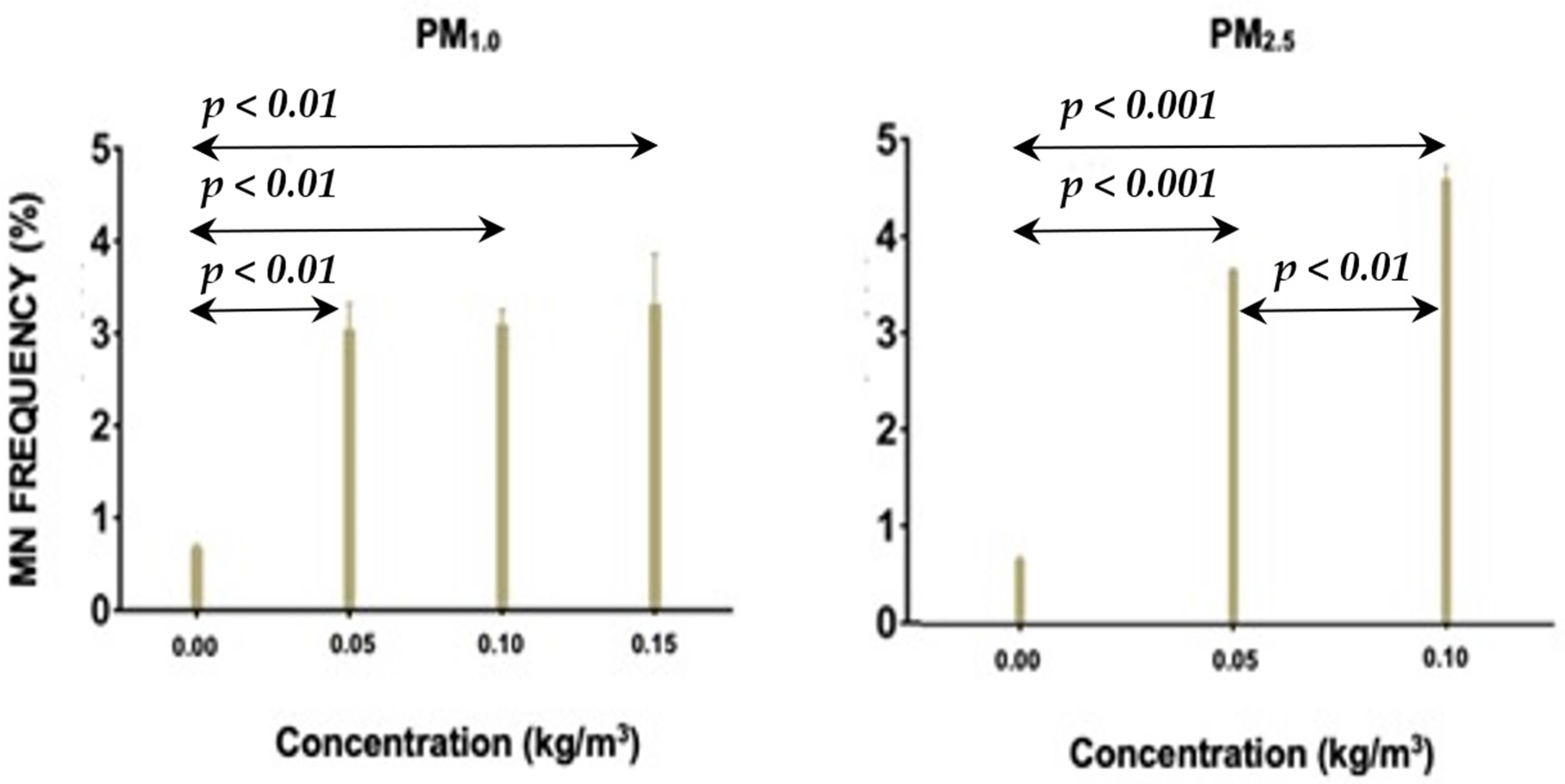
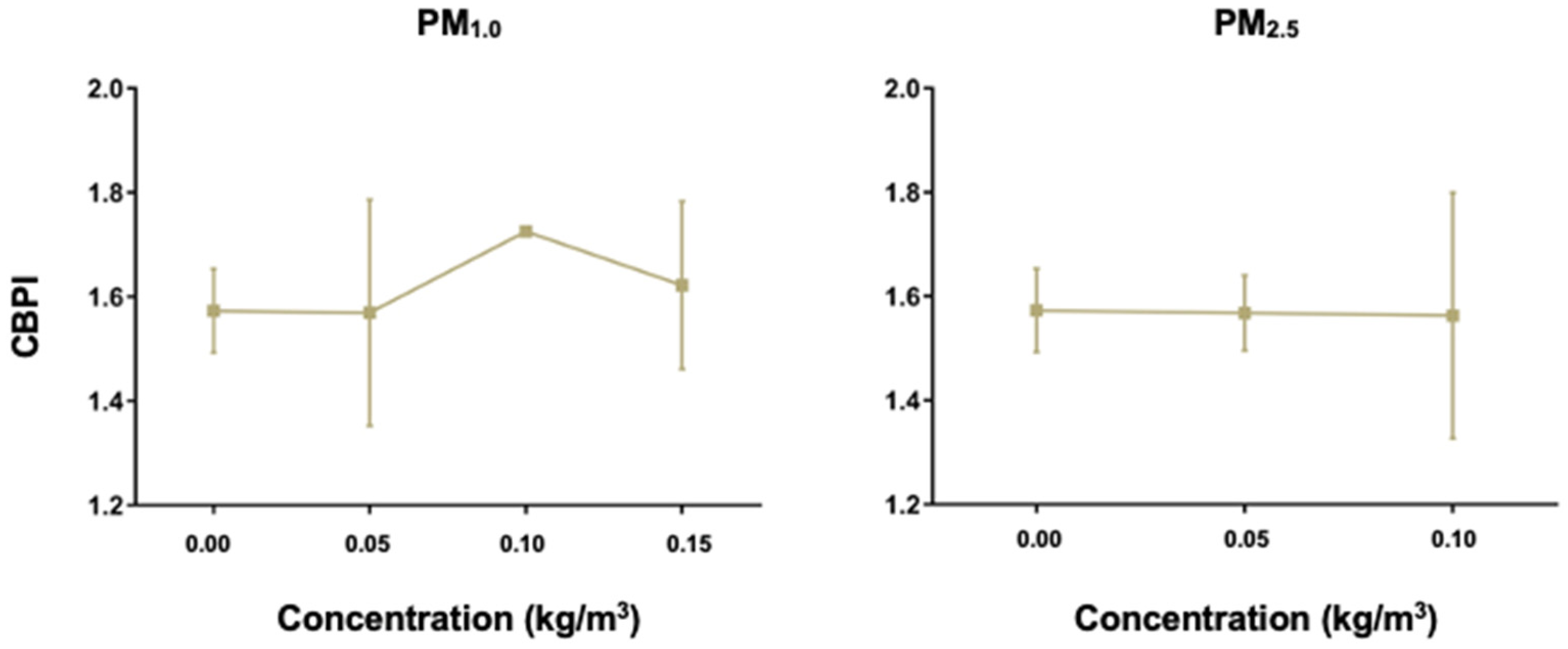
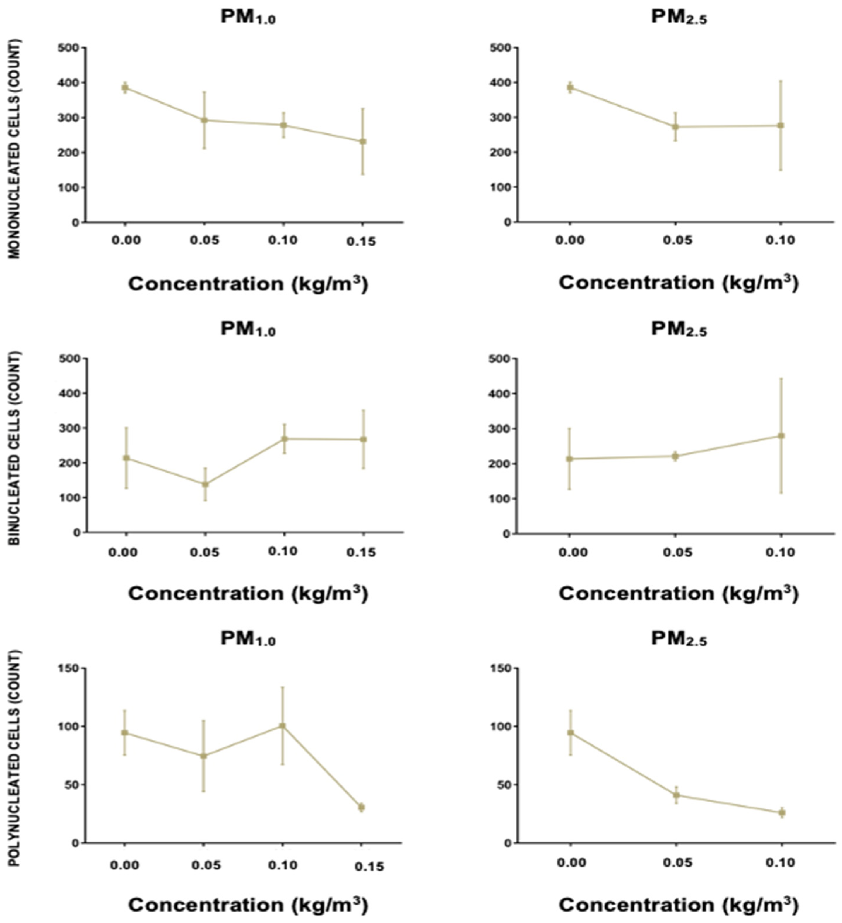
3.1. Differential Genotoxicity of PM2.5 and PM1.0

Figure 1 shows the MN frequencies of human peripheral blood lymphocytes that were exposed to different concentrations of PM1.0 and PM2.5. Based on the p-values obtained, there was a significant difference in the induction of micronuclei between PM1.0 and PM2.5. Two-way ANOVA showed that PM2.5 was more genotoxic compared to PM1.0 at p = 0.03. No data were obtained at the 0.15 kg/m3 concentration for PM2.5 because the concentration was cytotoxic and no cells were detected (Figure 2). This phenomenon may be attributed to cell death induced by the high concentration of PM2.5.
Figure 2 shows the cytokinesis-block proliferation index (CBPI) counts at each concentration of the PM size fractions. There was no significant difference between the CBPI at each treatment concentration, regardless of the size fraction. This observation indicates that the range of concentrations used for PM1.0 did not affect cell proliferation. PM2.5 was also non-toxic to the cells from 0.05–0.1 kg/m3.
To further confirm that cell proliferation was not affected throughout the concentration range used, we disaggregated the CBPI results by evaluating the nuclear morphologies of the cells. Consistent with the results from Figure 2, the combined total cell counts of mono-, bi-, and poly-nucleated lymphocytes did not statistically differ across the different concentrations, except at the highest dose of PM2.5 at which no cells were detected (Figure 3). Frequencies of nuclear abnormalities are widely used as biomarkers of exposure to genotoxic pollutants. Our results particularly validate the differential genotoxic effects from the two classes of PMs as polynucleated cells predominated at 0.05–0.10 kg/m3 for PM2.5 compared to PM1.0. Frequencies of these cell types after PM2.5 exposures point to a potential genotoxic environment which can lead to the development of cancer [17]. Additionally, PM2.5 has also been shown to induce inflammation and produce reactive oxygen species. This process leads to DNA damage and oxidized DNA base modifications as well as single- and double-strand breaks [18]. Similar reported observations indicate a direct correlation between the individual dose of PM2.5 and lymphocyte micronucleus frequency in the urban areas of São Paulo (Brazil) [19].

3.2. Differences in the Metal Compositions

The exact chemical nature of the PM fractions that showed different genotoxic activities is still unclear. Since particulate matter is able to transport metals aggravating health-related problems due to air pollution, the presence of metals in the samples was also assessed. Even at low doses, airborne metal ions are known to be significant contributors to the detrimental health effects of air pollution, such as aggravating reactive oxygen species (ROS) generation that result to inflammation, DNA damage, and apoptosis [20,21]. As a triggering pathway for the risk of developing various diseases, toxic metal species induce signal transduction pathways involving NF-κB, NRF2, JAK-STAT, JNK, and FOXO [22]. Trace levels of cadmium (Cd), lead (Pb), and zinc (Zn) were found in both PM size fractions although the peaks were lower than the EMDL/ELOQ. Calcium (Ca), potassium (K), and sodium (Na) were also found in the samples used for the AAS analysis. Consistent with the pattern of genotoxicity, the concentrations of the metals, particularly of Pb, are higher in PM2.5 compared to PM1.0 (Table 1). Table 2 lists the EMDL and ELOQ for each element in the setup used. Since the Philippines adopted unleaded fuel in 2001, it is suspected that the trace amounts of lead may originate from resuspended soil and wear-and-tear of engines. Future studies may focus on source apportionment of these metals that may have affected the genotoxicity profile of the air.

4. Conclusions

There was an increase in MN frequencies for all treatments (0.050 kg/m3, 0.10 kg/m3, and 0.15 kg/m3) for both PM1.0 and PM2.5. Trace concentrations of Cd, Ca, Pb, K, Na, and Zn were found in samples of both PM1.0 and PM2.5. It is noteworthy that the concentration of Pb was higher in PM2.5 compared to PM1.0 which warrants further investigation into its contribution to the genotoxicity of this fraction. Based on the dose-dependent increase in micronuclei frequency of lymphocytes exposed to PM1.0 and PM2.5, air pollutants in Manila may have the potential to lead to long-term DNA damage.

Author Contributions

Conceptualization, M.K.G.E., J.A.C., C.J.V., E.V., and C.D.; methodology, G.C., G.J.R., and C.A.; formal analysis, M.K.G.E., J.A.C., C.J.V., E.V., M.C.G., and C.D.; investigation, M.K.G.E., J.A.C., C.J.V., E.V., and C.D.; resources, M.K.G.E., J.A.C., C.J.V., and E.V.; writing—original draft preparation, M.K.G.E., J.A.C., and C.J.V.; writing—review and editing, E.V., M.C.G., and C.D.; supervision, E.V. All authors have read and agreed to the published version of the manuscript.

Funding

This research received no external funding.

Institutional Review Board Statement

Ethical review and approval were waived for this study.

Informed Consent Statement

Informed consent was obtained from the subjects involved in the study.

Data Availability Statement

Not applicable.

Conflicts of Interest

The authors declare no conflict of interest.

References

  1. Kyung, S.; Jeong, S. Particulate-Matter Related Respiratory Diseases. Tuberc. Respir. Dis. 2020, 83, 116–121. [Google Scholar] [CrossRef] [PubMed]
  2. Hahad, O.; Lelieveld, J.; Birklein, F.; Lieb, K.; Daiber, A.; Münzel, T. Ambient Air Pollution Increases the Risk of Cerebrovascular and Neuropsychiatric Disorders through Induction of Inflammation and Oxidative Stress. Int. J. Mol. Sci. 2020, 21, 4306. [Google Scholar] [CrossRef] [PubMed]
  3. Miller, M. Oxidative stress and the cardiovascular effects of air pollution. Free Radic. Biol. Med. 2020, 151, 69–87. [Google Scholar] [CrossRef] [PubMed]
  4. World Health Organization. WHO Global Air Quality Guidelines: Particulate Matter (PM2.5 and PM10), Ozone, Nitrogen Dioxide, Sulfur Dioxide and Carbon Monoxide. Available online: https://www.who.int/publications/i/item/9789240034228 (accessed on 20 November 2021).
  5. Narain, U.; Sall, C. Methodology for Valuing the Health Impacts of Air Pollution: Discussion of Challenges and Proposed Solutions; World Bank: Washington, DC, USA, 2016; Available online: https://openknowledge.worldbank.org/handle/10986/24440 (accessed on 20 December 2021).
  6. Macatangay, L.; Hernandez, R. A Deep Learning-Based Prediction and Simulator of Harmful Air Pollutants: A Case from the Philippines. In Proceedings of the 2020 11th IEEE Control and System Graduate Research Colloquium (ICSGRC), Shah Alam, Malaysia, 8 August 2020; pp. 381–386. [Google Scholar] [CrossRef]
  7. Stahl, C.; Cruz, M.T.; Bañaga, P.A.; Betito, G.; Braun, R.A.; Aghdam, M.A.; Cambaliza, M.O.; Lorenzo, G.R.; MacDonald, A.B.; Pabroa, P.C.; et al. An annual time series of weekly size-resolved aerosol properties in the megacity of Metro Manila, Philippines. Sci. Data 2020, 7, 128. [Google Scholar] [CrossRef] [PubMed]
  8. Recoleto, K.; Villarino, A. Micronucleus Test in Exfoliated Buccal Cells of Female Street Vendors Exposed to Vehicular Exhaust in Iligan City, Philippines. Int. J. Humanit. Soc. Sci. 2017, 9, 119–130. [Google Scholar]
  9. Hallare, A.; Gervasio, M.; Gervasio, P.; Acacio-Claro, P. Monitoring genotoxicity among gasoline station attendants and traffic enforcers in the City of Manila using the micronucleus assay with exfoliated epithelial cells. Environ. Monit. Assess. 2008, 156, 331–341. [Google Scholar] [CrossRef] [PubMed]
  10. Xing, Y.; Xu, Y.; Shi, M.; Lian, Y. The impact of PM2.5 on the human respiratory system. J. Thorac. Dis. 2016, 8, E69–E74. [Google Scholar] [CrossRef] [PubMed]
  11. Huang, F.; Pan, B.; Wu, J.; Chen, E.; Chen, L. Relationship between exposure to PM2.5 and lung cancer incidence and mortality: A meta-analysis. Oncotarget 2017, 8, 43322–43331. [Google Scholar] [CrossRef] [Green Version]
  12. Sanchez-Guerra, M.; Zheng, Y.; Osorio-Yanez, C.; Zhong, J.; Chervona, Y.; Wang, S.; Chang, D.; McCracken, J.; Diaz, A.; Bertazzi, P.; et al. Effects of particulate matter exposure on blood 5-hydroxymethylation: Results from the Beijing truck driver air pollution study. Epigenetics 2015, 10, 633–642. [Google Scholar] [CrossRef]
  13. Manila Climate Weather Averages. Available online: https://www.worldweatheronline.com/manila-weather-averages/manila/ph.aspx (accessed on 29 November 2021).
  14. Specifications E-SAMPLER. Available online: https://metone.com/wp-content/uploads/pdfs/e-sampler.pdf (accessed on 2 October 2021).
  15. 1405-D TEOM™, Continuous Dichotomous Ambient Particulate Monitor. Available online: https://www.thermofisher.com/order/catalog/product/TEOM1405D?ICID=search-%20product#/TEOM1405D?ICID=search-%20product (accessed on 2 October 2021).
  16. Test No. 487: In Vitro Mammalian Cell Micronucleus Test. 2014. “OECD Guidelines for the Testing of Chemicals”, Section 4.; OECD: Paris, France, 2014. [CrossRef]
  17. Zhang, X.; Duan, H.; Gao, F.; Li, Y.; Huang, C.; Niu, Y.; Gao, W.; Yu, S.; Zheng, Y. Increased micronucleus, nucleoplasmic bridge, and nuclear bud frequencies in the peripheral blood lymphocytes of diesel engine exhaust-exposed workers. Toxicol. Sci. 2015, 143, 408–417. [Google Scholar] [CrossRef] [Green Version]
  18. Schins, R.P.F.; Knaapen, A.M. Genotoxicity of poorly soluble particles. Inhal. Toxicol. 2007, 19, 189–198. [Google Scholar] [CrossRef] [PubMed]
  19. Boas, D.S.V.; Matsuda, M.; Toffoletto, O.; Garcia, M.L.B.; Saldiva, P.H.N.; Marquezini, M.V. Workers of São Paulo City, Brazil, exposed to air pollution: Assessment of genotoxicity. Mutat. Res. Genet. Toxicol. Environ. Mutagen. 2018, 834, 18–24. [Google Scholar] [CrossRef] [PubMed]
  20. Jiang, N.; Wen, H.; Zhou, M.; Lei, T.; Shen, J.; Zhang, D.; Wang, R.; Wu, H.; Jiang, S.; Li, W. Low-dose combined exposure of carboxylated black carbon and heavy metal lead induced potentiation of oxidative stress, DNA damage, inflammation, and apoptosis in BEAS-2B cells. Ecotoxicol. Environ. Saf. 2020, 206, 111388. [Google Scholar] [CrossRef] [PubMed]
  21. Zeng, X.; Xu, X.; Zheng, X.; Reponen, T.; Chen, A.; Huo, X. Heavy metals in PM2.5 and in blood, and children’s respiratory symptoms and asthma from an e-waste recycling area. Environ. Pollut. 2016, 210, 346–353. [Google Scholar] [CrossRef] [PubMed]
  22. Paithankar, J.G.; Saini, S.; Dwivedi, S.; Sharma, A.; Chowdhuri, D.K. Heavy metal associated health hazards: An interplay of oxidative stress and signal transduction. Chemosphere 2021, 262, 128350. [Google Scholar] [CrossRef] [PubMed]
  23. Eaton, A.D.; Clesceri, L.S.; Franson, M.A.H.; American Public Health Association; Rice, E.W.; Greenberg, A.E.; American Water Works Association and Water Environment Federation. Standard Methods for the Examination of Water and Wastewater, 21st ed.; American Public Health Association, APHA: Washington, DC, USA, 2005. [Google Scholar]
  24. AOAC. Official Methods of Analysis, 17th ed.; Association of Official Analytical Chemists, AOAC: Arlington, VA, USA, 2002. [Google Scholar]
Figure 1. MN frequency of the two PM size fractions. Only the p-values which yielded a significant difference are shown.
Atmosphere 13 00006 g001
Figure 2. CBPI counts obtained from both PM size fractions.
Atmosphere 13 00006 g002
Figure 3. Cell count profile for both PM1.0 and PM2.5.
Atmosphere 13 00006 g003
Table 1. Comparison of the elemental composition. Estimated method detection limit (EMDL) indicates the lowest possible concentration that can be detected. Estimated limit of quantitation (ELOQ) is an indication of a high probability of the element in the sample [23,24]. Legend: * means based on EMDL.
ElementsPM1.0PM2.5
Cd (mg/L)<0.5 *<0.5 *
Ca (mg/L)5040
Pb (mg/L)<2 *5
K (mg/L)14402100
Na (mg/L)28002770
Zn (mg/L)33
Table 2. Summary of EMDL and ELOQ.
ElementsWavelength (Nanometers, nm)EMDL a (µg/mL)ELOQ b (µg/mL)
Cd228.80.52
Ca422.72050
Pb283.327
K766.5cd
Na589cd
Zn213.914
a,b Estimated from the instrument detection limit of each element and incorporating the factors of sample volume and final volume of the sample. c,d EMDL and ELOQ values are not provided as the concentrations in the samples are above the EMDL and ELOQ values. Note 1: EMDL and ELOQ values are proprietary data of the Research and Analytical Services Laboratory of the Natural Sciences Research Institute (NSRI-RASL). Note 2: Initial sample volume used is 1 mL per trial. Sample volume submitted is 2–3 mL per sample.
Publisher’s Note: MDPI stays neutral with regard to jurisdictional claims in published maps and institutional affiliations.

