Article

Effect of Bovine MEF2A Gene Expression on Proliferation and Apoptosis of Myoblast Cells

1
Key Laboratory of Animal Genetics, Breeding and Reproduction in the Plateau Mountainous Region, Ministry of Education, Guizhou University, Guiyang 550025, China
2
College of Animal Science, Guizhou University, Guiyang 550025, China
*
Author to whom correspondence should be addressed.
Genes 2023, 14(7), 1498; https://doi.org/10.3390/genes14071498
Submission received: 23 June 2023 / Revised: 17 July 2023 / Accepted: 21 July 2023 / Published: 22 July 2023
(This article belongs to the Special Issue Bovine Functional Genomics and Epigenetics)

Abstract

:
Myocyte enhancer factor 2A (MEF2A) is a member of the myocyte enhancer factor 2 family. MEF2A is widely distributed in various tissues and organs and participates in various physiological processes. This study aimed to investigate the effect of MEF2A expression on the proliferation and apoptosis of bovine myoblasts. CCK8, ELISA, cell cycle, and apoptosis analyses were conducted to assess cell status. In addition, the mRNA expression levels of genes associated with bovine myoblast proliferation and apoptosis were evaluated using RT-qPCR. The results showed that the upregulation of MEF2A mRNA promoted the proliferation rate of myoblasts, shortened the cycle process, and increased the anti-apoptotic rate. Furthermore, the RT-qPCR results showed that the upregulation of MEF2A mRNA significantly increased the cell proliferation factors MyoD1 and IGF1, cell cycle factors CDK2 and CCNA2, and the apoptotic factors Bcl2 and BAD (p < 0.01). These results show that the MEF2A gene can positively regulate myoblast proliferation and anti-apoptosis, providing a basis for the analysis of the regulatory mechanism of the MEF2A gene on bovine growth and development.

1. Introduction

Muscle yield and the quality of livestock are key indicators of the quality of livestock products. The growth and development of skeletal muscle and genetic characteristics significantly influence muscle yield and the quality of livestock [1]. Meanwhile, the productive performance of livestock is determined by the proportion of skeletal muscle. The type and amount of protein within skeletal muscle fibres and the content of intramuscular fat also affect muscle tenderness [2,3]. In addition, skeletal muscle is crucial for body coordination, metabolism, and homeostasis in mammals and is associated with a strong regenerative capacity [4,5]. Myogenesis (myoblast proliferation, differentiation, and fusion) is crucial for skeletal muscle development, including embryonic development, postnatal growth, and muscle regeneration after injury [6].
Myocyte enhancer factor 2 (MEF2), belonging to the MADS transcription factor superfamily, can regulate muscle-specific gene expression [7]. The MEF2 protein was first identified in developing the skeletal muscle myotubes of vertebrates. The MEF2 protein can anchor A/T-rich DNA sequences in the muscle creatine kinase (MCK) gene promoter [8]. The MEF2 gene family has four members in vertebrates: MEF2A, MEF2B, MEF2C, and MEF2D. However, this family is a single gene in Drosophila and nematodes [9,10]. MEF2 family members have similar structures, containing the MADS-box region, MEF2 central structural domain, and C-terminal transcriptional activation region [11]. The MADS-box region mainly binds to abundant A/T DNA sequences, the MEF2 central structural domain enhances the binding of A/T sequences in the regulatory regions of target DNA, and the C-terminal region participates in many key transcriptional processes during cell growth and development. Furthermore, the structural differences among MEF2 family members are mainly found in the C-terminal transcriptional region [12,13]. MEF2 is involved in some biological processes, including musculogenesis, skeletal development, and neurological development [14]. MEF2A activates many muscle-specific, growth factor-induced, and stress-induced genes. Moreover, MEF2A participates in life processes, such as cell proliferation and differentiation, apoptosis, and morphological changes [15,16]. MEF2A is located downstream of the signalling pathway regulating the expression of musculogenic genes. MEF2A functions as a regulatory factor and a structural protein carrier. Moreover, MEF2A performs a transcriptional activity not carried out by any other factors in the family [17]. Clark et al. found that the MEF2A gene specifically binds to the Gtl2/Dio3 locus, upregulates transcriptional cofactor CITED2, and promotes cardiomyocyte proliferation [18]. The MEF2A gene is one of the first genes detected during muscle regeneration in mice. Liu et al. showed that toxin injection in mouse tibialis anterior muscle can significantly downregulate MEF2A and MEF2C on day 2, and significantly upregulate MEF2A during later muscle regeneration [19]. Zhou et al. found that interference with MEF2A expression can promote atherosclerotic lesions in apoE knockout mice [20]. Studies have also shown that MEF2A can regulate vascular endothelial cell migration and anti-apoptotic cell death [21]. Besides the key activating and regulatory role of MEF2A in myogenesis, MEF2A is also involved in lipid metabolic pathways and plays a role in various biological processes. Moreover, MEF2A regulates muscle tissue growth and development, depending on tissue type [22,23].
However, no study has evaluated MEF2A in large domestic animals. Furthermore, systematic MEF2A studies on cattle are lacking. Guanling cattle are a unique breed in Guizhou Province, China, which have the advantages of strong physique and disease resistance, but their growth is slow and meat yield is low. In this study, Guanling cattle were used as experimental subjects with the aim of investigating the effects of changes in MEF2A gene expression on the proliferation and apoptosis of bovine myoblasts. Therefore, this study can help to investigate the regulation mechanism of beef qualitative traits and provide basic theoretical references for bovine growth and development.

2. Materials and Methods

2.1. Ethical Approval

Animal experiments were approved by the Laboratory Animal Ethics of Guizhou University (No. EAE-GZU-2021-E023, Guiyang, China; 1 November 2021).

2.2. MEF2A Overexpression and Construction of Interference Vector

The full-length CDS region sequence of bovine MEF2A (NM_001083638.2) was amplified and ligated to the PMD-19T vector. The recombinant MEF2A vector and pEGFP-C1 vector were double-enzymatically digested using EcoR Ⅰ and Sal Ⅰ. The recombinant digest product was obtained to obtain the MEF2A overexpression vector and negative control (NC) was supplied by our laboratory. Four interfering group target sequences and one NC group sequence were set based on the sequence of the coding region of MEF2A and shRNA design principles. The vector with the best interfering effect was selected for further analysis. The primer sequences were synthesized by GEMA Ltd. (Shanghai, China). The detailed shRNA target sequences are shown in Table 1.

2.3. Cell Identification and Transfection

Myoblasts were isolated and cultured from three healthy 3-day-old Guanling calves. The calves were born in Guanling Cattle Industrial Park, Anshun (Guizhou, China) then slaughtered. The longest dorsal muscle tissue was obtained, cut, and digested with type II collagenase. The filtrate was centrifuged, and the supernatant was discarded. The cells were then resuspended in DMEM-F/12 (Gibco, San Diego, CA, USA) containing 12% foetal bovine serum and 1.5% penicillin-streptomycin. The purity of the myocytes was determined using immunofluorescence. α-actin (Bioss, Beijing, China) was used as primary antibody and Alexa Fluor594 (proteintech, Suzhou, China) as secondary antibody. Cells were inoculated into 6-well cell plates. Each vector was transfected into cells using Lipofectamine 3000 (Thermo Fisher Scientific, Waltham, MA, USA) transfection reagent when cell density reached about 80%. The cells were harvested after 48 h of transient transfection for subsequent assays, including CCK8, ELISA, RT-qPCR, flow cytometry, cell cycle, and apoptosis analyses.

2.4. Cell Proliferation Activity Assay

The effect of MEF2A overexpression and the expression interference on the proliferative capacity of adult myoblasts was examined using CCK8 reagent (APExBIO, Houston, TX, USA). Briefly, myoblasts were inoculated in 96-well plates and incubated at 37 °C in 5% CO2 for 0, 6, 12, 24, 48, and 72 h, followed by the addition of CCK8 reagent (10 μL) to each well. An enzyme marker (Thermo Fisher Scientific, Waltham, MA, USA) was used to detect absorbance at 450 nm.

2.5. ELISA for GH and INS

First, each vector was transfected into myoblasts for 48 h. The cells were washed with PBS, then 180 μL of RIPA lysis solution (Solarbio, Beijing, China) was added, and they were lysed on ice for 5 min. Total protein was collected by vortexing at 12,000 rpm for 15 min. Absorbance was measured at 450 nm using an enzyme marker (Thermo Fisher Scientific, Waltham, MA, USA) according to the instructions of the GH and INS kits. Linear regression equations were calculated for the standards based on OD values.

2.6. Flow Cytometry for Cycle Assay

Cells were cultured in 6-well plates, and each vector was transfected into myoblasts for 48 h. The cells were collected via centrifugation after trypsin digestion, followed by the addition of 2 mL of 70% ethanol, then fixed at 4 °C overnight. the fixed cells were centrifuged and the supernatant was discarded. The cells were washed with PBS, resuspended with 100 μL of RNase A, and placed in a 37 °C water bath for 30 min. Then, 500 μL of PI staining solution was added, and the sample was left to react for 30 min at 4 °C away from light. The DNA content of the different transfected and control groups was measured via flow cytometry (CytoFLEX, Beckman, Brea, CA, USA).

2.7. Flow Cytometry Assay for Apoptosis Detection

Cells were collected and prepared, as described above. The cells were centrifuged and washed with precooled PBS, resuspended with 100 μL binding buffer, and incubated with propidium iodide (PI) solution and FITC Annexin V (Thermo Fisher Scientific) at room temperature for 15 min while away from light. The samples were analysed via flow cytometry.

2.8. Real-Time Quantitative PCR

Total RNA was extracted using TRIZOL reagent (Solarbio, Beijing, China). The total RNA was reverse-transcribed using StarScrip II First Strand cDNA Kit (Genstar, Beijing, China) to obtain cDNA. The effects of MEF2A expression changes on the expression levels of cell cycle factors CDK2 and CCNA2, apoptosis factors Bcl2 and BAD, and cell growth factors IGF1 and MyoD1 were detected via RT-qPCR analysis. qRT-PCR primer information is shown in Table 2. The RT-qPCR program was run via Bio-Rad CFX96™ (Thermo Fisher Scientific, Waltham, MA, USA). The reaction system consists of 5 μL of SsoFASTTM EvaGreen® SuperMix, 0.5 μL each of upstream and downstream primers, 0.5 μL of cDNA, and 4 μL of ddH2O. Each sample had three triplicates. The reaction program consisted of pre-denaturation at 95 °C for 30 s, denaturation at 95 °C for 5 s, and annealing for 5 s (39 cycles). GAPDH was used as the internal reference gene. Gene expression was calculated via the 2−ΔΔCt method.

2.9. Statistical Analysis

Data are expressed as mean ± SD of three biological replicates. One-way analysis of variance (ANOVA) and the independent t-test were used to assess differences between groups via SPSS 18.0 software (IBM SPSS Statistics 18, Inc., Chicago, IL, USA). Statistical significance was analysed at p < 0.05.

3. Results

3.1. Cell Purity

The cells were identified via indirect immunofluorescence (Figure 1). Myocytes appeared red when bound to the anti-α-actin, while the DAPI nuclear stain was blue. The cytoplasmic and nuclear staining overlapped perfectly, indicating that the cultured cells were myoblasts.

3.2. Carrier Efficiency

The expression of the MEF2A gene and the efficiency of the interfering vector were analysed using qRT-PCR. The gene expression of the overexpressed OE-MEF2A was about 100 times (p < 0.01) higher than that of the control OE-NC (Figure 2A). Moreover, the expression of sh-RNA3 significantly decreased in the interference group compared with the control sh-NC (Figure 2B)(interference efficiency; 94.66%) (p < 0.01). The sh-RNA3 vector (sh-MEF2A) was selected for subsequent experiments.

3.3. Cell Viability Assay

The CCK8 results showed that OE-MEF2A significantly promoted the proliferative activity of myogenic cells after 6 h compared with OE-NC (Figure 3A). However, sh-MEF2A significantly reduced the proliferative activity of myoblasts cells after 6 h onwards compared with sh-NC (Figure 3B).

3.4. GH and INS Analysis

The ELISA results showed that MEF2A overexpression slightly increased GH content (p < 0.01) and INS content in myoblasts (p > 0.05) compared with OE-NC (Figure 4A). However, MEF2A interference decreased GH and INS content in myoblasts (p < 0.01) (Figure 4B), suggesting that the MEF2A gene can promote cellular activity.

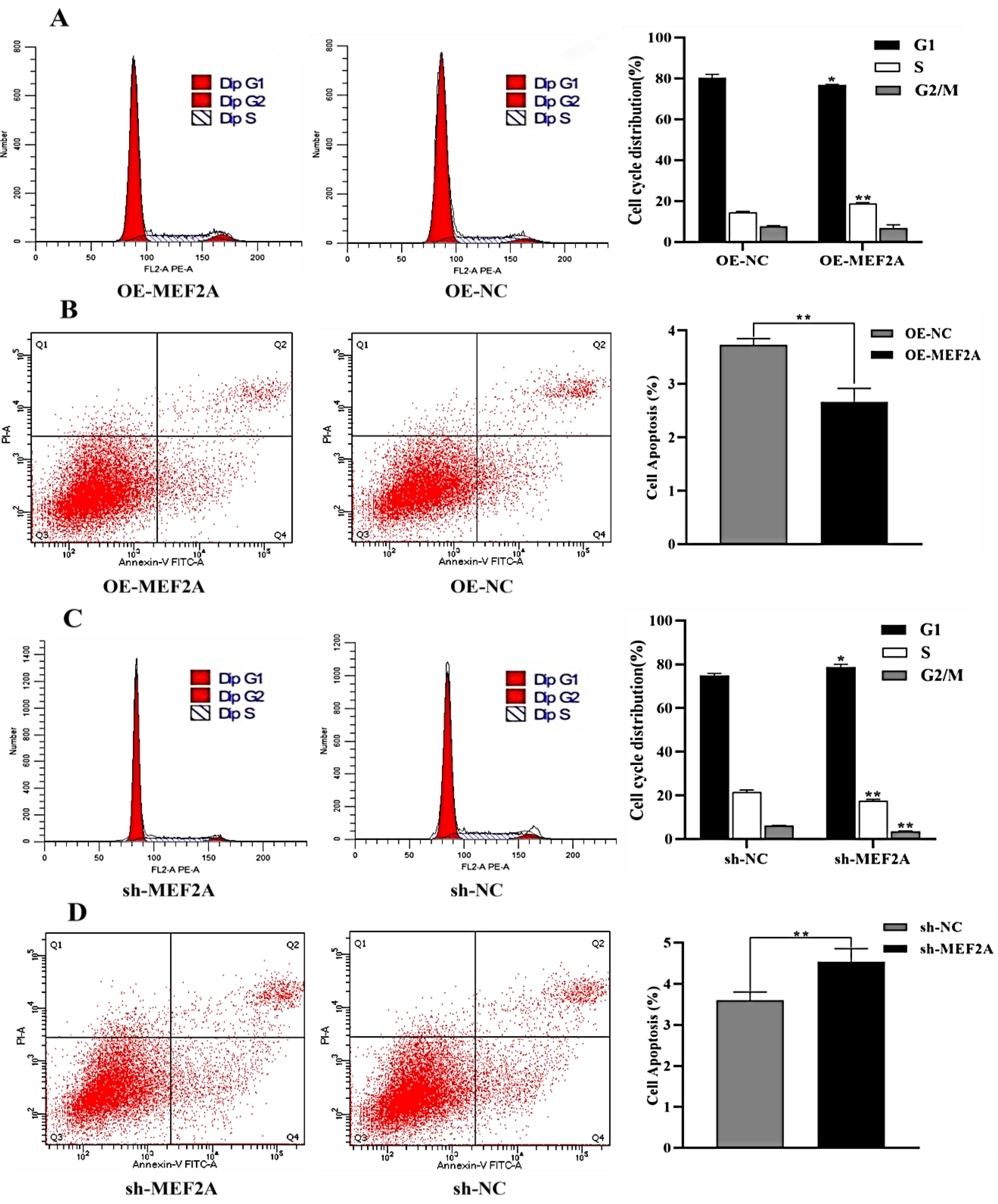
3.5. Cell Cycle and Apoptosis
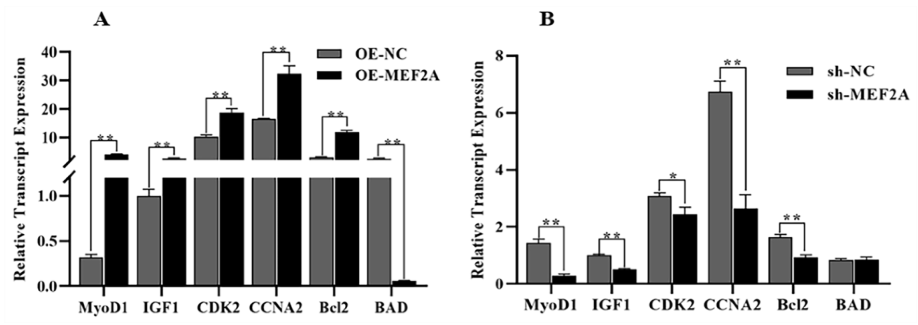
The effect of MEF2A overexpression and interference on myoblast cell cycle and apoptosis was examined via flow cytometry. Compared with OE-NC, the number of OE-MEF2A myoblast cells was significantly reduced in the G1 phase (p < 0.05). Furthermore, the number of S-phase myoblast cells was significantly increased (p < 0.01). Furthermore, G2/M was not significantly changed in the S-phase (p > 0.05) (Figure 5A), while the apoptosis rate of myoblast cells was reduced (p < 0.01) (Figure 5B). Compared with the sh-NC group, the number of myofibroblasts in the sh-MEF2A group significantly increased (p < 0.05) in the G1 phase and significantly decreased (p < 0.01) in the S and G2/M phases (Figure 5C). Moreover, the apoptosis rate significantly increased after the suppression of the MEF2A gene (p < 0.01). Furthermore, the inhibition of the MEF2A gene significantly increased the apoptosis rate of myoblast cells (p < 0.01) (Figure 5D). These findings suggest that the MEF2A gene can promote the proliferation of adult myoblasts. Moreover, MEF2A can stabilize cell activity.

3.6. RT-qPCR

MEF2A overexpression significantly increased the expression of the MyoD1, IGF1, CCNA2, CDK2, and Bcl2 genes (p < 0.01) and significantly decreased the expression of the BAD gene (p < 0.05) in myoblasts (Figure 6A). In contrast, MEF2A interference significantly downregulated the MyoD1, IGF1, CCNA2, and Bcl2 genes in myoblast cells (p < 0.01), significantly reduced the expression of the CDK2 gene (p < 0.05), and slightly upregulated the BAD gene (p > 0.05) (Figure 6B). These results further confirm the effect of the MEF2A gene on myoblast cells.

4. Discussion

The protein encoded by MEF2A can participate in various cellular processes as homo/heterodimers, including muscle development, cell growth control, and apoptosis. The proliferation and differentiation of myoblast cells are crucial in muscle tissue growth through cell proliferation and protein deposition [24]. MEF2A promotes myofibril formation and participates in the regeneration and differentiation of skeletal muscle stem cells and myoblasts cells [25]. Wang et al. reported that the MEF2A-MEG3/DIO3-PP2A axis promotes bovine muscle regeneration [26]. Chen et al. also demonstrated that MEF2A can promote postnatal muscle growth and development in goat kids. Moreover, MEF2A significantly increases muscle fibre diameter in the loin [27]. CDK2 is a cell cycle regulator that promotes the transition from the G1 to the S phase by forming a complex with CCNA2, thereby regulating cell cycle progression [28]. In this study, MEF2A upregulation or downregulation changed CDK2 and CCNA2 expression. MEF2A overexpression significantly decreased the number of G1-phase cells and significantly increased the number of S-phase cells, thus greatly shortening the G1-S phase process. Moreover, MEF2A interference did not affect the G1-S phase process, indicating that MEF2A accelerates the cell cycle process, consistent with the study of Wang et al. [29].
MyoD1 positively regulates skeletal muscle growth and development, thus affecting the development and repair of muscle tissue [30]. IGF1 can play a synergistic role with growth hormone (GH) to accelerate cell proliferation and differentiation and other physiological functions [31]. Herein, the CCK8 results showed that MEF2A upregulation significantly increased the viability of myoblast cells. Other assays also showed that MEF2A upregulation increased the expression of the MyoD1 and IGF1 genes, while MEF2A inhibition decreased these expressions. Growth hormone promotes growth, cell division, and proliferation, thus accelerating metabolism [32]. Insulin can be a long-term regulator of feed intake in ruminants, promoting glucose uptake from the blood. The glucose is then converted to glycogen for storage in the liver and muscle [33]. In this study, ELISA showed that MEF2A overexpression significantly increased GH levels and slightly increased INS levels. Moreover, MEF2A inhibition significantly decreased both INS and GH levels.
Zhou et al. reported that siRNA inhibition of MEF2A expression in apoE−/− mice increased the levels of pro-inflammatory cytokines, such as IL-6, MCP-1, TNF-α, and MMP-8, and atherosclerosis in apoE−/−mice [20]. Lu et al. also found that MEF2A deletion increased the risk of haemorrhage, inflammation, and hypercoagulable state in mice [34]. Resveratrol can delay the senescence and apoptosis of vascular endothelial cells (VECs). Resveratrol can also significantly upregulate the expression of MEF2A in VECs, thereby preventing apoptosis-induced oxidative stress [35]. In this study, MEF2A upregulation increased the activity of myogenic cells and had an anti-apoptotic effect. RT-qPCR also showed that MEF2A overexpression inhibited the expression of the pro-apoptogenic gene BAD and increased the expression of the anti-apoptogenic gene Bcl2. Furthermore, MEF2A inhibition increased the rate of apoptosis. One study reported that MEF2A reduced the expression level of Hspb7 in mouse skeletal muscle, which was effective in preventing muscle atrophy [36]. However, studies have also shown that MEF2A overexpression can disrupt myoblast differentiation patterns and apoptosis [29].
In addition, several studies have shown that MEF2A, MEF2C, and MEF2D have synergistic roles in muscle growth and development, especially MEF2A [37,38]. Synder et al. reported that mice with an overall deletion of MEF2A are defective in terms of skeletal muscle regeneration [38]. Deletion-of-function mutations in MEF2A, or MEF2A knockout, can significantly cause a disease phenotype in mice [39,40], suggesting that MEF2A has an irreplaceable function. Wu et al. found that MEF2A is evolutionarily higher on the list than MEF2C through phylogenetic analysis [41]. These findings reveal the importance and specificity of the MEF2A gene among the MEF2 family members.

5. Conclusions

In conclusion, MEF2A can positively regulate the proliferation and anti-apoptosis of bovine myoblasts through the regulation of key genes of the cycle (CDK2, CCNA2), growth (MyoD1, IGF1), and apoptosis (Bcl2, BAD). These results provide a basic theoretical reference for analysing the mechanisms through which MEF2A regulates bovine growth and development.

Author Contributions

Funding acquisition, H.X.; writing—original draft, J.S.; data curation, methodology, Y.R.; writing—review and editing, J.X.; investigation, P.S. All authors have read and agreed to the published version of the manuscript.

Funding

This study was supported by the Ministry of Education’s Research Cooperation Promotion Programme, the American University Area High-level Talent Cultivation Project ([2015] 2062), and the National Natural Science Foundation of China (31571279).

Institutional Review Board Statement

The research protocols and animals used in this study strictly comply with the guidelines of the Animal Welfare Committee of Guizhou University (EAE-GZU-2021-E023, 3 November 2021).

Informed Consent Statement

Not applicable.

Data Availability Statement

The data presented in this study are available on request from the corresponding author.

Conflicts of Interest

The authors declare no conflict of interest.

References

  1. Picard, B.; Berri, C.; Lefaucheur, L.; Molette, C.; Sayd, T.; Terlouw, C. Skeletal muscle proteomics in livestockproduction. Brief. Funct. Genom. 2010, 9, 259–278. [Google Scholar] [CrossRef]
  2. Motoyama, M.; Sasaki, K.; Watanabe, A. Wagyu and the factors contributing to its beef quality: A Japanese industry overview. Meat Sci. 2016, 120, 10–18. [Google Scholar] [CrossRef] [PubMed]
  3. Kruk, Z.A.; Bottema, M.J.; Reyes-Veliz, L.; Forder, R.E.A.; Pitchford, W.S.; Bottema, C.D.K. Vitamin A and marbling attributes: Intramuscular fat hyperplasia effects in cattle. Meat Sci. 2018, 137, 139–146. [Google Scholar] [CrossRef] [PubMed] [Green Version]
  4. Li, Y.; Chen, X.; Sun, H.; Wang, H. Long non-coding RNAs in the regulation of skeletal myogenesis andmuscle diseases. Cancer Lett. 2018, 417, 58–64. [Google Scholar] [CrossRef] [PubMed]
  5. Ge, L.; Dong, X.; Gong, X.; Kang, J.; Zhang, Y.; Quan, F. Mutation in myostatin 3′UTR promotes C2C12 myoblast proliferation and differentiation by blocking thetranslation of MSTN. Int. J. Biol. Macromol. 2020, 154, 634–643. [Google Scholar] [CrossRef]
  6. Bentzinger, C.F.; Wang, Y.X.; Rudnicki, M.A. Building muscle: Molecular regulation of myogenesis. Cold Spring Harbor Perspect. Biol. 2012, 4, a008342. [Google Scholar] [CrossRef] [Green Version]
  7. Mckinsey, T.A.; Zhang, C.L.; Olson, E.N. MEF2: A calcium-dependent regulator of cell division, differentiation and death. Trends Biochem. Sci. 2002, 27, 40–47. [Google Scholar] [CrossRef]
  8. Brand, N.J. Myocyte enhancer factor 2(MEF2). Int. J. Biochem. Cell Biol. 1997, 29, 1467–1470. [Google Scholar] [CrossRef]
  9. Black, B. Transcriptional control of muscle development by myocyte enhancer factor-2 (MEF2) proteins. Annu. Rev. Cell Dev. Biol. 1998, 14, 167–196. [Google Scholar] [CrossRef]
  10. Akhtar, M.W.; Kim, M.S.; Adachi, M.; Morris, M.J.; Qi, X.; Richardson, J.A.; Bassel-Duby, R.; Olson, E.N.; Kavalali, E.T.; Monteggia, L.M. In vivo analysis of MEF2 transcription factors in synapse regulation and neuronal survival. PLoS ONE 2012, 7, e34863. [Google Scholar] [CrossRef] [Green Version]
  11. Martin, J.F.; Miano, J.M.; Hustad, C.M.; Copeland, N.G.; Jenkins, N.A.; Olson, E.N. A Mef2 gene that generates a muscle-specific isoform via alternative mRNA splicing. Mol. Cell. Biol. 1994, 14, 1647–1656. [Google Scholar]
  12. Molkentin, J.D.; Olson, E.N. Combinatorial control of muscle development by basic helix-loop-helixand MADS-box transcription factors. Proc. Natl. Acad. Sci. USA 1996, 93, 9366–9373. [Google Scholar] [CrossRef] [PubMed]
  13. Zhu, B.; Carmichael, R.E.; Valois, L.S.; Wilkinson, K.A.; Henley, J.M. The transcription factor MEF2A plays a key role in the differentiation/maturation of rat neural stem cellsinto neurons. Biochem. Biophys. Res. Commun. 2018, 500, 645–649. [Google Scholar] [CrossRef]
  14. Edmondson, D.G.; Lyons, G.E.; Martin, J.F.; Olson, E.N. Mef2 gene expression marks the cardiac and skeletal muscle lineages during mouse embryogenesis. Development 1994, 120, 1251–1263. [Google Scholar] [CrossRef] [PubMed]
  15. Wu, Y.; Dey, R.; Han, A.; Jayathilaka, N.; Philips, M.; Ye, J.; Chen, L. Structure of the MADS-box/MEF2 domainof MEF2A bound to DNA and its implication for myocardin recruitment. J. Mol. Biol. 2010, 397, 520–533. [Google Scholar] [CrossRef] [PubMed] [Green Version]
  16. Dai, Y.; Zhang, S.; Wu, W. Analysis of MEF2A mutations in a Chinese population with premature coronary artery disease. Genet. Test. Mol. Biomark. 2013, 17, 352–355. [Google Scholar] [CrossRef]
  17. Blixt, N.; Norton, A.; Zhang, A.; Aparicio, C.; Prasad, H.; Gopalakrishnan, R.; Jensen, E.D.; Mansky, K.C. Loss of myocyte enhancer factor 2 expression in osteoclasts leads to opposing skeletal phenotypes. Bone 2020, 138, 115466. [Google Scholar] [CrossRef] [PubMed]
  18. Clark, A.L.; Naya, F.J. MicroRNAs in the Myocyte Enhancer Factor 2(MEF2)regulated MEG3-DIO3 Noncoding RNA Locus Promote Cardiomyocyte Proliferation by Targeting the Transcriptional Coactivator Cited2. J. Biol. Chem. 2015, 290, 23162–23172. [Google Scholar] [CrossRef] [Green Version]
  19. Liu, N.; Nelson, B.R.; Bezprozvannaya, S.; Shelton, J.M.; Richardson, J.A.; Bassel-Duby, R.; Olson, E.N. Requirement of MEF2A, C, and D for skeletal muscle regeneration. Proc. Natl. Acad. Sci. USA 2014, 111, 4109–4114. [Google Scholar] [CrossRef]
  20. Zhou, W.P.; Zhang, H.; Zhao, Y.X.; Liu, G.Q.; Zhang, J.Y. RNA interference of myocyte enhancer factor 2A accelerates atherosclerosis in apolipoprotein E-deficient mice. PLoS One. 2015, 10, e0121823. [Google Scholar] [CrossRef]
  21. Liu, C.; Yao, M.D.; Li, C.P.; Shan, K.; Yang, H.; Wang, J.J.; Liu, B.; Li, X.M.; Yao, J.; Jiang, Q.; et al. Silencing of circular RNA-ZNF609 ameliorates vascular endothelial dysfunction. Theranostics 2017, 7, 2863–2877. [Google Scholar] [CrossRef] [PubMed]
  22. Potthoff, M.J.; Olson, E.N. MEF2: A central regulator of diverse developmental programs. Development 2007, 134, 4131–4140. [Google Scholar] [CrossRef] [Green Version]
  23. Ma, Q.; Telese, F. Genome wide epigenetic analysis of MEF2A and MEF2C transcription factors in mouse cortical neurons. Commun. Integr. Biol. 2015, 8, e1087624. [Google Scholar] [CrossRef] [Green Version]
  24. Shahjahan, M. Skeletal muscle development in vertebrate animals. Asian J. Med. Biol. Res. 2015, 1, 139–148. [Google Scholar] [CrossRef] [Green Version]
  25. Medrano, J.L.; Naya, F.J. The transcription factor MEF2A fine-tunes gene expression in the atrial and ventricular chambers of the adult heart. J. Biol. Chem. 2017, 292, 20975–20988. [Google Scholar] [CrossRef]
  26. Wang, Y.; Mei, C.; Su, X.; Wang, H.; Yang, W.; Zan, L. MEF2A regulates the MEG3-DIO3 miRNA megacluster-targeted PP2A signaling in bovine skeletal myoblast differentiation. Int. J. Mol. Sci. 2019, 20, 27–48. [Google Scholar]
  27. Chen, L.; Cheng, B.; Li, L.; Zhan, S.; Wang, L.; Zhong, T.; Chen, Y.; Zhang, H. The molecular characterization and temporal spatial expression of myocyte enhancer factor 2 genes in the goat and their association with myofiber traits. Gene 2015, 555, 223–230. [Google Scholar] [CrossRef] [PubMed]
  28. Harashima, H.; Dissmeyer, N.; Schnittger, A. Cell cycle control across the eukaryotic kingdom. Trends Cell Biol. 2013, 23, 345–356. [Google Scholar] [CrossRef] [PubMed]
  29. Wang, Y.N.; Yang, W.C.; Li, P.W.; Wang, H.B.; Zhang, Y.Y.; Zan, L.S. Myocyte enhancer factor 2A promotes proliferation and its inhibition attenuates myogenic differentiation via myozenin 2 in bovine skeletal muscle myoblast. PLoS ONE 2018, 13, e0196255. [Google Scholar] [CrossRef] [Green Version]
  30. Hamed, M.; Khilji, S.; Chen, J.; Li, Q. Stepwise acetyltransferase association and histone acetylation at the Myod1 locus during myogenic differentiation. Sci. Rep. 2013, 3, 2390. [Google Scholar] [CrossRef] [Green Version]
  31. Lai, Z.; Wu, F.; Li, M.; Bai, F.; Gao, Y.; Yu, J.; Li, H.; Lei, C.; Dang, R. Tissue expression profile, polymorphism of IGF1 gene and its effect on body size traits of Dezhou donkey. Gene 2021, 766, 145118. [Google Scholar] [CrossRef] [PubMed]
  32. Berryman, D.E.; Glad CA, M.; List, E.O.; Johannsson, G. The GH/IGF-1 axis in obesity: Pathophysiology and therapeutic considerations. Nat. Rev. Endocrinol. 2013, 9, 346–356. [Google Scholar] [CrossRef] [PubMed]
  33. Takahashi, J.S.; Hong, H.K.; Ko, C.H.; McDearmon, E.L. The genetics of mammalian circadian order and disorder: Implications for physiology and disease. Nat. Rev. Genet. 2008, 9, 764–775. [Google Scholar] [CrossRef]
  34. Lu, Y.W.; Martino, N.; Gerlach, B.D.; Lamar, J.M.; Vincent, P.A.; Adam, A.P.; Schwarz, J.J. MEF2 (Myocyte Enhancer Factor 2) is essential for endothelial homeostasis and the atheroprotective gene expression program. Arterioscler. Thromb. Vasc. Biol. 2021, 41, 1105–1123. [Google Scholar] [CrossRef]
  35. Liu, B.R.; Pang, L.H.; Ji, Y.; Fang, L.; Tian, C.W.; Chen, J.; Chen, C.N.; Zhong, Y.; Ou, W.C.; Xiong, Y.J.; et al. MEF2A Is the Trigger of Resveratrol Exerting Protection on Vascular Endothelial Cell. Front. Cardiovasc. Med. 2022, 8, 775392. [Google Scholar] [CrossRef]
  36. Tobin, S.W.; Yang, D.; Girgis, J.; Farahzad, A.; Blais, A.; McDermott, J.C. Regulation of Hspb7 by MEF2 and AP-1: Implications for Hspb7 in muscle atrophy. J. Cell Sci. 2016, 129, 4076–4090. [Google Scholar]
  37. Snyder, C.M.; Rice, A.L.; Estrella, N.L.; Held, A.; Kandarian, S.C.; Naya, F.J. MEF2A regulates theMEG3-DIO3 microRNA mega-cluster to modulate WNT signaling in skeletal muscle regeneration. Development 2012, 140, 31–42. [Google Scholar] [CrossRef] [Green Version]
  38. Estrella, N.L.; Desjardins, C.A.; Nocco, S.E.; Clark, A.L.; Maksimenko, Y.; Naya, F.J. MEF2 transcription factors reguLate distinct gene programs in mammalian skeletal muscledifferentiation. J. Biol. Chem. 2015, 290, 1256–1268. [Google Scholar] [CrossRef] [Green Version]
  39. Wang, L.; Fan, C.; Topol, S.E.; Topol, E.J.; Wang, Q. Mutation of MEF2A in an inherited disorder with features of coronary artery disease. Science 2003, 302, 1578–1581. [Google Scholar] [CrossRef] [PubMed] [Green Version]
  40. Naya, F.J.; Black, B.L.; Wu, H.; Bassel-Duby, R.; Richardson, J.A.; Hill, J.A.; Olson, E.N. Mitochondrial deficiency and cardiac sudden death in mice lacking the MEF2A transcription factor. Nat. Med. 2002, 8, 1303–1309. [Google Scholar] [CrossRef]
  41. Wu, M.; Zhang, M.; Fan, Z.; Qin, X.; Zhu, X.; Ji, H.; Qin, Y.; Wang, Q.; Wu, L. Vertebrate paralogous MEF2 genes: Origin, conservation, and evolution. PLoS ONE 2011, 6, e17334. [Google Scholar] [CrossRef] [PubMed] [Green Version]
Figure 1. Indirect immunofluorescence detection of myoblasts bound to anti-α-actin (red, 1:100, Bioss, Beijing, China); nuclei stained with DAPI (blue) (200×).
Genes 14 01498 g001
Figure 2. Detection of MEF2A expression and interference vector efficiency. (A) qRT-PCR analysis of MEF2A overexpressed vector and (B) MEF2A interference vector. Two asterisks (**) represent highly significant differences (p < 0.01).
Genes 14 01498 g002
Figure 3. (A) CCK8 assay showing cell proliferation activity after transfection with OE-MEF2A and (B) sh-MEF2A. Two asterisks (**) represent highly significant differences (p < 0.01).
Genes 14 01498 g003
Figure 4. Effect of MEF2A expression on myoblast GH and INS. (A) ELISA showing the effect of OE-MEF2A transfection on the changes in GH and INS in myoblasts. (B) ELISA showing the effect of sh-MEF2A transfection on GH and INS changes in adult myoblasts. Two asterisks (**) represent highly significant differences (p < 0.01).
Genes 14 01498 g004
Figure 5. Detection of myoblast cycle and apoptosis via flow cytometry. (A) Characterization of myoblast cell cycle after OE-MEF2A transfection. (B) Characterization of apoptosis in myoblast cells after OE-MEF2A transfection. (C) Characterization of myoblast cell cycle after sh-MEF2A transfection. (D) Characterization of apoptosis in myoblasts cells through transfection with sh-MEF2A. One asterisk (*) indicates significant differences (p < 0.05). Two asterisks (**) represent highly significant differences (p < 0.01).
Genes 14 01498 g005
Figure 6. Effect of MEF2A expression on myoblast growth, proliferation, and apoptosis. (A) RT-qPCR showing the effect of OE-MEF2A transfection on the expression of MyoD1, IGF1, CDK2, CCNA2, Bcl2 and BAD. (B) qRT-PCR showing the effect of sh-MEF2A transfection on the expression of MyoD1, IGF1, CDK2, CCNA2, Bcl2, and BAD. One asterisk (*) indicates a significant difference (p < 0.05), while two asterisks (**) indicate extremely significant differences (p < 0.01).
Genes 14 01498 g006
Table 1. shRNA sequences.
NameSequence (5′-3′)
sh-NCTTCTCCGAACGTCTCACGT
sh-RNA1GCAGAACCAACTCGGATATTG
sh-RNA2GCCTCCACTGAATACCCAAAG
sh-RNA3GCAGCACCATTTAGGACAAGC
sh-RNA4GCAGTTATCTCAGGGTTCAAA
Table 2. Primer information.
GeneAccession NumbersPrimer Sequence(5′-3′)Product Size (bp)
MEF2ANM_001083638.2F: AATGAACCTCACGAAAGCAGAAC
R: TTAGCACATAGGAAGTATCAGGGTC
106
CDK2NM_001014934.1F: CCTGGATGAAGATGGACG
R: CTTGGAAGAAAGGGTGAG
101
CCNA2NM_001075123.1F: GCAGCCTTTCATTTAGCACTCT
R: ATTGACTGTTGTGCGTGCTG
155
Bcl2NM_001166486.1F: ATGTGTGTGGAGAGCGTCAA
R: ATACAGCTCCACAAAGGCGT
138
BADNM_001035459.2F: TCCCAGAGTTTGAGCAGAGTG
R: TTAGCCAGTGCTTGCTGAGAC
108
MyoD1NM_001040478.2F: AACCCCAACCCGATTTACC
R: CACAACAGTTCCTTCGCCTCT
162
IGF1NM_001077828.1F: TGCGGAGACAGGGGCTTTTATTTC
R: AAGCAGCACTCATCCACGATTCC
95
GAPDHNM_001034034.2F: TTGTGATGGGCGTGAACC
R: GTCTTCTGGGTGGCAGTGAT
169
Note: F represents the upstream primer and R represents the downstream primer.
Disclaimer/Publisher’s Note: The statements, opinions and data contained in all publications are solely those of the individual author(s) and contributor(s) and not of MDPI and/or the editor(s). MDPI and/or the editor(s) disclaim responsibility for any injury to people or property resulting from any ideas, methods, instructions or products referred to in the content.

