Article

Molecular Mechanism of MYL4 Regulation of Skeletal Muscle Development in Pigs

1
College of Animal Science and Technology, Hunan Agricultural University, Changsha 410128, China
2
Guangdong Laboratory for Lingnan Modern Agriculture, Guangzhou 510642, China
3
Institute of Subtropical Agriculture, Chinese Academy of Sciences, Changsha 410125, China
*
Authors to whom correspondence should be addressed.
Genes 2023, 14(6), 1267; https://doi.org/10.3390/genes14061267
Submission received: 17 May 2023 / Revised: 11 June 2023 / Accepted: 13 June 2023 / Published: 15 June 2023
(This article belongs to the Special Issue Genetics and Genomics of Pig Breeding)

Abstract

:
The processes of muscle growth and development, including myoblast proliferation, migration, differentiation, and fusion, are modified by a variety of regulatory factors. MYL4 plays an important role in atrial development, atrial cardiomyopathy, muscle-fiber size, and muscle development. The structural variation (SV) of MYL4 was found via the de novo sequencing of Ningxiang pigs, and the existence of SV was verified in the experiments. The genotype distribution of Ningxiang pigs and Large White pigs was detected, and it was found that Ningxiang pigs were mainly of the BB genotype and that Large White pigs were mainly of the AB genotype. However, the molecular mechanisms behind the MYL4-mediated regulation of skeletal muscle development need to be deeply explored. Therefore, RT-qPCR, 3′RACE, CCK8, EdU, Western blot, immunofluorescence, flow cytometry, and bioinformation analysis were used to explore the function of MYL4 in myoblast development. The cDNA of MYL4 was successfully cloned from Ningxiang pigs, and its physicochemical properties were predicted. The expression profiles in six tissues and four stages of Ningxiang pigs and Large White pigs were found to be the highest in the lungs and 30 days after birth. The expression of MYL4 increased gradually with the extension of the myogenic differentiation time. The myoblast function test showed that the overexpression of MYL4 inhibited proliferation and promoted apoptosis and differentiation. The knockdown of MYL4 showed the opposite result. These results enhance our understanding of the molecular mechanisms of muscle development and provide a solid theoretical foundation for further exploring the role of the MYL4 gene in muscle development.

1. Introduction

Due to rapid economic development and improved living standards, people have higher requirements in terms of the quality of livestock and poultry meat, and the demand for high-quality meat is increasing. Skeletal muscle is not only an important tissue in animals but also a source of daily meat products. Skeletal muscle consists of muscle fibers and connective tissue [1]. Muscle growth and development is closely related to meat production and is the main factor affecting overall growth. The maturation of muscle fibers goes through four stages: premyogenic progenitors, myoblasts, muscle tubes, and muscle fibers [2]. This complex process is precisely regulated by specific myogenic regulatory factors, such as the paired box family, the myocyte enhancer factor family, growth factors, cytokines, and other factors [3,4]. Genomic structural mutations are an important source of variation in many species and play an important role in phenotypic diversification and evolution [5]. Structural variation (SV) is defined as changes larger than 50 bp in the structure of a chromosome, and the main forms are deletion, insertion, repetition, inversion, and translocation [6]. Deletion is the most common type of SV, in which a nucleotide sequence is deleted on the chromosome, resulting in a decrease in the number of bases. Ma et al. identified 47 Chinese domestic pig-specific SVs; a 281 bp deletion in the first intron of the MYL4 gene was found in these SVs, and the existence of the SV of MYL4 was verified via experiments [7].
MYL4, also known as MLC1, encodes a kind of basic myosin light chain and is a member of the MYL family. The MYL family includes MYL2, MYL1, MYL3, and MYL4, and there is a high degree of sequence conservation among these genes. The MYL4 gene was found to be located on chromosome 12 in pigs and on chromosome 11 in mice [8,9]. Studies have shown that MYL4 was expressed in both adult atrial muscles and fetal skeletal muscles; two alternative splicing transcripts were found in the MYL4 gene, encoding the MLC1A subtype in adult atrial muscles and the MLClemb subtype in fetal skeletal muscles and showing the same protein-coding sequence [10]. At present, reports on MYL4 are mostly focused on atrial development and atrial cardiomyopathy [11,12,13,14]. Moreover, MYL4 can also play a regulatory role in the process of muscle development. MYL4 constitutes the structural component of muscles and can regulate the development of muscle fibers, and it is related to muscle development and striated muscle contraction [15,16]. Ghazizadeh et al. found increased retinoic acid synthesis and actin disorder in MYL4 mutant cell lines and zebrafish mutation models, indicating that MYL4 interacts with cytoskeletal actin both in vitro and in vivo [17]. Dong et al. identified nine SNPs in Pig’s 5′ flanking region of MYL4 genes; these nine SNPs increased the mRNA and protein expression of MYL4 in porcine muscles and leads to an increase in the number of muscle fibers in porcine Longissimus dorsi muscle [18]. Given that MYL4 is involved in the whole process of skeletal muscle, the effect of MYL4 on cells within muscle tissue requires further exploration.
In this experiment, we detected the distribution SV in MYL4 in Ningxiang pigs and Large White pigs and cloned the sequence of MYL4 in Ningxiang pigs and predicted its physicochemical properties. We also analyzed the tissue expression profiles of MYL4 and determined its role in C2C12 cells. Our results showed that MYL4 inhibits the proliferation of C2C12 cells and promotes the differentiation and apoptosis of C2C12 cells.

2. Materials and Methods

2.1. Animals and Samples

The animals used in the experiments were Ningxiang pigs and Large White pigs. Ningxiang pigs were purchased from Hunan Ningxiang Dalong Animal Husbandry Technology Co., Ltd. (Changsha, China). The Large White pigs were purchased from Xiangtan Weihong Food Co., Ltd. (Changsha, China). The ears of the Ningxiang pigs (n = 110) and the Large White pigs (n = 110) were collected and stored at −80 °C. Samples of the hearts, livers, spleens, lungs, fat, and Longissimus dorsi muscles of 30, 90, 150, and 210 days-after-birth Ningxiang pigs (n = 3) and Large White pigs (n = 3) were collected and stored in liquid nitrogen immediately. The Longissimus dorsi muscle of a 1-day-old Ningxiang pig was collected to clone the cDNA sequence of MYL4. All of the studies involving animals were conducted according to the ethics committee of the Animal Science and Technology College of Hunan Agriculture University (No. 2021–13).

2.2. Cell Culture and Transfection

The C2C12 cells were purchased from Anweisci (Shanghai, China). The cells were cultured in a complete medium containing 89% DMEM (Gibco, Waltham, MA, USA), 10% fetal bovine serum (Gibco, Waltham, MA, USA) and 1% penicillin–streptomycin (Gibco, Waltham, MA, USA) and cultured in a 37 °C incubator containing 5% CO2. For myogenic differentiation, the complete medium was changed into DMEM containing 2% horse serum (Hyclone, Logan, UT, USA). Three siRNAs, Si-NC, PCDNA3.1-MYL4 and PCDNA3.1, were purchased from JTS Scientific (Wuhan, China); the sequences were as follows (Table 1). The C2C12 cells were seeded into 6-well plates after reaching 80% confluence, and Si-MYL4, Si-NC, PCDNA3.1-MYL4 and PCDNA3.1 were transfected into the cells using Lipofectamine 2000 (Invitrogen, Waltham, MA, USA) after reaching 50% confluence.

2.3. PCR

The DNA was separated using the TIANamp genomic DNA Kit (TIANGEN, Beijing, China), following the recommendations of the manufacturer. NanoDrop 2000 (Thermo Scientific, USA) was used to determine the purity and concentration of the DNA. The sequences of the primer used to detect the SV in MYL4 were 5′TACTAGCTGCCACCTTGG 3′ (ssc-MYL4-1-F) and 5′ACAATGCCAGATCCTTAGCC 3′ (ssc-MYL4-1-R). The sequences of the primer used to clone the sequence of CDS were 5′CGCGTCTCTTGAGTCCTTCC3′ (MYL4-F) and 5′GTCTGCTTCACCCAGACATGA3′ (MYL4-R). PCR was performed using the Taq Master Mix buffer (Vazyme, Nanjing, China) on a PCR instrument (Bio-Rad, Hercules, CA, USA), and then the PCR production was detected using agarose gel electrophoresis. DL2000 was used as a DNA marker (Biodragon, Suzhou, China).

2.4. Real-Time Quantitative PCR

The RNA was extracted using the RNAsimple Total RNA kit (TIANGEN, Beijing, China), and the concentration and purity of the RNA were determined using agarose gel electrophoresis and NanoDrop 2000 (Thermo Scientific, Waltham, MA, USA). RNA reverse transcription was performed using a reverse transcription kit (Thermo Scientific, Waltham, MA, USA). PerfectStart® Green qPCR SuperMix (TransGen, Beijing, China) was used for real-time quantitative PCR (RT-qPCR), and RT-qPCR was performed on a CFX connect real-time system (Bio-Rad, Hercules, CA, USA) using Gapdh for the reference genes. The relative expression of the genes was calculated using 2−ΔΔCt. The sequences of the genes were downloaded from the NCBI database (https://www.ncbi.nlm.nih.gov) (accessed on 9 May 2023), and Primer 5.0 was used to design the primers. All of the primers were synthesized from Tsingke Biotechnology (Beijing, China), and the primer sequences are listed in the following table (Table 2).

2.5. Cloning of 3′ Sequence of MYL4 cDNA

We performed 3′ RACE using SMARTer RACE 5′/3′ Kit (Takara, Dalian, Chain), following the recommendations of the manufacturer. The sequence of the primer used for 3′ RACE was 5′-CCAAGCCAGAAGAAATGAATGCC-3′. The first-stand cDNA of Ningxiang pigs’ Longissimus dorsi was used to clone the 3′ sequence of MYL4. Then, the PCR reaction was performed, and the PCR product was gel-purified using the TIANgel purification kit (TIANGEN, Beijing, China). Next, the purified-PCR product was ligated into the pMD18-Tvector (Takara, Dalian, Chain) and then transformed into DH5α-competent cells. Positive clones were selected and sequenced by Tsingke Biotechnology (Beijing, China).

2.6. Cell Proliferation Assays

Cell proliferation was measured using CCK-8 assays and EdU staining. For CCK-8 assays, the transfected C2C12 cells were seeded into a 96-well plate, 10 μL of CCK-8 solution was added after incubation for 0 h (cell adhesion) 12 h, 24 h, 36 h and 48 h. Then, the cells were incubated for 4 h. Finally, a microplate reader (Multiskan FC, Thermo Scientific, Waltham, MA, USA) was used to measure the absorbance at 450 nm. For EdU staining, the cells were incubated with DMEM containing 20 μM EdU solution for 2 h after reaching 80% confluence. Next, EdU staining was performed using the EdU kit (Meilunbio, Dalian, China), following the recommendations of the manufacturer. Finally, fluorescence microscopy (Axio Vert A1, ZEISS, Germany) was used to capture the images.

2.7. Western Blot

The cells were collected, and 150 μL of RIPA lysis buffer (Beyotime, Shanghai, China) containing 1% protease inhibitor (Beyotime, Shanghai, China) was added to extract the protein. The concentration of protein was measured using a BCA protein assay kit (Meilunbio, Dalian, China) on a Multiskan FC microplate reader (Thermo Scientific, Waltham, MA, USA). Then, 2× protein loading buffer (Solarbio, Beijing, China) was added to the protein, and the protein was denatured by heating in 100 °C water for 10 min. PAGE Gel was made using a PAGE Gel Fast Preparation Kit (Epizyme, Shanghai, China). A total of 4 µg of protein in each well of PAGE Gel was electrophoresed and then transferred to a polyvinylidene difluoride (PVDF) membrane (Beyotime, Shanghai, China). The PVDF membrane was blocked in blocking buffer (Beyotime, Shanghai, China) for 2 h and then incubated with primary antibodies overnight at 4 °C. The primary antibodies used for Western blot were as follows: Anti-CDK4 (AF300822, 1:1000, AiFangBio, Changsha, China), Anti-PCNA (R25293, 1:1000, Zenbio, Chengdu, China), Anti-BAX (R22708, 1:1000, Zenbio, Chengdu, China), Anti-Caspase3 (R22811, 1:1000, Zenbio, Chengdu, China), Anti-β-actin (HC201, 1:10,000, TransGen, Beijing, China), Anti-MyoG (F5D, 1:200, DSHB, Iowa, IA, USA), Anti-MyHC (MF20, 1:500, DSHB, Iowa, IA, USA), Anti-MyoD (ER1913-45, 1:1000, Huabio, Hangzhou, China), and Anti-MYL4 (AF07872, 1:500, AiFangBio, Changsha, China). Next, The PVDF membrane was incubated with secondary antibodies (1:15,000, Zenbio, Chengdu, China) for 2 h. Finally, the PVDF membrane was exposed using an Image Quant LAS 4000 mini (GE, Boston, MA, USA). The ratio of the target protein to the reference protein β-actin is the relative expression of each protein.

2.8. Flow Cytometry

Flow cytometry was used to evaluate cell apoptosis. The cells after transfection with Si-MYL4, Si-NC, PCDNA3.1-MYL4, and PCDNA3.1 were collected, and then 1 mL of ice-cold PBS was added to wash the cells. Then, 100 μL of binding buffer was added to disperse the cells, and the collected cells were treated with 5 μL Annexin V-FITC and 10 μL propidium iodide (Yeasen, Shanghai, China) in the dark for 15 min at 37 °C. Finally, the treated cells were added to 400 μL 1 × Binding Buffer and measured using Cytek DxP Athena flow cytometry (Cytek, Fremont, CA, USA).

2.9. Immunofluorescence Analysis

C2C12 cell differentiation was evaluated via immunofluorescence analysis. The differentiated cells were fixed using 4% paraformaldehyde (Beyotime, Shanghai, China) for 30 min, 0.5% triton X-100 (Solarbio, Beijing, China) for 20 min, and blocked with 5% bovine serum albumin (BioFroxx, Frankfurt, Germany) for 2 h. For myogenic differentiation at 4 d after transfection with Si-MYL4, Si-NC, the cells were incubated with anti-MyHC monoclonal antibody (MF20, 1:300, DSHB, Iowa, IA, USA) overnight at 4 °C. For myogenic differentiation at 0 d, 2 d, 4 d, 6 d, and 8 d, the cells were incubated with anti-MYL4 polyclonal antibody (67533-1-Ig, 1:500, proteintech, Chicago, IL, USA) overnight at 4 °C. Then, the cells were incubated with DyLight 488 goat anti-mouse IgG (1:1000, Abbkine, Wuhan, China) for 2 h. Next, the nuclei were stained with DAPI (1:100, Beyotime, Shanghai, China) for 10 min. Finally, images were captured using a fluorescence microscope (Axio Vert A1, ZEISS, Oberkochen, Germany). The number of nucleus in myotubes to total nucleus was differentiation index. Myotubes were classified into three levels (<3 nucleus, 3–5 nucleus, >5 nucleus), and the fusion index was defined as the number of nucleus in MyHC-positive myotubes (<3 nucleus, 3–5 nucleus, >5 nucleus) to total nucleus within MyHC-positive myotubes.

2.10. Bioinformatics Analysis

An open reading frame (ORF) was found on an online website “https://www.ncbi.nlm.nih.gov/orffinder” (accessed on 8 May 2023). Sequence blast using DNAMAN (Version 9) and blastn suite was enabled through an online website “https://blast.ncbi.nlm.nih.gov/blast” (accessed on 8 May 2023). A phylogenetic tree was constructed with the MYL4 protein of different species using the neighbor-joining (NJ) method and MEGA version 11.0 software. The physicochemical properties were predicted using ProtParam at the following online website “https://web.expasy.org/protparam” (accessed on 9 May 2023). SOPMA online software, “http://npsa-pbil.ibcp.fr/cgi-bin” (accessed on 9 May 2023), was used to predict the secondary structure. The tertiary structural was predicted using SWISS-MODEL online software, “https://www.swissmodel.expasy.org” (accessed on 9 May 2023). SOPMA used PHD, GOR, Levin homology prediction, SOPMA, and dual prediction methods to predict the secondary structure of proteins, and synthesizes this into one result. SWISS-MODEL is a homology modelling of protein structures. STRING online software (version 11.5), “https://cn.string-db.org” (accessed on 9 May 2023), was used to analyze the protein interactions.

2.11. Statistical Analyses

Data statistical analysis was performed using one-way ANOVA or Student’s t-test using IBM SPSS 22.0 software. The data were indicated as mean ± standard error. GraphPad Prism 8.0 was used to draw the pictures. Statistically significant differences were considered at p < 0.05. * p < 0.05, and ** p < 0.01 indicates a significant difference, and ns means no significant difference. A Chi-square test was used to test whether the SV in the MYL4 gene was in a Hardy–Weinberg balance in Ningxiang pigs and Large White pigs. When p > 0.05, it was in a Hardy–Weinberg balance, and when p < 0.05, it was in a Hardy–Weinberg imbalance. When polymorphism information content (PIC) is >0.5, the locus is highly polymorphic. When 0.25 < PIC < 0.5, then the locus is moderately polymorphic, and when PIC is <0.25, the locus is considered to be low polymorphic. ImageJ (Version 1.49) software was used for the cell count and to calculate the relative gray value of the protein.

3. Results

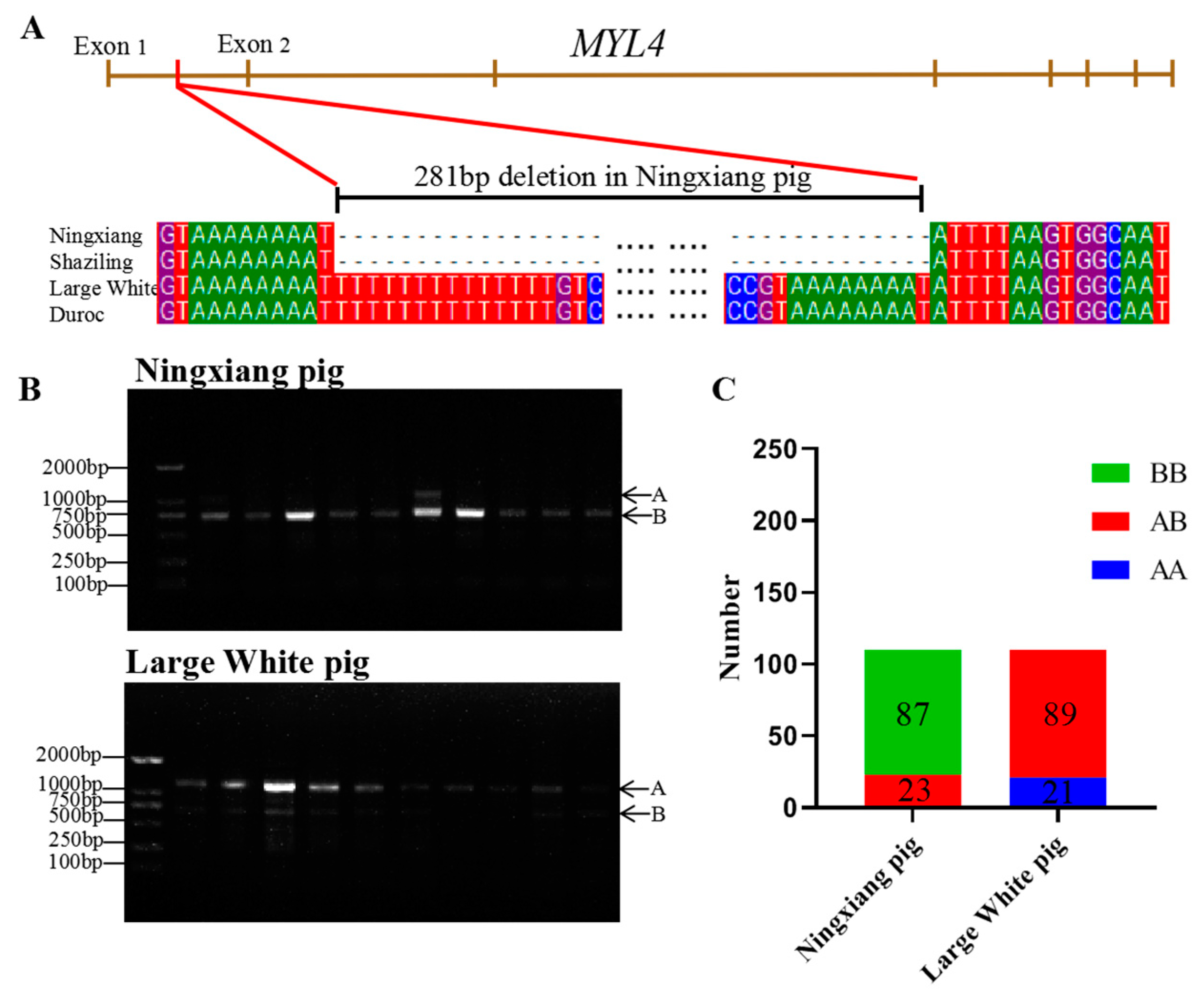
3.1. SV in MYL4 Gene of Ningxiang Pigs and Large White Pigs

As shown in Figure 1A, the 281 bp deletion in the first intronic of MYL4 of Ningxiang pig. The DNA of the Ningxiang pigs (n = 110) and Large White pigs (n = 110) were collected, and then the genotype distributions of the SV in the MYL4 gene of Ningxiang pigs and Large White pigs were detected using PCR and agarose gel electrophoresis. The production length of primer ssc-MYL4-1 is 1036 bp. Here, the genotype distributions of the SV in the MYL4 gene were defined as follows: the BB genotype represents a deletion homozygote (755 bp), the AB genotype represents deletion heterozygotes (1036 bp and 755 bp), and the AA genotype is a non-deletion homozygote (1036 bp). The typical agarose gel electrophoresis pictures of the genotype distribution of the SV in the MYL4 gene of Ningxiang pigs and Large White pigs are listed in Figure 1B. As shown in Figure 1B,C, the AB genotype (n = 23) and BB genotype (n = 87) were detected in Ningxiang pigs, and the AB genotype (n = 89) and AA genotype (n = 21) were detected in Large White pigs. The detailed detection results of the SVs in the MYL4 gene in Ningxiang pigs and Large White pigs are shown in Table 3. The dominant genotype was BB in Ningxiang pigs (79.09%), but it was AB in Large White pigs (80.91%). The frequency of allele A in Ningxiang pigs (10%) is lower than that in Large White pigs (59.55%). In addition, the genotype frequency of Ningxiang pigs was in a Hardy–Weinberg balance (p > 0.05), while the genotype frequency of Large White pigs was in a Hardy–Weinberg imbalance (p < 0.05). PIC analysis revealed that the PIC of Ningxiang pigs (PIC = 0.17) was lower than that of Large White pigs (PIC = 0.37).

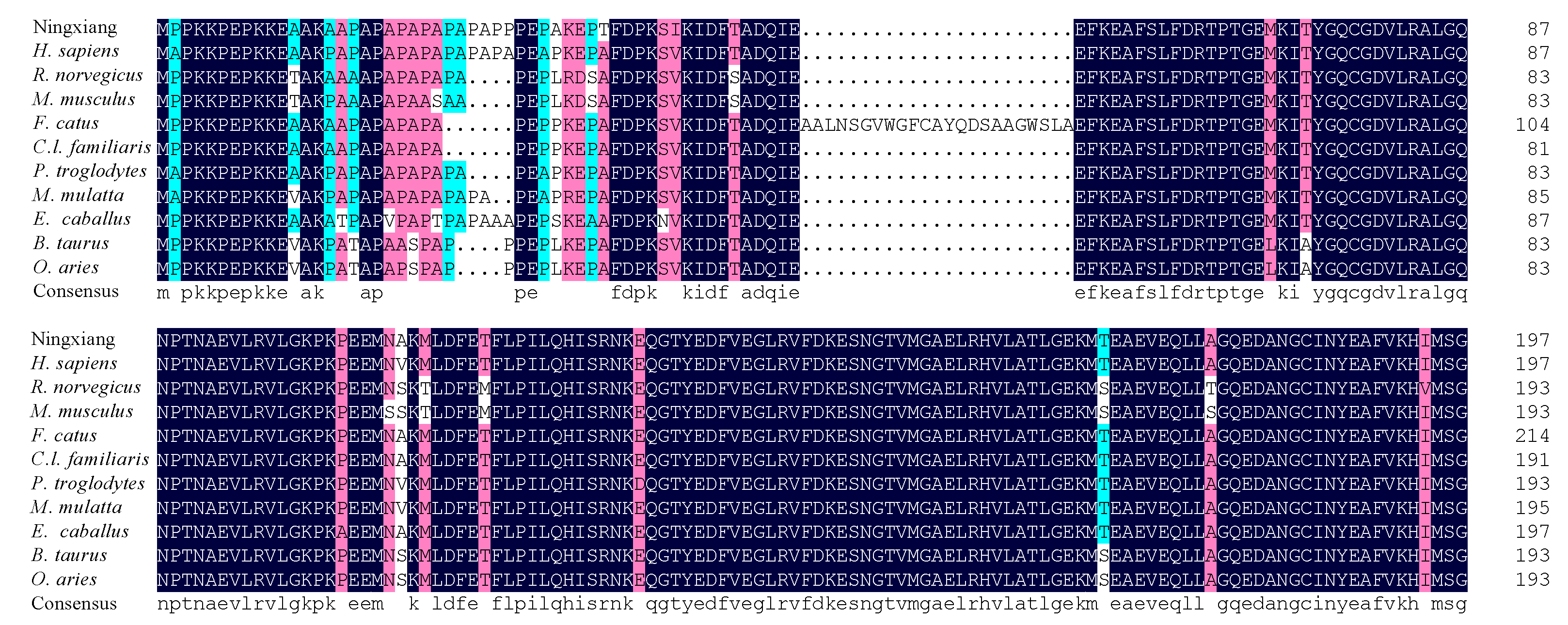
3.2. cDNA Cloning and Sequence Analysis of MYL4 in Ningxiang Pigs

Using 3′ RACE, 500 bp 3′ RACE products of the MYL4 gene were cloned (Figure 2A). In addition, 657 bp PCR products of the MYL4 gene were obtained (Figure 2B). Then, two fragments were stitched together, and 857 bp cDNA sequences were obtained. The sequence was consistent with the sequence of the porcine MYL4 gene in the NCBI database, encoding the same amino acid sequence. As shown in Figure 2C, the MYL4 cDNA contained a 594 bp ORF encoding a 197 amino acid peptide, the start codon is ATG, and the stop codon is TGA. Phylogenetic trees were constructed using MEGA. As shown in Figure 2D, the MYL4 protein evaluated in this study shared a close evolutionary position with Canis lupus familiaris and Felis catus MYL4 proteins. The amino acid sequences of MYL4 were aligned with the amino acid sequences of the other 10 species when using DNAMAN. The amino acid sequences used in this study were obtained from GeneBank, and the accession numbers are listed in (Supplementary Table S1). The homology analyses indicated that the MYL4 protein of Ningxiang pigs shared 95.94, 89.34, 87.31, 78.97, 95.43, 92.89, 89.34, 94.42, 92.39, and 92.89% sequence similarity with the MYL4 of Homo sapiens, Rattus norvegicus, Mus musculus, F. catus, C.l. familiaris, Pan troglodytes, Macaca mulatta, Bos taurus, Equus caballus, and Ovis aries (Figure 3).

3.3. Prediction of the Structures and Features of MYL4 Protein

The physicochemical properties of the MYL4 protein were predicted using ProtParam, and the results are shown in Table 4. The molecular weights were about 22 kDa, and the theoretical pI, aliphatic index, instability, and the grand average of hydropathicity were 4.98, 67.97, 58.38, and −0.535, respectively. Secondary protein structural analysis showed that the MYL4 protein of Ningxiang pigs comprised 46.70% α-helix, 5.58% β-turn, 5.58% extended strand, and 42.13% random coil (Figure 4A), indicating that α-helix and random coils were the dominant structural features of MYL4 proteins. The tertiary structure of the MYL4 protein was predicted using SWISS-MODEL, and the predictions are depicted in Figure 4B, which is consistent with the secondary structure prediction results. As shown in Figure 4C, MYL4 interacted with MYL3, MYL7, MYH3, MYH6, MYH7, MYH7B, ACTC1, MLC2V, MYLK, and MYLK4.

3.4. Profiles Analysis of MYL4 Gene Expression in Ningxiang Pigs and Large White Pigs

RT-qPCR was used to investigate the expression levels of MYL4 in different tissues and different development periods of Ningxiang pigs and Large White pigs. As shown by the results of RT-qPCR, MYL4 was widely expressed across tissues in Ningxiang pigs and Large White pigs. MYL4 is expressed in the lungs of Ningxiang pigs and Large White pigs, with the highest expression, but in the muscles, the lowest expression (Figure 5A–H). The temporal expression profiles of MYL4 in the muscle showed the highest expression levels 30 days after birth (Figure 5I,J). Further comparing the expression of MYL4 in different tissues after 30 days for NingXiang pigs and Large White pigs, the results indicated that the expression of MYL4 in the muscles, spleens, and hearts of Large White pigs was significantly higher than that of Ningxiang pigs (p < 0.01), while the expression of MYL4 in the fat and livers of Large White pigs was significantly lower than that of Ningxiang pigs (p < 0.01) (Figure 5K).

3.5. Knockdown and Overexpression of MYL4

The endogenous MYL4 in proliferating C2C12 cells was detected through the use of RT-qPCR, and the cells were collected at 8 h (8 h after the cells were seeded), 16 h, 24 h, and 32 h, respectively. From the results of the RT-qPCR, MYL4 was the most strongly expressed at 24 h, which then gradually downregulated (Figure 6A). MYL4 was successfully inhibited or overexpressed in C2C12 cells by transfecting the si-MYL4 or MYL4-plasmid. As shown in Figure 6B–D, MYL4 was successfully inhibited, and siRNA1 had the highest inhibition efficiency (p < 0.01). The transfection of 2.5 ug of PCDNA3.1-MYL4 had the highest overexpression efficiency (p < 0.01) (Figure 6F–H). Next, the mRNA expressions of MYL1, MYL2, and MYL3 were detected by means of RT-qPCR in C2C12 cells proliferating for 24 h, and it was found that MYL1 mRNA was expressed in C2C12 cells, while MYL2 and MYL3 were not detected. RT-qPCR was used to explore the effect of MYL4 on other light chain genes after the knockdown and overexpression of MYL4. As shown in the RT-qPCR results, the mRNA expression of MYL1 insignificantly decreased when MYL4 was inhibited (p < 0.01), and the mRNA expression of MYL1 also insignificantly decreased when MYL4 was overexpressed (Figure 6E) (p < 0.05).

3.6. MYL4 Inhibits C2C12 Cell Proliferation

Concerning the effect of MYL4 on C2C12 cell proliferation, and as shown by the result of RT-qPCR and Western blot, the knockdown of MYL4 in C2C12 cells caused a marked increase in the mRNA expression of CCND, PCNA, CDK4, and CCNE (p < 0.05) and the protein levels of PCNA (p < 0.01) (Figure 7A–C). The results of the CCK8 analysis indicated that the knockdown of MYL4 dramatically promoted C2C12 cell proliferation (p < 0.01) (Figure 7D). The overexpression of MYL4 showed that the mRNA expression of CCND, PCNA, CDK4 and CCNE significantly decreased (Figure 6E) (p < 0.05), and the protein of PCNA and CDK4 significantly decreased (Figure 6F,G) (p < 0.05). The CCK8 analysis results also indicated that an overexpression of MYL4 could inhibit C2C12 cells proliferation (Figure 7H) (p < 0.01). EdU staining was used to further confirm the effect of MYL4 on cell proliferation, and the results indicated that MYL4 knockdown significantly promoted C2C12 cell proliferation (p < 0.01) (Figure 7I), and the overexpression of MYL4 markedly inhibited C2C12 cell proliferation (p < 0.01) (Figure 7J). In general, these results suggested that MYL4 could inhibit C2C12 cell proliferation.

3.7. MYL4 Promotes C2C12 Cells Apoptosis

To explore the effect of MYL4 on C2C12 cell apoptosis, Si-MYL4, Si-NC, PCDNA3.1-MYL4, and PCDNA3.1 were transfected into C2C12 cells. As shown in Figure 8A, the mRNA expression of BAX and Caspase3 significantly decreased after transfection with Si-MYL4 (p < 0.01). The Western blot results showed that the protein expression of BAX and Caspase3 significantly decreased in Si-MYL4 when compared with Si-NC (p < 0.01) (Figure 8B,C). The overexpression of MYL4 showed an opposite result (Figure 8D–F). This result indicated that MYL4 promotes C2C12 cell apoptosis. To further confirm the effect of MYL4 on cell apoptosis, cell apoptosis was detected using flow cytometry. The flow cytometry results showed that the number of apoptosis cells (Q2 + Q3) in the Si-MYL4 group was significantly lower than that in Si-NC (p < 0.05) (Figure 8G), and the number of apoptosis cells in the PCDNA3.1-MYL4 group was much higher than that in the PCDNA3.1 group (p < 0.05) (Figure 8H). In general, the above results suggested that MYL4 could promote C2C12 cell apoptosis.

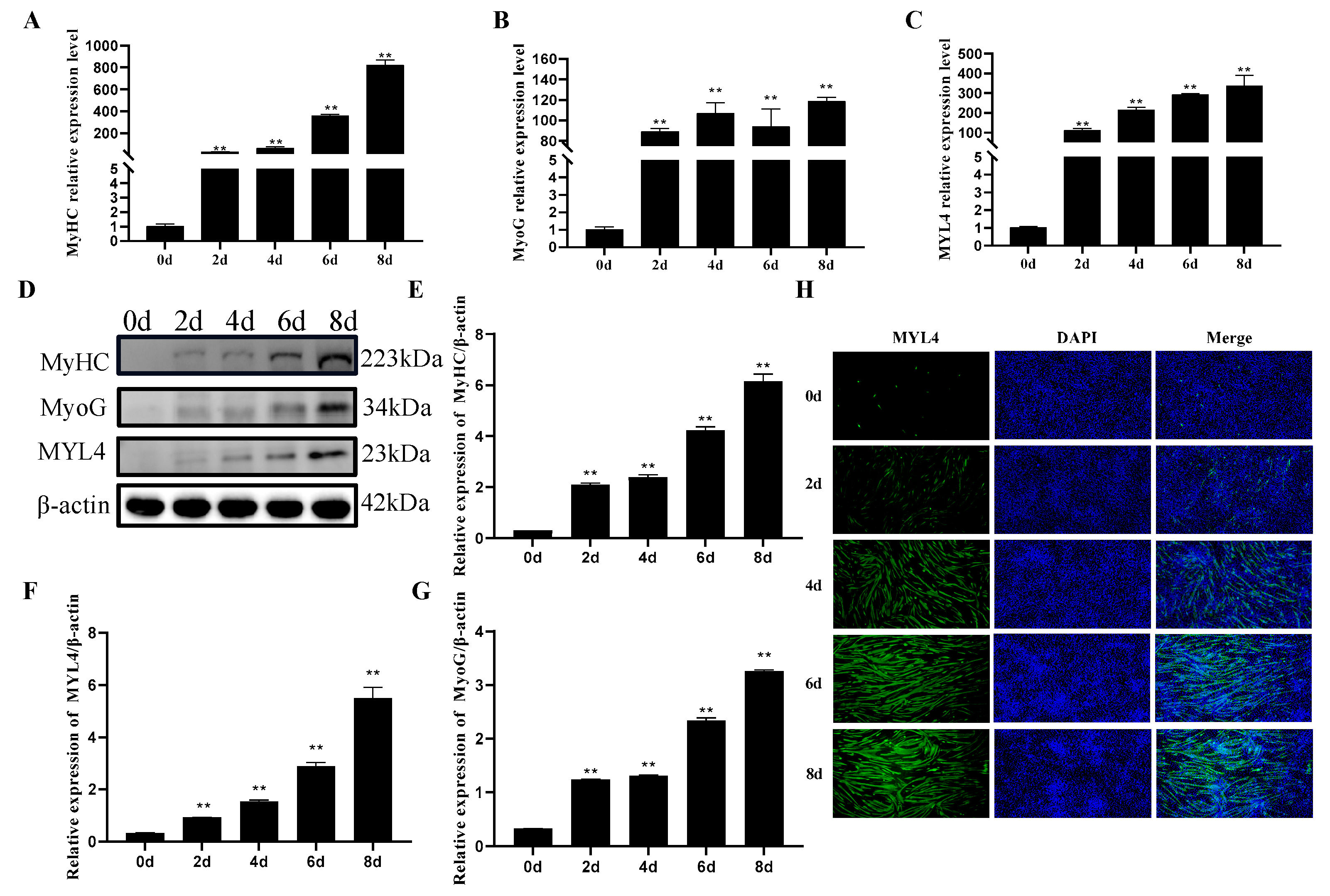
3.8. Expression Pattern of MYL4 during the Differentiation of C2C12 Cells

To detect the expression levels of MYL4 at different stages of C2C12 cell differentiation, C2C12 cells were collected at differentiation periods of 0 d, 2 d, 4 d, 6 d, and 8 d, respectively. RT-qPCR and Western blot were used to detect the mRNA expressions and protein expressions of MyHC, MyoG, and MYL4. As shown in Figure 9A,B, the mRNA expression levels of MyHC and MyoG were both gradually upregulated as C2C12 cell differentiation progressed. In addition, the result of the Western blot indicated that the expression levels of MyHC and MyoG increased significantly during C2C12 cell differentiation (Figure 9D–F). These results demonstrated that the C2C12 cells were well differentiated. As shown in Figure 9C,G, the mRNA and protein expression levels of MYL4 increased during C2C12 cell differentiation. The fluorescence intensity of MYL4 was enhanced with the extension of the differentiation time, as shown through immunofluorescence staining (Figure 9H). These results indicated that MYL4 may play an important role in C2C12 cell differentiation.

3.9. MYL4 Promotes C2C12 Cells Differentiation

To explore the effect of MYL4 on C2C12 cell differentiation, Si-MYL4, Si-NC, PCDNA3.1-MYL4, and PCDNA3.1 were transfected into C2C12 cells. The cells were induced to differentiate for 2 d and 4 d. As shown in the results of RT-qPCR and Western blot, the mRNA expression levels of MyoG, MyHC, Myf5, and MyoD significantly decreased (p < 0.05), and the protein expression levels of MyoG, MyHC, and MyoD significantly decreased after the knockdown of MYL4 (p < 0.05) (Figure 10A–D). The overexpression of MYL4 could upregulate the mRNA expression of MyoG, MyHC, Myf5, and MyoD (p < 0.05), and the protein expression levels of MyoG, MyHC, and MyoD (p < 0.01) (Figure 10E–H). These results indicated that MYL4 was a positive regulator of C2C12 cell differentiation. Then, immunofluorescence was used to further investigate the effect of MYL4 on C2C12 cell differentiation (Figure 10I), and the results indicated that the differentiation index decreased from 30.01% to 24.25% (p < 0.05) (Figure 10J). Fewer than three nuclear myotubes in the Si-MYL4 group was significantly higher than that in Si-NC (p < 0.01), and more than five nuclear myotubes in the Si-MYL4 group was significantly lower than that in Si-NC (p < 0.01) (Figure 10J,K). The above results indicate that MYL4 could promote C2C12 cell differentiation.

4. Discussion

SVs are large genomic alterations. It has been reported that large genomic SVs have a greater effect on gene expression than single-nucleotide variations and that SVs have a greater effect on gene expression and function [19]. Ma et al. proved that the deletion of 281 bp in the first intron of the MYL4 gene can promote fat deposition in Ningxiang pigs; thus, does the deletion of 281 bp in the first intron of the MYL4 gene affects the development of skeletal muscle in Ningxiang pigs? In this study, genotype BB was found to exist mainly in Ningxiang pigs, and genotype AB existed mainly in Large White pigs, and the frequency of A allele in Ningxiang pigs was lower than that in Large White pigs. In addition, the genotype frequency of Ningxiang pigs was in a state of Hardy–Weinberg balance, but the genotype frequency of Large White pigs was in a state of Hardy–Weinberg imbalance, which may be associated with population loss or the degree of inbreeding. The PIC values of the SV in the MYL4 gene of Ningxiang pigs is 0.17, and those results indicate that the variation in the SV in the MYL4 gene of Ningxiang pigs is stable. In recent decades, more and more full-length sequences of genes have been obtained through the use of the RACE method [20]. Wu et al. cloned the cDNA sequence of porcine MYL4 for the first time, which provided a basis for revealing the structure and function of the porcine MYL4 gene [21]. This research showed that the amino acid sequence of the MYL4 in Ningxiang pigs was consistent with the amino acid sequence of the porcine MYL4 in the NCBI database, and the MYL4 protein of Ningxiang pigs shared 87.31% sequence similarity with M. musculus. 3′-UTR of mRNA is not only the hub of post-transcriptional control but also the target of binding to miRNA. It may specifically cleave mRNA when the 3′-UTR sequence of the mRNA is completely complementary to that of the miRNA [22]. This research cloned the cDNA sequence of the MYL4 gene of Ningxiang pigs, obtained the 3′-UTR sequence, and predicted its physicochemical properties. It provides a basis for further obtaining the full-length sequence of MYL4 in Ningxiang pigs, finding its combined miRNA, and exploring its regulatory mechanism. MYL4 is ubiquitously present in fetal and neonatal cardiac muscle and is also involved in muscle development and growth in pigs [23,24]. This research found that MYL4 was widely expressed in the tissues of Ningxiang pigs and Large White pigs and highly expressed in the liver and lung, but it is rarely expressed in the Longissimus dorsi. The results of this study are similar to other MYL4-related studies [25,26,27]. Ningxiang pigs are fatty-type pigs with a high IMF content, while Large White pigs are lean-type pigs with a high lean meat content [28,29]. SV can influence the gene dosage directly or indirectly through different mechanisms, thereby causing phenotypic variation and even disease in livestock and poultry [30]. In this research, the expression of MYL4 in the muscle of 30-d Ningxiang pigs was lower than that of 30-d Large White pigs; therefore, we speculate that the deletion of 281 bp in the first intron of the MYL4 gene in Ningxiang pigs potentially drives a decrease in the transcription of MYL4 in the muscle of Ningxiang pigs. Our results are similar to the results of Zhou [31]. In addition, there may be genetic differences other than the SV between the two pigs that cause the expression differences in MYL4.
Given that C2C12 cells are the classical model used in the study of skeletal muscle growth and development, this study explored the role of MYL4 in skeletal muscle development in C2C12 cells [32]. Studies have shown that MYL4 begins to be expressed at the beginning of myoblast differentiation, and other studies have shown that MYL4 is expressed prior to the shaping of the myotubes [33,34]. This research showed that a high level of MYL4 gene mRNA expression was detected at 8 h after the proliferation of C2C12 cells, and MYL4 protein expression was also detected at 48 h after proliferation, which is consistent with the results of Zhan [35]. The mRNA expression of MYL1 was detected at 8 h after the proliferation of C2C12 cells, but MYL2 and MYL3 were not detected in C2C12 cells. Previous studies reported that MYL2 and MYL3 were expressed only in mature muscles [9,36,37], and our results were consistent with the existing literature. Myoblast proliferation is the key step to muscle regeneration. PCNA, CDK4, CCND, and CCNE are the key genes in cell proliferation; the expression of these genes reflects cell proliferation [38,39]. In this research, the function of MYL4 in C2C12 cells was determined using overexpression and knockdown experiments. The results showed that the knockdown of MYL4 could promote C2C12 cell proliferation, and the overexpression of MYL4 showed the opposite result. This research indicated that MYL4 could promote C2C12 cell apoptosis. MyoD, MyoG, Myf5, and MyHC are muscle differentiation marker genes [40,41]. Our results showed that the mRNA and protein expression levels of MyoG and MyHC increased upon myogenic stimuli, indicating that the C2C12 cells were well-differentiated. Subsequently, the mRNA and protein expression levels of MYL4 were found to be rarely expressed on the 0th day and increased upon myogenic stimuli. Given that MYL4 increased during myogenic differentiation, we further explored the effect of MYL4 on C2C12 cell differentiation. In this research, the knockdown of MYL4 could inhibit C2C12 cell differentiation, and the overexpression of MYL4 promotes C2C12 cell differentiation. These results indicated that MYL4 is a positive regulator of skeletal muscle differentiation. In summary, the above results indicate that MYL4 inhibits C2C12 proliferation and promotes C2C12 cell apoptosis and differentiation.

5. Conclusions

In this study, we verified the presence of SV in the MYL4 gene. The genotype distribution of SV in MYL4 was detected in Ningxiang pigs and Large White pigs. The main genotypes of Ningxiang pigs and Large White pigs were BB and AB, respectively. The cDNA sequence of MYL4 was cloned, and the physicochemical properties of MYL4 were analyzed. MYL4 has been demonstrated to inhibit proliferation and promote apoptosis and differentiation in myoblasts. These results laid a foundation for the molecular mechanism of pork quality improvement and muscle development.

Supplementary Materials

The following supporting information can be downloaded at: https://www.mdpi.com/article/10.3390/genes14061267/s1, Table S1: The accession numbers of MYL4 in different species.

Author Contributions

Conceptualization, X.L. (Xiaolin Liu) and B.C.; data curation, Z.Y. and N.A.; methodology, S.L.; software, X.L. (Xintong Li); supervision, J.J. and Y.Z.; writing—original draft, X.X.; writing—review and editing, H.M. and Y.Y. All authors have read and agreed to the published version of the manuscript.

Funding

This research was funded by the Laboratory of Lingnan Modern Agriculture Project (NT2021005), the Hunan Provincial Natural Science Joint Foundation (2022JJ50018), the Changsha Municipal Natural Science Foundation (kq2202230), and the Major Science and Technology Projects in Yunnan Province (202202AE090032).

Institutional Review Board Statement

All studies involving animals were conducted according to the ethics committee of Animal Science and Technology College of Hunan Agriculture University (No. 2021–13).

Informed Consent Statement

Not applicable.

Data Availability Statement

The data analyzed during the current study are available from the corresponding author on reasonable request.

Acknowledgments

We express thanks to all the members of the lab for their help.

Conflicts of Interest

The authors declare no conflict of interest.

References

  1. Frontera, W.R.; Ochala, J. Skeletal Muscle: A Brief Review of Structure and Function. Calcif. Tissue Int. 2015, 96, 183–195. [Google Scholar] [CrossRef]
  2. Chal, J.; Pourquié, O. Making Muscle: Skeletal Myogenesis in Vivo and in Vitro. Development 2017, 144, 2104–2122. [Google Scholar] [CrossRef] [PubMed] [Green Version]
  3. Kim, S.Y.; Lee, J.H.; Kang, N.; Kim, K.N.; Jeon, Y.J. The Effects of marine algal polyphenols, phlorotannins, on skeletal muscle growth in C2C12 muscle cells via Smad and IGF-1 signaling pathways. Mar. Drugs. 2021, 19, 266. [Google Scholar] [CrossRef] [PubMed]
  4. Relaix, F.; Bencze, M.; Borok, M.J.; Der Vartanian, A.; Gattazzo, F.; Mademtzoglou, D.; Perez-Diaz, S.; Prola, A.; Reyes-Fernandez, P.C.; Rotini, A.; et al. Perspectives on skeletal muscle stem cells. Nat. Commun. 2021, 12, 692. [Google Scholar] [CrossRef] [PubMed]
  5. Songsomboon, K.; Brenton, Z.; Heuser, J.; Kresovich, S.; Shakoor, N.; Mockler, T.; Cooper, E.A. Genomic patterns of structural variation among diverse genotypes of Sorghum bicolor and a potential role for deletions in local adaptation. G3 2021, 11, jkab154. [Google Scholar] [CrossRef]
  6. Conrad, D.F.; Pinto, D.; Redon, R.; Feuk, L.; Gokcumen, O.; Zhang, Y.; Aerts, J.; Andrews, T.D.; Barnes, C.; Campbell, P.; et al. Origins and Functional Impact of Copy Number Variation in the Human Genome. Nature 2010, 464, 704–712. [Google Scholar] [CrossRef] [Green Version]
  7. Ma, H.; Jiang, J.; He, J.; Liu, H.; Han, L.; Gong, Y.; Li, B.; Yu, Z.; Tang, S.; Zhang, Y.; et al. Long-read Assembly of the Chinese Indigenous Ningxiang Pig Genome and Identification of Genetic Variations in fat Metabolism Among Different Breeds. Mol. Ecol. Resour. 2022, 22, 1508–1520. [Google Scholar] [CrossRef]
  8. Larkin, D.M.; Astakhova, I.M.; Kozhin, A.Y.; Karamysheva, T.V.; Rubtsov, N.B.; Kuznetsov, S.B.; Zhdanova, N.S. The Structure of a Conserved Region of Porcine Genome, Represented in Human Genome by Chromosome 17. Genetika 2004, 40, 961–967. [Google Scholar] [CrossRef]
  9. Cohen-Haguenauer, O.; Barton, P.J.; Van Cong, N.; Cohen, A.; Masset, M.; Buckingham, M.; Frézal, J. Chromosomal Assignment of two Myosin Alkali Light-chain Genes Encoding the Ventricular/slow Skeletal Muscle Isoform and the Atrial/fetal Muscle Isoform (MYL3, MYL4). Hum. Genet. 1989, 81, 278–282. [Google Scholar] [CrossRef]
  10. Barton, P.J.; Robert, B.; Fiszman, M.Y.; Leader, D.P.; Buckingham, M.E. The Same Myosin Alkali Light Chain Gene is Expressed in Adult Cardiac Atria and in Fetal Skeletal Muscle. J. Muscle Res. Cell Motil. 1985, 6, 461–475. [Google Scholar] [CrossRef]
  11. Wang, T.Y.; Arking, D.E.; Maleszewski, J.J.; Fox-Talbot, K.; Nieuwenhuis, T.O.; Santhanam, L.; Virmani, R.; Rosenberg, A.Z.; Halushka, M.K. Human Cardiac Myosin Light Chain 4 (MYL4) Mosaic Expression Patterns Vary by Sex. Sci. Rep. 2019, 9, 12681. [Google Scholar] [CrossRef] [Green Version]
  12. Gudbjartsson, D.F.; Holm, H.; Sulem, P.; Masson, G.; Oddsson, A.; Magnusson, O.T.; Saemundsdottir, J.; Helgadottir, H.T.; Helgason, H.; Johannsdottir, H.; et al. A Frameshift Deletion in the Sarcomere Gene MYL4 Causes Early-onset Familial Atrial Fibrillation. Eur. Heart J. 2017, 38, 27–34. [Google Scholar] [CrossRef] [Green Version]
  13. Liu, Y.; Zhou, H.; Tao, Y.; Xu, Z.; Lai, H. Relationship Between Serum miR-106 and MYL4 Levels and the Prevalence, Risk Stratification, and Prognosis of Atrial Fibrillation. J. Immunol. Res. 2022, 2022, 1069866. [Google Scholar] [CrossRef]
  14. Morano, I.; Hadicke, K.; Haase, H.; Böhm, M.; Erdmann, E.; Schaub, M.C. Changes in Essential Myosin Light Chain Isoform Expression Provide a Molecular Basis for Isometric Force Regulation in the Failing Human Heart. J. Mol. Cell. Cardiol. 1997, 29, 1177–1187. [Google Scholar] [CrossRef]
  15. Periasamy, M.; Strehler, E.E.; Garfinkel, L.I.; Gubits, R.M.; Ruiz-Opazo, N.; Nadal-Ginard, B. Fast Skeletal Muscle Myosin Light Chains 1 and 3 are Produced From a Single Gene by a Combined Process of Differential RNA Transcription and Splicing. J. Biol. Chem. 1984, 259, 13595–13604. [Google Scholar] [CrossRef]
  16. Ye, M.; Ye, F.; He, L.; Luo, B.; Yang, F.; Cui, C.; Zhao, X.; Yin, H.; Li, D.; Xu, H.; et al. Transcriptomic Analysis of Chicken Myozenin 3 Regulation Reveals its Potential Role in Cell Proliferation. PLoS ONE 2017, 12, e189476. [Google Scholar] [CrossRef] [Green Version]
  17. Ghazizadeh, Z.; Kiviniemi, T.; Olafsson, S.; Plotnick, D.; Beerens, M.E.; Zhang, K.; Gillon, L.; Steinbaugh, M.J.; Barrera, V.; Sui, S.H.; et al. Metastable Atrial State Underlies the Primary Genetic Substrate for MYL4 Mutation-Associated Atrial Fibrillation. Circulation 2020, 141, 301–312. [Google Scholar] [CrossRef]
  18. Dong, S.; Han, Y.; Zhang, J.; Ye, Y.; Duan, M.; Wang, K.; Wei, M.; Chamba, Y.; Shang, P. Haplotypes Within the Regulatory Region of MYL4 are Associated with Pig Muscle Fiber Size. Gene 2023, 850, 146934. [Google Scholar] [CrossRef]
  19. Tang, Z.; Li, Y.; Wan, P.; Li, X.; Zhao, S.; Liu, B.; Fan, B.; Zhu, M.; Yu, M.; Li, K. LongSAGE Analysis of Skeletal Muscle at Three Prenatal Stages in Tongcheng and Landrace Pigs. Genome Biol. 2007, 8, R115. [Google Scholar] [CrossRef] [Green Version]
  20. Nataf, Y.; Bahari, L.; Kahel-Raifer, H.; Borovok, I.; Lamed, R.; Bayer, E.A.; Sonenshein, A.L.; Shoham, Y. Clostridium Thermocellum Cellulosomal Genes are Regulated by Extracytoplasmic Polysaccharides via Alternative Sigma Factors. Proc. Natl. Acad. Sci. USA 2010, 107, 18646–18651. [Google Scholar] [CrossRef] [Green Version]
  21. Wu, X.X.; Tang, Z.L.; Deng, H.; Li, Y.; Chu, M.X.; Ma, Y.H.; Li, K. Clone of MYL4 Gene and Its Expression Analysis in Pig Embryo Skeletal Muscle. J. Agric. Biotechnol. 2008, 04, 580–585. [Google Scholar]
  22. Bartel, D.P. MicroRNAs: Target Recognition and Regulatory Functions. Cell 2009, 136, 215–233. [Google Scholar] [CrossRef] [PubMed] [Green Version]
  23. Budsuren, U.; Ulaangerel, T.; Shen, Y.; Liu, G.; Davshilt, T.; Yi, M.; Bold, D.; Zhang, X.; Bai, D.; Dorjgotov, D.; et al. MSTN Regulatory Network in Mongolian Horse Muscle Satellite Cells Revealed with miRNA Interference Technologies. Genes 2022, 13, 1836. [Google Scholar] [CrossRef] [PubMed]
  24. Schaub, M.C.; Hefti, M.A.; Zuellig, R.A.; Morano, I. Modulation of Contractility in Human Cardiac Hypertrophy by Myosin Essential Light Chain Isoforms. Cardiovasc. Res. 1998, 37, 381–404. [Google Scholar] [CrossRef] [Green Version]
  25. Zhang, C.; Wang, J.; Wang, G.; Ji, Z.; Hou, L.; Liu, Z.; Chao, T. Molecular Cloning and mRNA Expression Analysis of Sheep MYL3 and MYL4 Genes. Gene 2016, 577, 209–214. [Google Scholar] [CrossRef]
  26. Soussi-Yanicostas, N.; Butler-Browne, G.S. Transcription of the Embryonic Myosin Light Chain Gene is Restricted to type II Muscle Fibers in Human Adult Masseter. Dev. Biol. 1991, 147, 374–380. [Google Scholar] [CrossRef]
  27. Chiang, C.; Scott, A.J.; Davis, J.R.; Tsang, E.K.; Li, X.; Kim, Y.; Hadzic, T.; Damani, F.N.; Ganel, L.; GTEx Consortium; et al. The Impact of Structural Variation on Human Gene Expression. Nat. Genet. 2017, 49, 692–699. [Google Scholar] [CrossRef] [Green Version]
  28. Xing, Y.; Wu, X.; Xie, C.; Xiao, D.; Zhang, B. Meat Quality and Fatty Acid Profiles of Chinese Ningxiang Pigs Following Supplementation with N-Carbamylglutamate. Animals 2020, 10, 88. [Google Scholar] [CrossRef] [Green Version]
  29. Velotto, S.; Claudia, V.; Antonio, C. Muscle Fibre Types, Fat Deposition and Fatty Acid Profile of Casertana Versus Large White Pig. Anim. Sci. Pap. Rep. 2012, 30, 35–44. [Google Scholar]
  30. Sharp, A.J.; Cheng, Z.; Eichler, E.E. Structural Variation of the Human Genome. Annu. Rev. Genomics Hum. Genet. 2006, 7, 407–442. [Google Scholar] [CrossRef] [Green Version]
  31. Zhou, R.; Li, S.T.; Yao, W.Y.; Xie, C.D.; Chen, Z.; Zeng, Z.J.; Wang, D.; Xu, K.; Shen, Z.J.; Mu, Y.; et al. The Meishan pig Genome Reveals Structural Variation-mediated Gene Expression and Phenotypic Divergence Underlying Asian Pig Domestication. Mol. Ecol. Resour. 2021, 21, 2077–2092. [Google Scholar] [CrossRef]
  32. Morgan, J.E.; Moore, S.E.; Walsh, F.S.; Partridge, T.A. Formation of skeletal muscle in vivo from the mouse C2 cell line. J. Cell Sci. 1992, 102, 779–787. [Google Scholar] [CrossRef]
  33. Lyons, G.E.; Ontell, M.; Cox, R.; Buckingham, M. The Expression of Myosin Genes in Developing Skeletal Muscle in the Mouse Embryo. J. Cell Biol. 1990, 111, 1465–1476. [Google Scholar] [CrossRef] [Green Version]
  34. Keller, L.R.; Emerson, C.J. Synthesis of Adult Myosin Light Chains by Embryonic Muscle Cultures. Proc. Natl. Acad. Sci. USA 1980, 77, 1020–1024. [Google Scholar] [CrossRef] [Green Version]
  35. Zhang, S.; Xie, H.; Xu, Y.; Li, X.; Deng, L.; Chen, X.; Qiu, L.; Yang, Z. An Experimental Study of the Role of Myosin Light Chain in Myogenesis in Vitro. Chin. J. Reparative Reconstr. Surg. 2008, 22, 753–758. [Google Scholar]
  36. Sutherland, C.J.; Esser, K.A.; Elsom, V.L.; Gordon, M.L.; Hardeman, E.C. Identification of a Program of Contractile Protein Gene Expression Initiated Upon Skeletal Muscle Differentiation. Dev. Dyn. 1993, 196, 25–36. [Google Scholar] [CrossRef]
  37. Sutherland, C.J.; Elsom, V.L.; Gordon, M.L.; Dunwoodie, S.L.; Hardeman, E.C. Coordination of Skeletal Muscle Gene Expression Occurs Late in Mammalian Development. Dev. Biol. 1991, 146, 167–178. [Google Scholar] [CrossRef]
  38. Moldovan, G.L.; Pfander, B.; Jentsch, S. PCNA, the Maestro of the Replication Fork. Cell 2007, 129, 665–679. [Google Scholar] [CrossRef] [Green Version]
  39. Kowalczyk, A.; Filipkowski, R.K.; Rylski, M.; Wilczynski, G.M.; Konopacki, F.A.; Jaworski, J.; Ciemerych, M.A.; Sicinski, P.; Kaczmarek, L. The Critical Role of cyclin D2 in Adult Neurogenesis. J. Cell Biol. 2004, 167, 209–213. [Google Scholar] [CrossRef]
  40. Zhang, J.; Hua, C.; Zhang, Y.; Wei, P.; Tu, Y.; Wei, T. KAP1-associated Transcriptional Inhibitory Complex Regulates C2C12 Myoblasts Differentiation and Mitochondrial Biogenesis via miR-133a Repression. Cell Death Dis. 2020, 11, 732. [Google Scholar] [CrossRef]
  41. Du, C.; Jin, Y.Q.; Qi, J.J.; Ji, Z.X.; Li, S.Y.; An, G.S.; Jia, H.T.; Ni, J.H. Effects of Myogenin on Expression of Late Muscle Genes Through MyoD-dependent Chromatin Remodeling Ability of Myogenin. Mol. Cells 2012, 34, 133–142. [Google Scholar] [CrossRef] [PubMed] [Green Version]
Figure 1. Genotype distribution of the SV of the MYL4 gene in Ningxiang pigs and Large White pigs. (A) 281 bp deletion in the first intronic of MYL4 of Ningxiang pig. (B) The typical agarose gel electrophoresisand analysis result, A represent 1036 bp, B means 755 bp. (C) The genotype distribution of the SV in the MYL4 gene of Ningxiang pigs and Large White pigs.
Genes 14 01267 g001
Figure 2. Acquisition of the cDNA sequence of the MYL4 gene of Ningxiang pigs. (A) The 500 bp 3′ RACE products of the MYL4 gene. (B) The 657 bp PCR products of the MYL4 gene. (C) Nucleotide and amino acid sequences of the MYL4 of Ningxiang pigs, ATG is start codon, TGA is stop codon, underline part is 3′-UTR. (D) Phylogenetic tree of MYL4 protein.
Genes 14 01267 g002
Figure 3. Multiple amino acid sequence alignments of MYL4. Black indicates 100% conserved sequences, pink means the homology is ≥75%, and blue means the homology is ≥50%.
Genes 14 01267 g003
Figure 4. Prediction of the structures and features of MYL4 protein of Ningxiang pigs. (A) Predicted secondary structures of the MYL4 protein. (B) Predicted tertiary structures of the MYL4 protein. (C) Protein interaction analysis of the MYL4 protein.
Genes 14 01267 g004
Figure 5. The expressions of MYL4 in different tissues of Ningxiang pigs and Large White pigs. (AD) The expression profiles of MYL4 in different tissues of 30-, 90-, 150-, and 210-day-old Ningxiang pigs. (EH) The expression profiles of MYL4 in different tissues of 30-, 90-, 150-, and 210-day-old Large White pigs (I) The expression levels of MYL4 in Longissimus dorsi muscles of Ningxiang pigs at 30, 90, 150 and 210 days old. (J) The expression levels of MYL4 in Longissimus dorsi muscles of Large White pigs at 30, 90, 150 and 210 days old. (K) The expression profiles of MYL4 in 6 tissues of 30 days old Ningxiang pigs and 30 days old Large White pigs. ** p < 0.01, ns means no significant difference. n = 3.
Genes 14 01267 g005
Figure 6. MYL4 was successfully inhibited or overexpressed in C2C12 cells. (A) Relative expression levels of MYL4 in C2C12 cells after proliferation for 8 h, 16 h, 24 h, and 32 h. (B) Relative expression levels of MYL4 were detected using RT-qPCR 24 h after transfection with Si-MYL4, and Si-NC. (C,D) The protein expression of MYL4 was detected using Western blot in C2C12 cells 48 h after transfection with Si-MYL4 and Si-NC. (E) Relative expression levels of MYL1 were detected using RT-qPCRafter transfection with Si-MYL4, Si-NC, PCDNA3.1-MYL4 and PCDNA3.1. (FH) Relative expression levels of MYL4 were detected using RT-qPCR, and the protein expression was detected by Western blot after transfection with PCDNA3.1-MYL4 and PCDNA3.1. * p < 0.05 and ** p < 0.01.
Genes 14 01267 g006
Figure 7. MYL4 inhibits C2C12 cell proliferation. (AC) Relative expression levels of CCND, PCNA, CDK4, and CCNE were detected through the use of RT-qPCR, and the protein expressions of PCNA, CDK4 were detected using Western blot after transfection with Si-MYL4 and Si-NC. (D) Cell viability was measured using a CCK8 kit after transfection with Si-MYL4 and Si-NC. (EG) Relative expression levels of CCND, PCNA, CDK4, and CCNE were detected through the use of RT-qPCR, and the protein expressions of PCNA and CDK4 were detected using Western blot after transfection with PCDNA3.1-MYL4 and PCDNA3.1. (H) Cell viability was measured using a CCK8 kit after transfection with PCDNA3.1-MYL4 and PCDNA3.1. (I,J) Cell proliferation was measured using an EdU assay after transfection with Si-MYL4/Si-NC, PCDNA3.1-MYL4 and PCDNA3.1. * p < 0.05 and ** p < 0.01. n = 3.
Genes 14 01267 g007
Figure 8. MYL4 promotes C2C12 cell apoptosis. (A) The mRNA expression of BAX and Caspase3 were detected through the use of RT-qPCR 24 h after transfection with Si-MYL4 and Si-NC. (B,C) The protein expressions of BAX and Caspase3 in C2C12 cells were detected using Western blot after transfection with Si-MYL4 and Si-NC. (D) Relative expression levels of BAX and Caspase3 were detected through the use of RT-qPCR after transfection with PCDNA3.1-MYL4 and PCDNA3.1. (E,F) The protein expressions of BAX and Caspase3 in C2C12 cells were detected via Western blot after transfection with Si-MYL4 and Si-NC. (G,H) The rate of C2C12 cell apoptosis was determined through the use of flow cytometry after transfection with Si-MYL4/Si-NC, PCDNA3.1-MYL4/PCDNA3.1. * p < 0.05 and ** p < 0.01. n = 3.
Genes 14 01267 g008
Figure 9. The expression patterns of MyHC, MyoG, and MYL4 in C2C12 cell differentiation for 0, 2, 4, 6, and 8 days. (AC) Relative expression levels of MyHC, MyoG, and MYL4 in C2C12 cell differentiation for 0–8 d, as detected through the use of RT-qPCR. (D) Change in MyHC, MyoG, and MYL4 expression levels in C2C12 cell differentiation for 0–8 d, as detected via Western blot. (EG) Gray scanning of MyHC, MyoG, and MYL4 is shown in Figure 8D. (H) MYL4 protein expression in C2C12 cell differentiation for 0–8 d, as detected via immunofluorescence. ** p < 0.01. n = 3.
Genes 14 01267 g009
Figure 10. MYL4 promotes C2C12 cell differentiation. (A,B) Relative expression levels of MyoG, MyHC, Myf5, and MyoD were detected via RT-qPCR at the 2nd and 4th days of myoblast differentiation after transfection with Si-MYL4 and Si-NC. (C,D) The protein expression levels of MyHC, MyoG, and MyoD were detected via Western blot at the 4th day of differentiation after transfection with Si-MYL4 and Si-NC. (E,F) Relative expression levels of MyoG, MyHC, Myf5, and MyoD were detected through the use of RT-qPCR at the 2nd and 4th days of myoblast differentiation after transfection with PCDNA3.1-MYL4 and PCDNA3.1. (G,H) The protein expression levels of MyHC, MyoG, and MyoD were detected via Western blot at the 4th day of differentiation after transfection with PCDNA3.1-MYL4 and PCDNA3.1. (I) Immunofluorescence analysis of MyHC in C2C12 myoblasts at the 4th day of differentiation after transfection with Si-MYL4 and Si-NC. (J,K) The differentiation index and fusion index of myoblasts. * p < 0.05 and ** p < 0.01. n = 3.
Genes 14 01267 g010
Table 1. Sequences of siRNA.
siRNAForward (5′→3′)Reverse (5′→3′)
siRNA1GCUGACCAGAUCGAAGAAUTTAUUCUUCGAUCUGGUCAGCTT
siRNA2GCUGCGGGUCUUUGACAAATTUUUGUCAAACACCCGCAGCTT
siRNA3GCAUCAACUAUGAAGCCUUTTAAGGCUUCAUAGUUGAUGCTT
Table 2. Primers for RT-qPCR.
GenePrimer Sequence (5′→3′)Tm (°C)Length (bp)
ssc-MYL4F: CAGCCCAGTCTCCCATCT60375
R: GCAGCACCTCGGCATTAG
mmu-MYL4F: GAAACCCGAGCCTAAGAA60175
R: AGTCCGGTCAAACAATGAA
CCNEF: CCTCTGCTCGGGTGTTGTAG6072
R: TCTGCATCCCACACTTGCTC
CCNDF: TCAAGTGTGACCCGGACTG60235
R: GCTCCTTCCTCTTTGCGGG
PCNAF: GCCGAGACCTTAGCCACATT60229
R: GTAGGAGACAGTGGAGTGGC
CDK4F: CGAGCGTAAGGCTGATGGAT60177
R: CCAGGCCGCTTAGAAACTGA
Caspase3F: GCTTGGAACGGTACGCTAAG60112
R: CCACTGACTTGCTCCCATGT
BAXF: CCAGGATGCGTCCACCAA60196
R: AAAGTAGAAGAGGGCAACCAC
MyoGF: CAATGCACTGGAGTTCGGT60134
R: CTGGGAAGGCAACAGACAT
MyHCF: CGGTCGAAGTTGCATCCCT60141
R: GAGCCTCGATTCGCTCCTTT
Myf5F: CAGGAATGCCATCCGCTACA6078
R: CCCGGCAGGCTGTAATAGTT
MyoDF: AAGACGACTCTCACGGCTTG60169
R: GCAGGTCTGGTGAGTCGAAA
GapdhF: AGGGCATCCTGGGCTACACT60166
R: TCCACCACCCTGTTGCTGTAG
MYL1F: GGGAACCCCAGCAATGAAGA60132
R: GAAGACACGCAGACCCTCAA
MYL2F: CTGACGTCACCGGCAATCTT60195
R: GGCAACTCCCATCTTCTCCT
MYL3F: GCCAAGCATCTCCCAACCAT60115
R: GGGCCAGGAAAGACTACCAC
Table 3. The analysis of the genetic diversity of MYL4 in Ningxiang pigs and Large white pigs.
BreedsGenotype Frequency (%)Gene Frequency (%)χ2pPIC
AAABBBAB
Ningxiang020.9179.0910901.50>0.050.17
Large White19.0980.91059.5540.4550.77<0.050.37
Table 4. Physicochemical properties of MYL4 protein.
Physicochemical PropertiesMYL4
Amino acid197
Molecular weight (Da)21,606.64
Theoretical pI4.98
Number of atoms3031
Asp + Glu32
Arg + Lys23
Extinction coefficients4595
Aliphatic index67.97
Instability index58.38
Grand average of hydropathicity−0.535
Disclaimer/Publisher’s Note: The statements, opinions and data contained in all publications are solely those of the individual author(s) and contributor(s) and not of MDPI and/or the editor(s). MDPI and/or the editor(s) disclaim responsibility for any injury to people or property resulting from any ideas, methods, instructions or products referred to in the content.

