Review

Sclerotinia sclerotiorum (Lib.) de Bary: Insights into the Pathogenomic Features of a Global Pathogen

by
Md. Motaher Hossain
1,
Farjana Sultana
2,
Weiqiang Li
3,
Lam-Son Phan Tran
4,* and
Mohammad Golam Mostofa
5,*
1
Department of Plant Pathology, Bangabandhu Sheikh Mujibur Rahman Agricultural University, Gazipur 1706, Bangladesh
2
College of Agricultural Sciences, International University of Business Agriculture and Technology, Dhaka 1230, Bangladesh
3
Jilin Da’an Agro-Ecosystem National Observation Research Station, Changchun Jingyuetan Remote Sensing Experiment Station, State Key Laboratory of Black Soils Conservation and Utilization, Northeast Institute of Geography and Agroecology, Chinese Academy of Sciences, Changchun 130102, China
4
Institute of Genomics for Crop Abiotic Stress Tolerance, Department of Plant and Soil Science, Texas Tech University, Lubbock, TX 79409, USA
5
MSU-DOE Plant Research Laboratory, Michigan State University, East Lansing, MI 48824, USA
*
Authors to whom correspondence should be addressed.
Cells 2023, 12(7), 1063; https://doi.org/10.3390/cells12071063
Submission received: 14 January 2023 / Revised: 11 March 2023 / Accepted: 23 March 2023 / Published: 31 March 2023

Abstract

:
Sclerotinia sclerotiorum (Lib.) de Bary is a broad host-range fungus that infects an inclusive array of plant species and afflicts significant yield losses globally. Despite being a notorious pathogen, it has an uncomplicated life cycle consisting of either basal infection from myceliogenically germinated sclerotia or aerial infection from ascospores of carpogenically germinated sclerotia. This fungus is unique among necrotrophic pathogens in that it inevitably colonizes aging tissues to initiate an infection, where a saprophytic stage follows the pathogenic phase. The release of cell wall-degrading enzymes, oxalic acid, and effector proteins are considered critical virulence factors necessary for the effective pathogenesis of S. sclerotiorum. Nevertheless, the molecular basis of S. sclerotiorum pathogenesis is still imprecise and remains a topic of continuing research. Previous comprehensive sequencing of the S. sclerotiorum genome has revealed new insights into its genome organization and provided a deeper comprehension of the sophisticated processes involved in its growth, development, and virulence. This review focuses on the genetic and genomic aspects of fungal biology and molecular pathogenicity to summarize current knowledge of the processes utilized by S. sclerotiorum to parasitize its hosts. Understanding the molecular mechanisms regulating the infection process of S. sclerotiorum will contribute to devising strategies for preventing infections caused by this destructive pathogen.

1. Introduction

Sclerotinia sclerotiorum (Lib.) de Bary is among the world’s most virulent and widespread plant-killing organisms. This fungus is widely distributed throughout temperate, tropic, and arid regions [1]. The disease caused by this fungal pathogen is named by various terms, including white mold, watery soft rot, cottony rot, cottony soft rot, drop, stem rot, bloom blight, crown rot, and Sclerotinia rot [2]. Among these terms, white mold is most frequently used to refer to the disease caused by S. sclerotiorum. S. sclerotiorum possesses profound parasitic abilities and can colonize nearly all plant tissues with its mycelia. Critically, this fungus does not have a specificity for any particular host and infects over a few hundred monocotyledonous and dicotyledonous plant species, including some economically important crops, flowers, and weeds [2,3,4,5,6,7,8,9]. While S. sclerotiorum is primarily a plant parasite, a recent study has found that it can also grow endophytically in monocots, including rice (Oryza sativa), wheat (Triticum aestivum), maize (Zea mays), barley (Hordeum vulgare), and oat (Avena sativa) [10].
As a plant pathogen, S. sclerotiorum is highly destructive, and its infection frequently results in significant crop damage and yield loss. Infected vegetable crops become entirely unvendible, and infected grain and oilseed crops suffer from decreased seed weight, number, and/or quality, eventuating substantial economic losses [3,11]. Losses caused by this pathogen can vary significantly based on geographic locations and crops. In favorable environments, yield losses often surpass 20–35%, although cases of over 50% and as high as 80–100% have been documented in various places, particularly in temperate climates [12]. The lack of effective host resistance, vast host range, and general difficulties in dealing with this disease are the primary reason for S. sclerotiorum-caused plant damage.
S. sclerotiorum, being a belligerent necrotroph, can effectively cause cell death in plant tissues. The fungus produces numerous lytic enzymes, including endo- and exo-pectinases, cellulases, hemicellulases, and proteases, which aid in colonization and cause the breakdown of host cell walls [13]. Alongside cell wall-degrading enzymes (CWDE), oxalic acid (OA) has a central role in pathogenesis [14]. Pathogen proteins, or effectors, are secreted by the fungus and are also considered essential factors for its pathogenesis. After infection, a series of carefully regulated events results in programmed cell death of the hosts and, thus, disease resistance. Significant research has concentrated on the mechanisms and processes used by S. sclerotiorum to infect host cells. The annotated S. sclerotiorum genome is a significant treasure, which can substantially be used to elucidate the complicated mechanisms of pathogenesis caused by this fungus. This review aims to focus on recent advances in understanding the genetic and genomic elements that contribute to S. sclerotiorum pathogenicity in host plants.

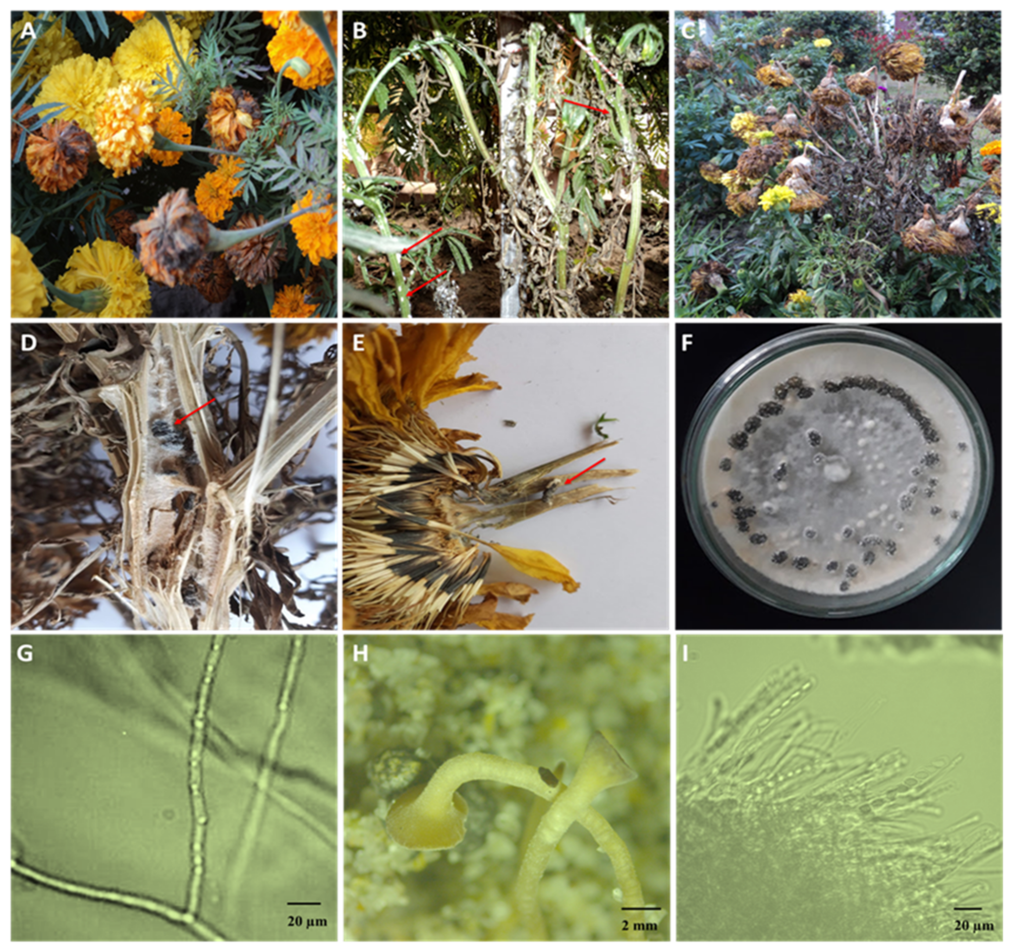
2. Unique Signs and Symptoms That Constitute Clinical Features of S. sclerotiorum

Correct disease identification at the onset is a crucial step in pathogen control and disease management. Traditionally, agricultural extension workers and other experts identify disease by observing signs and symptoms produced by a pathogen. Since S. sclerotiorum has a broad host range, no symptoms are exclusive to all infected plants. White mold is easily identifiable on any host by white cottony S. sclerotiorum mycelium on affected aerial tissues [15]. Stems, leaves, petioles, and reproductive organs may initially show water-soaked lesions with a distinct margin, followed by wilting, bleaching, and shredding. The cottony hyphae congregate into pea-sized clumps of mycelium, which gradually mature into hard black sclerotia, typically on the infected tissue’s surface and inside soft host tissues or cavities [15]. In sunflowers, the first aboveground symptom may be sudden wilting before or during flowering, resulting from root rot or basal stalk rot near the soil line [16]. Infections may occur on stems at any time of growth, leading to stem rot. The fungus also attacks sunflower head, causing the inner head to rot, disintegrate, and shred, leaving behind large sclerotia [16]. The symptoms of canola include the appearance of bleached, greyish lesions on the main stem, branches, or pods; the presence of hard, melanized, black sclerotia within the cortex of infected stems; and the premature flowering and wilting of plant tissues in the terminal parts of infected stems [11]. Infected stems are often prone to lodging during flowering and seed filling. In marigolds (Tagetes erecta), the first symptoms are typically visible during flowering. The symptoms appear on the flower petals and then spread to the entire flower and the lower section of the flower (Figure 1A). Infection may migrate from the infected flowers to stems or leaves in close proximity to the infected flowers. Leaves often develop water-soaked lesions that rapidly grow and become gray-green before turning dark brown and dying. Some infected plants may acquire whitish fungal growth on their stems and exhibit water-soaked lesions (Figure 1B). Infected stems may die prematurely, turn tan, and eventually bleach. The severe infection makes the plant weak and can cause it to wilt, fall over, and eventually die (Figure 1C). During humid conditions, infected plant parts often show signs of S. sclerotiorum, such as whitish, fluffy mycelium and big, irregularly shaped sclerotia on the host surface or embedded in the infected tissues (Figure 1D,E). Sclerotia are variable in shape but are generally globose to cylindrical, ranging from 2.0 to 20.0 mm [2,5,17]. On PDA, colonies rapidly expand and have white, cottony, aerial mycelia with a salmon-buff hue on the back. At the expanding edges of the colonies, sclerotia form radiating lines and concentric rings (Figure 1F). The hyphae are hyaline, branched, and multinucleate (Figure 1G). A sclerotium produces one or multiple apothecia of a tan to amber hue (Figure 1H). Apothecian asci are cylinder-shaped and have eight spores (Figure 1E). The ascospores are uniseriate, unicellular, hyaline, and elliptical (Figure 1F), whereas the paraphyses are abundant.

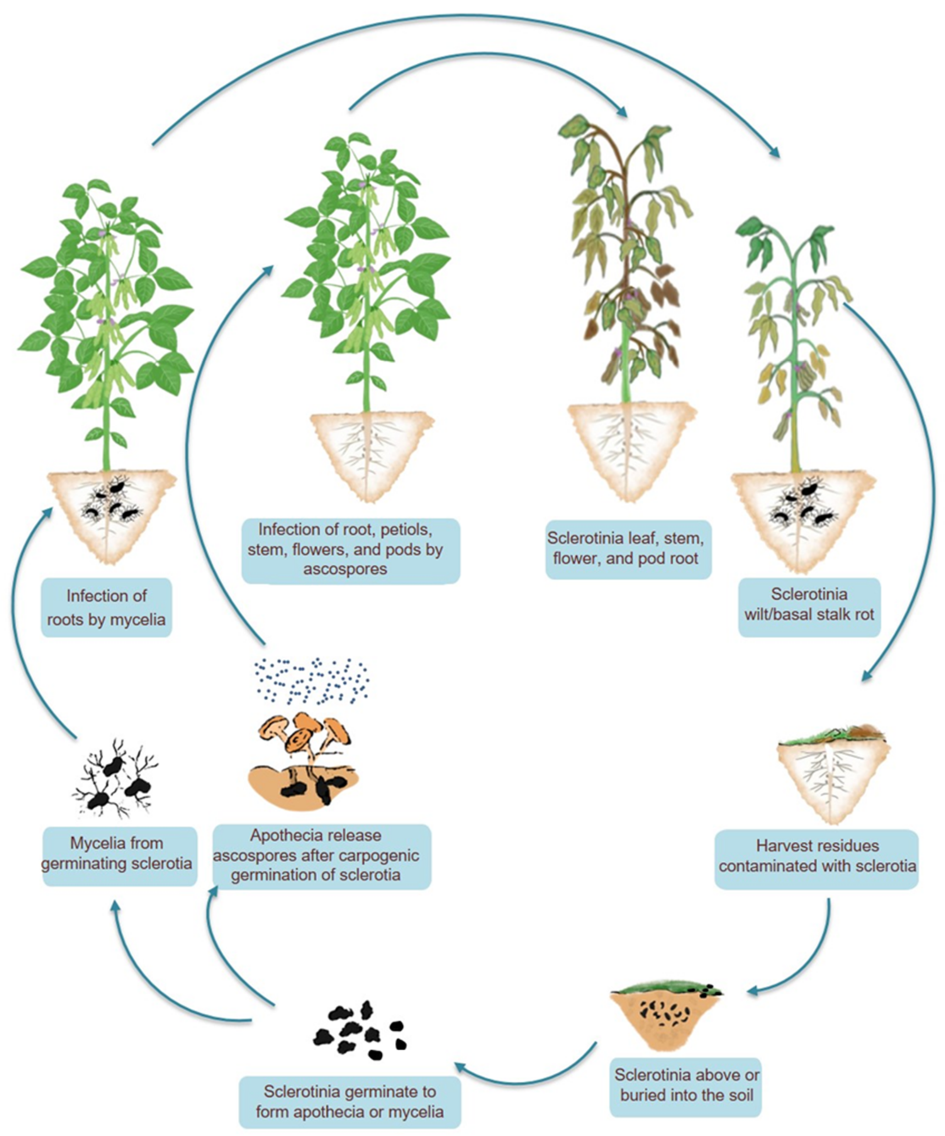
3. A Naive Lifecycle of a Deadly Pathogen

Because there is only one cycle of inoculum production during the course of infection, S. sclerotiorum is a simple monocyclic pathogen (Figure 2). The S. sclerotiorum lifecycle is primarily anchored by the formation of sclerotium. Sclerotia remain dormant in the soil for most of the S. sclerotiorum lifecycle and serve as the principal surviving structure. Sclerotium germinates directly (myceliogenic) or indirectly (carpogenic) and signals the inception of a new cycle of disease. Myceliogenic germination (by mycelium), possibly triggered by root exudates, is the primary mode of infection and is important from an epidemiological standpoint [18]. Mycelia from soilborne sclerotia may directly initiate infection on roots and basal stem. Basal stem rot and wilt, and in extreme cases, plant death, are the outcomes of the colonization of lateral roots, tap roots, and stem bases [18]. Ascospores released following carpogenic germination of sclerotia may also induce infection in plants. Despite being vulnerable to predation and deterioration from microorganisms, each sclerotium has the ability to produce carpogenic germination and several stipitate apothecia over multiple years. Each apothecium generates clusters of ascospores that are forcibly expelled into the air and disseminated within and between fields of vulnerable crops. The airborne ascospores are primarily associated with the infection of aerial plant parts [19].
In flower plants, ascospores that land in the sugary secretions of the extrafloral nectaries adjacent to the leaf margins germinate and colonize the leaf, leafstalk, and, eventually, the stem, causing stem rot [18]. During blossoming, the deposition of ascospores among the florets can result in the colonization of the capitulum, which causes bud rot (Figure 1). When conditions are optimum, the infection extends from the site of initial infection to the rest of the plant and then to uninfected plants. The potential of ascospores for airborne distribution and infection presents a unique limitation for the fungus since ascospores cannot infect healthy tissues. Ascospores are able to germinate, even invade host tissues, and cause compatible infections only when dead or senescent tissues are colonized saprotrophically before infecting healthy tissues [20]. In the field, this is typically done by colonizing leaves and flower petals that have senesced, damaged, or abscised but are still in physical contact with uninfected healthy host tissue (Figure 2). Mycelial growth from sclerotia on the soil surface can also initiate infection. The germinated hyphae colonize dead organic matter and then infect neighboring alive plants [21]. The life cycle is completed by forming sclerotia outside or inside hollow stems, flowers, buds, and fruits that have been colonized by the fungus (Figure 2). Due to the extended soil survival of sclerotia, even in adverse conditions, controlling white mold is difficult [22]. The sclerotium of S. sclerotiorum consists of three distinct layers, making this multihyphal structure hardy and enduring. The thick outer black rind contains melanin, a substance considered significant in the protection against unfavorable circumstances and microbial decomposition, and in some fungi, it has functions in virulence [23]. However, for S. sclerotiorum, no connection between melanin and virulence has so far been known.
The medulla, the innermost section, is embedded in a fibrillar matrix and is mainly made of proteins and glucans. Three phases of sclerotial formation have been identified: the first phase is initiation or commencement (hyphae aggregate to generate a white sclerotial mass or initials); the second phase is development (sclerotial initials increase in size); and the third phase is maturation (surface delimits, melanin deposits in peripheral rind cells, and internal cells consolidate) [2,5,17]. Several factors, including many dietary elements, environmental changes, secondary messengers, and primary metabolism, affect sclerotial formation and development. Sclerotia are typically formed when mycelial development confronts a nutrient-limited environment [17]. Glucose, yeast extract, peptone, ascorbic acid, and casein are the most suitable sources of nutrients for sclerotial formation in S. sclerotiorum [2,5,17]. Moreover, sclerotial development is favored in media enriched with calcium, magnesium, potassium, and sodium [2]. Temperature alone or in conjunction with other variables may affect sclerotial growth and survival. A temperature of 20 °C promotes maximum sclerotial development, while sclerotial production is significantly constrained at temperatures above 25 °C and, to a lesser degree, below 20 °C [2]. Consequently, low temperatures have been linked to the development of Sclerotinia infections in various plants [24]. pH has been demonstrated to affect sclerotial growth significantly. Sclerotial production is hindered at a neutral or alkaline pH [14], while it is favored at an acidic pH [2,5]. In contrast, fungus accumulation of OA lowers the pH and promotes sclerotial formation [14]. In line with this notion, S. sclerotiorum mutants incapable of producing OA do not form sclerotia in vitro and infect planta [25]. However, reducing the ambient pH in these mutants does not restore their ability to form sclerotia [14], indicating the involvement of more factors than pH alone. A number of genetic regulators are known to control the formation and development of sclerotia, which will be discussed in the later part of the current review.

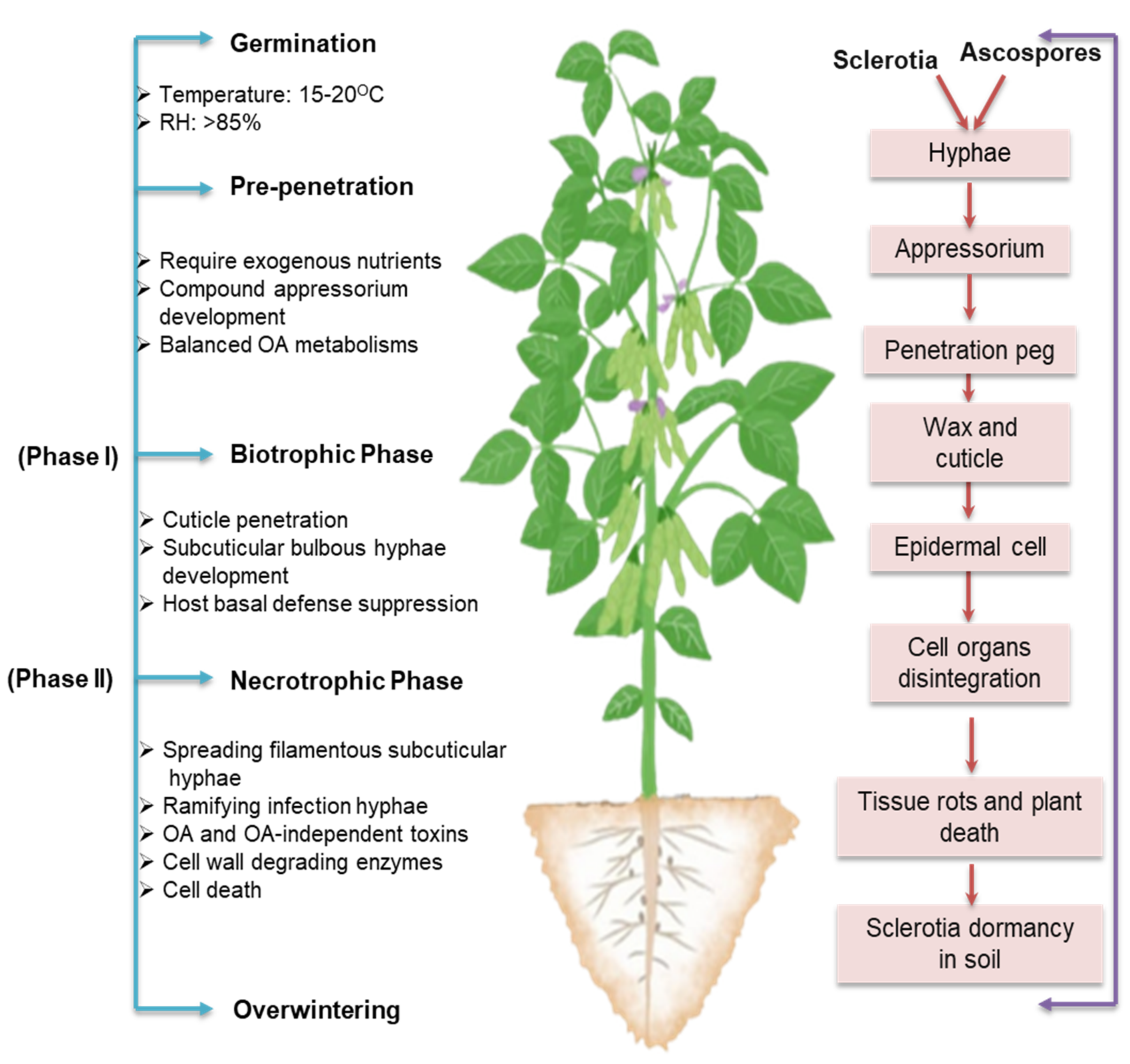
4. Two-Phase Infection Model: The Transition from Biotrophy to Necrotrophy

Although S. sclerotiorum has been widely accepted as a necrotrophic pathogen, recent evidence demonstrates that it is, in fact, a hemibiotroph [26]. In the early phases of infection, once S. sclerotiorum has penetrated the cuticle, the spread does not cause host cell necrosis. There is a brief period for biotrophic development in the apoplastic gaps prior to the necrotization of host cells. In this two-phase infection model, as described by Liang and Rollins [26], S. sclerotiorum evades, thwarts, and suppresses host basal defense responses before killing and destroying host cells (Figure 3).
The development of modified hyphae into appressoria is crucial for the infection of healthy tissues. During field infection, these hyphae are produced either directly from sclerotia or indirectly from ascospores in the presence of external nutrients, primarily from flowers or other aging tissues [27]. Depending on the host surface, simple or complex appressoria can be formed. Simple appressoria originate from dichotomous terminal branching of hyphae growing on the host surface. These appressoria are made up of a pad of broad, multiseptate, and short hyphae that are fixed perpendicularly to the surface of the host by mucilage [21]. A compound appressorium or infection cushion forms from a hyphal tip during recurring processes, such as swelling, growth retardation, and successive bifurcation [26]. The development of infection cushions is thought to improve the adherence of the pathogen to the host surface [28]. Previous research has suggested that infection cushion aids in penetrating the cuticle barrier of the host epidermis through mechanical pressure and/or enzymatic disintegration of the host surface during compatible interaction [29]. While in incompatible interaction, the most important component of the defense mechanisms by which the host avoids penetration and colonization by S. sclerotiorum is the active suppression of infection cushions [30], indicating the core role of infection cushions in the infection process of the fungus. Hyphae that form infection cushions frequently become flattened and larger in diameter, from which thin penetration pegs develop. Penetration of a host tissue only occurs beneath infection cushions and/or compound appressorium [28,30]. Hyphal tips are compactly aligned within a compound appressorium [28]. Separate hyphal tips initiate separate penetrations. The localized grouping of multiple hyphal points within a continuous plane, as opposed to a single-celled appressorium, likely increases the local amassing of toxins, hydrolytic enzymes, and defense-inhibitory substances [26]. With mechanical force and enzymatic digestion, penetration pegs derived from appressorium penetrate the cuticle layer, but not the underlying epidermal cells of the host [28,31]. Bulbous and multilobed subcuticular vesicles arising from these penetration pegs are responsible for the production of subcuticular infection hyphae, which grow in a horizontal direction beneath the cuticle to form the leading front of the colonization process [26].
Subcuticular hyphae of S. sclerotiorum spread through several cell layers before epidermal cells are killed [31,32], implying that S. sclerotiorum infection includes a transitory biotrophic phase, which begins around 12–24 h after infection [33]. During this phase, compatibility is established between the host and the pathogen primarily through suppressing or subverting host physical and chemical defense barriers. Effector genes in the pathogen S. sclerotiorum are upregulated, which may inhibit host–pathogen recognition and host defense mechanisms [34]. This initial phase is crucial for the “necrotrophic” success of the pathogen. After the initial growth of subcuticular hyphae, smaller hyphae ramify from the subapical ends and grow between and inside epidermal and mesophyll cells [26]. With the successful colonization by ramifying hyphae, S. sclerotiorum enters the necrotrophic phase [34], during which it produces significant amounts of reactive oxygen species (ROS), toxins, and CWDEs, which result in the dissolution of the subcuticular epidermal walls, rapid cell death, and development of necrotic symptoms [33,35,36].

5. Privileges of Massive Arsenal to Subsist and Succeed against Host Defense

S. sclerotiorum possesses an immense arsenal of disease weaponry to subsist and succeed under widespread environmental conditions. The massive pathogenic arsenal of S. sclerotiorum, including oxalic acid (OA), CWDEs, and small secretory proteins (effectors), has long been associated with virulence [26]. Recent research reveals that a subtle interplay between these virulence factors serves various regulatory functions in host cells, allowing S. sclerotiorum to colonize the host, evade or inhibit the host defense system, and cause disease (Figure 4). Understanding and studying these mechanisms is crucial for detecting pathways of genetic interventions that could result in improved control of this disease.

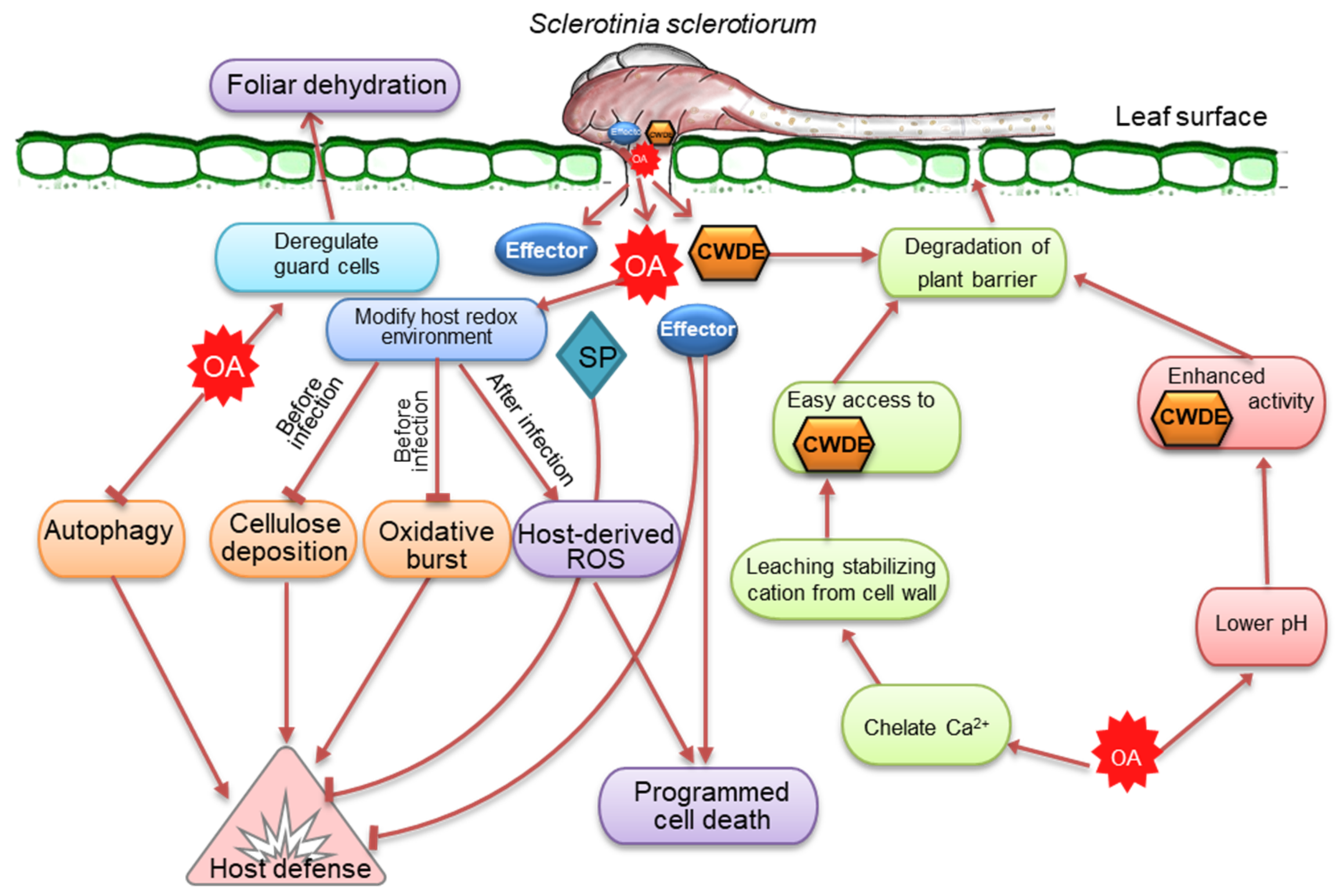
5.1. Oxalic Acid, a Versatile Toxin and Broad-Spectrum Pathogenicity Factor

Of the various virulence factors, extensive research has been conducted on the relationship between white mold disease and the production of OA by S. sclerotiorum [37,38]. This fungus is well-known for its ability to acidify the surroundings via OA secretion. The OA-deficient mutants of the fungus have been shown to be poorly pathogenic [39], suggesting that the pathogenesis of S. sclerotiorum requires accumulating high quantities of OA. While OA-minus mutants can still infect and colonize hosts to variable degrees, it is appropriate to view OA more as a colonization factor rather than a crucial compatibility element [31]. OA is not directly toxic to plant cells but plays a more sophisticated role as a signaling molecule that seizes control of host cells and creates an environment favorable for S. sclerotiorum growth. OA alters a number of physiological processes in host plants. OA promotes cell wall degradation by increasing the activity of polygalacturonases, inhibiting plant-protection enzymes, subduing oxidative burst, deregulating stomatal guard cell closure, mediating pH signaling, inducing apoptosis-like cell death, and altering cellular redox status in plants (Figure 4) [14,35,39,40,41,42,43]. The production of OA seems to suit the overall strategy of S. sclerotiorum to acidify its ambient environment to facilitate infection in several ways. Many of the fungal enzymes (such as polygalacturonase) secreted during the invasion of plant tissues have maximal activities at a low pH [40]. It is postulated that OA could increase Sclerotinia virulence by adjusting the apoplastic pH to a level conducive to the enzymatic breakdown of plant cell walls. OA can also weaken plant cell walls via acidity, thereby facilitating invasion [40]. Interestingly, no ROS production is detected at acidic pH [43]. It is evident that S. sclerotinia uses OA to elicit apoptosis-like programmed cell death, and the induction of this process is commenced by a reducing environment generated by the fungus in the host cell [35]. As a direct consequence of this redox manipulation, the fungus subverts host defense responses, inhibits oxidative burst, and prepares the infection court for disease establishment [35]. These findings imply that the effect of OA on the reduction of pH in the ambient environment is significantly greater than that of OA alone as an essential component of the infection process caused by S. sclerotiorum. Intriguingly, the regulation of host defense by OA appears to be biphasic. In the early phases of infection, OA suppresses host defenses by culminating ROS buildup and callose deposition in the host [35]. OA sequesters Ca2+ liberated during cell wall collapse, thereby protecting the development of S. sclerotiorum hyphae from hazardous Ca2+ concentrations [44]. In the later stages of infection, OA quashes antioxidant enzyme activities and stimulates ROS generation, concurrently damaging the membranes of host cells [45]. These findings suggest that OA is a cross-functional toxin crucial to the widespread pathogenic success of S. sclerotiorum.
The dicarboxylic acid OA can be produced through the oxidation of glyoxylate or glycolaldehyde or the hydrolysis of oxaloacetate [46]. However, current research indicates that OA biosynthesis entirely depends on the oxaloacetate acetylhydrolase (OAH) activity [26]. OAH mediates the C-C cleavage of oxaloacetate during the OA biosynthesis process [31]. Depending on the growth substrate, either the tricarboxylic acid (TCA) cycle or the glyoxylate cycle may produce oxaloacetate precursor [47]. Experimental evidence from S. sclerotiorum and B. cinerea gene-deletion mutants supports the hypothesis that the expressed enzyme activity is responsible for catalyzing the last step in the OA biosynthesis process [48]. Three types of OA-degrading enzymes are found throughout evolutionary lineages: OxDC (EC 4.1.1.2), OxOx (EC 1.2.3.4), and oxalylCoA decarboxylase (OXC, EC 4.1.1.8). S. sclerotiorum exhibits oxalate decarboxylase (OxDCs) activity but not OxOx [49], while only bacterial species have been identified to possess OXC activity. OxDC catalyzes the production of formate and CO2 from OA, and its activity is widespread among fungi and bacteria [50].

5.2. Arsenal of Cell Wall-Degrading Enzymes Reflects Broad Host Preference

An essential component of the lifecycle of S. sclerotiorum is its capacity to break down a wide range of complex plant polysaccharides by CWDEs (Figure 4). Cellulose, hemicellulose, and pectin form a complex network of polysaccharides that constitute plant cell walls [51]. This network is the target of CAZymes, a group of proteins and enzymes that break down plant cell wall polysaccharides into simple monomers [52]. These monomers serve as carbon sources and allow access to internal plant tissues. At least 118 CAZymes are explicitly associated with plant cell wall degradation by S. sclerotiorum; however, many other CAZymes presumably contribute to this activity [53]. Several endo- and exo-polygalacturonases (PGs) are known to break down unesterified pectate polymers, middle lamella structural polysaccharides, and primary plant cell wall components. Endo-PGs catalyze the hydrolysis of homogalacturonan, whereas exo-PGs fragment the substrate and release potential nutrients by removing monomeric or dimeric glycosyl groups from pectic cell wall polysaccharides [54]. PGs are also recognized as potential virulence factors in several pathosystems [55,56,57]; however, convincing proof for their universal involvement in virulence is missing. The principal hemicellulose constituent of plant cell walls, xylan, is comprised of xylose and arabinose connected by β-(1–4)-linked xylopyranose. Endo β-1, 4-xylanase is the major enzyme that degrades xylan [58]. Studies have highlighted the critical functions of endo-β-1, 4-xylanase in the development and pathogenicity of S. sclerotiorum [59]. Other important groups of mannan- and cellulose-degrading enzymes are also highly upregulated [53]. These indicate that S. sclerotiorum utilizes a diverse array of enzymes that play significant roles in its infection strategy across many host species.

5.3. Secretory Effectors, Critical Components in the Necrotrophic Infection Strategy

Numerous small, secreted proteins of S. sclerotiorum have been reported as having effector-like features or effector-like activities, which are released either extracellularly or within the cytoplasm of plant cells to disarm host defense mechanisms and enhance fungal infection (Figure 4) [33,60]. Until now, a number of S. sclerotiorum effectors associated with virulence have been identified. During the infection of soybean seedlings, S. sclerotiorum produces a basic endopolygalacturonase (PG) isoform early in the process that elicits Ca2+-mediated signaling and programmed cell death [61]. A polygalacturonase (PG) enzyme, called Sspg1d, reacts with proteins that have a C2 domain in Brassica napus [62]. Necrosis and ethylene-inducing peptides encoded by two genes, SsNep1 and SsNep2, induce necrosis in tobacco (Nicotiana tabacum) leaves during heterologous expression [63]. A protein with a signal peptide and 302 amino acid residues in the integrin alpha N-terminal domain superfamily, SSITL, inhibits defense reactions in Arabidopsis thaliana [64]. A putative-secreted protein disrupting pathogen virulence has also been reported [65]. A cutinase encoded by SsCut induces cell death in an array of plants, including rice (Oryzae staiva), G. max, maize (Zea mays), B. napus, wheat (Triticum aestivum), and A. thaliana [66]. A small cysteine-rich protein containing a cyanoviron-N homology (CVNH) domain, SsSSVP1, is required for pathogen virulence [67]. This putative effector is believed to alter plant energy metabolism in order to promote S. sclerotiorum infection. A small virulence-related protein 1, without obvious functional domains, has been found explicitly in S. sclerotiorum and B. cinerea [68].
In addition, several other effectors, such as chorismate mutase (SsCM1) [69], compound appressorium formation-related protein 1 (SsCaf1) [70], BAX inhibitor-1 (SsBI1) [71], superoxide dismutase (SsSodI) [72], catalase (SsCat1) [73], a protein analogous to a Magnaporthe grisea protein elicitor (SsPemG1) [74], and ceratoplantanin effector (SsCP1) [75], have been documented in S. sclerotiorum. The vast majority of the putative proteinaceous effectors characterized in S. sclerotiorum cause cell death in host tissue. S. sclerotiorum genome analysis has recently identified a novel class of necrosis-inducing effector 2 (SsNE2) [76]. On the contrary, a few biotrophy-related candidate effectors have been spotted, such as salicylate hydroxylase and a lysin motif (LysM) domain protein [33]. These effector genes were found to be upregulated within the first 24 h after infection. In biotrophic and hemibiotrophic pathogens, recognizing the effectors often leads to a hypersensitive reaction (HR) to contain the pathogens (Jones and Dangl, 2006) [77]. Contrariwise, for necrotrophic infections, the elicitation of HR responses by effectors delivers substrates to stimulate host colonization [26,78]. Additionally, S. sclerotiorum exhibits a host-specific expression of candidate effectors, as Guyon et al. [79] showed that the expression patterns of 16 effector candidates varied significantly between the hosts S. lycospersicum, Nicotiana benthamiana, and susceptible and resistant accessions of A. thaliana. According to previous studies, the host-specific expression of putative effectors was more limited than anticipated. Only one of the effector candidate genes (sscle 08g064180) identified by Derbyshire et al. [80] and Amselem et al. [53] was expressed differently between B. napus and Lupinus angustifolius, where the expression was significantly higher in B. napus compared to L. angustifolius. This putative effector is anticipated to have a coiled-coil domain, although little is known about the role of such domain in mediating effectors’ functions.

6. An Array of Genes Regulating Growth, Development, and Virulence

Molecular underpinnings of the genes that regulate mycelial growth, reproduction, and virulence in S. sclerotiorum have been the subject of several recent studies [26,37]. Because many of these genes have already been reviewed, the current review primarily focuses on works that have not been discussed in previous reviews [26,37]. A large number of genes required for S. sclerotiorum growth, development, and virulence have already been identified using the genome sequence and subsequent mutant analysis. These include Sspac1 (pH-responsive transcription factor) [25], Sscna1 (catalytic subunit calcineurin-encoding gene) [81], PP2B (type 2B phosphatase) [81], Ssnox1, Ssnox2 (NADPH oxidase) [82], SsITL (integrin-like protein) [64], Sssod1 (Cu/Zn superoxide dismutase) [83], SsMADS (MADS-box transcription factor) [84], Scat1 (type A catalase) [73], Shk1 (histidine kinases) [85], SsCVNH (cysteine-rich protein with a carbohydrate-binding module) [86], Ssoah1 (oxaloacetate acetylhydrolase) [31], Ssodc1 and Ssodc2 (oxalate decarboxylase) [87], SsBi1 (BAX inhibitor-1) [71], Ss-Fkh1 (forkhead transcription factor) [88], Sssop1 (microbial opsin homolog gene) [68], SsXyl1 (Endo-β-1, 4-xylanase) [59], SsSSVP1 (small secreted virulence-related protein) [67], rhs1 (rearrangement hotspot repeat-containing protein) [89], THR1 (trihydroxynaphthalene reductase) [90], SCD1 (scytalone dehydratase) [26], Ssams2 (GATA-box domain) [91], Sssfh1 (GATA transcription factor with an SNF5 domain) [92], SsSte12 (Ste12 transcription factor [37], Sssvf1 (survival factor 1 homologue) [93], Ssrhs1 (Rhs repeat-containing protein) [94], and SsAGM1 (N-acetylglucosamine-phosphate mutase) [95] (Table 1). In mutant or transformant strains of S. sclerotiorum, knocking out or silencing these genes causes severe deficiencies in radial growth, sclerotial development, appressorium formation, virulence, and/or stress tolerance. For instance, the pacC gene homolog, a Sspac1 replacement mutant, exhibited abnormal sclerotial development and maturation, implying that Sspac1 is essential for the proper sclerotial development of S. sclerotiorum [25].
Likewise, Ssoah1 mutants displayed a significant radial growth defect on buffered neutral pH substrate and failed to generate sclerotium or compound appressorium [31]. The nox (NADPH oxidase)-encoding gene, Ssnox1, contributes toward fungal virulence and sclerotial formation [82]. The MADS-box transcription factor, SsMADS, is essential for the growth and pathogenicity of S. sclerotiorum [84]. The S. sclerotiorum sop1, a microbial opsin homolog gene, is significantly upregulated during sclerotial development and infection relative to the vegetative growth stage [68]. Moreover, sop1 is required for S. sclerotiorum growth, sclerotial development, full virulence, and stress responses. Because microbial opsins are missing in animals and most higher plant genomes, sop1 is a candidate fungicide target for S. sclerotiorum control. The S. sclerotiorum integrin-like gene, SsITL, was intensely suppressed in the presence of SsDRV, a mycovirus gene linked with reduced virulence [64].
Silencing of SsITL decreased virulence, hyphal polarity, and qualitative and quantitative production of sclerotia in S. sclerotiorum. Sclerotia that were formed exhibited carpogenic germination deficiencies. In addition, SsITL encodes an effector involved in inhibiting jasmonate/ethylene-mediated host resistance during the initial phases of infection [64]. The function of SsAMS2 in the development and growth of S. sclerotiorum has been investigated [91]. AMS2 protein is predicted to contain the GATA-box domain. S. sclerotiorum needs the transcription factor SsAMS2 for growth, appressorium production, pathogenicity, and chromosome segregation [91]. Another gene with a GATA transcription factor with an SNF5 domain, SsSFH1, has also been analyzed and is known to influence growth, ROS accumulation, and pathogenicity of S. sclerotiorum [92].
According to recent research, vegetative development and sclerotial production in S. sclerotiorum are intimately tied to cell wall integrity. SsFkh1 contributes to cell wall integrity, and the deletion mutants are significantly deficient in hyphal development and sclerotial formation [96]. SsAGM1 has been isolated from several fungi, identified as necessary for chitin production, and known to play a role in preserving the integrity of cell wall [97,98]. Silencing SsAGM1 substantially decreased the hyphal and sclerotial development of S. sclerotiorum [95]. The silenced transformants did not produce sclerotia, irrespective of the duration of the culture. In fact, SsAGM1 mediates UDP-GlcNAc (uridine diphosphate-N-acetylglucosamine) synthesis, which influences mycelial development, cell wall integrity, cell wall chitin content, sclerotial production, infection cushion formation, and pathogenicity of S. sclerotiorum [95]. An Rhs repeat-containing protein (Ssrhs1) is shown to be imperative for hyphal infection and sclerotial development [94]. Some of these genes are components of a regulatory mechanism that mediates pH-responsive gene expression, indicating that pH-regulated gene expression is an essential feature of S. sclerotiorum growth, development, and virulence [25,31].

7. cAMP as a Key Modulator

Secondary messenger such as cyclic adenosine monophosphate (cAMP) also influences the growth, development, and virulence of S. sclerotiorum. cAMP is involved in the shift from mycelia to sclerotial growth [99]. An increase in either endogenous or exogenous cAMP suppresses sclerotial formation and stimulates OA accumulation [100]. In a ∆bcg3 mutant of B. cinerea deficient in the Gα3 subunit, a drop in cAMP levels induced a substantial amount of sclerotial production under conditions that did not normally induce sclerotial production. On the contrary, sclerotial production in the ∆bcg3 mutant was reduced by adding cAMP to the growth medium in a manner comparable to that seen in the wild type, suggesting that cAMP has a detrimental effect on sclerotial development [101]. cAMP-dependent PKA signaling was investigated to identify its potential functions in the pathogenesis, growth, and development of S. sclerotiorum [102]. In the sac1-disrupted mutant, the intracellular cAMP level was lowered by obliterating the single-copy adenylate cyclase gene (sac1) from the S. sclerotiorum genome. The disrupted mutant showed abnormal hyphal branching and the formation of excessive microconidia (spermatia) and aberrant sclerotia. Even though sclerotia generally form close to the outside edge of culture plates, the S. sclerotiorum sac1 mutant generated sclerotia without ever reaching the edge of the culture plates. This demonstrated a negative role for cAMP in regulating the initiation of sclerotial formation. cAMP is also necessary for the development of compound appressorium and the pathogenicity of S. sclerotiorum [103]. cAMP has been suggested as a possible intermediary in the cellular cascade required for the production of a pathogenesis-related fungal protease known as Acp1 in S. sclerotiorum [104].
In other fungi, cAMP has been shown to have the opposite effect on sclerotial production. Higher intracellular cAMP levels facilitate sclerotial production in Sclerotium rolfsii and Rhizoctonia solani [99,105,106]. The enhancement of sclerotial development in an R. solani wild-type isolate was achieved by supplementing its growth medium with exogenous cAMP [99]. Hence, cAMP may have a species-specific role in sclerotial development. When sclerotial formation is triggered, the initial events are hyphal branching and fusion. cAMP likely influences sclerotial initiation by altering hyphal elongation and the rates of hyphal branching, supporting the observation that Neurospora crassa grows more slowly and more radially when intracellular cAMP levels are low [107]. The fact that hyphal branching is impaired in the sac1 mutant, which has low cAMP levels and abnormal sclerotial formation, shows the association between cAMP levels, branching, and appropriate sclerotial development.

8. MAPK as Central Signaling Cascade

The mitogen-activated protein kinase (MAPK) is a major signaling cascade engaged in sclerotial formation and various processes related to fungal infection [37]. In S. sclerotiorum, an ERK-type MAPK, Smk1, was proven essential for sclerotiogenesis [108]. pH- and cAMP-dependent signaling mediates the regulation of sclerotial formation by MAPK [108]. The levels of cAMP-dependent protein kinase A (PKA) rise during sclerotial development in wild-type strains but remain low in mutants defective in sclerotial development [109]. However, similar pathogenicity, cAMP-responsiveness, and sclerotial development to wild-type strains were observed in pka1 knock-out mutants, indicating a PKA-independent mechanism or the participation of other PKA-encoding genes in these processes [102]. As specific PKA inhibitors do not affect sclerotial development or cAMP-dependent MAPK inhibition, cAMP-mediated sclerotial suppression is PKA-independent [100]. Class III histidine kinases (HKs) are known to transmit signals via the high-osmolarity glycerol MAPK (HOG MAPK). The S. sclerotiorum Shk1 gene, encoding a putative class III HK, plays a role in multiple processes, including vegetative differentiation, sclerotial formation, adaptation to stresses and fungicides, and glycerol accumulation in S. sclerotiorum [83]. Ras, a small GTPase that acts as an upstream activator of the MAPK pathway, is required for normal sclerotial growth because the loss of Ras function prevents MAPK activation [110]. Intriguingly, the suppression of a member of the Ras protein family, Rap-1, restores sclerotial growth and MAPK activation. These findings imply that sclerotial development depends on Ras/MAPK pathways, which are adversely controlled by Rap-1 in a PKA-independent cAMP signaling pathway [48]. In a recent study, the catalytic SsPKA and the regulatory SsPKAR subunits of the cAMP-dependent PKA signaling pathway in S. sclerotiorum were further characterized in terms of their functional properties [111]. Disruption of Sspka2 or SspkaR markedly reduced growth and virulence, together with defects in appressorium development.
Unexpectedly, both disruptions exhibit an increase in autophagy without nutrient deprivation, indicating that accurately synchronized PKA activity is necessary to control autophagy. These findings show that the cAMP-dependent PKA signaling pathway disrupts autophagy and is vital in S. sclerotiorum development and virulence [111]. Investigation has been carried out to examine the potential downstream transcription factor of the MAPK pathway. SsSte12 has been reported to be a transcription factor that acts as a downstream component of the MAPK pathway and is essential for a number of MAPK-dependent cellular processes, including penetration-dependent pathogenicity, mycelial growth, sclerotial development, and appressorium formation [37]. In reaction to different extracellular stimuli, MAPK is enzymatically stimulated by a series of phosphorylation events. It can also control the expression of genes and induce the phosphorylation of transcription factors [112]. Current research has demonstrated that the activity of protein phosphatase types 2A and 2B is reliant on MAPK and is needed for the development of sclerotia [81,113].

9. Sclerotinia Genome as a Treasure Trove

The first complete sequence of S. sclerotiorum genome was elucidated by Amselem and associates in 2011 [53]. This genome project used the isolate ‘1980’ collected from bean culls. Shotgun sequencing with Sanger sequencing chemistry and optical mapping was used to assemble the genome sequence. Genome annotation and gene validation employed expressed sequence tags (ESTs). These ESTs were obtained from key life cycle stages, such as sclerotium initials, mycelia, apothecia during the expansion of disc development, and compound appressoria. The assembled genome was 38 Mb, which covered 96% of the optical map. The majority of the unexplored areas on the map are likely centromeric locations. The number of predicted genes is 14,522, and the number of genes with a high degree of confidence is 11,860. The average GC contents found in the S. sclerotiorum genome are 41.8% [53]. Moreover, the S. sclerotiorum genome has eight duplicated blocks containing between four and twelve pairs of syntenic genes. Of the eight blocks, four contain genes encoding proteins. Approximately 7% of the genome is occupied by repetitive elements, with no apparent hotspots along the chromosomes. This expansion contains various categories of transposable components. LTR elements are commonly observed, followed by TIR, LINE, unknown, and MITE elements [53].
Derbyshire et al. [80] reported a complete and precisely annotated genome of the same S. sclerotiorum isolate ‘1980′. The new S. sclerotiorum genome assembly has an additional 805,146 base pairs compared to the previous genome assembly by Amselem et al. [53]. The majority of the extra bases constructed (78.7%) are within regions that are projected to be repeated. In addition, 12.96% of the genome is constituted of transposable elements, which is greater than the 7.7–9.0% previously estimated [80]. Though only a moderate improvement is shown in the new genome assembly, significant progress has been made in annotating new genes. If 11,860 unambiguous genes are considered in the previous genome version, there are yet 730 fewer in the new gene annotation set with 1130 predictions. Only 10,528 of the sequences with version one show reciprocal best BLAST matches, implying that an additional 602 sequences are either absent from the previous genome or have been altered significantly enough to prevent BLAST analysis from retrieving their corresponding loci as hits. The merging of previously separate loci is primarily responsible for the drop in filtered gene predictions. Recently, an upgraded genome assembly of S. sclerotiorum strain WH6 has been completed using the PacBio SMRT cell platform [114]. The assembled genome contains 10,512 predicted genes, with 685 secreted proteins and 65 candidate effectors, and is 38.96 Mbp in size with a contig N50 length of 1.90 Mbp. The S. sclerotiorum genome sequence from China had never been reported before the study of [114].
When comparing the related fungal genomes, S. sclerotiorum possesses a genome comparable in size and predicted gene number to B. cinerea, Scleromitrula shiraiana, and Magnaporthe oryzae, except for the biotrophic fungus Blumeria graminis f. sp. hordei (Table 2). The GC content in the S. sclerotiorum (41.55%) sequences is close to that of B. cinerea (42.0%) and S. shiraiana (38.9%). However, the percentage of repeated sequences in the S. sclerotiorum genome is greater than that of S. shiraiana (4.45%) and B. cinerea (4.4%). Similarly, the S. sclerotiorum genome (7%) contains more transposable elements than the genomes of S. shiraiana (0.07%) and B. cinerea (0.9%). The majority of the 11,860 predicted protein-coding genes are shared by S. sclerotiorum and B. cinerea., with an average similarity between these species of 84% for 8609 proteins [53]. Additionally, S. sclerotiorum shares 157,162 genes and 2605 ortholog families with 15 other fungi, including necrotrophs, biotrophs, and saprobes [115]. In the phylogenetic tree created with STRIDE (Species Tree Root Inference from gene Duplication Events) and STAG (Species Tree inference from All Genes) methods, S. sclerotiorum was grouped with the Leotiomycetes; B. cinerea, S. shiraiana, and B. graminis f. sp. hordei. Exclusively, S. sclerotiorum, S. shiraiana, and B. cinerea share 7402 ortholog families [115]. Although S. sclerotiorum is evolutionally nearer to Scleromitrula shiraiana and B. cinerea, the genome coverage remains at 72.3% [53] and 21.4% [115], respectively. Based on these results, it is plausible that S. sclerotiorum and B. cinerea diverged from their predecessors later in evolutionary time than S. shiraiana did.
A genome-wide search identified genes encoding key enzymes, including NRPS (non-ribosomal peptide synthetase), PKS (polyketide synthase), HYBRID (hybrid NRPS–PKS enzyme), DMATS (dimethylallyl tryptophan synthase) and TS (terpene synthase). Thirty-three key biosynthetic genes were found to encode these five enzymes in S. sclerotiorum, which were similar in number to those in S. shiraiana, but less than those in B. cinerea [115]. By analyzing the genes that are unique to B. cinerea and S. sclerotiorum and understanding the lack of resemblance to those of other fungi, it is determined that cytochrome p450 and transcription factors are the most prevalent [53]. In contrast to other fungi, S. sclerotiorum and B. cinerea lack terpene synthases, subtilases, zinc finger domains, and two glycosyl hydrolase families (GH2 and GH43) [48]. S. sclerotiorum has fewer genes encoding CAZymes than the majority of necrotrophic and hemibiotrophic fungi but considerably higher than the obligate biotrophic fungi B. graminis f. sp. hordei and P. graminis f. sp. tritici [115]. While entire CWDE profiles are comparable to those of saprotrophs, distinct CWDEs subsets/families may contribute to specific nectrotrophic habitats. Nevertheless, there is a correlation between gene conservation patterns and the emergence of a necrotrophic lifestyle. The expansion of gene families in S. sclerotiorum, B. cinerea, and two remotely related necrotrophic species, P. teres and P. nodorum, was investigated to identify traits that are shared among necrotrophic pathogens [53]. This analysis identified only heterokaryon incompatibility proteins.
Andrew and colleagues [128] investigated the possibility that members of the Sclerotiniaceae family have a group of genes related to necrotrophy in common. The phylogeny of 24 species reconstructed from the concatenated sequences of three housekeeping genes, cal, hsp60, and g3ph, agreed with the phylogenies derived from other loci [53]. The tree contained four major lineages. Both generalist and specialist host pathogens were found in two necrotrophic lineages: Sclerotinia/Dumontinia/Sclerotium cepivorum and Botryotinia/Botrytis. The third group comprises species that are either host specialists or facultative biotrophs (Myriosclerotinia). The necrotrophic, biotrophic, and host specialists, such as Monilinia, and the obligatorily biotrophic and host specialist Ciborinia whetzelii are members of the fourth lineage [128]. According to the conventional interpretation of this phylogeny, the progenitor of all members of the family Sclerotiniaceae was necrotrophyte, while biotrophy is considered a more recent, achieved state resulting from one or two modifications away from necrotrophy.

10. Conclusions

Over the past decade, uncovering the genomic features of S. sclerotiorum has facilitated comprehension of the intricacies of mechanisms underlying S. sclerotiorum growth, development, and virulence. Genetic evidence of the importance of OA, CWDEs, and effector proteins in various phases of pathogenesis has been unfolded. Comparative analyses demonstrate that necrotrophic infections do not encode much more quantity of degradative enzymes than other infection types, except for cutin degradation. Efforts are in progress to identify and characterize new genes involved in pathogen growth, development, pathogenesis, and stress adaptation. Moreover, the population of S. sclerotiorum appears to be more diverse than previously thought. Understanding the underlying reason for this divergence may help to understand Sclerotinaceae strategies for narrow versus broad host-range specialization. Previous Sclerotinia genome analysis may not have revealed definitive answers to all the underlying mechanisms of S. sclerotiorum biology and genetics, but it has already laid the groundwork for future research. Metabolomics studies that define fungal compounds, their regulation, and their roles in plant infection could help understand the specificity of plant–host connections even more. Functional genomic studies, comparative pathobiology and genome research, and a full assessment of population dynamics and structure are required to achieve further progress. Such efforts using genomic and genetic resources are expected to shed light on the intricate nature of the necrotrophic pathogenicity of S. sclerotiorum.

Author Contributions

Conceptualization, M.M.H., F.S. and M.G.M.; Data Curation, M.M.H., F.S. and W.L; Writing—Original Draft Preparation, M.M.H. and F.S.; Writing—Review and Editing, M.G.M., W.L. and L.-S.P.T.; Figure and Table, M.M.H. and M.G.M.; Supervision, M.G.M. and L.-S.P.T. All authors have read and agreed to the published version of the manuscript.

Funding

We sincerely thank the Strategic Priority Research Program of the Chinese Academy of Sciences (Grant No. XDA28110102) and Ministry of Science and Technology, Bangladesh, for supporting our research on Sclerotinia sclerotiorum.

Institutional Review Board Statement

Not applicable.

Informed Consent Statement

Not applicable.

Data Availability Statement

All data generated or analyzed during this study are included in this published article.

Conflicts of Interest

The authors declare no conflict of interest.

References

  1. Lehner, M.S.; De Paula Junior, T.J.; Del Ponte, E.M.; Mizubuti, E.S.G.; Pethybridge, S.J. Independently founded populations of Sclerotinia sclerotiorum from a tropical and a temperate region have similar genetic structure. PLoS ONE 2017, 12, e0173915. [Google Scholar] [CrossRef] [PubMed] [Green Version]
  2. Jahan, R.; Siddique, S.S.; Jannat, R.; Hossain, M.M. Cosmos white rot: First characterization, physiology, host range, disease resistance, and chemical control. J. Basic Microbiol. 2022, 62, 911–929. [Google Scholar] [CrossRef]
  3. Saharan, G.S.; Mehta, N. Sclerotinia Diseases of Crop Plants: Biology, Ecology and Disease Management; Springer: Berlin, Germany, 2008; ISBN 978-1-4020-8408-9. [Google Scholar]
  4. Prova, A.; Akanda, A.M.; Islam, S.; Hossain, M.M. First report of Sclerotinia sclerotiorum causing pod rot disease on okra in Bangladesh. Can. J. Plant Pathol. 2017, 39, 72–76. [Google Scholar] [CrossRef]
  5. Islam, M.R.; Akanda, A.M.; Hossain, M.M.; Hossain, M.M. First characterization of a newly emerging phytopathogen, Sclerotinia sclerotiorum causing white mold in pea. J. Basic Microbiol. 2021, 61, 923–939. [Google Scholar] [CrossRef] [PubMed]
  6. Prova, A.; Akanda, M.A.M.; Islam, S.; Sultana, F.; Islam, M.T.; Hossain, M.M. First report of stem and pod blight of hyacinth bean caused by Sclerotinia sclerotiorum in Bangladesh. J. Plant Pathol. 2014, 96, 603–611. [Google Scholar] [CrossRef]
  7. Choi, I.Y.; Kim, J.; Lee, W.H.; Cho, S.E.; Shin, H.D. First report of Sclerotinia stem rot caused by Sclerotinia sclerotiorum on Chinese chives in Korea. Plant Dis. 2017, 101, 1953. [Google Scholar] [CrossRef]
  8. Islam, M.R.; Prova, A.; Akanda, A.M.; Hossain, M.M. First report of white mould caused by Sclerotinia sclerotiorum on pea in Bangladesh. J. Plant. Pathol. 2020, 102, 941. [Google Scholar] [CrossRef] [Green Version]
  9. Boland, G.J.; Hall, R. Index of plant hosts of Sclerotinia sclerotiorum. Can. J. Plant Pathol. 1994, 16, 93–108. [Google Scholar] [CrossRef]
  10. Tian, B.; Xie, J.; Fu, Y.; Cheng, J.; Li, B.O.; Chen, T.; Zhao, Y.; Gao, Z.; Yang, P.; Barbetti, M.J.; et al. A cosmopolitan fungal pathogen of dicots adopts an endophytic lifestyle on cereal crops and protects them from major fungal diseases. ISME J. 2020, 14, 3120–3135. [Google Scholar] [CrossRef]
  11. Derbyshire, M.C.; Denton-Giles, M. The control of sclerotinia stem rot on oilseed rape (Brassica napus): Current practices and future opportunities. Plant Pathol. 2016, 65, 859–877. [Google Scholar] [CrossRef] [Green Version]
  12. Alkooranee, J.T.; Aledan, T.R.; Ali, A.K.; Lu, G.; Zhang, X.; Wu, J.; Fu, C.; Li, M. Detecting the hormonal pathways in oilseed rape behind induced systemic resistance by Trichoderma harzianum TH12 to Sclerotinia sclerotiorum. PLoS ONE 2017, 12, e0168850. [Google Scholar] [CrossRef] [Green Version]
  13. Albert, D.; Dumonceaux, T.; Carisse, O.; Beaulieu, C.; Filion, M. Combining desirable traits for a good biocontrol strategy against Sclerotinia sclerotiorum. Microorganisms 2022, 10, 1189. [Google Scholar] [CrossRef]
  14. Rollins, J.A.; Dickman, M.B. pH signaling in Sclerotinia sclerotiorum: Identification of a pacC/RIMI homolog. Appl. Environ. Microbiol. 2001, 67, 75–81. [Google Scholar] [CrossRef] [Green Version]
  15. Heffer Link, V.; Johnson, K.B. White Mold. Plant Health Instructor. 2007. [Google Scholar] [CrossRef]
  16. Mathew, F.; Harveson, R.; Block, C.; Gulya, T.; Ryley, M.; Thompson, S.; Markell, S. Sclerotinia sclerotiorum diseases of sunflower (white mold). Plant Health Instructor. 2020. [Google Scholar] [CrossRef]
  17. Prova, A.; Akanda, A.M.; Islam, S.; Hossain, M.M. Characterization of Sclerotinia sclerotiorum, an emerging fungal pathogen causing blight in Hyacinth Bean (Lablab purpureus). Plant Pathol. J. 2018, 34, 367–380. [Google Scholar] [CrossRef]
  18. Ekins, M.G.; Aitken, E.A.B.; Goulter, K.C. Carpogenic germination of Sclerotinia minor and potential distribution in Australia. Australas. Plant Pathol. 2002, 31, 259–265. [Google Scholar] [CrossRef]
  19. Smolinska, U.; Kowalska, B. Biological control of the soil-borne fungal pathogen Sclerotinia sclerotiorum—A review. J. Plant Pathol. 2018, 100, 1–12. [Google Scholar] [CrossRef] [Green Version]
  20. Sutton, D.C.; Deverall, B.J. Studies on infection of bean (Phaseolus vulgaris) and soybean (Glycine max) by ascospores of Sclerotinia sclerotiorum. Plant Pathol. 1983, 32, 251–261. [Google Scholar] [CrossRef]
  21. Hegedus, D.D.; Rimmer, S.R. Sclerotinia sclerotiorum: When “to be or not to be” a pathogen? FEMS Microbiol Lett. 2005, 251, 177–184. [Google Scholar] [CrossRef] [Green Version]
  22. Fernando, W.G.D.; Nakkeeran, S.; Zhang, Y. Ecofriendly Methods in Combating Sclerotinia sclerotiorum (Lib.) de Bary; Research Signpost: Trivandrum, India, 2004; Volume 661, pp. 329–347. ISBN 81-7736-217-8. [Google Scholar]
  23. Thomma, B.P.H.J. Alternaria spp. from general saprophyte to specific parasite. Mol. Plant Pathol. 2003, 4, 225–236. [Google Scholar] [CrossRef] [PubMed]
  24. Clarkson, J.P.; Fawcett, L.; Anthony, S.G.; Young, C. A model for Sclerotinia sclerotiorum Infection and disease development in lettuce, based on the effects of temperature, relative humidity and ascospore density. PLoS ONE 2014, 9, e94049. [Google Scholar] [CrossRef] [PubMed]
  25. Rollins, J.A. The Sclerotinia sclerotiorum pac1 gene is required for sclerotial development and virulence. Mol. Plant Microb. Interact. 2003, 16, 785–795. [Google Scholar] [CrossRef] [PubMed] [Green Version]
  26. Liang, X.F.; Rollins, J.A. Mechanisms of broad host range necrotrophic pathogenesis in Sclerotinia sclerotiorum. Phytopathology 2018, 108, 1128–1140. [Google Scholar] [CrossRef] [Green Version]
  27. Tariq, V.N.; Jeffries, P. Appressorium formation by Sclerotinia sclerotiorum: Scanning electron microscopy. Trans. Br. Mycol. Soc. 1984, 82, 645–651. [Google Scholar] [CrossRef]
  28. Huang, L.; Buchenauer, H.; Han, Q.; Zhang, X.; Kang, Z. Ultrastructural and cytochemical studies on the infection process of Sclerotinia sclerotiorum in oilseed rape. J. Plant Dis. Prot. 2008, 115, 9–16. [Google Scholar] [CrossRef]
  29. Tariq, V.N.; Jeffries, P. Ultrastructure of penetration of Phaseolus spp. by Sclerotinia sclerotiorum. Can. J. Bot. 1986, 64, 2909–2915. [Google Scholar] [CrossRef]
  30. Garg, H.; Li, H.; Sivasithamparam, K.; Kuo, J.; Barbetti, M.J. The infection processes of Sclerotinia sclerotiorum in cotyledon tissue of a resistant and a susceptible genotype of Brassica napus. Ann. Bot. 2010, 106, 897–908. [Google Scholar] [CrossRef] [Green Version]
  31. Liang, X.; Liberti, D.; Li, M.; Kim, Y.T.; Hutchens, A.; Wilson, R.; Rollins, J.A. Oxaloacetate acetylhydrolase gene mutants of Sclerotinia sclerotiorum do not accumulate oxalic acid but do produce limited lesions on host plants. Mol. Plant Pathol. 2015, 16, 559–571. [Google Scholar] [CrossRef]
  32. Kabbage, M.; Yarden, O.; Dickman, M.B. Pathogenic attributes of Sclerotinia sclerotiorum: Switching from a biotrophic to necrotrophic lifestyle. Plant Sci. 2015, 233, 53–60. [Google Scholar] [CrossRef]
  33. Seifbarghi, S.; Borhan, M.H.; Wei, Y.; Coutu, C.; Robinson, S.J.; Hegedus, D.D. Changes in the Sclerotinia sclerotiorum transcriptome during Infection of Brassica napus. BMC Genom. 2017, 18, 266. [Google Scholar] [CrossRef] [Green Version]
  34. Ding, L.N.; Li, T.; Guo, X.J.; Li, M.; Liu, X.Y.; Cao, J.; Tan, X.L. Sclerotinia stem rot resistance in rapeseed: Recent progress and future prospects. J. Agric. Food Chem. 2021, 69, 2965–2978. [Google Scholar] [CrossRef]
  35. Williams, B.; Kabbage, M.; Kim, H.J.; Britt, R.; Dickman, M.B. Tipping the balance: Sclerotinia sclerotiorum secreted oxalic acid suppresses host defenses by manipulating the host redox environment. PLoS Pathog. 2011, 7, e1002107. [Google Scholar] [CrossRef] [Green Version]
  36. Chittem, K.; Yajima, W.R.; Goswami, R.S.; Del Río Mendoza, L.E. Transcriptome analysis of the plant pathogen Sclerotinia sclerotiorum interaction with resistant and susceptible canola (Brassica napus) lines. PLoS ONE 2020, 15, e0229844. [Google Scholar] [CrossRef] [Green Version]
  37. Xu, T.; Li, J.; Yu, B.; Liu, L.; Zhang, X.; Liu, J.; Pan, H.; Zhang, Y. Transcription Factor SsSte12 was involved in mycelium growth and development in Sclerotinia sclerotiorum. Front. Microbiol. 2018, 9, 2476. [Google Scholar] [CrossRef]
  38. Derbyshire, M.C.; Newman, T.E.; Khentry, Y.; Owolabi, T.A. The evolutionary and molecular features of the broad-host-range plant pathogen Sclerotinia sclerotiorum. Mol. Plant Pathol. 2022, 23, 1075–1090. [Google Scholar] [CrossRef]
  39. Cessna, S.G.; Sears, V.E.; Low, P.S.; Dickman, M. Oxalic acid, a pathogenicity factor for Sclerotinia sclerotiorum, suppresses the oxidative burst of the host plant. Plant Cell. 2000, 12, 2191–2200. [Google Scholar] [CrossRef] [Green Version]
  40. Bateman, D.F.; Beer, S.V. Simultaneous production and synergistic action of oxalic acid and polygalacturonase during pathogenesis by Sclerotium rolfsii. Phytopathology 1965, 55, 204–211. [Google Scholar]
  41. Favaron, F.; Sella, L.; D’Ovidio, R. Relationships among endo-polygalacturonase, oxalate, pH, and plant polygalacturonase-inhibiting protein (PGIP) in the interaction between Sclerotinia sclerotiorum and soybean. Mol. Plant Microbe Interact. 2004, 17, 1402–1409. [Google Scholar] [CrossRef] [Green Version]
  42. Guimaraes, R.L.; Stotz, H.U. Oxalate production by Sclerotinia sclerotiorum deregulates guard cells during infection. Plant Physiol. 2004, 136, 3703–3711. [Google Scholar] [CrossRef] [Green Version]
  43. Kim, K.S.; Min, J.Y.; Dickman, M.B. Oxalic acid is an elicitor of plant programmed cell death during Sclerotinia sclerotiorum disease development. Mol. Plant Microb. Interact. 2008, 21, 605–612. [Google Scholar] [CrossRef] [PubMed] [Green Version]
  44. Heller, A.; Witt-Geiges, T. Oxalic acid has an additional, detoxifying function in Sclerotinia sclerotiorum pathogenesis. PLoS ONE 2013, 8, e72292. [Google Scholar] [CrossRef] [PubMed] [Green Version]
  45. Fagundes-Nacarath, I.R.F.; Debona, D.; Oliveira, A.T.H.; Hawerroth, C.; Rodrigues, F.A. Biochemical responses of common bean to white mold potentiated by phosphites. Plant Physiol. Biochem. 2018, 132, 308–319. [Google Scholar] [CrossRef] [PubMed]
  46. Dutton, M.V.; Evans, C.S. Oxalate production by fungi: Its role in pathogenicity and ecology in the soil environment. Can. J. Microbiol. 1996, 42, 881–895. [Google Scholar] [CrossRef]
  47. Liberti, D.; Rollins, J.A.; Dobinson, K.F. Peroxisomal carnitine acetyl transferase influences host colonization capacity in Sclerotinia sclerotiorum. Mol. Plant Microb. Interact. 2013, 26, 768–780. [Google Scholar] [CrossRef] [Green Version]
  48. Rollins, J.; Cuomo, C.; Dickman, M.; Kohn, L. Genomics of Sclerotinia sclerotiorum. In Genomics of Plant-Associated Fungi and Oomycetes: Dicot Pathogens; Dean, R.A., Lichens-Park, A., Kole, C., Eds.; Springer: Berlin/Heidelberg, Germany, 2014; pp. 1–17. ISBN 978-3-662-44056-8. [Google Scholar]
  49. Magro, P.; Marciano, P.; Lenna, P. Enzymatic oxalate decarboxylation in isolates of Sclerotinia sclerotiorum. FEMS Microbiol. Lett. 1988, 49, 49–52. [Google Scholar] [CrossRef]
  50. Mäkelä, M.R.; Hildén, K.; Lundell, T.K. Oxalate decarboxylase: Biotechnological update and prevalence of the enzyme in filamentous fungi. Appl. Microbiol. Biotechnol. 2010, 87, 801–814. [Google Scholar] [CrossRef]
  51. Held, M.A.; Jiang, N.; Basu, D.; Showalter, A.M.; Faik, A. Plant Cell Wall Polysaccharides: Structure and Biosynthesis. In Polysaccharides; Ramawat, K., Mérillon, J.M., Eds.; Springer: Cham, Switzerland, 2015; ISBN 978-3-319-03751-6. [Google Scholar]
  52. Blanco-Ulate, B.; Morales-Cruz, A.; Amrine, K.C.H.; Labavitch, J.M.; Powell, A.L.T.; Cantu, D. Genome-wide transcriptional profiling of Botrytis cinerea genes targeting plant cell walls during infections of different hosts. Front. Plant Sci. 2014, 5, 435. [Google Scholar] [CrossRef] [Green Version]
  53. Amselem, J.; Cuomo, J.C.A.; van Kan, J.A.L.; Viaud, M.; Benito, E.P.; Couloux, A.; Coutinho, P.M.; de Vries, R.P.; Dyer, P.S.; Fillinger, S.; et al. Genomic analysis of the necrotrophic fungal pathogens Sclerotinia sclerotiorum and Botrytis cinerea. PLoS Genet. 2011, 7, e1002230. [Google Scholar] [CrossRef] [Green Version]
  54. Kars, I.; Van Kan, J.A. Extracellular enzymes and metabolites involved in pathogenesis of Botrytis. In Botrytis: Biology, Pathology and Control; Elad, Y., Williamson, B., Tudzynski, P., Delen, N., Eds.; Springer: Dordrecht, The Netherlands, 2004; pp. 99–118. ISBN 978-1-4020-6586-6. [Google Scholar]
  55. Wagner, F.; Kusserow, H.; Schafer, W. Cloning and targeted disruption of two polygalacturonase genes in Penicillium olsonii. FEMS Microbiol. Lett. 2000, 186, 293–299. [Google Scholar] [CrossRef]
  56. Garcia-Maceira, F.I.; Di Pietro, A.; Huertas-Gonzalez, M.D.; Ruiz-Roldan, M.C.; Roncero, M.I.G. Molecular characterization of an endopolygalacturonase from Fusarium oxysporum expressed during early stages of infection. Appl. Environ. Microbiol. 2001, 67, 2191–2196. [Google Scholar] [CrossRef] [PubMed] [Green Version]
  57. Kars, I.; Krooshof, G.H.; Wagemakers, L.; Joosten, R.; Benen, J.A.; Van Kan, J.A. Necrotizing activity of five Botrytis cinerea endopolygalacturonases produced in Pichia pastoris. Plant J. 2005, 43, 213–225. [Google Scholar] [CrossRef] [PubMed]
  58. Collins, T.; Gerday, C.; Feller, G. Xylanases, xylanase families and extremophilic xylanases. FEMS Microbiol. Rev. 2005, 29, 3–23. [Google Scholar] [CrossRef] [PubMed] [Green Version]
  59. Yu, Y.; Xiao, J.; Du, J.; Yang, Y.; Bi, C.; Qing, L. Disruption of the gene encoding endo-β-1, 4-xylanase affects the growth and virulence of Sclerotinia sclerotiorum. Front. Microbiol. 2016, 7, 1787. [Google Scholar] [CrossRef] [PubMed] [Green Version]
  60. Heard, S.; Brown, N.A.; Hammond-Kosack, K. An interspecies comparative analysis of the predicted secretomes of the necrotrophic plant pathogens Sclerotinia sclerotiorum and Botrytis cinerea. PLoS ONE 2015, 10, e0130534. [Google Scholar] [CrossRef] [Green Version]
  61. Zuppini, A.; Navazio, L.; Sella, L.; Castiglioni, C.; Favaron, F.; Mariani, P. An endopolygalacturonase from Sclerotinia sclerotiorum induces calcium-mediated signaling and programmed cell death in soybean cells. Mol. Plant Microb. Interact. 2005, 18, 849–855. [Google Scholar] [CrossRef] [Green Version]
  62. Wang, J.X.; Ma, H.X.; Chen, Y. Sensitivity of Sclerotinia sclerotiorum to boscalid in Jiangsu Province of China. Crop Prot. 2009, 28, 882–886. [Google Scholar] [CrossRef]
  63. Dallal Bashi, Z.; Hegedus, D.D.; Buchwaldt, L.; Rimmer, S.R.; Borhan, M.H. Expression and regulation of Sclerotinia sclerotiorum necrosis and ethylene-inducing peptides (NEPs). Mol. Plant Pathol. 2010, 11, 43–53. [Google Scholar] [CrossRef]
  64. Zhu, W.; Wei, W.; Fu, Y.; Cheng, J.; Xie, J.; Li, G.; Yi, X.; Kang, Z.; Dickman, M.B.; Jiang, D. A secretory protein of necrotrophic fungus Sclerotinia sclerotiorum that suppresses host resistance. PLoS ONE 2013, 8, e53901. [Google Scholar] [CrossRef] [Green Version]
  65. Liang, X.; Rollins, J.A. An oxalate decarboxylase gene functions in the early infection processes of Sclerotinia sclerotiorum. Phytopathology 2013, 103, 81. [Google Scholar]
  66. Zhang, H.; Wu, Q.; Cao, S.; Zhao, T.; Chen, L.; Zhuang, P.; Zhou, X.; Gao, Z. A novel protein elicitor (SsCut) from Sclerotinia sclerotiorum induces multiple defense responses in plants. Plant Mol. Biol. 2014, 86, 495–511. [Google Scholar] [CrossRef]
  67. Lyu, X.; Shen, C.; Fu, Y.; Xie, J.; Jiang, D.; Li, G.; Cheng, J. A small secreted virulence-related protein is essential for the necrotrophic interactions of Sclerotinia sclerotiorum with its host plants. PLoS Pathog. 2016, 12, e1005435. [Google Scholar] [CrossRef]
  68. Lyu, X.; Shen, C.; Fu, Y.; Xie, J.; Jiang, D.; Li, G.; Cheng, J. The microbial opsin homolog Sop1 is involved in Sclerotinia sclerotiorum development and environmental stress response. Front. Microbiol. 2016, 6, 1504. [Google Scholar] [CrossRef] [Green Version]
  69. Kabbage, M.; Williams, B.; Dickman, M.B. Cell death control: The interplay of apoptosis and autophagy in the pathogenicity of Sclerotinia sclerotiorum. PLoS Pathog. 2013, 9, e1003287. [Google Scholar] [CrossRef] [Green Version]
  70. Xiao, X.; Xie, J.; Cheng, J.; Li, G.; Yi, X.; Jiang, D.; Fu, Y. Novel secretory protein Ss-Caf1 of the plant-pathogenic fungus Sclerotinia sclerotiorum is required for host penetration and normal sclerotial development. Mol. Plant Microb. Interact. 2014, 27, 40–55. [Google Scholar] [CrossRef] [Green Version]
  71. Yu, Y.; Xiao, J.; Yang, Y.; Bi, C.; Qing, L.; Tan, W. Ss-Bi1 encodes a putative BAX inhibitor-1 protein that is required for full virulence of Sclerotinia sclerotiorum. Physiol. Mol. Plant Pathol. 2015, 90, 115–122. [Google Scholar] [CrossRef]
  72. Xu, L.; Chen, W. Random T-DNA mutagenesis identifies a Cu/Zn superoxide dismutase gene as a virulence factor of Sclerotinia sclerotiorum. Mol. Plant Microb. Interact. 2013, 26, 431–441. [Google Scholar] [CrossRef] [Green Version]
  73. Yarden, O.; Veluchamy, S.; Dickman, M.B.; Kabbage, M. Sclerotinia sclerotiorum catalase SCAT1 affects oxidative stress tolerance, regulates ergosterol levels and controls pathogenic development. Physiol. Mol. Plant Pathol. 2014, 85, 34–41. [Google Scholar] [CrossRef]
  74. Pan, Y.; Xu, Y.; Li, X.; Yao, C.; Gao, Z. SsPemG1 encodes an elicitor-homologous protein and regulates pathogenicity in Sclerotinia sclerotiorum. Physiol. Mol. Plant Pathol. 2015, 92, 70–78. [Google Scholar] [CrossRef]
  75. Yang, G.; Tang, L.; Gong, Y.; Xie, J.; Fu, Y.; Jiang, D.; Li, G.; Collinge, D.B.; Chen, W.; Cheng, J. A cerato-platanin protein SsCP1 targets plant PR1 and contributes to virulence of Sclerotinia sclerotiorum. New Phytol. 2018, 217, 739–755. [Google Scholar] [CrossRef] [Green Version]
  76. Seifbarghi, S.; Borhan, M.H.; Wei, Y.; Ma, L.; Coutu, C.; Bekkaoui, D.; Hegedus, D.D. Receptor-Like Kinases BAK1 and SOBIR1 are required for necrotizing activity of a novel group of Sclerotinia sclerotiorum necrosis-inducing effectors. Front. Plant Sci. 2020, 11, 1021. [Google Scholar] [CrossRef] [PubMed]
  77. Jones, J.D.G.; Dangl, J.L. The plant immune system. Nature 2006, 444, 323. [Google Scholar] [CrossRef] [PubMed] [Green Version]
  78. Wang, Y.; Wang, Y. Trick or treat: Microbial pathogens evolved apoplastic effectors modulating plant susceptibility to infection. Mol. Plant Microb. Interact. 2018, 31, 6–12. [Google Scholar] [CrossRef] [PubMed] [Green Version]
  79. Guyon, K.; Balague, C.; Roby, D.; Raffaele, S. Secretome analysis reveals effector candidates associated with broad host range necrotrophy in the fungal plant pathogen Sclerotinia sclerotiorum. BMC Genom. 2014, 15, 336. [Google Scholar] [CrossRef] [Green Version]
  80. Derbyshire, M.; Denton-Giles, M.; Hegedus, D.; Seifbarghy, S.; Rollins, J.; Van Kan, J.; Seidl, M.F.; Faino, L.; Mbengue, M.; Navaud, O.; et al. The complete genome sequence of the Phytopathogenic fungus Sclerotinia sclerotiorum reveals insights into the genome architecture of broad host range pathogens. Genome Biol. Evol. 2017, 9, 593–618. [Google Scholar] [CrossRef]
  81. Harel, A.; Bercovich, S.; Yarden, O. Calcineurin is required for sclerotial development and pathogenicity of Sclerotinia sclerotiorum in an oxalic acid-independent manner. Mol. Plant Microb. Interact. 2006, 19, 682–693. [Google Scholar] [CrossRef] [Green Version]
  82. Kim, H.J.; Chen, C.; Kabbage, M.; Dickman, M.B. Identification and characterization of Sclerotinia sclerotiorum NADPH oxidases. Appl. Environ. Microbiol. 2011, 77, 7721–7729. [Google Scholar] [CrossRef] [Green Version]
  83. Duan, Y.; Ge, C.; Liu, S.; Wang, J.; Zhou, M. A two-component histidine kinase Shk1 controls stress response, sclerotial formation and fungicide resistance in Sclerotinia sclerotiorum. Mol. Plant Pathol. 2013, 14, 708–718. [Google Scholar] [CrossRef]
  84. Qu, X.; Yu, B.; Liu, J.; Zhang, X.; Li, G.; Zhang, D.; Li, L.; Wang, X.; Wang, L.; Chen, J.; et al. MADS-Box transcription factor SsMADS is involved in regulating growth and virulence in Sclerotinia sclerotiorum. Int. J. Mol. Sci. 2014, 15, 8049–8062. [Google Scholar] [CrossRef] [Green Version]
  85. Duan, Y.B.; Ge, C.Y.; Zhou, M.G. Molecular and biochemical characterization of Sclerotinia sclerotiorum laboratory mutants resistant to dicarboximide and phenylpyrrole fungicides. J. Pest Sci. 2014, 87, 221–230. [Google Scholar] [CrossRef]
  86. Lyu, X.; Shen, C.; Fu, Y.; Xie, J.; Jiang, D.; Li, G.; Cheng, J. Comparative genomic and transcriptional analyses of the carbohydrate-active enzymes and secretomes of phytopathogenic fungi reveal their significant roles during infection and development. Sci. Rep. 2015, 5, 15565. [Google Scholar] [CrossRef] [Green Version]
  87. Liang, X.; Moomaw, E.W.; Rollins, J.A. Fungal oxalate decarboxylase activity contributes to Sclerotinia sclerotiorum early infection by affecting both compound appressoria development and function. Mol. Plant Pathol. 2015, 16, 825–836. [Google Scholar] [CrossRef]
  88. Fan, H.; Yu, G.; Liu, Y.; Zhang, X.; Liu, J.; Zhang, Y.; Rollins, J.A.; Sun, F.; Pan, H. An atypical forkhead containing transcription factor SsFKH1 is involved in sclerotial formation and is essential for pathogenicity in Sclerotinia sclerotiorum. Mol. Plant Pathol. 2016, 18, 963–975. [Google Scholar] [CrossRef]
  89. Yu, Y.; Xiao, J.; Zhu, W.; Yang, Y.; Mei, J.; Bi, C.; Qian, W.; Qing, L.; Tan, W. Ss-Rhs1, a secretory Rhs repeat-containing protein, is required for the virulence of Sclerotinia sclerotiorum. Mol. Plant Pathol. 2017, 18, 1052–1061. [Google Scholar] [CrossRef]
  90. Liang, Y.; Xiong, W.; Steinkellner, S.; Feng, J. Deficiency of the melanin biosynthesis genes SCD1 and THR1 affects sclerotial development and vegetative growth, but not pathogenicity, in Sclerotinia sclerotiorum. Mol. Plant Pathol. 2018, 19, 1444–1453. [Google Scholar] [CrossRef] [Green Version]
  91. Liu, L.; Wang, Q.; Zhang, X.; Liu, J.; Zhang, Y.; Pan, H. Ssams2, a gene encoding GATA transcription factor, is required for appressoria formation and chromosome segregation in Sclerotinia sclerotiorum. Front. Microbiol. 2018, 9, 3031. [Google Scholar] [CrossRef] [Green Version]
  92. Liu, L.; Wang, Q.; Sun, Y.; Zhang, Y.; Zhang, X.; Liu, J.; Yu, G.; Pan, H. Sssfh1, a gene encoding a putative component of the RSC chromatin remodeling complex, is involved in hyphal growth, reactive oxygen species accumulation, and pathogenicity in Sclerotinia sclerotiorum. Front. Microbiol. 2018, 9, 1828. [Google Scholar] [CrossRef]
  93. Yu, Y.; Du, J.; Wang, Y.; Zhang, M.; Huang, Z.; Cai, J.; Fang, A.; Yang, Y.; Qing, L.; Bi, C.; et al. Survival factor 1 contributes to the oxidative stress response and is required for full virulence of Sclerotinia sclerotiorum. Mol. Plant Pathol. 2019, 20, 895–906. [Google Scholar] [CrossRef] [Green Version]
  94. Westrick, N.M.; Ranjan, A.; Jain, S.; Grau, C.R.; Smith, D.L.; Kabbage, M. Gene regulation of Sclerotinia sclerotiorum during Infection of Glycine max: On the road to pathogenesis. BMC Genom. 2019, 20, 157. [Google Scholar] [CrossRef] [Green Version]
  95. Zhang, J.; Xiao, K.; Li, M.; Hu, H.; Zhang, X.; Liu, J.; Pan, H.; Zhang, Y. SsAGM1-mediated uridine diphosphate-n-acetylglucosamine synthesis is essential for development, stress response, and pathogenicity of Sclerotinia sclerotiorum. Front. Microbiol. 2022, 13, 938784. [Google Scholar] [CrossRef]
  96. Cong, J.; Xiao, K.; Jiao, W.; Zhang, C.; Zhang, X.; Liu, J.; Zhang, Y.; Pan, H. The Coupling between cell wall integrity mediated by MAPK Kinases and SsFkh1 is involved in sclerotia formation and pathogenicity of Sclerotinia sclerotiorum. Front. Microbiol. 2022, 13, 816091. [Google Scholar] [CrossRef] [PubMed]
  97. Mio, T.; Yamada-Okabe, T.; Arisawa, M.; Yamada-Okabe, H. Functional cloning and mutational analysis of the human cDNA for phosphoacetylglucosamine mutase: Identification of the amino acid residues essential for the catalysis. Biochim. Biophys. Acta Gene Struct. Exp. 2000, 1492, 369–376. [Google Scholar] [CrossRef] [PubMed]
  98. Fang, W.; Du, T.; Raimi, O.G.; Hurtado-Guerrero, R.; Marino, K.; Ibrahim, A.F.; Albarbarawi, O.; Ferguson, M.A.J.; Jin, C.; Van Aalten, D.M.F. Genetic and structural validation of Aspergillus fumigatus N-acetylphosphoglucosamine mutase as an antifungal target. Biosci. Rep. 2013, 33, e00063. [Google Scholar] [CrossRef] [PubMed]
  99. Rollins, J.A.; Dickman, M.B. Increase in endogenous and exogenous cyclic AMP levels inhibits sclerotial development in Sclerotinia sclerotiorum. Appl. Environ. Microbiol. 1998, 64, 2539–2544. [Google Scholar] [CrossRef] [PubMed] [Green Version]
  100. Chen, C.; Dickman, M.B. cAMP blocks MAPK activation and sclerotial development via Rap-1 in a PKA-independent manner in Sclerotinia sclerotiorum. Mol. Microbiol. 2005, 55, 299–311. [Google Scholar] [CrossRef]
  101. Doehlemann, G.; Berndt, P.; Hahn, M. Different signalling pathways involving a G alpha protein, cAMP and a MAP kinase control germination of Botrytis cinerea conidia. Mol. Microbiol. 2006, 59, 821–835. [Google Scholar] [CrossRef]
  102. Jurick II, W.M.; Dickman, M.B.; Rollins, J.A. Characterization and functional analysis of a cAMP-dependent protein kinase A catalytic subunit gene (pka1) in Sclerotinia sclerotiorum. Physiol. Mol. Plant Pathol. 2004, 64, 155–163. [Google Scholar] [CrossRef]
  103. Jurick, W.M.; Rollins, J.A. Deletion of the adenylate cyclase (sac1) gene affects multiple developmental pathways and pathogenicity in Sclerotinia sclerotiorum. Fungal Genet. Biol. 2007, 44, 521–530. [Google Scholar] [CrossRef]
  104. Girard, V.; Fèvre, M.; Bruel, C. Involvement of cyclic AMP in the production of the acid protease Acp1 by Sclerotinia sclerotiorum. FEMS Microbiol. Lett. 2004, 237, 227–233. [Google Scholar] [CrossRef] [Green Version]
  105. Hashiba, T.; Ishikawa, T. Effect of adenosine 30, 50-cyclic monophosphate on induction of sclerotia in Rhizoctonia solani. Phytopathology 1978, 68, 1723–1727. [Google Scholar] [CrossRef]
  106. Hadar, Y.; Pines, M.; Chet, I.; Henis, Y. The regulation of sclerotium initiation in sclerotium rolfsii by glucose and cyclic-AMP. Can. J. Microbiol. 1983, 29, 21–26. [Google Scholar] [CrossRef]
  107. Rosenberg, G.B.; Pall, M.L. Properties of two cyclic-nucleotide deficient mutants of Neurospora crassa. J. Bacteriol. 1979, 137, 1140–1144. [Google Scholar] [CrossRef] [Green Version]
  108. Chen, C.; Harel, A.; Gorovoits, R.; Yarden, O.; Dickman, M.B. MAPK regulation of sclerotial development in Sclerotinia sclerotiorum is linked with pH and cAMP sensing. Mol. Plant Microb. Interact. 2004, 17, 404–413. [Google Scholar] [CrossRef] [Green Version]
  109. Harel, A.; Gorovits, R.; Yarden, O. Changes in protein kinase A activity accompany sclerotial development in Sclerotinia sclerotiorum. Phytopathology 2005, 95, 397–404. [Google Scholar] [CrossRef]
  110. Bolton, M.D.; Thomma, B.P.; Nelson, B.D. Sclerotinia sclerotiorum (Lib.) de Bary: Biology and molecular traits of a cosmopolitan pathogen. Mol. Plant Pathol. 2006, 7, 1–16. [Google Scholar] [CrossRef]
  111. Yu, P.L.; Rollins, J.A. The cAMP-dependent protein kinase A pathway perturbs autophagy and plays important roles in development and virulence of Sclerotinia sclerotiorum. Fungal Biol. 2022, 126, 20–34. [Google Scholar] [CrossRef]
  112. Erental, A.; Dickman, M.B.; Yarden, O. Sclerotial development in Sclerotinia sclerotiorum: Awakening molecular analysis of a “Dormant” structure. Fungal Bio. Rev. 2008, 22, 6–16. [Google Scholar] [CrossRef]
  113. Erental, A.; Harel, A.; Yarden, O. Type 2A phosphoprotein phosphatase is required for asexual development and pathogenesis of Sclerotinia sclerotiorum. Mol Plant Microb. Interact. 2007, 20, 944–954. [Google Scholar] [CrossRef] [Green Version]
  114. Zhang, X.; Cheng, X.; Liu, L.; Liu, S. Genome sequence resource for the plant pathogen Sclerotinia sclerotiorum WH6 isolated in China. Plant Dis. 2021, 105, 3720–3722. [Google Scholar] [CrossRef]
  115. Lv, Z.; He, Z.; Hao, L.; Kang, X.; Ma, B.; Li, H.; Luo, Y.; Yuan, J.; He, N. Genome sequencing analysis of Scleromitrula shiraiana, a causal agent of mulberry sclerotial disease with narrow host range. Front. Microbiol. 2021, 11, 603927. [Google Scholar] [CrossRef]
  116. Atwell, S.; Corwin, J.A.; Soltis, N.E.; Subedy, A.; Denby, K.J.; Kliebenstein, D.J. Whole genome resequencing of Botrytis cinerea isolates identifies high levels of standing diversity. Front. Microbiol. 2015, 6, 996. [Google Scholar] [CrossRef] [PubMed] [Green Version]
  117. Dean, R.A.; Talbot, N.J.; Ebbole, D.J.; Farman, M.L.; Mitchell, T.K.; Orbach, M.J.; Thon, M.; Kulkarni, R.; Xu, J.R.; Pan, H.; et al. The genome sequence of the rice blast fungus Magnaporthe grisea. Nature 2005, 434, 980–986. [Google Scholar] [CrossRef] [PubMed] [Green Version]
  118. Zampounis, A.; Pigné, S.; Dallery, J.F.; Wittenberg, A.H.; Zhou, S.; Schwartz, D.C.; Thon, M.R.; O’Connell, R.J. Genome sequence and annotation of Colletotrichum higginsianum, a causal agent of crucifer anthracnose disease. Genome Announc. 2016, 4, e00821-16. [Google Scholar] [CrossRef] [PubMed] [Green Version]
  119. Buiate, E.A.S.; Xavier, K.V.; Moore, N.; Torres, M.F.; Farman, M.L.; Schardl, C.L.; Vaillancourt, L.J. A comparative genomic analysis of putative pathogenicity genes in the host-specific sibling species Colletotrichum graminicola and Colletotrichum sublineola. BMC Genom. 2017, 18, 67. [Google Scholar] [CrossRef] [Green Version]
  120. Yin, Z.; Liu, H.; Li, Z.; Ke, X.; Dou, D.; Gao, X.; Song, N.; Dai, Q.; Wu, Y.; Xu, J.R.; et al. Genome sequence of Valsa canker pathogens uncovers a potential adaptation of colonization of woody bark. New Phytol. 2015, 208, 1202–1216. [Google Scholar] [CrossRef]
  121. Spanu, P.D.; Abbott, J.C.; Amselem, J.; Burgis, T.A.; Soanes, D.M.; Stüber, K.; Ver Loren van Themaat, E.; Brown, J.K.; Butcher, S.A.; Gurr, S.J.; et al. Genome expansion and gene loss in powdery mildew fungi reveal tradeoffs in extreme parasitism. Science 2010, 330, 1543–1546. [Google Scholar] [CrossRef]
  122. Galagan, J.E.; Calvo, S.E.; Borkovich, K.A.; Selker, E.U.; Read, N.D.; Jaffe, D.; FitzHugh, W.; Ma, L.J.; Smirnov, S.; Purcell, S.; et al. The genome sequence of the filamentous fungus Neurospora crassa. Nature 2003, 422, 859–868. [Google Scholar] [CrossRef] [PubMed] [Green Version]
  123. King, R.; Urban, M.; Hammond-Kosack, M.C.; Hassani-Pak, K.; Hammond-Kosack, K.E. The completed genome sequence of the pathogenic ascomycete fungus Fusarium graminearum. BMC Genom. 2015, 16, 544. [Google Scholar] [CrossRef] [Green Version]
  124. Galagan, J.E.; Calvo, S.E.; Cuomo, C.; Ma, L.J.; Wortman, J.R.; Batzoglou, S.; Lee, S.I.; Baştürkmen, M.; Spevak, C.C.; Clutterbuck, J.; et al. Sequencing of Aspergillus nidulans and comparative analysis with A. fumigatus and A. oryzae. Nature 2005, 438, 1105–1115. [Google Scholar] [CrossRef] [Green Version]
  125. Hane, J.K.; Lowe, R.G.; Solomon, P.S.; Tan, K.C.; Schoch, C.L.; Spatafora, J.W.; Crous, P.W.; Kodira, C.; Birren, B.W.; Galagan, J.E.; et al. Dothideomycete plant interactions illuminated by genome sequencing and EST analysis of the wheat pathogen Stagonospora nodorum. Plant Cell. 2007, 19, 3347–3368. [Google Scholar] [CrossRef] [Green Version]
  126. Wyatt, N.A.; Richards, J.K.; Brueggeman, R.S.; Friesen, T.L. Reference assembly and annotation of the Pyrenophora teres f. teres isolate 0-1. G3 2018, 8, 1–8. [Google Scholar] [CrossRef] [Green Version]
  127. Duplessis, S.; Cuomo, C.A.; Lin, Y.C.; Aerts, A.; Tisserant, E.; Veneault-Fourrey, C.; Joly, D.L.; Hacquard, S.; Amselem, J.; Cantarel, B.L.; et al. Obligate biotrophy features unraveled by the genomic analysis of rust fungi. Proc. Natl. Acad. Sci. USA 2011, 108, 9166–9171. [Google Scholar] [CrossRef] [Green Version]
  128. Andrew, M.; Barua, R.; Short, S.M.; Kohn, L.M. Evidence for a common toolbox based on necrotrophy in a fungal lineage spanning necrotrophs, biotrophs, endophytes, host generalists and specialists. PLoS ONE 2012, 7, e29943. [Google Scholar] [CrossRef] [Green Version]
Figure 1. Infected marigold plants and the isolated fungus. (A) Infected marigold plants showing white mycelium growth (indicated by arrows). (B) Infected flower buds showing white rot symptoms. (C) Severely infected garden showing infected, wilted, and dried plants. (D) Infected stem showing sclerotial development in the internal pith tissues (indicated by arrows). (E) Infected marigold flower with external and internal brown discoloration with embedded black sclerotia (indicated by arrows). (F) Pure culture of the infected fungus showing fluffy white mycelium and sclerotial ring. (G) Micrograph of the mycelium of Sclerotinia sclerotiorum isolated from marigold plants. (H) Induced formation of mature apothecia. (I) Rows of asci containing ascospores.
Cells 12 01063 g001
Figure 2. Life cycle and signs of infection of Sclerotinia sclerotiorum in soybean. The fungus has a monocyclic life cycle and does not produce secondary inoculum. It forms a survival structure known as a sclerotium on or inside host tissues, allowing it to thrive in soil. Sclerotia germinate and give rise to apothecia or mycelia. Mycelia infect roots or stem bases, whereas wind-transported ascospores, after being liberated from the asci, infect aerial parts, such as stems, foliage, buds, flowers, and fruits. At the end of the growing season, S. sclerotiorum produces sclerotia, which persist on the ground surface or in the soil, on either living or dead plant tissues, until the following season.
Cells 12 01063 g002
Figure 3. A two–phase infection model of Sclerotinia sclerotiorum presented schematically, with significant cellular events noted for each infection phase. During phase one, the pathogen employs well-planned techniques to overcome host basal defense mechanisms and establish fundamental compatibility. This is accomplished by forming infection structures, such as compound appressoria, bulbous subcuticular hyphae, and primary invasive hyphae. In phase two, the pathogen uses toxins, particularly oxalic acid (OA) and hydrolytic enzymes, to disrupt and degrade host tissues, resulting in rapid tissue maceration and death.
Cells 12 01063 g003
Figure 4. The frontline weapons or virulence factors of Sclerotinia sclerotiorum important for developing and regulating the infection process in plants. S. sclerotiorum releases oxalic acid (OA), which modulates reactive oxygen species (ROS) and the activity of cell wall-degrading enzymes (CWDEs) while dampening host defense responses and suppressing autophagy. At the early stage, OA suppresses plant defensive responses and modifies the host redox state by inhibiting callose deposition and host oxidative burst and establishing a reducing environment. At a later stage, OA-induced ROS causes apoptotic-like programmed cell death and disease. Because of its affinity for Ca2+, OA may degrade plant barriers by draining the stabilization of cations from the plant cell wall. OA accumulation also lowers the pH and activates CWDEs, which are necessary to destroy infection surfaces. Effectors and other secreted proteins (SP) have a similar role in cell death and host defensive responses.
Cells 12 01063 g004
Table 1. List of potential genes regulating growth, development, and virulence of Sclerotinia sclerotiorum.
Gene NameAnnotationGene Functions Reference
Sspac1pH-responsive transcription factor-encoding geneRadial growth, sclerotial development (having the melanized rind layer), and virulence[25]
Sscna1Catalytic subunit calcineurin-encoding geneVirulence[81]
PP2BType 2B phosphataseMaturation of sclerotium[81]
Ssnox1NADPH oxidaseSclerotial formation[82]
SsITLIntegrin-like proteinVirulence, hyphal polarity, and sclerotial formation[64]
Sssod1Cu/Zn-superoxide dismutaseHyphal growth, sclerotial formation, virulence, oxidative stress tolerance, host ROS repression, and fungicide resistance[83]
SsMADSMADS-box transcription factorAerial growth, sclerotial development, and virulence [84]
Scat1Type A catalaseRadial growth, larger and melanized sclerotia, and osmotic stress tolerance[73]
Shk1Histidine kinaseRadial growth, sclerotial formation, oxidative stresses, and glycerol accumulation[85]
SsCVNHCysteine-rich, small secreted protein with a carbohydrate-binding moduleNormal mycelial growth and sclerotial formation[86]
Ssoah1Oxaloacetate acetylhydrolaseRadial growth, compound appressorium formation, sclerotial development, and pathogenesis[31]
Ssodc1Oxalate decarboxylaseVegetative hyphae, apothecia, early phases of compound appressorium formation, and during plant colonization[87]
Ssodc2Oxalate decarboxylaseMiddle-to-late phases of compound appressorium formation[87]
Ss-Bi1BAX inhibitor-1Full virulence[71]
Ss-sop1Microbial opsin
homolog gene
Radial growth, sclerotial formation, tolerance to salt, and osmotic and cell wall stresses[68]
Ss-Xyl1Endo-β-1, 4-xylanaseNormal colony morphology, radial growth, apothecia producing fertile sclerotial formation, and pathogenicity[59]
Ss-SSVP1Small secreted virulence-related proteinSlightly necessary for radial growth[67]
rhs1Rearrangement hotspot repeat-containing proteinHyphal growth, sclerotial development, and virulence[89]
SCD1Scytalone dehydrataseVegetative growth and sclerotial development[26]
THR1Trihydroxynaphthalene reductaseVegetative growth and sclerotial development[26]
Ss-ams2A protein predicted to contain GATA-box domainRadial growth, appressorium formation, pathogenicity, and chromosome segregation[91]
Ss-sfh1GATA transcription factor with an SNF5 domainRadial growth, ROS accumulation, and virulence[92]
Ss-Ste12Ste12 transcription factorMycelial growth, sclerotial development, appressorium formation, and penetration-dependent pathogenicity.[37]
Ss-svf1Survival factor 1 homologueAppressorium formation and virulence[93]
Ss-rhs1Rhs repeat-containing proteinSclerotial development and hyphal infection[94]
Ss-Fkh1Forkhead
transcription factor
Hyphal growth, sclerotial formation, and tolerance to oxidative and osmotic stresses[88,96]
Ss-AGM1N-acetylglucosamine-phosphate mutaseChitin production, cell wall integrity of mycelia, infection cushions, sclerotial formation, and virulence[95]
Table 2. Comparative highlight of genomes of various fungal pathogens.
GenomesPhylumNutrition ModeGenome Size (Mb)GC (%)ChromosomeGenesProteinsGenes/
Genome
Reference
Sclerotinia sclerotiorum 1980AscomycotaNecrotroph38.941.551611,36811,130292.24[53]
Scleromitrula shiraiana SX-001AscomycotaNecrotroph39.038.85n/a11,32711,327290.44[115]
Botrytis cinerea B05.10AscomycotaNecrotroph42.642.001814,26213,703334.79[116]
Magnaporthe oryzae 70-15 (MG8)AscomycotaHemibiotroph41.051.59713,18412,989321.56[117]
Colletotrichum higginsianum IMI 349063AscomycotaHemibiotroph50.754.411114,65014,650288.95[118]
Colletotrichum graminicola M1.001AscomycotaHemibiotroph51.649.09n/a12,38712,020240.06[119]
Valsa mali 03-8AscomycotaNecrotroph44.749.351311,28411,284252.44[120]
Valsa pyri SXYL134AscomycotaNecrotroph35.751.70n/a10,85510,855304.06[120]
Blumeria graminis f. sp. hordei DH14AscomycotaObligate biotroph118.745.40n/a7061649559.49[121]
Neurospora crassa OR74AAscomycotaSaprotroph41.148.23710,78510,785262.41[122]
Fusarium graminearum PH-1AscomycotaHemibiotroph38.048.15414,14514,143372.24[123]
Aspergillus nidulans FGSC_A4AscomycotaSaprotroph29.850.37810,59710,534355.60[124]
Parastagonospora nodorum SN15AscomycotaNecrotroph37.250.37n/a12,77312,391343.36[125]
Pyrenophora teres f. sp. teres 0-1AscomycotaNecrotroph33.550.9n/a12,10311,799361.28[126]
Puccinia graminis f. sp. tritici CRL 75-36-700-3BasidiomycotaObligate biotroph88.743.80n/a16,30415,979183.81[127]
Melampsora larici-populina 98AG31BasidiomycotaObligate biotroph109.841.001817,14016,372178.05[128]
Disclaimer/Publisher’s Note: The statements, opinions and data contained in all publications are solely those of the individual author(s) and contributor(s) and not of MDPI and/or the editor(s). MDPI and/or the editor(s) disclaim responsibility for any injury to people or property resulting from any ideas, methods, instructions or products referred to in the content.

