Perspective

Metformin: A New Inhibitor of the Wnt Signaling Pathway in Cancer

1
URT Genomics of Diabetes, Institute of Endocrinology and Experimental Oncology, National Research Council & Department of Translational Medicine, University of Naples “Federico II”, 80131 Naples, Italy
2
Department of Advanced Biomedical Sciences, University of Naples “Federico II”, 80131 Naples, Italy
*
Author to whom correspondence should be addressed.
These authors contributed equally to this work.
Cells 2023, 12(17), 2182; https://doi.org/10.3390/cells12172182
Submission received: 19 July 2023 / Revised: 10 August 2023 / Accepted: 14 August 2023 / Published: 30 August 2023
(This article belongs to the Special Issue From Mechanisms to Therapeutics: Wnt Signaling in Cancer)

Abstract

:
The biguanide drug metformin is widely used in type 2 diabetes mellitus therapy, due to its ability to decrease serum glucose levels, mainly by reducing hepatic gluconeogenesis and glycogenolysis. A considerable number of studies have shown that metformin, besides its antidiabetic action, can improve other disease states, such as polycystic ovary disease, acute kidney injury, neurological disorders, cognitive impairment and renal damage. In addition, metformin is well known to suppress the growth and progression of different types of cancer cells both in vitro and in vivo. Accordingly, several epidemiological studies suggest that metformin is capable of lowering cancer risk and reducing the rate of cancer deaths among diabetic patients. The antitumoral effects of metformin have been proposed to be mainly mediated by the activation of the AMP-activated protein kinase (AMPK). However, a number of signaling pathways, both dependent and independent of AMPK activation, have been reported to be involved in metformin antitumoral action. Among these, the Wingless and Int signaling pathway have recently been included. Here, we will focus our attention on the main molecular mechanisms involved.
Keywords:
metformin; cancer; Wnt

1. Introduction

Metformin, a biguanide drug, is the most prescribed oral antidiabetic agent worldwide, taken by over 150 million people annually [1]. It is able, indeed, to lower the plasma glucose level mainly by inhibiting hepatic gluconeogenesis (HGP) and improving insulin resistance with negligible hypoglycemia side effects [2,3]. Many of both the systemic indirect and direct effects exerted by metformin are thought to be mediated through the activation of the Adenosine monophosphate (AMP)-activated protein kinase (AMPK), a regulator of energy metabolism induced by cellular stresses that cause a depletion of cellular ATP (Adenosine triphosphate) content, thus increasing the AMP/ATP ratio [4,5,6]. It has been shown, indeed, that the inhibition of AMPK signaling significantly reduces the efficiency of metformin in the treatment of type 2 diabetes mellitus (T2DM) and atherosclerosis [7]. Once activated, AMPK can inhibit the mammalian target of rapamycin (mTOR), thereby regulating different pathways involved in glucose, lipid and energy metabolism. Besides T2DM, metformin has been reported to be effective in the treatment of other diseases such as nephropathy [8], polycystic ovary syndrome [9], neurological disorders [10] and cardiovascular diseases [11], which are often associated with insulin resistance or diabetes. Evans and coworkers were the first to recognize the antitumor properties of metformin in 2005 [12]. They found, indeed, an inverse correlation between cancer occurrence and metformin treatment in diabetic patients, launching the investigation on the usage of metformin and other biguanides in both cancer therapy and prevention. Since then, a plethora of studies have been performed that support the antitumoral properties of metformin in different cancer types where diabetes represents an important risk factor, such as kidney, pancreas, liver, lung, non-Hodgkin lymphomas, prostate, rectum, endometrial, breast, and colon cancers [13,14]. In these cases, metformin reduces both the risk of cancer and the rate of cancer deaths [15]. As for diabetes, the anticancer properties of metformin are thought to be due to the activation of AMPK and the consequent inhibition of mTOR, leading to protein synthesis inhibition and, thus, decreasing the proliferation of cancer cells. However, although a considerable number of effects have been described, the mechanisms of action underlying the antitumoral activity of metformin remain only partially elucidated and it is becoming increasingly clear that metformin can modulate different molecular pathways [16]. Several studies have recently reported that the Wing-less and Int (Wnt) signaling pathway can be affected by metformin. This pathway regulates embryonic development and different physiologic functions such as stem cell regulation but also cell migration, wound healing, and maintenance of tissue homeostasis [17]. However, it plays a key role also in cancer, favoring the initiation, progression, and invasion of cancer cells [17]. Thus, a better understanding of the molecular mechanism through which metformin can modulate the Wnt pathway might be extremely relevant in the perspective of a therapeutic use of metformin, alone or in association with other antineoplastic agents, especially in those tumors in which this pathway is deregulated. Here, we will provide an overview of the main molecular mechanisms implicated in the Wnt-related anticancer effects of metformin.

2. The Wnt Signaling Pathway

2.1. The Canonical Wnt Signaling Pathway

The Wnt/β-catenin signaling pathway is traditionally known as the “canonical” Wnt pathway. It plays a crucial role in development and in many physiological aspects, but it also drives pivotal processes in cancer such as progression, invasion, metastasis, and drug resistance in hematological malignancies and solid tumors [17,18]. Under normal physiological conditions, the transcription factor β-catenin is destroyed by the β-catenin destruction complex formed by adenomatous polyposis (APC), casein kinase I (CK I), glycogen synthase kinase 3β (GSK3β) and Axin [19,20,21]. Axin works as a scaffolding protein whether β-catenin is phosphorylated by CK I and GSK3β in different residues [19,20,21]. At this point, the protein is degraded by the E3 ubiquitin ligase (β-TrCP) [22]. However, the binding of the Wnt protein ligands Wnt1, Wnt2, Wnt3, Wnt3a, Wnt8a, Wnt8b, Wnt10a, and Wnt10b to the complex formed by the cell membrane Frizzled protein (Fz) and the low-density lipoprotein receptor-associated protein (LRP5/6), changes the conformation of the complex itself, leading to LRP phosphorylation and to the association of the cytoplasmic disheveled proteins (DVL) to Fz, increasing the binding of Axin to heterodimers [23]. In turn, the stability of the degradation complex is decreased and the phosphorylation of β-catenin by CK1 and by unphosphorylated GSK-3β is blocked, as well as the targeting of the protein for ubiquitination and proteasome degradation. The result is the accumulation of unphosphorylated β-catenin in the cytoplasm and its translocation to the nucleus [24], where binding to the T-cell transcription factor/lymphoid enhancer-binding factor (TCF/LEF) occurs. This interaction, finally, determines the transcriptional upregulation of downstream target genes Axin2, c-Myc, Cyclin D1, Survivin, Matrix Metalloproteinases (MMPs) and others [25,26,27]. Wnt antagonists have been divided into two classes, characterized by different molecular mechanisms: the first class includes proteins able to bind Wnt ligands, such as Cerberus, Wnt inhibitory factor-1 (WIF-1), and secreted Frizzled-related proteins (SFRPs); the second class includes proteins able to bind Lrp5/6 and that are constituents of the Wnt receptor complex Dickkopf (Dkk) [28]. The canonical Wnt pathway has the capability to regulate the epithelial–mesenchymal transition (EMT), a process that mediates the invasion and progression of tumors [29]. The connections between E-cadherin and β-catenin play a key role in this process: E-cadherin deals, indeed, with cell adhesion, stabilizing the structure of tissues [30]. The downregulation of E-cadherin alters cellular architecture, weakening cellular junctions and favoring tumoral invasion [31]. Upon E-cadherin downregulation, β-catenin is free to translocate into the nucleus, thus exerting its transcriptional activity [32]. One of its target genes is ADAM10, a metalloprotease that, reducing cell adhesion, favors cell migration, while promoting the translocation of β-catenin to the nucleus [31]. Slug [33] or Twist [34], which play important roles in the destabilization of cell junction, besides pro-invasive factors that favor both the motility and loss of polarity of epithelial cells, such as laminin-5γ2 [31,35], are also targets of Wnt/β-catenin.

2.2. The Non-Canonical Wnt Signaling Pathway

The main non-canonical Wnt signaling pathways are the Wnt/Planar cell polarity (PCP) and Wnt/Ca2+ signaling pathways [36]. Wnt4, Wnt5a, Wnt5b, Wnt7a, Wnt7b, and Wnt11 activate non-canonical pathways by binding the Frizzled receptors. In particular, in the Wnt/Calcium (Ca2+) pathway, the activation of the Fzd/Dvl complex enables phospholipase C γ to convert phosphatidylinositol 4,5-bisphosphate (PIP2) into diacylglycerol (DAG) and inositol 1,4,5-triphosphate (IP3), leading to the increased release of intracellular Ca2+. The release of Ca2+ causes, in turn, the activation of calcium-dependent kinases, including Ca2+-dependent phosphatase calcineurin (CaN), Ca2+-calmodulin dependent kinase II (CAMKII), or protein kinase C (PKC) [37]. Once activated, CaMKII phosphorylates TGFβ-activated kinase 1 (TAK1), inducing Nemo-like kinase (NLK) activation. In turn, the TAK1/NLK pathway is able to counterbalance the canonical Wnt/β-catenin pathway [38]; CaN, instead, induces the nuclear factor of activated T-cells (NFAT) family proteins to move into the nucleus, increasing their transcriptional activity. In the Wnt/PCP pathway, the activated Fzd/Dvl complex determines the activation of the Rho family small GTPases RhoA, Rac and Cdc42 [39]. Cdc42 and Rac induce the activation of the c-Jun N-terminal kinase (JNK) signaling, leading to the activation of the activating protein-1 (AP-1) complex [40], while RhoA activates ROCK (Rho-associated kinase) [41]. These pathways mainly modulate cell motility and polarity [42,43].

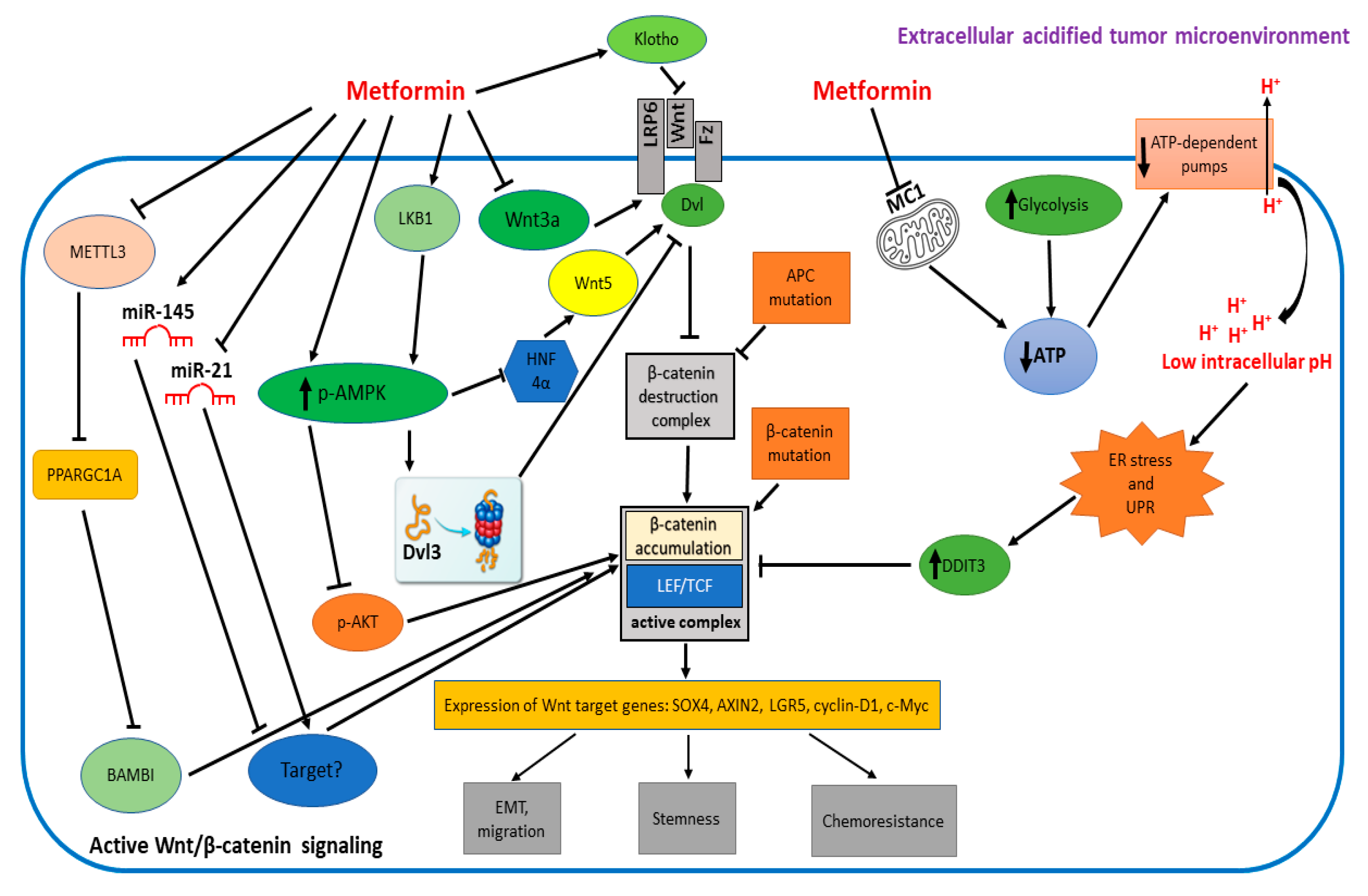
3. Molecular Players Involved in the Wnt/β-Catenin-Mediated Anticancer Activity of Metformin

3.1. DVL3

Upregulation of Wnt signaling is a strong cancer-driving force for multiple types of malignancies [36]. In most cases, Wnt signaling upregulation is due to loss-of-function mutations in the APC gene or stabilizing mutations in the β-catenin gene, both resulting in β-catenin accumulation. These events account for 95% of incidences of colorectal cancer (CRC) but are often also found in tumors of other origin, such as the liver [44], kidney [45], ovary [46], prostate [47], brain [48], endometrium [49], thyroid [50], and lung [51]. However, changes in the expression and/or function of any of the components of the Wnt signaling pathway might contribute to the onset and progression of different types of cancer. Kwan and coworkers [52] reported a significant link between DVL3 upregulation and increased Wnt/β-catenin activity in cervical cancer. Interestingly, they were the first to demonstrate that AMPK activators, including metformin, block the growth of cervical cancer cells by interfering with the DVL3-mediated Wnt/β-catenin signaling. They showed, indeed, that the increase in proteasomal degradation was the molecular mechanism of the reduction of DVL3 induced by AMPK activators, such as metformin, evidencing the importance of DVL3 in the oncogenesis of cervical cancer and highlighting the importance of targeting DVL3 in cervical cancer via AMPK activators. The implication of AMPK activation in this process was supported by the fact that the use of an AMPK inhibitor (Compound C) was able to prevent DVL3 reduction induced by metformin [52]. The role of DVL3 in the anticancer activity of metformin was also highlighted in breast cancer in a similar study by Zou and coworkers [53], where the reduction in DVL3 levels upon metformin treatment was paralleled by the downregulation of β-catenin levels and its transcriptional targets cyclin D1 and c-Myc. Also in this study, increased AMPK-dependent proteasomal degradation of DVL3 was reported to be responsible for the downregulation of Wnt signaling by metformin. Interestingly, the inhibition of the DVL-3/Wnt/β-catenin axis by metformin with the impairment of the nuclear translocation of β-catenin was reported to play a role not only in cancer but also in the development of neural crest cells, since it interferes with the epithelial mesenchymal transition (EMT) process, which is crucial for neural cell crest fate determination [54].

3.2. MMP26

Matrix metalloproteinases (MMPs) play a key role in regulating embryogenesis, tissue morphogenesis and wound healing processes. In addition, the members of this family of proteins have the ability to disintegrate the components of the extracellular Matrix (ECM), favoring invasion and metastasis. Consequently, increased levels of these proteins have been described in different types of cancer. Metformin has been described to inhibit the expression of several members of the MMP family in cancer, such as MMP11 in male germ tumor cells [55], MMP9 in ovarian cancer cells and in fibrosarcoma cells [56,57], MMP2 in cholangiocarcinoma cells [58], melanoma cells [59] and endometrial carcinoma cells [60], and MMP2 and MMP9 in breast cancer cells [61] and in esophageal squamous cell carcinoma [62]. In these studies, different molecular mechanisms were described to explain the inhibitory activity of metformin in MMPs expression. However, the involvement of the Wnt pathway was not reported. Xu and coworkers [63] have recently shown that the overexpression of the matrix metalloproteinase-26 (MMP26) increased the invasiveness of chondrosarcoma cells. This is the only study to show that metformin is able to limit the effects of MMP26 on the invasion of cancer cells through the inhibition of the Wnt pathway, possibly by increasing β-catenin phosphorylation.

3.3. HNF4α

Chang and coworkers [64] found hepatocyte nuclear factor-4α (HNF4α) as a key component among multiple expression datasets of gastric cancer (GC) in whole-transcriptome profiles in Caucasians. They showed that the knockdown of HNF4α exerted antitumorigenic effects both in vitro and in vivo. Interestingly, HNF4α has been previously described as a downstream target of AMPKα [65,66], since it is phosphorylated in its ligand-binding domain (Ser304), thereby blocking dimerization and its binding capability to DNA. They demonstrated that metformin induces the tumor suppressing liver kinase B1 (LKB1) and AMPK in different GC cell lines. Moreover, metformin treatment reduces HNF4α expression, suggesting that the activation of AMPK may be responsible for the downregulation of HNF4α. Interestingly, metformin treatment also decreased WNT5A expression and its downstream target genes TCF1 and β-catenin in both GC cell lines and in xenograft models. Silencing of HNF4α showed similar effects, downregulating WNT5A expression and TCF/LEF signaling, thus confirming that WNT5A is a direct target of HNF4α in GC [64] and that the LKB1/AMPK/HNF4α/WNT5A axis constitutes a signaling cascade that might play a main role in GC tumorigenesis.

3.4. Wnt3a

Cancer stem cells (CSCs) are tumor cells with the ability of self-renewal that can induce tumor generation, recurrence and chemoresistance [67]. Several studies have showed that the Wnt/β-catenin pathway plays a crucial role in stem cells formation in cancer [68]. Zhang and Wang [69] have recently shown that metformin impaired the capability to form a spheroid. Moreover, metformin was also able to inhibit the expression of different markers of stemness in HCT116 colorectal cancer cells, such as aldehyde dehydrogenase 1 (ALDH1), epithelial cell adhesion molecule (EpCAM), Nanog and CD44. This was paralleled by the attenuation of the epithelial mesenchymal transition (EMT), as demonstrated by the reduction in vimentin, a known mesenchymal marker, and by the increase in E-cadherin, an epithelial marker. More importantly, they showed that these changes were a direct consequence of the capability of metformin to downregulate the expression of both Wnt3a and β-catenin. Treating CSCs with the Wnt3a/β-catenin agonist SKL2001 or increasing the expression of Wnt3a, besides inhibiting EMT markers, also attenuated the inhibition of the size and number of sphere cells induced by metformin. Metformin was also able to attenuate 5-fluorouracil resistance of HCT116 sphere cells, confirming what was observed in other types of cancer such as hepatocellular carcinoma [70], pancreatic cancer [71] and non-small cell lung cancer (NSCLC) [72].

3.5. Intracellular Acidification and ER Stress

The tumor microenvironment is characterized by a low extracellular pH that can be reduced to levels near to ~pH 5.5. Consequently, acidosis might become an important stress factor, forcing the selection and the evolution of cancer cells [73]. A property of metformin and other biguanide-type drugs resides in their capability to decrease the cellular ATP amount by blocking the activity of mitochondrial complex I (MCI). This event is usually paralleled by a compensatory increase in glycolysis [74] and the imbalance between the leakage of H+ from the extracellular acidified microenvironment to intracellular compartments, thereby determining an alteration of proton pumps function and the fall of intracellular pH. Melnik and coworkers [75] have recently shown that intracellular acidification caused by metformin inhibits Wnt signaling induced by Wnt3a, preferentially in cancer cells. In particular, the rise of intracellular acidification in association with the drop of ATP levels would cause Endoplasmic Reticulum (ER) stress, followed by the activation of the Unfolded Protein Response (UPR), a homeostatic cellular response that involves a transcriptional reprogramming of stressed cells [76,77]. DDIT3 (DNA damage-inducible transcript 3, CHOP, GADD153), one of the genes more strongly induced by the UPR, is a transcriptional repressor whose expression has been shown to be driven by metformin [78] and which is also capable of inhibiting Wnt signaling through the binding to LEF/TCF [79]. Melnik and coworkers confirmed, indeed, this evidence, also showing that upregulation of DDIT3 determines the direct inhibition of SOX4, a gene that favors metastasis in different tumors [80], by disrupting the TCF4/β-catenin activation complex and, thus, impairing the binding of TCF4 and CBP/p300 to SOX4 promoter. These events limited Wnt signaling, reducing both cancer cells’ growth and invasion. A link between ER stress, the UPR and the Wnt pathway has been also recently reported by our group [16]. Metformin was able, indeed, to inhibit β-catenin expression and transcriptional activity in endometrial cancer cells. This was associated with the upregulation of DDIT3, as described by Melnik et al. [75], but also to the downregulation of HSPA5, a key player of the UPR that also exerts antiapoptotic functions [81,82,83]. We observed, indeed, a reduction in both growth and survival of endometrial cancer cells following metformin treatment. Furthermore, metformin effects on both β-catenin and UPR protein levels/activity were not influenced by a specific inhibitor of AMPK activation, supporting the importance of molecular mechanisms independent from AMPK.

3.6. PPARGC1A

Peroxisome proliferator-activated receptor-gamma coactivator 1 alpha (PPARGC1A) is a transcriptional coactivator that, interacting with PPAR gamma, is able to regulate the expression of genes related to energy metabolism. PPARGC1A is involved in the progression and prognosis of different types of cancer, such as clear cell renal cell carcinoma [84], pancreatic cancer [85], breast cancer [86] and, lately, also in hepatocellular carcinoma (HCC). It has been shown, indeed, that PPARGC1A l expression is significantly reduced in HCC samples and that this represents a risk factor for overall survival of patients [87], suggesting that PPARGC1A might play an important role in HCC. Zuo et al. [88] reported that PPARGC1A acted as a tumor suppressor, inhibiting metastasis by suppressing glycolysis and suggested the involvement of the WNT/β-catenin/PDK1 axis. Zhang and coworkers [89] further investigated these aspects and found, via RNA-seq analysis, that the expression of BAMBI (bone morphogenetic protein and activin membrane-bound inhibitor), a survival-related gene target of the Wnt/β-catenin signaling pathway, was significantly upregulated in HCC cells when PPARGC1A was knocked down. Furthermore, in most of the HCC cohorts analyzed, they discovered a negative correlation between PPARGC1A and BAMBI expression. The downregulation of PPARGC1A in HCC and also resistance of HCC cells to lenvatininb were due to N6-Methyladenosine (m6A) modification of PPARGC1A mRNA by methyltransferase 3 (METTL3), a key m6A writer that they found highly expressed in HCC. Low PPARGC1A levels fail to repress BAMBI and, thus, the WNT/β-catenin pathway. Metformin was, instead, able to restore PPARGC1A expression by inhibiting METTL3 and, thus, reducing m6A modification of the protein, therefore inhibiting the WNT/β-catenin pathway.

3.7. Klotho

Klotho is mainly known as an antiaging gene, due to its ability to suppress senescence, oxidative stress, and inflammation. Thus, Klotho insufficiency seems to be involved in human aging and, specifically, in several aging-related diseases, including cancer [90]. There are two different forms of Klotho: a membrane-bound coreceptor form for fibroblast growth factor 23 (FGF23) [91] or a soluble form, exerting the functions of an endocrine mediator [92]. Klotho acts as an antitumor protein, blocking cancer cell proliferation and migration by modulating different signaling pathways usually involved in cancer, such as the Wnt/β-catenin and phosphoinositide 3-kinase (PI3K)/Akt pathways [93]. Klotho expression is reduced or silenced in several cancers, due mostly to epigenetic changes such as histone modifications, DNA hypermethylation at promoter sites and miRNAs activity, as already observed for other tumor suppressor genes [94]. Wang et al. showed, indeed, that the use of a demethylating agent, such as 5-azacytidine, led to increased expression of Klotho [95]. Klotho, in its soluble form, can prevent Wnt activation by sponging different Wnt ligands, such as Wnt3, Wnt1, Wnt5a, and Wnt4 [96,97]. In preclinical studies, metformin, besides other antidiabetic drugs (PPAR-γ agonists, GLP-1-based, GABA) has been reported to enhance Klotho expression [90]. Thus, metformin would be able to antagonize aberrant Wnt signaling due to deregulated production of Wnt ligands.

3.8. miRNAs

Metformin has been recognized to be effective in the prevention and treatment of cancer through multiple mechanisms, including micro-RNA (miRNA) regulation. Metformin is able, indeed, to modify miRNAs expression, thereby affecting specific downstream pathways. In particular, metformin can exert its anticancer effects by inhibiting the expression of oncogenic miRNAs and/or by upregulating miRNAs that display tumor suppressor activity, as extensively reviewed by Alimoradi et al. [98]. Oncogenic miR-21 has been shown to be upregulated in different types of cancer, such as breast, colorectal, renal, and skin cancer [98]. It has been described that metformin can inhibit the expression of miR-21, thereby negatively affecting several oncogenic pathways such as the TGF-β and the PTEN/Akt pathways [98]. However, only the study of Nangia-Makker et al. [99] evidenced a link between miR-21 downregulation by metformin and the Wnt pathway. They show the effects of metformin on survival of chemo-resistant colon cancer cells that are highly enriched in CSCs/CSLCs (Cancer Stem-Like Cells). In particular, they report that metformin can act synergistically with FuOx, a combination of 5-fluoruracil and oxaliplatin, to promote apoptosis in chemo-resistant colon cancer cells HT-29 and HCT-116. Furthermore, they observed that this drug combination was also able to inhibit colonospheres formation and to enhance their destruction. Moreover, the combinatorial treatment was able to inhibit migration of CR colon cancer cells. This in vitro evidence was confirmed in vivo, since the combination of metformin and FuOX for 5 weeks was capable of inhibiting the growth of tumor xenografts obtained by implanting chemo-resistant HCT-116 and HT-29 cells in SCID mice by almost 50%, when compared with the vehicle-treated controls. These effects were associated with the variation of specific microRNAs (miRNAs). They showed, indeed, that tumor suppressor miR-145 levels were increased while oncomiR miR-21 levels were reduced following metformin treatment, alone or in combination with FuOX. miR21, in particular, was previously shown to be upregulated in colorectal cancer, where it induces stemness in chemo-resistant colon cancer cells [100]. Furthermore, over-expression of miR-21 in HCT-116 cells resulted in increased β-catenin activity [33] and c-myc levels. Thus, the capability of metformin to downregulate miR-21 and the Wnt/β-catenin signaling pathway suggests a possible role of miR-21 in targeting not yet identified key regulators of this pathway.

4. Conclusions and Future Perspectives

Metformin is still the most prescribed antidiabetic agent worldwide. However, a number of different pathologic states, including cancer, have been described to benefit from metformin administration. The more extensively investigated molecular pathway is the AMPK-dependent pathway. However, in recent years, a number of other pathways have been reported to contribute to the anticancer potential of metformin. One of the more interesting and promising, from a therapeutic point of view, is the Wnt pathway. It is, indeed, often deregulated in cancer cells, where it drives important processes, such as EMT, invasion, stemness and chemoresistance. As described in this “Perspective”, metformin appears to affect the Wnt pathway at various levels (Wnt signaling complex formation, β-catenin accumulation, β-catenin transcriptional complex formation) and, often, in a cancer-specific manner (Table 1 and Figure 1). Of particular interest is the therapeutic strategy that might arise from the entrapment of cancer cells in a sort of a “Warburg Trap”, a vicious cycle determined by MC1 inhibitors such as metformin and ionophores combination, able to cause both an increase in intracellular acidification and a drop in ATP concentration, ultimately leading to apoptosis of cancer cells. Thus, a better understanding of the different molecular players that can be modulated by metformin in the Wnt pathway might be relevant in the therapy of different types of cancer.

Author Contributions

L.U. was responsible for writing—review and editing, and designed the figures, D.C. wrote the original draft, P.M. made substantial contributions to the conception, F.F., A.N. and L.I. helped to shape and revised the manuscript, F.B. provided critical feedback. All authors have read and agreed to the published version of the manuscript.

Funding

This research was funded, in part, by the Ministero dell’Università e della Ricerca (PNRR, Missione 4 Componente 2 Investimento 1.4 finanziato dall’Unione europea—NextGenerationEU—Progetto “National Center for Gene Therapy and Drugs based on RNA Technology”—CN00000041—CUP E63C22000940007), by the Ministero dell’Università e della Ricerca (PNRR, Missione 4 Componente 2 Investimento 1.3 finanziato dall’Unione europea—NextGenerationEU—Progetto “Ageing Well in an ageing society, Age-It”—PE0000015—CUP B83C22004880006), by the Ministero dell’Università e della Ricerca (Progetti di Rilevante Interesse Nazionale—Anno 2020—Progetto “Cognitive dysfunction in dysmetabolic obesity and diabetes: role of inter-organ crosstalk and cellular ageing”—2020N5WK98), by the Ministero dell’Università e della Ricerca (Progetti di Rilevante Interesse Nazionale—Anno 2017—Progetto “The Incretin Hormones and their analogues as physiological and pharmacological regulators of a complex multi-organ network”—2017CPLH32), by the Ministero dell’Università e della Ricerca (PNRR, Missione 4 Componente 2, Linea di investimento 3.1, finanziato dall’Unione europea—NextGenerationEU—Progetto IR0000031 “Strengthening of the Biobanking and Biomolecular Resources Research Infrastructure of Italy”), by the Ministero della Salute (Piano Operativo Salute Traiettoria 3—Linea di azione 3.1—Progetto “GENOMED” T3-AN-09—CUP E63C22001440001) and by the Regione Campania (POR FESR 2014-2020—Manifestazioni di interesse per la realizzazione di servizi di ricerca e sviluppo per la lotta contro il COVID-19—DD 19/2022 Project SHARCODE.

Institutional Review Board Statement

Not applicable.

Informed Consent Statement

Not applicable.

Data Availability Statement

Not applicable.

Acknowledgments

The authors thank Antonio D’Andrea for the excellent technical assistance.

Conflicts of Interest

The authors declare no conflict of interest.

References

  1. He, L.; Wondisford, F.E. Metformin Action: Concentrations Matter. Cell Metab. 2015, 21, 159–162. [Google Scholar] [CrossRef] [PubMed]
  2. King, P.; Peacock, I.; Donnelly, R. The UK Prospective Diabetes Study (UKPDS): Clinical and therapeutic implications for type 2 diabetes. Br. J. Clin. Pharmacol. 1999, 48, 643. [Google Scholar] [CrossRef] [PubMed]
  3. Sanchez-Rangel, E.; Inzucchi, S.E. Metformin: Clinical use in type 2 diabetes. Diabetologia 2017, 60, 1586–1593. [Google Scholar] [CrossRef] [PubMed]
  4. Hou, W.-L.; Yin, J.; Alimujiang, M.; Yu, X.-Y.; Ai, L.-G.; Bao, Y.-Q.; Liu, F.; Jia, W.-P. Inhibition of mitochondrial complex I improves glucose metabolism independently of AMPK activation. J. Cell. Mol. Med. 2018, 22, 1316–1328. [Google Scholar] [CrossRef] [PubMed]
  5. Ben Sahra, I.; Regazzetti, C.; Robert, G.; Laurent, K.; Le Marchand-Brustel, Y.; Auberger, P.; Tanti, J.-F.; Giorgetti-Peraldi, S.; Bost, F. Metformin, Independent of AMPK, Induces mTOR Inhibition and Cell-Cycle Arrest through REDD1. Cancer Res. 2011, 71, 4366–4372. [Google Scholar] [CrossRef]
  6. Musi, N.; Hirshman, M.F.; Nygren, J.; Svanfeldt, M.; Bavenholm, P.; Rooyackers, O.; Zhou, G.; Williamson, J.M.; Ljunqvist, O.; Efendic, S.; et al. Metformin Increases AMP-Activated Protein Kinase Activity in Skeletal Muscle of Subjects With Type 2 Diabetes. Diabetes 2002, 51, 2074–2081. [Google Scholar] [CrossRef]
  7. Vasamsetti, S.B.; Karnewar, S.; Kanugula, A.K.; Thatipalli, A.R.; Kumar, J.M.; Kotamraju, S. Metformin Inhibits Monocyte-to-Macrophage Differentiation via AMPK-Mediated Inhibition of STAT3 Activation: Potential Role in Atherosclerosis. Diabetes 2015, 64, 2028–2041. [Google Scholar] [CrossRef]
  8. Eisenreich, A.; Leppert, U. Update on the Protective Renal Effects of Metformin in Diabetic Nephropathy. Curr. Med. Chem. 2017, 24, 3397–3412. [Google Scholar] [CrossRef]
  9. Lord, J.M.; Flight, I.H.K.; Norman, R.J. Metformin in polycystic ovary syndrome: Systematic review and meta-analysis. BMJ 2003, 327, 951–953. [Google Scholar] [CrossRef]
  10. El Massry, M.; Alaeddine, L.M.; Ali, L.; Saad, C.; Eid, A.A. Metformin: A Growing Journey from Glycemic Control to the Treatment of Alzheimer’s Disease and Depression. Curr. Med. Chem. 2021, 28, 2328–2345. [Google Scholar] [CrossRef]
  11. Salvatore, T.; Galiero, R.; Caturano, A.; Vetrano, E.; Rinaldi, L.; Coviello, F.; Di Martino, A.; Albanese, G.; Marfella, R.; Sardu, C.; et al. Effects of Metformin in Heart Failure: From Pathophysiological Rationale to Clinical Evidence. Biomolecules 2021, 11, 1834. [Google Scholar] [CrossRef] [PubMed]
  12. Evans, J.M.M.; Donnelly, L.A.; Emslie-Smith, A.M.; Alessi, D.R.; Morris, A.D. Metformin and reduced risk of cancer in diabetic patients. Br. Med. J. 2005, 330, 1304–1305. [Google Scholar] [CrossRef] [PubMed]
  13. Zi, F.; Zi, H.; Li, Y.; He, J.; Shi, Q.; Cai, Z. Metformin and cancer: An existing drug for cancer prevention and therapy. Oncol. Lett. 2018, 15, 683–690. [Google Scholar] [CrossRef]
  14. Kasznicki, J.; Sliwinska, A.; Drzewoski, J. Metformin in cancer prevention and therapy. Ann. Transl. Med. 2014, 2, 57. [Google Scholar] [CrossRef] [PubMed]
  15. Kim, H.J.; Lee, S.; Chun, K.H.; Jeon, J.Y.; Han, S.J.; Kim, D.J.; Kim, Y.S.; Woo, J.T.; Nam, M.S.; Baik, S.H.; et al. Metformin reduces the risk of cancer in patients with type 2 diabetes: An analysis based on the Korean National Diabetes Program Cohort. Medicine 2018, 97, e0036. [Google Scholar] [CrossRef]
  16. Conza, D.; Mirra, P.; Calì, G.; Insabato, L.; Fiory, F.; Beguinot, F.; Ulianich, L. Metformin Dysregulates the Unfolded Protein Response and the WNT/β-Catenin Pathway in Endometrial Cancer Cells through an AMPK-Independent Mechanism. Cells 2021, 10, 1067. [Google Scholar] [CrossRef]
  17. Zhu, Y.; Li, X. Advances of Wnt Signalling Pathway in Colorectal Cancer. Cells 2023, 12, 447. [Google Scholar] [CrossRef]
  18. Clevers, H.; Nusse, R. Wnt/Beta-Catenin Signaling and Disease. Cell 2012, 149, 1192–1205. [Google Scholar] [CrossRef]
  19. Cheng, X.; Xu, X.; Chen, D.; Zhao, F.; Wang, W. Therapeutic Potential of Targeting the Wnt/Beta-Catenin Signaling Pathway in Colorectal Cancer. Biomed Pharmacoth. 2019, 110, 473–481. [Google Scholar] [CrossRef]
  20. Li, V.S.W.; Ng, S.S.; Boersema, P.J.; Low, T.Y.; Karthaus, W.R.; Gerlach, J.P.; Mohammed, S.; Heck, A.J.R.; Maurice, M.M.; Mahmoudi, T.; et al. Wnt Signaling through Inhibition of Beta-Catenin Degradation in an Intact Axin1 Complex. Cell 2012, 149, 1245–1256. [Google Scholar] [CrossRef]
  21. Mantilla, C.; Suárez Mellado, I.; Duque Jaramillo, A.; Navas, M.C. β-catenin signaling mechanisms and its role in carcinogenesis. CES Med. 2015, 29, 109–127. [Google Scholar]
  22. Kim, N.G.; Xu, C.; Gumbiner, B.M. Identification of Targets of the Wnt Pathway Destruction Complex in Addition to Beta-Catenin. Proc. Natl. Acad. Sci. USA 2009, 106, 5165–5170. [Google Scholar] [CrossRef]
  23. Latres, E.; Chiaur, D.S.; Pagano, M. The Human F Box Protein Beta-Trcp Associates with the Cul1/Skp1 Complex and Regulates the Stability of Beta-Catenin. Oncogene 1999, 18, 849–854. [Google Scholar] [CrossRef] [PubMed]
  24. Koch, S.; Acebron, S.P.; Herbst, J.; Hatiboglu, G.; Niehrs, C. Post-transcriptional Wnt Signaling Governs Epididymal Sperm Maturation. Cell 2015, 163, 1225–1236. [Google Scholar] [CrossRef] [PubMed]
  25. Behrens, J.; Von Kries, J.P.; Kühl, M.; Bruhn, L.; Wedlich, D.; Grosschedl, R.; Birchmeier, W. Functional interaction of β-catenin with the transcription factor LEF-1. Nature 1996, 382, 638–642. [Google Scholar] [CrossRef] [PubMed]
  26. Lustig, B.; Jerchow, B.; Sachs, M.; Weiler, S.; Pietsch, T.; Karsten, U.; van de Wetering, M.; Clevers, H.; Schlag, P.M.; Birchmeier, W.; et al. Negative Feedback Loop of Wnt Signaling through Upregulation of Conductin/Axin2 in Colorectal and Liver Tumors. Mol. Cell. Biol. 2002, 22, 1184–1193. [Google Scholar] [CrossRef]
  27. Liu, C.; Li, Y.; Semenov, M.; Han, C.; Baeg, G.H.; Tan, Y.; Zhang, Z.; Lin, X.; He, X. Control of beta-catenin phosphorylation/degradation by a dual-kinase mechanism. Cell 2002, 108, 837–847. [Google Scholar] [CrossRef]
  28. He, T.-C.; Sparks, A.B.; Rago, C.; Hermeking, H.; Zawel, L.; da Costa, L.T.; Morin, P.J.; Vogelstein, B.; Kinzler, K.W. Identification of c- MYC as a Target of the APC Pathway. Science 1998, 281, 1509–1512. [Google Scholar] [CrossRef] [PubMed]
  29. Kawano, Y.; Kypta, R. Secreted antagonists of the Wnt signalling pathway. J. Cell Sci. 2003, 116, 2627–2634. [Google Scholar] [CrossRef]
  30. Li, X.; Zhang, C.; Yuan, Y.; Wang, Y.; Lu, S.; Zhou, Z.; Zhen, P.; Zhou, M. Downregulation of ARMC8 promotes tumorigenesis through activating Wnt/β-catenin pathway and EMT in cutaneous squamous cell carcinomas. J. Dermatol. Sci. 2021, 102, 184–192. [Google Scholar] [CrossRef]
  31. Heuberger, J.; Birchmeier, W. Interplay of Cadherin-Mediated Cell Adhesion and Canonical Wnt Signaling. Cold Spring Harb. Perspect. Biol. 2010, 2, a002915. [Google Scholar] [CrossRef] [PubMed]
  32. Margulis, A.; Zhang, W.; Alt-Holland, A.; Crawford, H.C.; Fusenig, N.E.; Garlick, J.A. E-cadherin Suppression Accelerates Squamous Cell Carcinoma Progression in Three-Dimensional, Human Tissue Constructs. Cancer Res 2005, 65, 1783–1791. [Google Scholar] [CrossRef]
  33. Conacci-Sorrell, M.; Simcha, I.; Ben-Yedidia, T.; Blechman, J.; Savagner, P.; Ben-Ze’ev, A. Autoregulation of E-cadherin expression by cadherin-cadherin interactions: The roles of beta-catenin signaling, Slug, and MAPK. J. Cell Biol. 2003, 163, 847–857. [Google Scholar] [CrossRef]
  34. Khan, M.A.; Chen, H.-C.; Zhang, D.; Fu, J. Twist: A molecular target in cancer therapeutics. Tumour Biol. 2013, 34, 2497–2506. [Google Scholar] [CrossRef]
  35. Hlubek, F.; Spaderna, S.; Jung, A.; Kirchner, T.; Brabletz, T. Beta-catenin activates a coordinated expression of the proinvasive factors laminin-5 gamma2 chain and MT1-MMP in colorectal carcinomas. Int. J. Cancer 2004, 108, 321–326. [Google Scholar] [CrossRef]
  36. Zhan, T.; Rindtorff, N.; Boutros, M. Wnt Signaling in Cancer. Oncogene 2017, 36, 1461–1473. [Google Scholar] [CrossRef] [PubMed]
  37. Sheldahl, L.C.; Slusarski, D.C.; Pandur, P.; Miller, J.R.; Kühl, M.; Moon, R.T. Dishevelled activates Ca2+ flux, PKC, and CamKII in vertebrate embryos. J. Cell Biol. 2003, 161, 769–777. [Google Scholar] [CrossRef] [PubMed]
  38. Ishitani, T.; Kishida, S.; Hyodo-Miura, J.; Ueno, N.; Yasuda, J.; Waterman, M.; Shibuya, H.; Moon, R.T.; Ninomiya-Tsuji, J.; Matsumoto, K. The TAK1-NLK mitogen-activated protein kinase cascade functions in the Wnt-5a/Ca(2+) pathway to antagonize Wnt/beta-catenin signaling. Mol. Cell Biol. 2003, 23, 131–139. [Google Scholar] [CrossRef]
  39. Schlessinger, K.; Hall, A.; Tolwinski, N. Wnt signaling pathways meet Rho GTPases. Genes Dev. 2009, 23, 265–277. [Google Scholar] [CrossRef]
  40. Yamanaka, H.; Moriguchi, T.; Masuyama, N.; Kusakabe, M.; Hanafusa, H.; Takada, R.; Takada, S.; Nishida, E. JNK functions in the non-canonical Wnt pathway to regulate convergent extension movements in vertebrates. EMBO Rep. 2002, 3, 69–75. [Google Scholar] [CrossRef]
  41. Habas, R.; Kato, Y.; He, X. Wnt/Frizzled Activation of Rho Regulates Vertebrate Gastrulation and Requires a Novel Formin Homology Protein Daam1. Cell 2001, 107, 843–854. [Google Scholar] [CrossRef]
  42. Endo, Y.; Wolf, V.; Muraiso, K.; Kamijo, K.; Soon, L.; Üren, A.; Barshishat-Küpper, M.; Rubin, J.S. Wnt-3a-dependent Cell Motility Involves RhoA Activation and Is Specifically Regulated by Dishevelled-2. J. Biol. Chem. 2005, 280, 777–786. [Google Scholar] [CrossRef] [PubMed]
  43. Schlessinger, K.; McManus, E.J.; Hall, A. Cdc42 and noncanonical Wnt signal transduction pathways cooperate to promote cell polarity. J. Cell Biol. 2007, 178, 355–361. [Google Scholar] [CrossRef] [PubMed]
  44. Nhieu, J.T.; Renard, C.A.; Wei, Y.; Cherqui, D.; Zafrani, E.S.; Buendia, M.A. Nuclear accumulation of mutated beta-catenin in hepatocellular carcinoma is associated with increased cell proliferation. Am. J. Pathol. 1999, 155, 703–710. [Google Scholar] [CrossRef]
  45. Maiti, S.; Alam, R.; Amos, C.I.; Huff, V. Frequent association of beta-catenin and WT1 mutations in Wilms tumors. Cancer Res. 2000, 60, 6288–6292. [Google Scholar]
  46. Palacios, J.; Gamallo, C. Mutations in the beta-catenin gene (CTNNB1) in endometrioid ovarian carcinomas. Cancer Res. 1998, 58, 1344–1347. [Google Scholar] [PubMed]
  47. Voeller, H.J.; Truica, C.I.; Gelmann, E.P. Beta-catenin mutations in human prostate cancer. Cancer Res. 1998, 58, 2520–2523. [Google Scholar]
  48. Zurawel, R.H.; Chiappa, S.A.; Allen, C.; Raffel, C. Sporadic medulloblastomas contain oncogenic beta-catenin mutations. Cancer Res. 1998, 58, 896–899. [Google Scholar]
  49. Zaman, G.J.R.; de Roos, J.A.D.M.; Libouban, M.A.A.; Prinsen, M.B.W.; de Man, J.; Buijsman, R.C.; Uitdehaag, J.C.M. TTK inhibitors as a targeted therapy for CTNNB1 (beta-catenin) mutant cancers. Mol. Cancer Ther. 2017, 16, 2609–2617. [Google Scholar] [CrossRef]
  50. Garcia-Rostan, G.; Tallini, G.; Herrero, A.; D’Aquila, T.G.; Carcangiu, M.L.; Rimm, D.L. Frequent mutation and nuclear localization of beta-catenin in anaplastic thyroid carcinoma. Cancer Res 1999, 59, 1811–1815. [Google Scholar]
  51. Tammela, T.; Sanchez-Rivera, F.J.; Cetinbas, N.M.; Wu, K.; Joshi, N.S.; Helenius, K.; Park, Y.; Azimi, R.; Kerper, N.R.; Wesselhoeft, R.A.; et al. A Wnt-producing niche drives proliferative potential and progression in lung adenocarcinoma. Nature 2017, 545, 355–359. [Google Scholar] [CrossRef]
  52. Kwan, H.T.; Chan, D.W.; Cai, P.C.H.; Mak, C.S.L.; Yung, M.M.H.; Leung, T.H.Y.; Wong, O.G.W.; Cheung, A.N.Y.; Ngan, H.Y.S. AMPK Activators Suppress Cervical Cancer Cell Growth through Inhibition of DVL3 Mediated Wnt/β-Catenin Signaling Activity. PLoS ONE 2013, 8, e53597. [Google Scholar] [CrossRef]
  53. Zou, Y.-F.; Xie, C.-W.; Yang, S.-X.; Xiong, J.-P. AMPK activators suppress breast cancer cell growth by inhibiting DVL3-facilitated Wnt/β-catenin signaling pathway activity. Mol. Med. Rep. 2017, 1, 899–907. [Google Scholar] [CrossRef]
  54. Banerjee, P.; Dutta, S.; Rajarshi, P. Dysregulation of Wnt-Signaling and a Candidate Set of miRNAs Underlie the Effect of Metformin on Neural Crest Cell Development. Stem Cells 2016, 34, 334–345. [Google Scholar] [CrossRef]
  55. Salatino, A.; Mirabelli, M.; Chiefari, E.; Greco, M.; Di Vito, A.; Bonapace, G.; Brunetti, F.S.; Crocerossa, F.; Epstein, A.L.; Foti, D.P.; et al. The anticancer effects of Metformin in the male germ tumor SEM-1 cell line are mediated by HMGA1. Front. Endocrinol. 2022, 13, 1051988. [Google Scholar] [CrossRef]
  56. Zheng, Y.; Zhang, H.; Sun, H. Metformin inhibits the proliferation and invasion of ovarian cancer cells by suppressing tripartite motif-containing 37-induced tumor necrosis factor receptor-associated factor 2 ubiquitination. Cancer Sci. 2022, 113, 3776–3786. [Google Scholar] [CrossRef]
  57. Hwang, Y.P.; Jeong, H.G. Metformin blocks migration and invasion of tumour cells by inhibition of matrix metalloproteinase-9 activation through a calcium and protein kinase Calpha-dependent pathway: Phorbol-12-myristate-13-acetate-induced/extracellular signal-regulated kinase/activator protein-1. Br. J. Pharmacol. 2010, 160, 1195–1211. [Google Scholar]
  58. Trinh, S.X.; Nguyen, H.T.B.; Saimuang, K.; Prachayasittikul, V.; Chan-On, W. Metformin Inhibits Migration and Invasion of Cholangiocarcinoma Cells. Asian Pac. J. Cancer Prev. 2017, 18, 473–477. [Google Scholar] [CrossRef]
  59. Cerezo, M.; Tichet, M.; Abbe, P.; Ohanna, M.; Lehraiki, A.; Rouaud, F.; Allegra, M.; Giacchero, D.; Bahadoran, P.; Bertolotto, C.; et al. Metformin blocks melanoma invasion and metastasis development in AMPK/p53-dependent manner. Mol. Cancer Ther. 2013, 12, 1605–1615. [Google Scholar] [CrossRef]
  60. Tan, B.K.; Adya, R.; Chen, J.; Lehnert, H.; Cassia, L.J.S.; Randeva, H.S. Metformin Treatment Exerts Antiinvasive and Antimetastatic Effects in Human Endometrial Carcinoma Cells. J. Clin. Endocrinol. Metab. 2011, 96, 808–816. [Google Scholar] [CrossRef]
  61. Yenmis, G.; Sarac, E.Y.; Besli, N.; Soydas, T.; Tastan, C.; Kancagi, D.D.; Yilanci, M.; Senol, K.; Karagulle, O.O.; Ekmekci, C.G.; et al. Anti-cancer effect of metformin on the metastasis and invasion of primary breast cancer cells through mediating NF-kB activity. Acta Histochem. 2021, 123, 151709. [Google Scholar] [CrossRef]
  62. Liang, F.; Wang, Y.-G.; Wang, C. Metformin Inhibited Growth, Invasion and Metastasis of Esophageal Squamous Cell Carcinoma in Vitro and in Vivo. Cell. Physiol. Biochem. 2018, 51, 1276–1286. [Google Scholar] [CrossRef]
  63. Xu, X.; Ma, J.; Li, C.; Zhao, W.; Xu, Y. Regulation of chondrosarcoma invasion by MMP26. Tumor Biol. 2014, 36, 365–369. [Google Scholar] [CrossRef] [PubMed]
  64. Chang, H.R.; Nam, S.; Kook, M.C.; Kim, K.T.; Liu, X.; Yao, H.; Jung, H.R.; Jr, L.R.; Seo, H.H.; Park, H.S.; et al. HNF4α is a therapeutic target that links AMPK to WNT signalling in early-stage gastric cancer. Gut 2016, 65, 19–32. [Google Scholar] [CrossRef]
  65. Hong, Y.H.; Varanasi, U.S.; Yang, W.; Leff, T. AMP-activated Protein Kinase Regulates HNF4α Transcriptional Activity by Inhibiting Dimer Formation and Decreasing Protein Stability. J. Biol. Chem. 2003, 278, 27495–27501. [Google Scholar] [CrossRef]
  66. Ribeiro, A.; Archer, A.; Le Beyec, J.; Cattin, A.-L.; Saint-Just, S.; Pinçon-Raymond, M.; Chambaz, J.; Lacasa, M.; Cardot, P. Hepatic Nuclear Factor-4, a Key Transcription Factor at the Crossroads Between Architecture and Function of Epithelia. Recent Pat. Endocr. Metab. Immune Drug Discov. 2007, 1, 166–175. [Google Scholar] [CrossRef]
  67. Kim, D.Y.; Park, E.Y.; Chang, E.; Kang, H.G.; Koo, Y.; Lee, E.J.; Ko, J.Y.; Kong, H.K.; Chun, K.H.; Park, J.H. A novel mir-34a target, protein kinase d1, stimulates cancer stemness and drug resistance through gsk3/beta-catenin signaling in breast cancer. Oncotarget 2016, 7, 14791–14802. [Google Scholar] [CrossRef] [PubMed]
  68. Wiese, M.; Walther, N.; Diederichs, C.; Schill, F.; Monecke, S.; Salinas, G.; Sturm, D.; Pfister, S.M.; Dressel, R.; Johnsen, S.A.; et al. The beta-catenin/cbp-antagonist icg-001 inhibits pediatric glioma tumorigenicity in a wnt-independent manner. Oncotarget 2017, 8, 27300–27313. [Google Scholar] [CrossRef]
  69. Zhang, C.; Wang, Y. Metformin attenuates cells stemness and epithelial-mesenchymal transition in colorectal cancer cells by inhibiting the Wnt3a/β-catenin pathway. Mol. Med. Rep. 2019, 19, 1203–1209. [Google Scholar] [CrossRef]
  70. Tian, Y.; Tang, B.; Wang, C.; Sun, D.; Zhang, R.; Luo, N.; Han, Z.; Liang, R.; Gao, Z.; Wang, L. Metformin mediates resensitivity to 5-fluorouracil in hepatocellular carcinoma via the suppression of YAP. Oncotarget 2016, 7, 46230–46241. [Google Scholar] [CrossRef] [PubMed]
  71. Cheng, G.; Lanza-Jacoby, S. Metformin decreases growth of pancreatic cancer cells by decreasing reactive oxygen species: Role of NOX4. Biochem. Biophys. Res. Commun. 2015, 465, 41–46. [Google Scholar] [CrossRef]
  72. Liu, Y.; He, C.; Huang, X. Metformin partially reverses the carboplatin-resistance in NSCLC by inhibiting glucose metabolism. Oncotarget 2017, 8, 75206–75216. [Google Scholar] [CrossRef]
  73. Justus, C.R.; Dong, L.; Yang, L.V. Acidic tumor microenvironment and pH sensing G protein-coupled receptors. Front. Physiol. 2013, 4, 354. [Google Scholar] [CrossRef]
  74. Janzer, A.; German, N.J.; Gonzalez-Herrera, K.N.; Asara, J.M.; Haigis, M.C.; Struhl, K. Metformin and phenformin deplete tricarboxylic acid cycle and glycolytic intermediates during cell transformation and NTPs in cancer stem cells. Proc. Natl. Acad. Sci. USA 2014, 111, 10574–10579. [Google Scholar] [CrossRef]
  75. Melnik, S.; Dvornikov, M.; Müller-Decker, K.; Depner, S.; Stannek, P.; Meister, M.; Warth, A.; Thomas, M.; Muley, T.; Risch, A.; et al. Cancer cell specific inhibition of Wnt/β-catenin signaling by forced intracellular acidification. Cell Discov. 2018, 4, 37. [Google Scholar] [PubMed]
  76. Schröder, M.; Kaufman, R. The mammalian unfolded protein response. Annu. Rev. Biochem. 2005, 74, 739–789. [Google Scholar] [CrossRef]
  77. Treglia, A.S.; Turco, S.; Ulianich, L.; Ausiello, P.; Lofrumento, D.D.; Nicolardi, G.; Miele, C.; Garbi, C.; Beguinot, F.; Di Jeso, B. Cell fate following ER stress: Just a matter of “quo ante” recovery or death? Histol. Histopathol. 2012, 27, 1–12. [Google Scholar] [PubMed]
  78. Quentin, T.; Steinmetz, M.; Poppe, A.; Thoms, S. Metformin differentially activates ER stress signaling pathways without inducing apoptosis. Dis. Model. Mech. 2012, 5, 259–269. [Google Scholar] [CrossRef] [PubMed]
  79. Horndasch, M.; Lienkamp, S.; Springer, E.; Schmitt, A.; Pavenstädt, H.; Walz, G.; Gloy, J. The C/EBP homologous protein CHOP (GADD153) is an inhibitor of Wnt/TCF signals. Oncogene 2006, 25, 3397–3407. [Google Scholar]
  80. Vervoort, S.J.; Lourenco, A.R.; van Boxtel, R.; Coffer, P.J. SOX4 mediates TGF-beta-induced expression of mesenchymal markers during mammary cell epithelial to mesenchymal transition. PLoS ONE. 2013, 8, e53238. [Google Scholar]
  81. Bifulco, G.; Miele, C.; Di Jeso, B.; Beguinot, F.; Nappi, C.; Di Carlo, C.; Capuozzo, S.; Terrazzano, G.; Insabato, L.; Ulianich, L. Endoplasmic reticulum stress is activated in endometrial adenocarcinoma. Gynecol. Oncol. 2012, 125, 220–225. [Google Scholar] [CrossRef] [PubMed]
  82. Calì, G.; Insabato, L.; Conza, D.; Bifulco, G.; Parrillo, L.; Mirra, P.; Fiory, F.; Miele, C.; Raciti, G.A.; Di Jeso, B.; et al. GRP78 Mediates Cell Growth and Invasiveness in Endometrial Cancer. J. Cell. Physiol. 2014, 229, 1417–1426. [Google Scholar] [CrossRef]
  83. Ulianich, L.; Insabato, L. Endoplasmic Reticulum Stress in Endometrial Cancer. Front. Med. 2014, 1, 55. [Google Scholar] [CrossRef]
  84. Ma, T.; Meng, L.; Wang, X.; Tian, Z.; Wang, J.; Liu, X.; Zhang, W.; Zhang, Y. TNFSF13B and PPARGC1A expression is associated with tumor-infiltrating immune cell abundance and prognosis in clear cell renal cell carcinoma. Am. J. Transl. Res. 2021, 13, 11048–11064. [Google Scholar]
  85. Huang, X.; Pan, L.; Zuo, Z.; Li, M.; Zeng, L.; Li, R.; Ye, Y.; Zhang, J.; Wu, G.; Bai, R.; et al. LINC00842 inactivates transcription co-regulator PGC-1α to promote pancreatic cancer malignancy through metabolic remodelling. Nat. Commun. 2021, 12, 3830. [Google Scholar] [CrossRef] [PubMed]
  86. Andrzejewski, S.; Klimcakova, E.; Johnson, R.M.; Tabariès, S.; Annis, M.G.; McGuirk, S.; Northey, J.J.; Chénard, V.; Sriram, U.; Papadopoli, D.J.; et al. PGC-1α Promotes Breast Cancer Metastasis and Confers Bioenergetic Flexibility against Metabolic Drugs. Cell Metab. 2017, 26, 778–787.e5. [Google Scholar] [CrossRef] [PubMed]
  87. Zhang, Q.; Zhang, Y.; Guo, Y.; Tang, H.; Li, M.; Liu, L. A novel machine learning derived RNA-binding protein gene–based score system predicts prognosis of hepatocellular carcinoma patients. PeerJ 2021, 9, e12572. [Google Scholar] [CrossRef]
  88. Zuo, Q.; He, J.; Zhang, S.; Wang, H.; Jin, G.; Jin, H.; Cheng, Z.; Tao, X.; Yu, C.; Li, B.; et al. PPARgamma Coactivator-1alpha suppresses metastasis of hepatocellular carcinoma by inhibiting Warburg effect by PPARgamma-dependent WNT/beta-Catenin/Pyruvate Dehydrogenase Kinase Isozyme 1 Axis. Hepatology 2021, 73, 644–660. [Google Scholar]
  89. Zhang, Q.; Xiong, L.; Wei, T.; Liu, Q.; Yan, L.; Chen, J.; Dai, L.; Shi, L.; Zhang, W.; Yang, J.; et al. Hypoxia-responsive PPARGC1A/BAMBI/ACSL5 axis promotes progression and resistance to lenvatinib in hepatocellular carcinoma. Oncogene 2023, 42, 1509–1523. [Google Scholar] [CrossRef]
  90. Prud’homme, G.J.; Kurt, M.; Wang, Q. Pathobiology of the Klotho Antiaging Protein and Therapeutic Considerations. Front. Aging 2022, 3, 931331. [Google Scholar] [CrossRef]
  91. Urakawa, I.; Yamazaki, Y.; Shimada, T.; Iijima, K.; Hasegawa, H.; Okawa, K.; Fujita, T.; Fukumoto, S.; Yamashita, T. Klotho converts canonical FGF receptor into a specific receptor for FGF23. Nature 2006, 444, 770–774. [Google Scholar] [CrossRef]
  92. Dalton, G.D.; Xie, J.; An, S.-W.; Huang, C.-L. New Insights into the Mechanism of Action of Soluble Klotho. Front. Endocrinol. 2017, 8, 323. [Google Scholar] [CrossRef]
  93. Xuan, N.T.; Hai, N.V. Changes in expression of klotho affect physiological processes, diseases, and cancer. Iran. J. Basic Med. Sci. 2018, 21, 3–8. [Google Scholar]
  94. Abolghasemi, M.; Yousefi, T.; Maniati, M.; Qujeq, D. The interplay of Klotho with signaling pathway and microRNAs in cancers. J. Cell. Biochem. 2019, 120, 14306–14317. [Google Scholar] [CrossRef] [PubMed]
  95. Wang, L.; Wang, X.; Wang, X.; Jie, P.; Lu, H.; Zhang, S.; Lin, X.; Lam, E.K.; Cui, Y.; Yu, J.; et al. Klotho is silenced through promoter hypermethylation in gastric cancer. Am. J. Cancer Res. 2010, 1, 111–119. [Google Scholar]
  96. Liu, H.; Fergusson, M.M.; Castilho, R.M.; Liu, J.; Cao, L.; Chen, J.; Malide, D.; Rovira, I.I.; Schimel, D.; Kuo, C.J.; et al. Augmented Wnt Signaling in a Mammalian Model of Accelerated Aging. Science 2007, 317, 803–806. [Google Scholar] [CrossRef] [PubMed]
  97. Wang, Y.; Sun, Z. Current understanding of klotho. Ageing Res. Rev. 2009, 8, 43–51. [Google Scholar] [CrossRef]
  98. Alimoradi, N.; Firouzabadi, N.; Fatehi, R. How metformin affects various malignancies by means of microRNAs: A brief review. Cancer Cell Int. 2021, 21, 207. [Google Scholar]
  99. Nangia-Makker, P.; Yu, Y.; Vasudevan, A.; Farhana, L.; Rajendra, S.G.; Levi, E.; Majumdar, A.P.N. Metformin: A Potential Therapeutic Agent for Recurrent Colon Cancer. PLoS ONE 2014, 9, e84369. [Google Scholar] [PubMed]
  100. Yu, Y.; Kanwar, S.S.; Patel, B.B.; Oh, P.S.; Nautiyal, J.; Fazlul, H.S.; Adhip, P.N.M. miRNA-21 induces stemness by downregulating transforming growth factor beta receptor 2 (TGFbetaR2) in colon cancer cells. Carcinogenesis 2012, 33, 68–76. [Google Scholar]
Figure 1. Metformin inhibits the Wnt pathway through multiple mechanisms. Wnt/β-catenin signaling can be initiated either by Wnt ligands, or by inactivating mutations of APC, or by stabilizing mutations of β-catenin, all resulting in β-catenin accumulation. β-catenin binds LEF/TCF transcription factors and induces target genes, regulating EMT, migration, stemness and chemoresistance of cancer cells. In the right section of the picture, metformin impairs ATP production and, thus, the activity of the ATP-dependent H+ pumps, leading to low intracellular pH, ER stress with UPR induction, an increase in DDIT3 and inhibition of the β-catenin/LEF/TCF complex formation. In the left section of the picture, metformin can upregulate Klotho, preventing Wnt ligand binding; inhibit directly Wnt3a; activate AMPK that can inhibit AKT, interfering with β-catenin accumulation; activate the LKB1/AMPK axis causing proteasomal destruction of Dvl3; inhibit, via AMPK, the HNF4α-dependent transcription of Wnt5; inhibit the METTL3 dependent m6A modification of PPARGC1A, causing BAMBI and β-catenin inhibition; increase tumor suppressor miR-145 and inhibit oncomiR miR-121, having inhibitory or stimulatory effects, respectively, on β-catenin activity, through still unidentified mediators.
Cells 12 02182 g001
Table 1. The molecular mediators of the inhibitory activity of metformin on the Wnt pathway are shown, along with the cancer type, the molecular mechanism, and the citing references.
MediatorCancer TypeMechanismReference
Dv13Cervical, breastIncreased Dv13 proteasomal degradation[53,54]
MMP26ChondrosarcomaIncreased β-catenin phosphorylation[57]
HNF4αGastricWnt5 downregulation[65]
Wnt3aColorectalWnt3a and β-catenin downregulation[70]
DDIT3Lung, breast, colon, prostate, melanoma, glioblastomaInhibitory binding to LEF/TCF complex[76,79,80]
PPARGC1AHepatocellularBAMBI repression[89,90]
KlothoGastricCompetitive binding to Wnt ligands[97,98]
miR-21ColonReduced β-catenin activity[33,92]
Disclaimer/Publisher’s Note: The statements, opinions and data contained in all publications are solely those of the individual author(s) and contributor(s) and not of MDPI and/or the editor(s). MDPI and/or the editor(s) disclaim responsibility for any injury to people or property resulting from any ideas, methods, instructions or products referred to in the content.

