Article

Overexpression of HVA1 Enhances Drought and Heat Stress Tolerance in Triticum aestivum Doubled Haploid Plants

Department of Plant Molecular Biology, University of Delhi South Campus, Benito Juarez Road, New Delhi 110021, India
*
Author to whom correspondence should be addressed.
Cells 2022, 11(5), 912; https://doi.org/10.3390/cells11050912
Submission received: 22 February 2022 / Accepted: 28 February 2022 / Published: 7 March 2022
(This article belongs to the Special Issue Drought and Heat Stress Signalling Responses in Plants)

Abstract

:
Plant responses to multiple environmental stresses include various signaling pathways that allow plant acclimation and survival. Amongst different stresses, drought and heat stress severely affect growth and productivity of wheat. HVA1, a member of the group 3 LEA protein, has been well known to provide protection against drought stress. However, its mechanism of action and its role in other stresses such as heat remain unexplored. In this study, doubled haploid (DH) wheat plants overexpressing the HVA1 gene were analyzed and found to be both drought-and heat stress-tolerant. The transcriptome analysis revealed the upregulation of transcription factors such as DREB and HsfA6 under drought and heat stress, respectively, which contribute toward the tolerance mechanism. Particularly under heat stress conditions, the transgenic plants had a lower oxidative load and showed enhanced yield. The overexpression lines were found to be ABA-sensitive, therefore suggesting the role of HsfA6 in providing heat tolerance via the ABA-mediated pathway. Thus, apart from its known involvement in drought stress, this study highlights the potential role of HVA1 in the heat stress signaling pathway. This can further facilitate the engineering of multiple stress tolerance in crop plants, such as wheat.

1. Introduction

Different abiotic stresses such as drought, salinity, cold, and heat are the major factors that affect plant growth and development. In response to these stresses, plants have evolved defense mechanisms that consist of proteins that directly or indirectly aid in abiotic stress tolerance. The late embryogenesis abundant (LEA) protein belongs to one such family of proteins that decrease the cell damage and protect the cells under abiotic stress conditions [1,2]. The isolation of the first LEA protein was achieved from cotyledons of cotton and, as it accumulated in the late embryonic stage, it was named as LEA [3]. Thereafter, LEAs have been found to express not only during the late stage of seed maturation but also in various vegetative organs such as root, stem, leaves, and other tissues throughout the plant development [4,5]. Moreover, LEA proteins have been reported in different organisms such as Cyanobacteria, Arabidopsis thaliana, Oryza sativa, and Triticum aestivum and in prokaryotes such as Rotifers, which highlights their wide distribution [1,6].
On the basis of conserved motifs, amino acid sequences, and the phylogenetic relationship, LEA proteins in plants have been classified into eight groups: LEA1, LEA2, LEA3, LEA4, LEA5, LEA6, dehydrin (DHN), and seed maturation protein (SMP) [7]. Most LEA proteins range from 10 to 30 kDa and are mainly composed of a repeated arrangement of hydrophilic amino acids that lead to the formation of highly hydrophilic structures [8]. As they have a high net charge and low hydrophobicity, this allows them to function as molecular chaperones and help prevent the formation of damaging protein aggregates during water stress [7,9]. LEAs belonging to each group have a unique conserved motif that is preserved through the course of evolution and are essential for their protective role(s) under different abiotic stresses [1,10].
Different members of LEA appear to localize in different cellular compartments such as the cytosol, nucleus, mitochondria, endoplasmic reticulum, and chloroplast. This highlights that every compartment of the cell requires LEA to provide protection against the stress conditions [11]. LEAs have been found to be expressed under various abiotic stresses such as drought, heat, and cold [1]. The overexpression of Group I LEA (OsEm1) in rice increased the ABA sensitivity of the transgenics and provided osmotic stress tolerance [12]. The role of LEA14 has been linked with salt tolerance in Arabidopsis thaliana and Setaria italica [13,14].The role of CaLEA1 has been reported in the sealing of stomata, and an increased expressions of drought and salt-responsive genes were observed in CaLEA1 overexpression lines [15]. Recently, the role of Drought-Induced Late embryogenesis abundant protein 1 (DIL1) from Capsicum annum has been demonstrated to positively regulate ABA signaling and drought stress tolerance [16]. Interestingly, LEA3 has been reported to function in lipid accumulation in Brassica napus and Arabidopsis thaliana by improving the photosynthetic efficiency of plants and reducing ROS levels [17].
Amongst all the LEA members, Hordeum vulgare aleurone 1 (HVA1), which belongs to group 3 LEA, is one of the most studied members. It has been extensively used in the transformation of numerous plants to improve their abiotic stress tolerance. The overexpression of HVA1 in Triticum aestivum caused higher biomass production and water use efficiency under drought stress [18]. Similarly, Bahieldin et al. in 2005, reported an enhanced drought tolerance of transgenic wheat stably expressing the HVA1 gene under field conditions [19]. When overexpressed in rice cv. Nippponbare, an increased tolerance to water deficit and salinity was observed; analysis of T2 generation further indicated that better cell membrane protection might contribute toward the tolerance against prolonged drought stress [20,21]. Its overexpression in rice cv. Pusa Basmati 1 also highlighted improved cell integrity as a factor contributing to salt and drought tolerance mechanisms [22]. Besides this, HVA1 overexpression under the control of a stress-inducible promoter not only led to tolerance toward various abiotic stresses but also improved the root architecture [23]. The transformation of three oat cultivars with HVA1 led to the osmotic stress tolerance [24]. Similarly, a constitutive or stress-inducible expression of HVA1 in creeping bentgrass enhanced the turf quality and caused lower leaf wilting under water-deficient conditions [25]. Lal et al. in 2008 reported that HVA1 constitutive overexpression in mulberry provided protection and stability of the plasma membrane and chloroplastic membranes under drought and salt stress [26]. Moreover, these plants were found to be tolerant to even cold stress conditions [27]. Enhanced drought tolerance and root length were reported in the case of transgenic Phaseolus vulgaris harboring the HVA1 gene [28]. All these studies together highlight the potential role of HVA1 in stress regulatory pathways. However, none of these studies have provided the molecular mechanism that lead to the stress tolerance. Thus, it is important to investigate the genes that function downstream to HVA1 in imparting resistance against multiple stresses.
Doubled haploid (DH) plants represent pure homozygous lines and are thus an important tool for biological studies and breeding. These plants are produced by culturing microspores and megaspores followed by chromosomal doubling [29]. The advantage of achieving complete homozygosity in one generation makes DH technology better than conventional breeding. This significantly reduces the time required for the production of pure lines. The complete homozygous nature of DH plants allows more precise phenotyping and helps in genetic mapping and gene function studies [29]. Wheat is one of the important cereal crops and thus a prime target for the improvement of stress tolerance. Previously, Chauhan and Khurana in 2011 demonstrated the use of anther culture for the production of DH plants overexpressing the HVA1 gene [30]. These transgenic DH plants were found to be stable and showed a higher tolerance toward simulated water stress. Moreover, these DH lines represent true isogenic lines that are an ideal material to investigate the influence of a single gene on the functioning of the recipient plant. Therefore, for identifying the underlying molecular mechanisms involved in providing tolerance to these transgenics, they were further characterized. In the present study, the mechanism behind the drought tolerance of the DH plants (T7-T8 generation) was analyzed by using RNA-seq analysis. Apart from drought, the transgenic plants also showed heat stress tolerance at both vegetative and reproductive stages. The transcriptome analysis identified the upregulation of many heat-stress-responsive genes, which were validated by RT-PCR and were seen to contribute toward thermotolerance of the transgenic plants. Thus, this study indicates the potential role of HVA1 in conferring heat stress tolerance together with drought stress in an important crop such as wheat.

2. Material and Methods

2.1. Phylogenetic Analysis

The multiple sequence alignment of the HVA1 protein along with the LEA3 protein sequences of Triticum aestivum, Zea mays, Oryza sativa, Arabidopsis thaliana, and Solanum tuberosum was performed using the MUSCLE program. The protein sequences were downloaded from the NCBI database and the neighbor-joining (NJ) phylogenetic tree was constructed by using MEGA 7 software.

2.2. Plant Material and Stress Treatments

Bread wheat (Triticum aestivum) cultivar CPAN was used in this study. DH transgenic plants overexpressing HVA1 were generated by an anther culture-based approach [30]. The protocol briefly consisted of a liquid culture phase for haploid embryo induction followed by Agrobacterium-mediated transformation and plantlet regeneration on gelled medium. The transgenic plants were confirmed and then multiplied for several generations [30]. Seeds of both T7- and T8-generation DH and wild type (WT) were surface-sterilized by using 4% sodium hypochlorite for 15 min followed by 4–5 washes with autoclaved water. For drought stress treatment, the seeds were germinated on a cotton bed in a growth chamber maintained at a 24/20 °C daily temperature under a daily cycle of 16 h light/8 h dark photoperiod having 200–300 μmol m−2 s−1 of light intensity. Ten-day-old seedlings were subjected to 200 mM mannitol for 24 h for drought stress and then allowed to recover [31,32]. The plants were photographed after 2 days of recovery. For the leaf-disc assay, the leaves of one-month-old transgenic and WT plants were cut into small pieces of equal sizes and were then floated onto 200 mM mannitol solution [33]. The segments were then incubated at a 24/20 °C daily temperature under a daily cycle of 16 h light/8 h dark photoperiod. The photographs of the leaf segments were taken after the 4 days of incubation. For giving drought stress at the vegetative and reproductive stage, potted plants (both transgenic and the WT) at Zadok stage Z30 and at the anthesis stage, i.e., Zadok stage 60–64, were selected and the plants were supplemented with 200 mM mannitol solution for 24 h [31]. After the drought stress, the leaf and the spike tissue were harvested and frozen in liquid nitrogen immediately for the RNA-seq analysis. Similarly, for heat stress treatments at vegetative and reproductive stages, plants at Zadok stage 30 and Zadok stage 60 were subjected to 42 °C for 2 h and 4 h, respectively [31,34]. The leaf and the spike tissue were harvested immediately after stress for the RNA-seq analysis. For further analysis at the reproductive stage, the plants were allowed to recover for 2 days after which photographs of the plants and leaves were recorded. Photographs were also recorded at the time of seed harvesting to observe the difference between the spikes and seeds of DH and WT plants. For expression profiling of HVA1 in barley, ten-day-old seedlings were given drought stress by subjecting them to 200 mM mannitol solution for 1 h and 3 h [35,36]. For heat stress treatment, the seedlings were given 42 °C for 1 h and 3 h [37]. Seedlings were immediately frozen in liquid nitrogen after the stress treatments for RNA isolation.
For ABA treatment, seeds of both WT and transgenic DH were surface-sterilized by using 4% sodium hypochlorite for 15 min followed by 4–5 washes with autoclaved water. The seeds were allowed to dry inside the laminar air flow and were then plated onto media containing 5 µM ABA and 7 µM ABA [38,39]. The phenotype was observed after 4 and 7 days, respectively.

2.3. Histochemical ROS Detection and Quantification of H2O2

For analyzing the superoxide levels in the DH and the WT plants after heat stress, staining with Nitro Blue Tetrazolium (NBT) was performed according to Meena et al. (2020). For this, one-month-old plants of wheat were subjected to 42 °C for 2 h and then allowed to recover for 2 days after which the overnight staining of the small pieces of the leaves was performed with NBT (2 mM NBT powder, 20 mM phosphate buffer). The leaves were then washed with water and subjected to chlorophyll removal by dipping them in bleaching solution (ethanol, acetic acid, and glycerol in a ratio of 3:1:1). Similarly for hydrogen peroxide (H2O2) detection, DAB staining was performed according to [40]. The leaves after heat stress recovery were incubated in freshly prepared 3,3′-diaminobenzidine (DAB) solution (1 mg mL−1 DAB in Tris acetate buffer (pH 3.8)) in the dark for 18 h at 25 °C. The stained samples were then subjected to bleaching solution. The leaves were visualized under a bright field light microscope (Leica) and pictures were taken for comparison of ROS in the transgenic and WT plants. H2O2 was quantified according to [41]. For this, one hundred milligrams of the leaf tissue was ground using liquid nitrogen and was homogenized in 2 mL of 0.1% TCA. The homogenate was then centrifuged at 13,000× g for 20 min at 4°C. The supernatant was taken and mixed with an equal volume of 10 mM phosphate buffer (pH 7.0) and double volume of 1 M potassium iodide. The reaction was then incubated in the dark for 1 h at room temperature, after which its absorption was measured at 390 nm. The amount of H2O2 was calculated by using the standard curve.

2.4. RNA Isolation and RNA-Seq Analysis

Total RNA was extracted using the RNeasy plant mini kit (Qiagen, Germany) according to the manufacturer’s protocol, which included DNaseI treatment and removal of genomic DNA contamination. Equal quantities of total RNA from three biological replicates for both treated and untreated DH plants were then pooled. Paired-end cDNA library preparation and sequencing was carried out by SciGenom, India. Briefly, the quality and quantity of the RNA samples were analyzed with an Agilent 2100 Bioanalyzer (Agilent Technologies, Santa Clara, CA, USA). Samples with RNA integrity values between 6.0 and 10.0 (RIN) were used for the RNA-seq analysis. After passing all the quality criteria, 1µg of the RNA per sample was used to purify polyA-tailed mRNA using polyT oligo-attached magnetic beads. The mRNA was fragmented (100–140 bp) and the first and second strand of cDNA synthesis was performed. The cDNA fragments were end-repaired followed by adaptor ligation for PCR purification and enrichment to create the final cDNA library. The library was then sequenced in the Hiseq 4000 platform instrument (Illumina, San Diego, CA, USA).
The raw reads obtained after sequencing were filtered to remove adapter sequences and low-quality sequences by using Cutadapt. The RNA-seq reads were then mapped to the Triticum aestivum genome sequence obtained from Ensembl (IWGSC) using Trinity 2.0.6 software with default parameters [42]. The transcripts generated by Trinity were used for differential expression analysis. RSEM 1.2.7 software was utilized for the transcript abundance estimation and the FPKM (fragment per kilobase of exon fragments per million mapped) values were obtained. Differential expression was carried out by EdgeR 2.14 software with default parameters [43]. The expression pattern of the transcripts in each sample were restricted to transcripts with significant differential expressions (p-value 0.05, Fold change log2 scale).

2.5. Validation of Selected Genes by qRT-PCR

Total RNA was extracted using the TRIzol Reagent (Ambion) according to the manufacturer’s protocol. An amount of 2 µg of the isolated RNA was converted into cDNA using the Applied BiosystemsTM High-Capacity cDNA Reverse transcription kit (Thermo Fisher Scientific, Lithuania). Quantitative Real-Time PCR (qRT-PCR) was conducted using SYBR Green (Applied Biosystems) in the QuantStudioTM 3 Real-Time PCR system (Thermo Fisher Scientific) to study the expression of selected genes (primer sequences provided in Supplementary Table S1) with three biological and three technical replicates. GAPDH and RNase L inhibitor like protein (RLI) were used as the internal control for the analysis [44,45]. Relative gene expression was calculated according to the 2−ΔΔCT method [46]. The real time data presented in graphs depict the mean ± standard deviation of mean (SD). The distribution of the data was assumed to be normal and, thus, the paired Student’s t-test was used for the statistical analyses of results. Statistical significant differences were shown at p ≤ 0.05 (marked *), p ≤ 0.01 (marked **), and p ≤ 0.001 (marked ***).

3. Results

3.1. Phylogenetic Analysis and Expression Profile of HVA1

In order to understand the molecular evolution of the HVA1 gene, a phylogenetic tree was constructed of LEA3 protein sequences from different plant species such as Triticum aestivum, Zea mays, Oryza sativa, Arabidopsis thaliana, and Solanum tuberosum (Figure 1A). HVA1 was found to be closest to Triticum aestivum LEA3 proteins. Although 15 members of LEA3 were identified in wheat, HVA1 was observed to group with three members of TaLEA3, i.e., (TaLEA3-13, TaLEA3-14, and TaLEA3-15). Moreover, LEA3 proteins from various plant species are known to predominantly have five conserved motifs [47]. Sequence alignment showed that among the five conserved motifs, all the TaLEA3 proteins had the W-motif and the EDVMP motif (Figure 1B,C); however, the sequence of these two motifs was not fully conserved in the case of HVA1. This might have occurred due to the evolutionary differences between Triticum aestivum and Hordeum vulgare.
To investigate the expression pattern of HVA1 in barley under stress conditions, ten-day-old seedlings were subjected to drought and heat stress. As observed in Figure 2, HVA1 was found to express significantly within 1 h of drought stress and it subsequently peaked at 3 h. Interestingly, in the case of heat stress as well, levels of HVA1 were found to be significantly increased within 1 h and 3 h of the treatment. This indicated that HVA1 is inducible by both drought and heat stress.

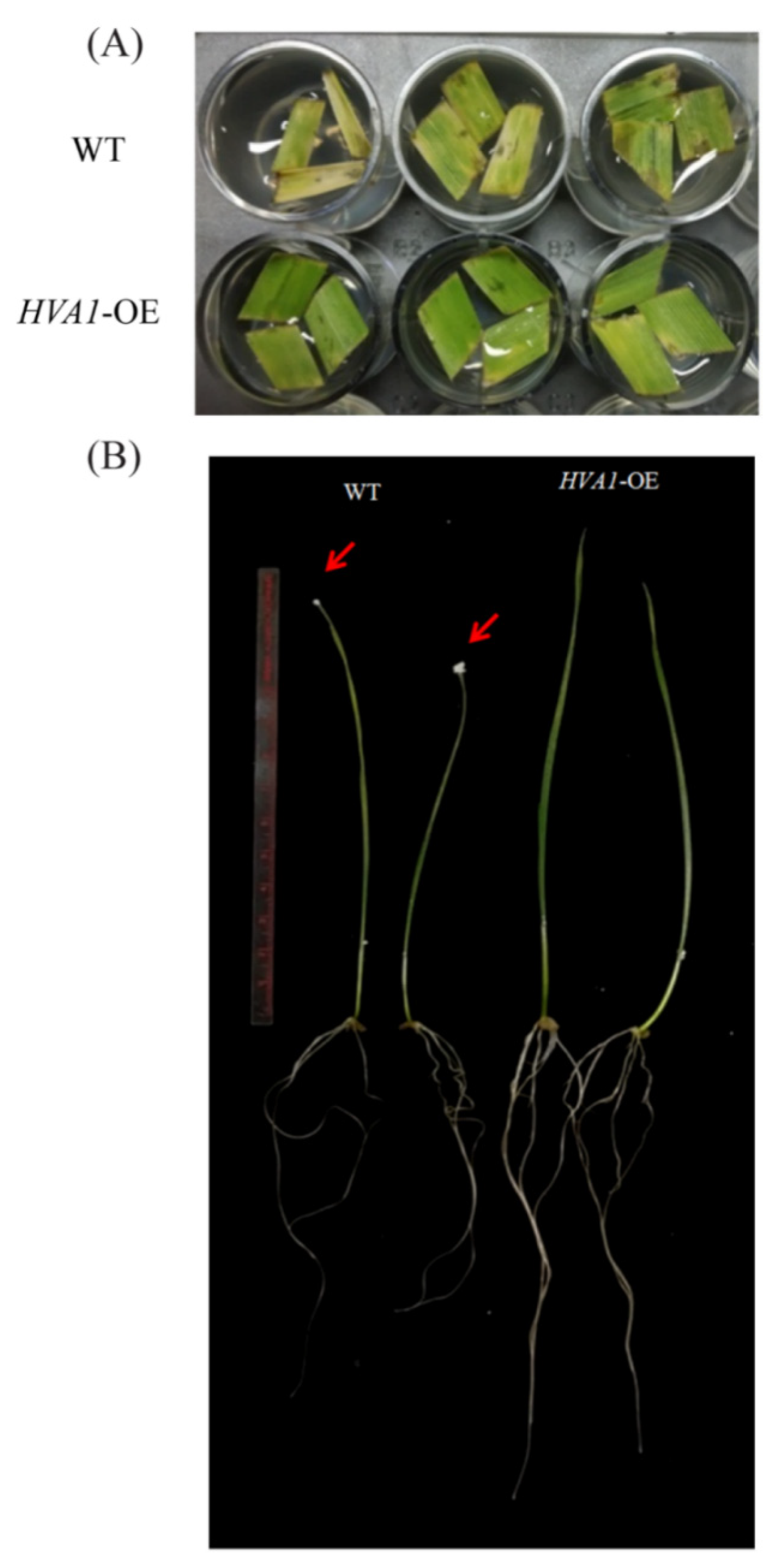
3.2. Phenotypic Analysis of HVA1 DH Plants Showed Improved Drought Tolerance

In wheat, HVA1 DH plants at T4 generation have been reported to show tolerance against stimulated water stress [30]. To further assess the drought tolerance of these plants, a leaf senescence assay was performed at T7 and T8 generation. As observed in Figure 3A, faster senescence was observed in WT as compared to the HVA1 overexpression lines. Apart from this, the ten-day-old seedlings were given drought stress with 200 mM mannitol. In the case of WT, the leaf showed senescence, root growth was inhibited, and it had salt deposited on the leaf tips. On the other hand, the overexpression lines performed better as evident by its greener leaf, robust root growth, and almost no salt deposition on the leaf tips (Figure 3B). Thus, the wheat DH plants showed significant drought tolerance.

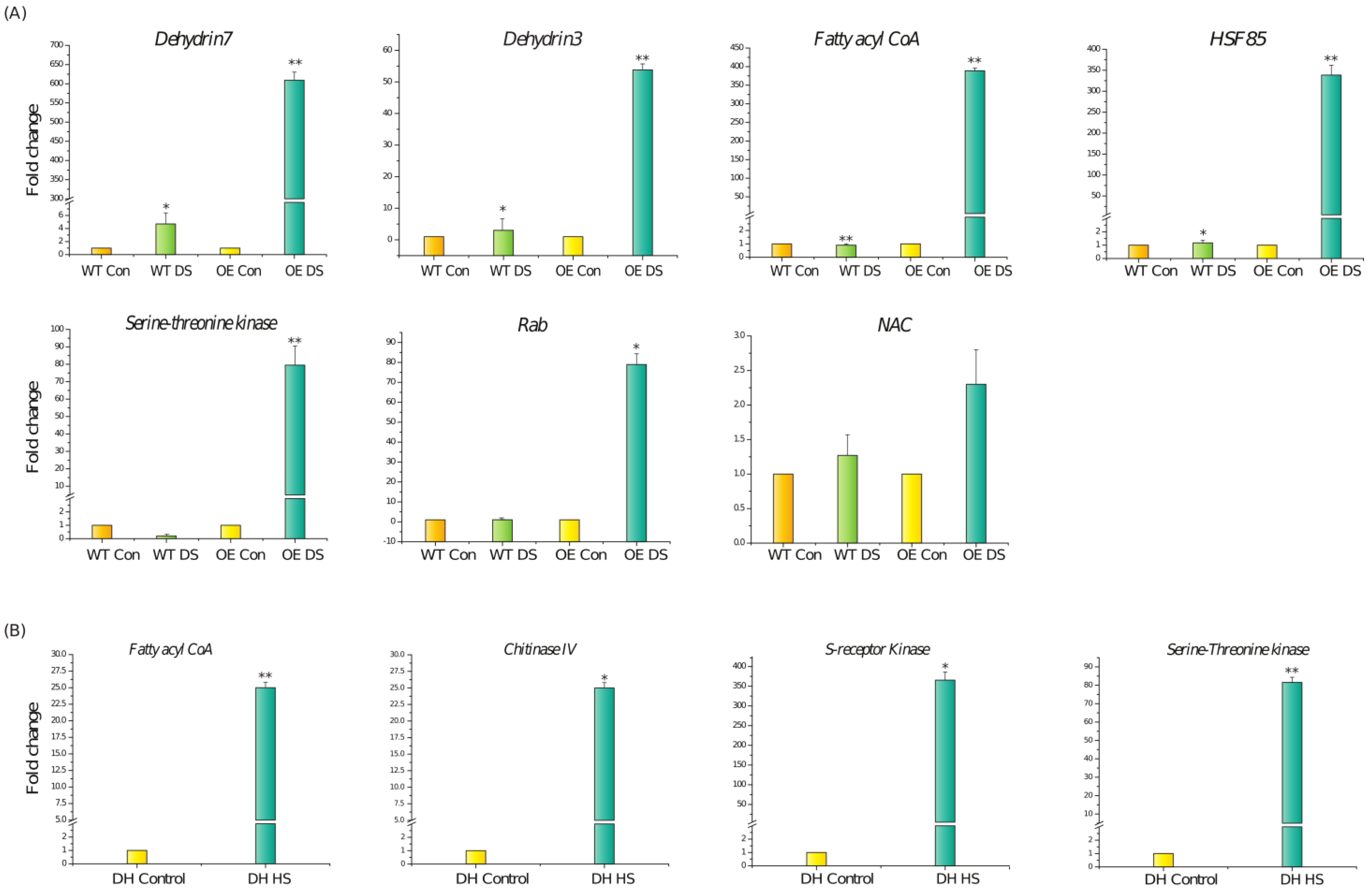
3.3. Transcriptome Analysis of DH Plants after Drought Stress

To understand the drought tolerance mechanism in the HVA1 overexpression lines, transcriptome analysis was performed both at the vegetative stage and at the reproductive stages in the drought-stressed plants. Comparison between non-stressed and stressed DH plants at the vegetative stage showed a total of 426 Differentially Expressed Genes (DEGs). The upregulated genes encoded for various types of proteins such as dehydrins (TraesCS6D02G332900; TraesCS6B02G383800), Rab (TraesCS6B02G383500), AP2 domain containing CBF protein (known as DREB) (TraesCS7A02G057800), Glycosyltransferase (TraesCS2B02G132700), O-methyltransferase (TraesCS5D02G488300), pathogenesis-related protein (TraesCS2D02G476400), Zinc finger protein 1 (TraesCS4D02G141500), NAC domain-containing protein (TraesCS4B02G212000), heat-responsive transcription factor 85 (TraesCS4D02G358900), and LRR receptor-like serine/threonine-protein kinase (TaresCS6B02G019200) (Figure 4). In contrast, the downregulated genes list included genes such as Auxin-responsive protein SAUR36 (TraesCS7D02G273500), various genes coding for histones such as Histone H2B (TraesCS6D02G040200), Histone H4 (TraesCS1B02G380200) and Histone H2A (TraesCS6B02G058400), and enzymes such as Phosphatase (TraesCS4A02G400600), Arginine decarboxylase (TraesCS1D02G012300), Esterase/lipase (TraesCS6D02G372000), Pectin acetylesterase (TraesCS3D02G539100), MYB75 (TraesCS1D02G084400), and Non-specific lipid transfer protein (TraesCS3D02G338300) (Figure S1).
At the reproductive stage, the upregulated genes included Chintinase IV (TraesCS2B02G369100), Fatty acyl-CoA reductase (TraesCSU02G10500), Serine-threonine protein kinase (TraesCS1A02G362900), Glutathione-S-transferase (TraesCS4B02G059300), and Endo-beta-1,3-glucanase (TraesCS3B02G528500) (Figure 5). The downregulated genes included Flavin-containing monooxygenase (TraesCS3A02G149500) and Endo-1,4- beta-Xylanase C (TraesCS2A02G584800) (Figure S2).
Amongst the upregulated genes, some of them were validated by quantitative real-time PCR. At the vegetative stage, a significant upregulation of the Dehydrin 3, Dehydrin 7, and Rab genes was observed under drought stress in DH plants. However, their expression did not increase much in the WT plants under drought stress. Transcript levels of HSF85, Fattyacyl CoA, NAC, and Serine-threonine protein kinase were also found to be higher in the case of transgenic as compared to WT under drought stress conditions (Figure 6A). At the reproductive stage, the expression of genes such as Chitinase IV, Fattyacyl CoA, S-receptor kinase, and Ser-threonine kinase were found to be significantly upregulated in the DH plants in comparison to the DH control plants, thereby validating the results of RNA-seq data (Figure 6B).

3.4. HVA1 Overexpression Promoted Thermotolerance in Wheat

A recent genome-wide study by [48] identified TaLEA3 members to be upregulated by heat stress and provide thermotolerance when overexpressed in E. coli and yeast. Taking hint from this, we checked the thermotolerance of the DH plants. At the vegetative stage, when exposed to 42 °C, the DH plants recovered better in comparison to the WT plants (Figure 7A). To assess the recovery of plants, the level of ROS (superoxide anions and hydrogen peroxide) was checked in both WT and DH plants by NBT and DAB staining. Lower ROS levels were observed in the DH leaves as compared to the WT (Figure 7B,C). Moreover, the level of hydrogen peroxide was also quantified in the transgenic and WT plants (Figure 7D). The DH plants showed a lower hydrogen peroxide accumulation after heat stress as compared to the WT.
At the reproductive stage when the plants were subjected to heat stress conditions, a better performance of DH plants was also observed, as depicted in Figure 8. The leaves of the WT showed more oxidative damage as compared to the DH plants (Figure 8A(b)). Moreover, the transgenic plants had larger spikes as compared to the heat-stressed WT plants (Figure 8A(c)). Transgenic DH plants were found to have a larger seed size and more seed weight in comparison to WT (Figure 8A(d),B).

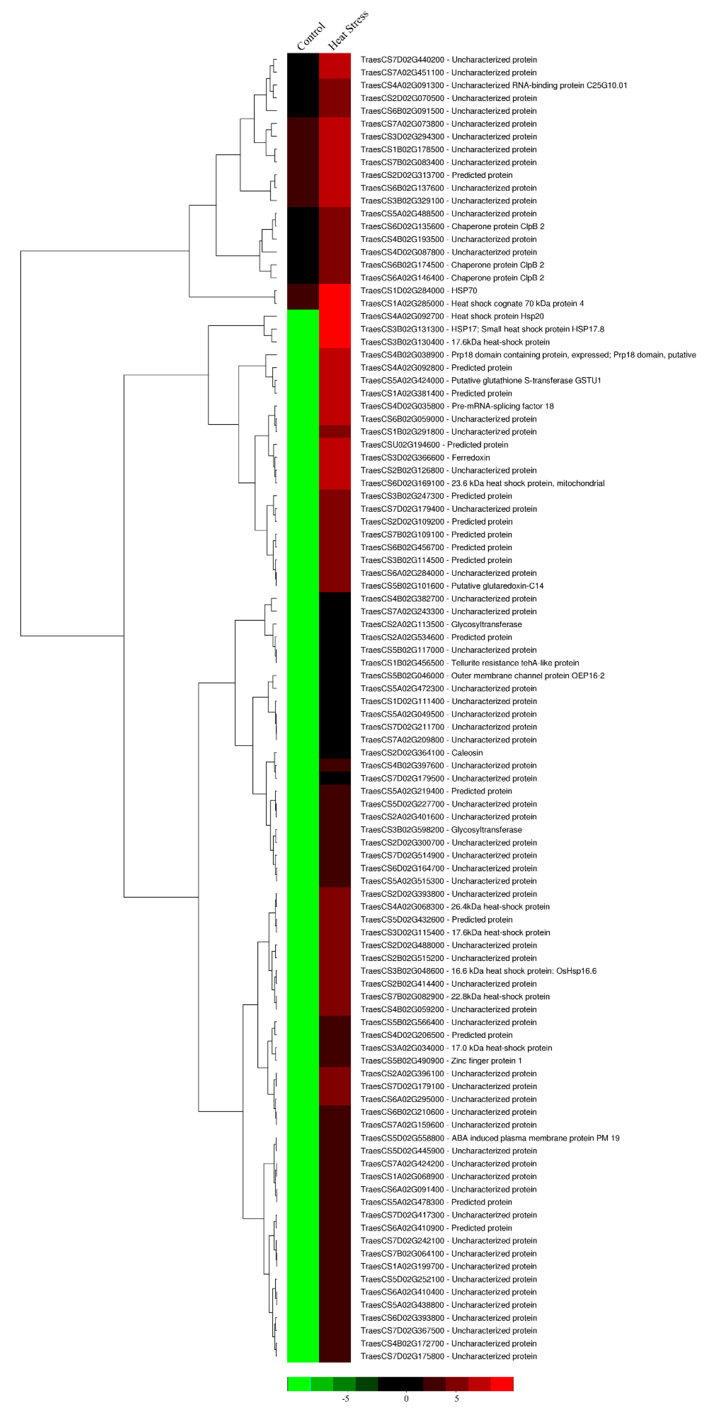
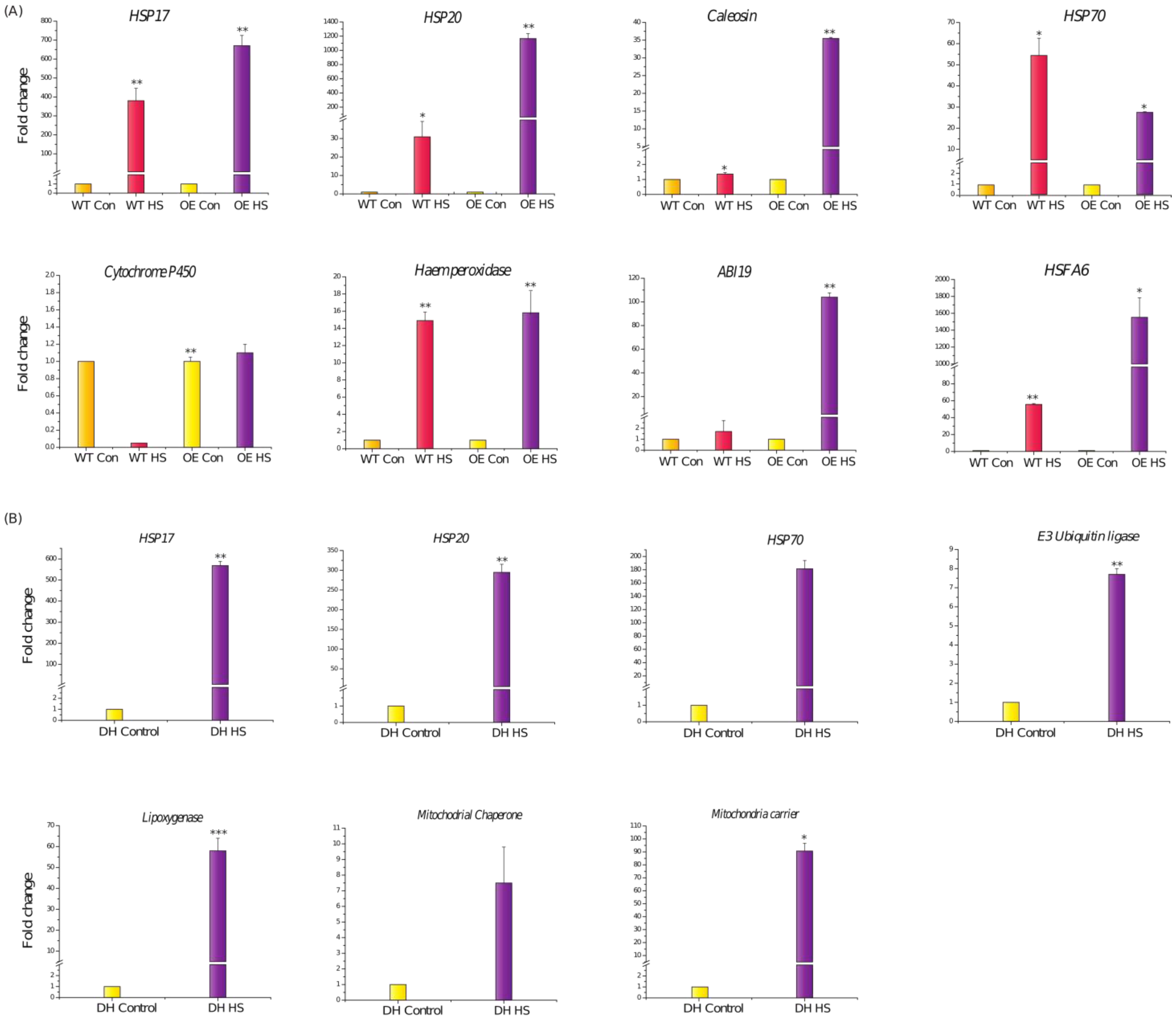
3.5. Transcriptome Analysis of DH Plants after Heat Stress

To investigate the molecular mechanisms contributing toward the heat stress tolerance, RNA-seq analysis of the transgenic DH plants after heat stress treatment was undertaken and was compared with the transgenic DH control plants. A total of 51 genes were found to be differentially expressed under control and heat stress conditions at the vegetative stage. The upregulated genes included various HSPs such as HSP70 (TraesCS1D02G284000), HSP17 (TraesCS3B02G131300), HSP20 (TraesCS4A02G092700), ClpB 2 (TraesCS6A02G146400), Glutathione -S-transferase (TraesCS5A02G424000), Ferrodoxin (TraesCS3D02G366600), ABA-induced plasma membrane protein PM19 (TraesCS5D02G558800), Caleosin (TraesCS2D02G364100), Cytochrome P450 (TraesCS2A02G534600), and Haem peroxidase (TraesCS2D02G070500) (Figure 9). The downregulated genes were found to be uncharacterized as they had no special domain to identify their function (Figure S3A).
At the reproductive stage, the upregulated genes included Lipoxygenase (TraesCS1B02G226400), Chaperonin CPN60-2 (TraesCS4A02G409100), Mitochondrial carrier protein (TraesCS7A02G209800), E3 ubiquitin ligase RNF5 (TraesCS4D02G021200), and Peptidylprolyl isomerase (TraesCS2D02G276000) (Figure 10). The downregulated genes mainly had uncharacterized proteins (Figure S3B).
Some of the genes identified in the transcriptome data were further confirmed by qRT-PCR. At the vegetative stage, a significant upregulation of the TaHSP17, TaHSP20, and TaHsfA6 genes under heat stress was observed in the DH transgenic plants in comparison to their expression in WT plants (Figure 11A). Moreover, higher expression levels of Caleosin, Haem peroxidase, and ABA-induced plasma membrane protein PM 19 (ABI19) were also observed in the heat-stressed DH in comparison to their almost negligible expression in the heat-stressed WT plants (Figure 11A). Even at reproductive stage, the expression profiles of various HSPs such as TaHSP17, TaHSP20, and TaHSP70 were found to upregulated in the spike tissue of DH after the heat stress treatment as compared to the control (Figure 11B). Apart from HSPs, higher transcript levels of Mitochondrial carrier, Lipoxygenase, E3-Ubiquitin ligase RNF5, and Mitochondrial chaperonin CPN60-2 were also found (Figure 11B).

3.6. Enhanced Sensitivity of HVA1-Overexpression Plants to ABA

ABA is one of the key hormones known to accumulate during stress conditions, particularly drought stress [49]. As the DH plants showed the stress-tolerant phenotype, they were screened on ABA containing medium. The seeds of both WT and overexpression lines were germinated on 5 µM and 7 µM of ABA. As observed in Figure 12, the DH seeds were found to be ABA-sensitive on both concentrations of ABA as compared to the WT seeds that showed germination and growth after 4 and 7 days respectively. This suggests that HVA1 positively modulates the drought and heat stress response via the ABA-mediated signaling pathway.

4. Discussion

LEA comprises one of the largest protein families that is widely diverse across the plant kingdom [50]. HVA1 is one of the first characterized and the most well studied LEA3 proteins from Hordeum vulgare [51]. Previous work by Chauhan and Khurana (2011) overexpressed HVA1 in wheat by the use of DH technology, and the transgenic plants were found to be drought-tolerant [30]. However, the molecular mechanisms involved in providing tolerance to the plants remained unknown. DH technology provides the fastest and most efficient way of generating complete homozygous lines as compared to conventional inbred lines, which are known have residual heterozygosity. Complete homozygosity is not only important for breeding and studying gene functions, but it also offers a higher phenotype to genotype correlation [52]. Therefore, in this study, the DH plants that can be considered as true isogenic lines were analyzed and found to be both drought- and heat-tolerant. Moreover, as the genetic variation for heat and drought stress tolerance in elite wheat germplasm is limited, it may not provide sufficient resistance to develop cultivars through traditional breeding. Thus, genetic engineering provides a way to enhance both heat and drought stress resistance in wheat. Although there exist many differences between the wheat and barley genomes such as ploidy levels and other genetic differences, the heterologous overexpression of the HVA1 gene provided stress tolerance in wheat. Therefore, it can be speculated that the overexpression of TaLEA3 in wheat itself would also provide stress tolerance in a more effective way (due to the same genetic background). Thus, this study highlights LEA3 as a potential gene for incorporating the simultaneous heat and drought tolerance trait in wheat.
Drought stress is known to accelerate leaf senescence, leading to a loss in photosynthesis and reduction in yield [53,54]. The drought stress tolerance of the DH plants was analyzed by a leaf disc senescence assay, which clearly showed that the leaves of the transgenic plants showed delayed senescence as compared to the WT plants (Figure 3A). Moreover, at the seedling stage, the DH plants showed the tolerant phenotype as compared to the WT when subjected to simulated drought stress (Figure 3B). These results suggested that the overexpression of HVA1 contributes toward drought tolerance. This was found to be consistent with earlier reports wherein the overexpression of LEA3 provided drought tolerance to various plants (such as creeping bentgrass, mulberry, Arabidopsis thaliana, and Brassica napus) and freezing tolerance in the case of yeast [17,25,27,55]. Thus, it could be concluded that LEA3 has a conserved functional role in providing tolerance toward drought stress. To gain insight into its mechanism of action, transcriptome data were analyzed after drought stress treatment. Interestingly, the upregulation of transcription factors such as NAC and DREB was observed in the DH plants, whereas their expression was found to be low in the WT plants after the drought stress treatment (Figure 6A). This could be supported by the evidence in Oryza sativa and Arabidopsis, wherein, the NAC transcription factor by activating DREB2A contributes toward drought tolerance of the plants [56,57]. Interestingly, the levels of both NAC and AP2 CBF/DREB were found to be upregulated more in the transgenic DH plants as compared to the WT plants after drought stress. Therefore, it could be speculated that HVA1 by interacting with other transcription factors either regulates the expression of both NAC and DREB independently, or it only regulates NAC, which then activates DREB to contribute toward the drought tolerance. Apart from transcription factors, expressions of Rab and Dehydrin genes were also found to be high in the DH plants. Rab and Dehydrin proteins have been reported to be involved in the drought signaling pathway and in protective functions against ROS generated during drought stress, respectively [58,59]. Therefore, higher expressions of Rab and Dehydrin genes might also provide drought tolerance to the DH plants. However, how HVA1 is able to regulate these genes under drought stress conditions has become an area of future research.
Apart from drought stress, there are limited reports that have highlighted the role of LEA in the heat stress response. Therefore, the heat tolerance of DH plants was also analyzed at both the vegetative and reproductive stage. In comparison to WT plants, the DH plants recovered better after the heat stress (Figure 7A). ROS accumulation is known to occur after heat stress, which often leads to cellular damage [60,61]. The DH plants had a lower oxidative load as compared to the WT (Figure 7D). This observation could further be corroborated with the upregulation of Haem peroxidase in the transgenic plants as compared to the WT (Figure 11A). This suggest that the higher transcript levels of this antioxidant enzyme leads to accumulation of the enzyme which help the HVA1 overexpression lines to survive better under heat stress by maintaining lower ROS levels. Consistent with this, Ascorbate peroxidase (APX), which belongs to class I Haem-peroxidase, has been shown to be heat-responsive in different plants, and its absence in Arabidopsis leads to sensitivity to heat stress [62,63,64]. Apart from this, group 3 LEA proteins are known to possess the 11-mer amino acid motif with the consensus sequence of TAQAAKEKAGE. Interestingly, this motif occurs nine times in the HVA1 protein, leading to alpha helical dimer formation, which is suitable to accommodate both positively and negatively charged ions [26].The ZmLEA3 protein has been found to bind metal ions, and the authors speculated that the binding of ions by ZmLEA3 contributes toward reducing oxidative damage and ion toxicity during abiotic stress [65]. Therefore, it is probable that HVA1 might reduce oxidative load inside the cell by binding to the metal ions through the alpha helical dimer.
Transgenic DH plants were found to be tolerant to heat stress even at the reproductive stage, as they had better spikes and higher seed weight in comparison to the WT (Figure 8A(c),B). This result was further supported by the transcriptome data wherein the upregulation of various HSPs was observed in the transgenic plants after heat stress (Figure 10). Moreover, the levels of expression of HSP17 and HSP20 were found to be higher in the DH plants (Figure 10 and Figure 11B). The role of small HSPs such as TaHSP26 has been earlier documented to provide heat stress tolerance to the overexpression plants [66,67]. Therefore, significant upregulation of the small HSPs plays a major role in providing thermotolerance to the HVA1-overexpression plants. Apart from stress, it is well known that HSPs accumulate during seed maturation [68]. As seed formation was observed to be better in the case of DH plants after heat stress, it is possible that the higher expression of these HSPs in the seeds might have caused enhanced seed development under heat stress conditions. Further, higher expression levels of mitochondrial carriers and mitochondrial chaperone proteins in HVA1-overexpression plants highlight their function in the heat stress response to protect the mitochondrial components from degradation and proteolysis [69,70].
Interestingly, ABI19, one of the ABA-responsive genes, was found to be upregulated in the DH plants after heat stress (Figure 9). Its expression levels were found to be higher in the transgenic plants as compared to the WT (Figure 11A). A report by Huang and co-workers highlighted the role of HsfA6b connecting ABA signaling and the ABA-mediated heat stress response [71]. Therefore, the expression of TaHsfA6 was also analyzed in the transgenic and WT plants under heat stress conditions. Interestingly, more transcript levels of TaHsfA6 were observed in the case of DH plants (Figure 11A). Therefore, it could be speculated that TaHsfA6 leads to the ABA-mediated heat stress response in the transgenic plants. This could be further supported by the ABA-sensitive phenotype of the transgenic plants (Figure 12). Thus, these results suggest that HVA1 via the ABA-mediated pathway plays a role in heat stress.
In conclusion, HVA1, which belongs to the LEA3 group, was found to play a major role in response to both drought and heat stress. However, as HVA1 lacks DNA binding domain, it is suspected that it might interact with some transcription factor to activate master regulators such as NAC, DREB, and HsfA6 to bring about both drought and heat stress tolerance. A hypothetical model summarizing the probable role of HVA1 under both drought and heat stress conditions is presented in Figure 13. Upon drought stress, HVA1 leads to the activation of genes such as NAC, DREB, and Rab. NAC and DREB, in turn, lead to the transcription of other drought-responsive genes such as dehydrins, which help in providing drought tolerance to plants. Similarly, under heat stress conditions, HVA1 via the ABA-mediated pathway activates the expression of TaHsfA6, which leads to the accumulation of small Hsps, thereby attributing heat stress tolerance. Apart from this, HVA1 also activates ABI19 and Haem peroxidase, which also contributes toward the abiotic stress tolerance mechanism. However, the identification of HVA1-interacting partners (transcription factors) needs to be investigated further. Moreover, the mechanism of regulation of HVA1 itself needs to be explored to understand how it works under both stresses. Overall, HVA1 appears to play a role in providing multiple stress tolerance to the plant both at the vegetative and reproductive stages, by modulating the transcriptome at multiple levels.

Supplementary Materials

The following supporting information can be downloaded at https://www.mdpi.com/article/10.3390/cells11050912/s1, Figure S1: Heatmap displaying the differentially expressed genes down-regulated in HVA1 overexpression wheat DH transgenics under drought stress conditions (200 mM mannitol) at the vegetative stage based on the FPKM data; Figure S2: Heatmap displaying the differentially expressed genes down-regulated in HVA1 overexpression wheat DH transgenics under drought stress conditions (200 mM mannitol) at the reproductive stage based on the FPKM data; Figure S3: (A) Heatmap displaying the differentially expressed genes down-regulated in HVA1 overexpression wheat DH transgenics under heat stress conditions at the vegetative stage based on the FPKM data. (B) Heatmap displaying the differentially expressed genes down-regulated in HVA1 overexpression wheat DH transgenics under heat stress conditions at the reproductive stage based on the FPKM data; Table S1: List of primers used for the qPCR analysis.

Author Contributions

P.K., H.S. and A.S. conceptualized the idea of the research. H.S. and A.S. performed the experiments. P.K. provided all the facilities for the experiments. H.S. and A.S. wrote the manuscript. All authors have read and agreed to the published version of the manuscript.

Funding

H.S. and A.S. acknowledge the University Grant Commission (UGC) for research fellowships. This work has been supported by a grant from J.C. Bose fellowship award, Science and Engineering Research Board, Government of India, for research support to P.K.

Institutional Review Board Statement

Not applicable.

Informed Consent Statement

Not applicable.

Data Availability Statement

The RNA-seq data reported in this article have been submitted in the NCBI database (Bioproject PRJNA811517).

Conflicts of Interest

The authors declare that the research was conducted in absence of any commercial or financial relationship that could be construed as a potential conflict of interest.

Abbreviations

ABAabscisic acid
DHdoubled haploid
HVA1Hordeum vulgare aleurone 1
LEAlate embryogenesis abundant
ROSreactive oxygen species
WTwildtype

References

  1. Chen, Y.; Li, C.; Zhang, B.; Yi, J.; Yang, Y.; Kong, C.; Lei, C.; Gong, M. The Role of the Late Embryogenesis-Abundant (LEA) Protein Family in Development and the Abiotic Stress Response: A Comprehensive Expression Analysis of Potato (Solanum Tuberosum). Genes 2019, 10, 148. [Google Scholar] [CrossRef] [PubMed] [Green Version]
  2. Battaglia, M.; Covarrubias, A.A. Late Embryogenesis Abundant (LEA) proteins in legumes. Front. Plant Sci. 2013, 4, 190. [Google Scholar] [CrossRef] [PubMed] [Green Version]
  3. Dure, L.; Greenway, S.C.; Galau, G.A. Developmental biochemistry of cottonseed embryogenesis and germination: Changing messenger ribonucleic acid populations as shown by in vitro and in vivo protein synthesis. Biochemistry 1981, 20, 4162–4168. [Google Scholar] [CrossRef]
  4. Hatanaka, R.; Gusev, O.; Cornette, R.; Shimura, S.; Kikuta, S.; Okada, J.; Okuda, T.; Kikawada, T. Diversity of the expression profiles of late embryogenesis abundant (LEA) protein encoding genes in the anhydrobiotic midge Polypedilum vanderplanki. Planta 2015, 242, 451–459. [Google Scholar] [CrossRef]
  5. Zheng, J.; Su, H.; Lin, R.; Zhang, H.; Xia, K.; Jian, S.; Zhang, M. Isolation and characterization of an atypical LEA gene (IpLEA) from Ipomoea pes-caprae conferring salt/drought and oxidative stress tolerance. Sci. Rep. 2019, 9, 14838. [Google Scholar] [CrossRef]
  6. Xu, M.; Tong, Q.; Wang, Y.; Wang, Z.; Xu, G.; Elias, G.K.; Li, S.; Liang, Z. Transcriptomic Analysis of the Grapevine LEA Gene Family in Response to Osmotic and Cold Stress Reveals a Key Role for VamDHN3. Plant Cell Physiol. 2020, 61, 775–786. [Google Scholar] [CrossRef] [PubMed]
  7. Artur, M.A.S.; Zhao, T.; Ligterink, W.; Schranz, E.; Hilhorst, H.W.M. Dissecting the Genomic Diversification of Late Embryogenesis Abundant (LEA) Protein Gene Families in Plants. Genome Biol. Evol. 2019, 11, 459–471. [Google Scholar] [CrossRef] [PubMed]
  8. Battaglia, M.; Olvera-Carrillo, Y.; Garciarrubio, A.; Campos, F.; Covarrubias, A.A. The enigmatic LEA proteins and other hydrophilins. Plant Physiol. 2008, 148, 6–24. [Google Scholar] [CrossRef] [Green Version]
  9. Goyal, K.; Walton, L.J.; Tunnacliffe, A. LEA proteins prevent protein aggregation due to water stress. Biochem. J. 2005, 388, 151–157. [Google Scholar] [CrossRef] [Green Version]
  10. Hong-Bo, S.; Zong-Suo, L.; Ming-An, S. LEA proteins in higher plants: Structure, function, gene expression and regulation. Colloids Surf. B. Biointerfaces 2005, 45, 131–135. [Google Scholar] [CrossRef]
  11. Candat, A.; Paszkiewicz, G.; Neveu, M.; Gautier, R.; Logan, D.C.; Avelange-Macherel, M.-H.; Macherel, D. The ubiquitous distribution of late embryogenesis abundant proteins across cell compartments in Arabidopsis offers tailored protection against abiotic stress. Plant Cell 2014, 26, 3148–3166. [Google Scholar] [CrossRef] [PubMed] [Green Version]
  12. Yu, J.; Lai, Y.; Wu, X.; Wu, G.; Guo, C. Overexpression of OsEm1 encoding a group I LEA protein confers enhanced drought tolerance in rice. Biochem. Biophys. Res. Commun. 2016, 478, 703–709. [Google Scholar] [CrossRef] [PubMed]
  13. Jia, F.; Qi, S.; Li, H.; Liu, P.; Li, P.; Wu, C.; Zheng, C.; Huang, J. Overexpression of Late Embryogenesis Abundant 14 enhances Arabidopsis salt stress tolerance. Biochem. Biophys. Res. Commun. 2014, 454, 505–511. [Google Scholar] [CrossRef] [PubMed]
  14. Wang, M.; Li, P.; Li, C.; Pan, Y.; Jiang, X.; Zhu, D.; Zhao, Q.; Yu, J. SiLEA14, a novel atypical LEA protein, confers abiotic stress resistance in foxtail millet. BMC Plant Biol. 2014, 14, 290. [Google Scholar] [CrossRef]
  15. Lim, C.W.; Lim, S.; Baek, W.; Lee, S.C. The pepper late embryogenesis abundant protein CaLEA1 acts in regulating abscisic acid signaling, drought and salt stress response. Physiol. Plant. 2015, 154, 526–542. [Google Scholar] [CrossRef]
  16. Lim, J.; Lim, C.W.; Lee, S.C. The Pepper Late Embryogenesis Abundant Protein, CaDIL1, Positively Regulates Drought Tolerance and ABA Signaling. Front. Plant Sci. 2018, 9, 1301. [Google Scholar] [CrossRef]
  17. Liang, Y.; Kang, K.; Gan, L.; Ning, S.; Xiong, J.; Song, S.; Xi, L.; Lai, S.; Yin, Y.; Gu, J.; et al. Drought-responsive genes, late embryogenesis abundant group3 (LEA3) and vicinal oxygen chelate, function in lipid accumulation in Brassica napus and Arabidopsis mainly via enhancing photosynthetic efficiency and reducing ROS. Plant Biotechnol. J. 2019, 17, 2123–2142. [Google Scholar] [CrossRef] [Green Version]
  18. Sivamani, E.; Bahieldin1, A.; Wraith, J.M.; Al-Niemi, T.; Dyer, W.E.; Ho, T.H.; Qu, R. Improved biomass productivity and water use efficiency under water deficit conditions in transgenic wheat constitutively expressing the barley HVA1 gene. Plant Sci. 2000, 155, 1–9. [Google Scholar] [CrossRef]
  19. Bahieldin, A.; Mahfouz, H.T.; Eissa, H.F.; Saleh, O.M.; Ramadan, A.M.; Ahmed, I.A.; Dyer, W.E.; El-Itriby, H.A.; Madkour, M.A. Field evaluation of transgenic wheat plants stably expressing the HVA1 gene for drought tolerance. Physiol. Plant. 2005, 123, 421–427. [Google Scholar] [CrossRef]
  20. Xu, D.; Duan, X.; Wang, B.; Hong, B.; Ho, T.; Wu, R. Expression of a Late Embryogenesis Abundant Protein Gene, HVA1, from Barley Confers Tolerance to Water Deficit and Salt Stress in Transgenic Rice. Plant Physiol. 1996, 110, 249–257. [Google Scholar] [CrossRef] [Green Version]
  21. Chandra Babu, R.; Zhang, J.; Blum, A.; David Ho, T.-H.; Wu, R.; Nguyen, H. HVA1, a LEA gene from barley confers dehydration tolerance in transgenic rice (Oryza sativa L.) via cell membrane protection. Plant Sci. 2004, 166, 855–862. [Google Scholar] [CrossRef]
  22. Rohila, J.S.; Jain, R.K.; Wu, R. Genetic improvement of Basmati rice for salt and drought tolerance by regulated expression of a barley HVA1 cDNA. Plant Sci. 2002, 163, 525–532. [Google Scholar] [CrossRef]
  23. Chen, Y.-S.; Lo, S.-F.; Sun, P.-K.; Lu, C.-A.; Ho, T.-H.D.; Yu, S.-M. A late embryogenesis abundant protein HVA1 regulated by an inducible promoter enhances root growth and abiotic stress tolerance in rice without yield penalty. Plant Biotechnol. J. 2015, 13, 105–116. [Google Scholar] [CrossRef] [PubMed]
  24. Maqbool, B.; Zhong, H.; El-Maghraby, Y.; Ahmad, A.; Chai, B.; Wang, W.; Sabzikar, R.; Sticklen, B. Competence of oat ( Avena sativa L.) shoot apical meristems for integrative transformation, inherited expression, and osmotic tolerance of transgenic lines containing HVA1. Theor. Appl. Genet. 2002, 105, 201–208. [Google Scholar] [CrossRef] [PubMed]
  25. Fu, D.; Huang, B.; Xiao, Y.; Muthukrishnan, S.; Liang, G.H. Overexpression of barley HVA1 gene in creeping bentgrass for improving drought tolerance. Plant Cell Rep. 2007, 26, 467–477. [Google Scholar] [CrossRef] [PubMed]
  26. Lal, S.; Gulyani, V.; Khurana, P. Overexpression of HVA1 gene from barley generates tolerance to salinity and water stress in transgenic mulberry (Morus indica). Transgenic Res. 2008, 17, 651–663. [Google Scholar] [CrossRef]
  27. Checker, V.G.; Chhibbar, A.K.; Khurana, P. Stress-inducible expression of barley HVA1 gene in transgenic mulberry displays enhanced tolerance against drought, salinity and cold stress. Transgenic Res. 2012, 21, 939–957. [Google Scholar] [CrossRef]
  28. Kwapata, K.; Nguyen, T.; Sticklen, M. Genetic Transformation of Common Bean ( Phaseolus vulgaris L.) with the Gus Color Marker, the Bar Herbicide Resistance, and the Barley ( Hordeum vulgare) HVA1 Drought Tolerance Genes. Int. J. Agron. 2012, 2012, 198960. [Google Scholar] [CrossRef]
  29. Yan, G.; Liu, H.; Wang, H.; Lu, Z.; Wang, Y.; Mullan, D.; Hamblin, J.; Liu, C. Accelerated Generation of Selfed Pure Line Plants for Gene Identification and Crop Breeding. Front. Plant Sci. 2017, 8, 1786. [Google Scholar] [CrossRef] [Green Version]
  30. Chauhan, H.; Khurana, P. Use of doubled haploid technology for development of stable drought tolerant bread wheat (Triticum aestivum L.) transgenics. Plant Biotechnol. J. 2011, 9, 408–417. [Google Scholar] [CrossRef] [Green Version]
  31. Meena, S.; Deb, S.; Samtani, H.; Khurana, P. Dissecting the Molecular Function of Triticum aestivum STI Family Members Under Heat Stress. Front. Genet. 2020, 11, 873. [Google Scholar] [CrossRef] [PubMed]
  32. Peng, Z.; Wang, M.; Li, F.; Lv, H.; Li, C.; Xia, G. A Proteomic Study of the Response to Salinity and Drought Stress in an Introgression Strain of Bread Wheat. Mol. Cell. Proteom. 2009, 8, 2676–2686. [Google Scholar] [CrossRef] [PubMed] [Green Version]
  33. Hezema, Y.S.; Shukla, M.R.; Goel, A.; Ayyanath, M.M.; Sherif, S.M.; Saxena, P.K. Rootstocks Overexpressing StNPR1 and StDREB1 Improve Osmotic Stress Tolerance of Wild-Type Scion in Transgrafted Tobacco Plants. Int. J. Mol. Sci. 2021, 22, 8398. [Google Scholar] [CrossRef] [PubMed]
  34. Sharma, N.; Chaudhary, C.; Khurana, P. Wheat Myo-inositol phosphate synthase influences plant growth and stress responses via ethylene mediated signaling. Sci. Rep. 2020, 10, 10766. [Google Scholar] [CrossRef]
  35. Dudziak, K.; Zapalska, M.; Börner, A.; Szczerba, H.; Kowalczyk, K.; Nowak, M. Analysis of wheat gene expression related to the oxidative stress response and signal transduction under short-term osmotic stress. Sci. Rep. 2019, 9, 2743. [Google Scholar] [CrossRef] [Green Version]
  36. Lan, P.; Li, W.; Wang, H.; Ma, W. Characterization, sub-cellular localization and expression profiling of the isoprenylcysteine methylesterase gene family in Arabidopsis thaliana. BMC Plant Biol. 2010, 10, 212. [Google Scholar] [CrossRef] [Green Version]
  37. Nguyen, C.C.; Nakaminami, K.; Matsui, A.; Watanabe, S.; Kanno, Y.; Seo, M.; Seki, M. Overexpression of oligouridylate binding protein 1b results in ABA hypersensitivity. Plant Signal. Behav. 2017, 12, e1282591. [Google Scholar] [CrossRef] [Green Version]
  38. Huang, S.; Hu, L.; Zhang, S.; Zhang, M.; Jiang, W.; Wu, T.; Du, X. Rice OsWRKY50 Mediates ABA-Dependent Seed Germination and Seedling Growth, and ABA-Independent Salt Stress Tolerance. Int. J. Mol. Sci. 2021, 22, 8625. [Google Scholar] [CrossRef]
  39. Cui, L.H.; Min, H.J.; Byun, M.Y.; Oh, H.G.; Kim, W.T. OsDIRP1, a Putative RING E3 Ligase, Plays an Opposite Role in Drought and Cold Stress Responses as a Negative and Positive Factor, Respectively, in Rice (Oryza sativa L.). Front. Plant Sci. 2018, 9, 1797. [Google Scholar] [CrossRef] [Green Version]
  40. Ghosh, S.; Kamble, N.U.; Verma, P.; Salvi, P.; Petla, B.P.; Roy, S.; Rao, V.; Hazra, A.; Varshney, V.; Kaur, H.; et al. Arabidopsis protein l-ISOASPARTYL METHYLTRANSFERASE repairs isoaspartyl damage to antioxidant enzymes and increases heat and oxidative stress tolerance. J. Biol. Chem. 2020, 295, 783–799. [Google Scholar] [CrossRef]
  41. Alexieva, V.; Sergiev, I.; Mapelli, S.; Karanov, E. The effect of drought and ultraviolet radiation on growth and stress markers in pea and wheat. Plant. Cell Environ. 2001, 24, 1337–1344. [Google Scholar] [CrossRef]
  42. Haas, B.J.; Papanicolaou, A.; Yassour, M.; Grabherr, M.; Blood, P.D.; Bowden, J.; Couger, M.B.; Eccles, D.; Li, B.; Lieber, M.; et al. De novo transcript sequence reconstruction from RNA-seq using the Trinity platform for reference generation and analysis. Nat. Protoc. 2013, 8, 1494–1512. [Google Scholar] [CrossRef] [PubMed]
  43. Robinson, M.D.; McCarthy, D.J.; Smyth, G.K. edgeR: A Bioconductor package for differential expression analysis of digital gene expression data. Bioinformatics 2010, 26, 139–140. [Google Scholar] [CrossRef] [Green Version]
  44. Paolacci, A.R.; Tanzarella, O.A.; Porceddu, E.; Ciaffi, M. Identification and validation of reference genes for quantitative RT-PCR normalization in wheat. BMC Mol. Biol. 2009, 10, 11. [Google Scholar] [CrossRef] [PubMed] [Green Version]
  45. Chaudhary, C.; Sharma, N.; Khurana, P. Decoding the wheat awn transcriptome and overexpressing TaRca1β in rice for heat stress tolerance. Plant Mol. Biol. 2021, 105, 133–146. [Google Scholar] [CrossRef] [PubMed]
  46. Livak, K.J.; Schmittgen, T.D. Analysis of relative gene expression data using real-time quantitative PCR and the 2−ΔΔCT method. Methods 2001, 25, 402–408. [Google Scholar] [CrossRef] [PubMed]
  47. Singh, K.K.; Graether, S.P. Conserved sequence motifs in the abiotic stress response protein late embryogenesis abundant 3. PLoS ONE 2020, 15, e0237177. [Google Scholar] [CrossRef]
  48. Liu, H.; Xing, M.; Yang, W.; Mu, X.; Wang, X.; Lu, F.; Wang, Y.; Zhang, L. Genome-wide identification of and functional insights into the late embryogenesis abundant (LEA) gene family in bread wheat (Triticum aestivum). Sci. Rep. 2019, 9, 13375. [Google Scholar] [CrossRef] [Green Version]
  49. Ali, A.; Pardo, J.M.; Yun, D.-J. Desensitization of ABA-Signaling: The Swing From Activation to Degradation. Front. Plant Sci. 2020, 11, 379. [Google Scholar] [CrossRef] [Green Version]
  50. Gao, J.; Lan, T. Functional characterization of the late embryogenesis abundant (LEA) protein gene family from Pinus tabuliformis (Pinaceae) in Escherichia coli. Sci. Rep. 2016, 6, 19467. [Google Scholar] [CrossRef] [Green Version]
  51. Gürel, F.; Öztürk, Z.N.; Uçarlı, C.; Rosellini, D. Barley Genes as Tools to Confer Abiotic Stress Tolerance in Crops. Front. Plant Sci. 2016, 7, 1137. [Google Scholar] [CrossRef] [PubMed]
  52. Chaikam, V.; Molenaar, W.; Melchinger, A.E.; Boddupalli, P.M. Doubled haploid technology for line development in maize: Technical advances and prospects. Theor. Appl. Genet. 2019, 132, 3227–3243. [Google Scholar] [CrossRef] [PubMed] [Green Version]
  53. Rivero, R.M.; Kojima, M.; Gepstein, A.; Sakakibara, H.; Mittler, R.; Gepstein, S.; Blumwald, E. Delayed leaf senescence induces extreme drought tolerance in a flowering plant. Proc. Natl. Acad. Sci. USA 2007, 104, 19631–19636. [Google Scholar] [CrossRef] [Green Version]
  54. Agarwal, P.; Baranwal, V.K.; Khurana, P. Genome-wide Analysis of bZIP Transcription Factors in wheat and Functional Characterization of a TabZIP under Abiotic Stress. Sci. Rep. 2019, 9, 4608. [Google Scholar] [CrossRef] [PubMed] [Green Version]
  55. Yu, J.-N.; Zhang, J.-S.; Shan, L.; Chen, S.-Y. Two New Group 3 LEA Genes of Wheat and Their Functional Analysis in Yeast. J. Integr. Plant Biol. 2005, 47, 1372–1381. [Google Scholar] [CrossRef]
  56. Yuan, X.; Wang, H.; Cai, J.; Bi, Y.; Li, D.; Song, F. Rice NAC transcription factor ONAC066 functions as a positive regulator of drought and oxidative stress response. BMC Plant Biol. 2019, 19, 278. [Google Scholar] [CrossRef]
  57. Yang, X.; Kim, M.Y.; Ha, J.; Lee, S.-H. Overexpression of the Soybean NAC Gene GmNAC109 Increases Lateral Root Formation and Abiotic Stress Tolerance in Transgenic Arabidopsis Plants. Front. Plant Sci. 2019, 10, 1036. [Google Scholar] [CrossRef] [Green Version]
  58. Wang, X.; Cai, X.; Xu, C.; Wang, Q.; Dai, S. Drought-Responsive Mechanisms in Plant Leaves Revealed by Proteomics. Int. J. Mol. Sci. 2016, 17, 1706. [Google Scholar] [CrossRef] [Green Version]
  59. Liu, Y.; Song, Q.; Li, D.; Yang, X.; Li, D. Multifunctional Roles of Plant Dehydrins in Response to Environmental Stresses. Front. Plant Sci. 2017, 8, 1018. [Google Scholar] [CrossRef] [Green Version]
  60. Driedonks, N.; Xu, J.; Peters, J.L.; Park, S.; Rieu, I. Multi-Level Interactions Between Heat Shock Factors, Heat Shock Proteins, and the Redox System Regulate Acclimation to Heat. Front. Plant Sci. 2015, 6, 999. [Google Scholar] [CrossRef] [Green Version]
  61. Agarwal, P.; Khurana, P. Characterization of a novel zinc finger transcription factor (TaZnF) from wheat conferring heat stress tolerance in Arabidopsis. Cell Stress Chaperones 2018, 23, 253–267. [Google Scholar] [CrossRef] [PubMed]
  62. Pandey, S.; Fartyal, D.; Agarwal, A.; Shukla, T.; James, D.; Kaul, T.; Negi, Y.K.; Arora, S.; Reddy, M.K. Abiotic Stress Tolerance in Plants: Myriad Roles of Ascorbate Peroxidase. Front. Plant Sci. 2017, 8, 581. [Google Scholar] [CrossRef] [PubMed] [Green Version]
  63. Agarwal, P.; Khurana, P. Overexpression of TaMADS from wheat promotes flowering by upregulating expression of floral promoters and provides protection against thermal stress. Plant Gene 2019, 17, 100168. [Google Scholar] [CrossRef]
  64. Suzuki, N.; Miller, G.; Sejima, H.; Harper, J.; Mittler, R. Enhanced seed production under prolonged heat stress conditions in Arabidopsis thaliana plants deficient in cytosolic ascorbate peroxidase 2. J. Exp. Bot. 2013, 64, 253–263. [Google Scholar] [CrossRef] [PubMed] [Green Version]
  65. Liu, Y.; Wang, L.; Xing, X.; Sun, L.; Pan, J.; Kong, X.; Zhang, M.; Li, D. ZmLEA3, a Multifunctional Group 3 LEA Protein from Maize (Zea mays L.), is Involved in Biotic and Abiotic Stresses. Plant Cell Physiol. 2013, 54, 944–959. [Google Scholar] [CrossRef] [PubMed] [Green Version]
  66. Chauhan, H.; Khurana, N.; Nijhavan, A.; Khurana, J.P.; Khurana, P. The wheat chloroplastic small heat shock protein (sHSP26) is involved in seed maturation and germination and imparts tolerance to heat stress. Plant. Cell Environ. 2012, 35, 1912–1931. [Google Scholar] [CrossRef]
  67. Khurana, N.; Chauhan, H.; Khurana, P. Wheat Chloroplast Targeted sHSP26 Promoter Confers Heat and Abiotic Stress Inducible Expression in Transgenic Arabidopsis Plants. PLoS ONE 2013, 8, e54418. [Google Scholar] [CrossRef] [Green Version]
  68. Wehmeyer, N.; Vierling, E. The expression of small heat shock proteins in seeds responds to discrete developmental signals and suggests a general protective role in desiccation tolerance. Plant Physiol. 2000, 122, 1099–1108. [Google Scholar] [CrossRef] [Green Version]
  69. Ma, W.; Guan, X.; Li, J.; Pan, R.; Wang, L.; Liu, F.; Ma, H.; Zhu, S.; Hu, J.; Ruan, Y.-L.; et al. Mitochondrial small heat shock protein mediates seed germination via thermal sensing. Proc. Natl. Acad. Sci. USA 2019, 116, 4716–4721. [Google Scholar] [CrossRef] [Green Version]
  70. Van Aken, O.; Zhang, B.; Carrie, C.; Uggalla, V.; Paynter, E.; Giraud, E.; Whelan, J. Defining the Mitochondrial Stress Response in Arabidopsis thaliana. Mol. Plant 2009, 2, 1310–1324. [Google Scholar] [CrossRef]
  71. Huang, Y.C.; Niu, C.Y.; Yang, C.R.; Jinn, T.L. The heat stress factor HSFA6b connects ABA signaling and ABA-mediated heat responses. Plant Physiol. 2016, 172, 1182–1199. [Google Scholar] [CrossRef] [PubMed]
Figure 1. Phylogenetic analysis and sequence alignment of HVA1 and LEA3 proteins from other plant species. (A) Phylogenetic tree of HVA1 protein and LEA3 proteins from other plants. The unrooted tree was made using MEGA (version 7) with the neighbor joining method (1000 replicates). (B,C) Amino acid sequence alignment of W motif and EDVMP motif of HVA1 and TaLEA3 proteins.
Cells 11 00912 g001
Figure 2. Expression analysis of HVA1 under drought and heat stress conditions in Hordeum vulgare seedlings at different time points. Relative fold change was checked by qRT-PCR. Graph was plotted using three biological and three technical replicates. Error bars indicate values ± SD. Asterisks on the top of the error bars represent the significance value (Student’s t-test; p value ≤ 0.05 (marked *); p ≤ 0.01 (marked **)). (DS—Drought stress; HS—Heat stress).
Cells 11 00912 g002
Figure 3. Phenotype of HVA1-overexpression transgenic wheat DH plants under drought stress. (A) Leaf senescence assay. Leaf segments of WT and transgenic plants were incubated in 200 mM mannitol. (B) Ten-day-old seedlings of WT and transgenic DH plants were supplemented with 200 mM mannitol for 24 h and then allowed to recover for 2 days.
Cells 11 00912 g003
Figure 4. Heatmap displaying the differentially expressed genes upregulated in HVA1-overexpression wheat DH transgenics under drought stress conditions (200 mM mannitol) at the vegetative stage based on the FPKM data. Column corresponds to the experimental design and row corresponds to the gene. The color key represents FPKM-normalized log10-transformed counts.
Cells 11 00912 g004
Figure 5. Heatmap displaying the differentially expressed genes upregulated in HVA1-overexpression wheat DH transgenics under drought stress conditions (200 mM mannitol) at the reproductive stage based on the FPKM data. Column corresponds to the experimental design and row corresponds to the gene. The color key represents FPKM-normalized log10-transformed counts.
Cells 11 00912 g005
Figure 6. (A) Validation and expression analysis of genes in WT and wheat transgenic DH plants under control and drought stress conditions at the vegetative stage. Expression was checked in the leaf tissue using qRT-PCR. (B)Validation and expression analysis of genes in wheat transgenic DH plants under control and drought stress conditions at the reproductive stage. Expression was checked in the spike tissue using qRT-PCR. Relative fold change was calculated. Graphs were plotted using three biological and three technical replicates. Error bars indicate values ± SD. Asterisks on the top of the error bars represent the significance value (Student’s t-test; p value ≤ 0.05 (marked *); p ≤ 0.01 (marked **)).
Cells 11 00912 g006
Figure 7. Phenotype of HVA1-overexpression wheat transgenic DH plants under heat stress at vegetative stage. (A) Photograph of the one-month-old WT and transgenic DH plants after 2 days of recovery following heat stress. (B) Detection of superoxide ions in WT and transgenic DH plants by NBT staining. (C) Detection of hydrogen peroxide in WT and transgenic DH plants by DAB staining. The presence of blue formazan precipitate and dark brown DAB precipitate indicates higher superoxide and hydrogen peroxide accumulation, respectively. (D) Quantification of the hydrogen peroxide levels in the WT and DH plants. The data represent mean ± SE of three biological replicates. Asterisks on the top of the error bars represent the significance value (Student’s t-test; p value ≤ 0.05 (marked *)).
Cells 11 00912 g007
Figure 8. (A)Phenotype of HVA1-overexpression wheat transgenic DH plants under heat stress at reproductive stage. (a) Photograph of the WT and transgenic DH plants (heading stage) after 2 days of recovery following heat stress. (b) Photograph of the WT and transgenic DH leaves showing the oxidative damage after recovery. (c) Photograph of the WT and transgenic DH spikes. (d) Photograph of the WT and transgenic DH spikes and seeds under control and heat stress conditions. (B) Graph representing seed weight of WT and transgenic DH plants under control and heat stress conditions. The data represent the mean ± SE of three biological replicates. Asterisks on the top of the error bars represent the significance value (Students t-test; p ≤ 0.01 (marked **); p ≤ 0.001 (marked ***)).
Cells 11 00912 g008
Figure 9. Heatmap displaying the differentially expressed genes upregulated in HVA1-overexpression wheat DH transgenics under heat stress conditions at the vegetative stage based on the FPKM data. Column corresponds to the experimental design and row corresponds to the gene. The color key represents FPKM-normalized log10-transformed counts.
Cells 11 00912 g009
Figure 10. Heatmap displaying the differentially expressed genes upregulated in HVA1-overexpression wheat DH transgenics under heat stress conditions at the reproductive stage based on the FPKM data. Column corresponds to the experimental design and row corresponds to the gene. The color key represents FPKM-normalized log10-transformed counts.
Cells 11 00912 g010
Figure 11. (A) Validation and expression analysis of genes in WT and wheat transgenic DH plants under control and heat stress conditions at the vegetative stage. Expression was checked in the leaf tissue using qRT-PCR. (B) Validation and expression analysis of genes in transgenic wheat DH plants under control and heat stress conditions at the reproductive stage. Expression was checked in the spike tissue using qRT-PCR. Relative fold change was calculated. Graphs were plotted using three biological and three technical replicates. Error bars indicate values ± SD. Asterisks on the top of the error bars represent the significance value (Student’s t-test; p value ≤ 0.05 (marked *); p ≤ 0.01 (marked **); p ≤ 0.001 (marked ***)).
Cells 11 00912 g011
Figure 12. Phenotype of HVA1-overexpression wheat transgenic DH plants on ABA containing medium. (A) Seeds of WT and transgenic DH were germinated on medium containing 5 µM of ABA. Phenotype was observed after 4 days. (B) Seeds of WT and transgenic DH were germinated on medium containing 7 µM of ABA. Phenotype was observed after 7 days.
Cells 11 00912 g012
Figure 13. Hypothetical model for HVA1 response under drought and heat stress. Under drought stress conditions, HVA1, by interacting with another unknown transcription factor, leads to the transcription of genes such as NAC, DREB, and Rab. These transcription factors further activate Dehydrin and other drought-responsive genes, which help in acquiring drought stress tolerance in plants. In the case of heat stress conditions, HVA1 activates HsfA6 and ABI19 via the ABA-mediated pathway. HsfA6 then leads to the expression of HSPs, which play a major role in providing protection against heat stress.
Cells 11 00912 g013
Publisher’s Note: MDPI stays neutral with regard to jurisdictional claims in published maps and institutional affiliations.

