Next Issue
Volume 11, March-2
Previous Issue
Volume 11, February-2


Cells, Volume 11, Issue 5 (March-1 2022) – 163 articles

Cover Story (view full-size image): 
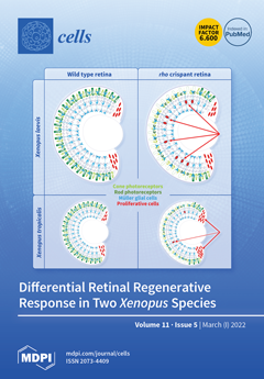
With the aim of comparing Müller cell-dependent regeneration of the retina in Xenopus laevis and tropicalis, we used CRISPR/Cas9-mediated rhodopsin gene editing to generate models of retinitis pigmentosa, a genetic condition that inescapably leads to vision loss. As expected, crispant individuals exhibit extensive rod degeneration. However, Müller glia response to this pathological environment differs tremendously between the two species. While these glial cells actively re-enter the cell cycle in Xenopus laevis, their proliferative response remains highly limited in Xenopus tropicalis, which instead mobilizes stem/progenitor cells from the ciliary marginal zone. This work thus further highlights how regenerative cellular processes can tremendously vary in vertebrates, even among closely related species. View this paper
  • Issues are regarded as officially published after their release is announced to the table of contents alert mailing list.
  • You may sign up for e-mail alerts to receive table of contents of newly released issues.
  • PDF is the official format for papers published in both, html and pdf forms. To view the papers in pdf format, click on the "PDF Full-text" link, and use the free Adobe Reader to open them.
Order results
Result details
Section
Select all
Export citation of selected articles as:
15 pages, 4958 KiB  
Review
Waste Clearance in the Brain and Neuroinflammation: A Novel Perspective on Biomarker and Drug Target Discovery in Alzheimer’s Disease
by Kazuhiko Uchida
Cells 2022, 11(5), 919; https://doi.org/10.3390/cells11050919 - 07 Mar 2022
Cited by 11 | Viewed by 4763
Abstract
Alzheimer’s disease (AD) is a multifactorial disease with a heterogeneous etiology. The pathology of Alzheimer’s disease is characterized by amyloid-beta and hyperphosphorylated tau, which are necessary for disease progression. Many clinical trials on disease-modifying drugs for AD have failed to indicate their clinical [...] Read more.
Alzheimer’s disease (AD) is a multifactorial disease with a heterogeneous etiology. The pathology of Alzheimer’s disease is characterized by amyloid-beta and hyperphosphorylated tau, which are necessary for disease progression. Many clinical trials on disease-modifying drugs for AD have failed to indicate their clinical benefits. Recent advances in fundamental research have indicated that neuroinflammation plays an important pathological role in AD. Damage- and pathogen-associated molecular patterns in the brain induce neuroinflammation and inflammasome activation, causing caspase-1-dependent glial and neuronal cell death. These waste products in the brain are eliminated by the glymphatic system via perivascular spaces, the blood-brain barrier, and the blood–cerebrospinal fluid barrier. Age-related vascular dysfunction is associated with an impairment of clearance and barrier functions, leading to neuroinflammation. The proteins involved in waste clearance in the brain and peripheral circulation may be potential biomarkers and drug targets in the early stages of cognitive impairment. This short review focuses on waste clearance dysfunction in AD pathobiology and discusses the improvement of waste clearance as an early intervention in prodromal AD and preclinical stages of dementia. Full article
Show Figures

Figure 1

14 pages, 12871 KiB  
Article
Non-Invasive Optical Motion Tracking Allows Monitoring of Respiratory Dynamics in Dystrophin-Deficient Mice
by Angelika Svetlove, Jonas Albers, Swen Hülsmann, Marietta Andrea Markus, Jana Zschüntzsch, Frauke Alves and Christian Dullin
Cells 2022, 11(5), 918; https://doi.org/10.3390/cells11050918 - 07 Mar 2022
Cited by 3 | Viewed by 2076
Abstract
Duchenne muscular dystrophy (DMD) is the most common x-chromosomal inherited dystrophinopathy which leads to progressive muscle weakness and a premature death due to cardiorespiratory dysfunction. The mdx mouse lacks functional dystrophin protein and has a comparatively human-like diaphragm phenotype. To date, diaphragm function [...] Read more.
Duchenne muscular dystrophy (DMD) is the most common x-chromosomal inherited dystrophinopathy which leads to progressive muscle weakness and a premature death due to cardiorespiratory dysfunction. The mdx mouse lacks functional dystrophin protein and has a comparatively human-like diaphragm phenotype. To date, diaphragm function can only be inadequately mapped in preclinical studies and a simple reliable translatable method of tracking the severity of the disease still lacks. We aimed to establish a sensitive, reliable, harmless and easy way to assess the effects of respiratory muscle weakness and subsequent irregularity in breathing pattern. Optical respiratory dynamics tracking (ORDT) was developed utilising a camera to track the movement of paper markers placed on the thoracic-abdominal region of the mouse. ORDT successfully distinguished diseased mdx phenotype from healthy controls by measuring significantly higher expiration constants (k) in mdx mice compared to wildtype (wt), which were also observed in the established X-ray based lung function (XLF). In contrast to XLF, with ORDT we were able to distinguish distinct fast and slow expiratory phases. In mdx mice, a larger part of the expiratory marker displacement was achieved in this initial fast phase as compared to wt mice. This phenomenon could not be observed in the XLF measurements. We further validated the simplicity and reliability of our approach by demonstrating that it can be performed using free-hand smartphone acquisition. We conclude that ORDT has a great preclinical potential to monitor DMD and other neuromuscular diseases based on changes in the breathing patterns with the future possibility to track therapy response. Full article
Show Figures

Figure 1

34 pages, 2165 KiB  
Review
A Mechanistic Theory of Development-Aging Continuity in Humans and Other Mammals
by Richard F. Walker
Cells 2022, 11(5), 917; https://doi.org/10.3390/cells11050917 - 07 Mar 2022
Cited by 3 | Viewed by 7607
Abstract
There is consensus among biogerontologists that aging occurs either as the result of a purposeful genome-based, evolved program or due to spontaneous, randomly occurring, maladaptive events. Neither concept has yet identified a specific mechanism to explain aging’s emergence and acceleration during mid-life and [...] Read more.
There is consensus among biogerontologists that aging occurs either as the result of a purposeful genome-based, evolved program or due to spontaneous, randomly occurring, maladaptive events. Neither concept has yet identified a specific mechanism to explain aging’s emergence and acceleration during mid-life and beyond. Presented herein is a novel, unifying mechanism with empirical evidence that describes how aging becomes continuous with development. It assumes that aging emerges from deterioration of a regulatory process that directs morphogenesis and morphostasis. The regulatory system consists of a genome-wide “backbone” within which its specific genes are differentially expressed by the local epigenetic landscapes of cells and tissues within which they reside, thereby explaining its holistic nature. Morphostasis evolved in humans to ensure the nurturing of dependent offspring during the first decade of young adulthood when peak parental vitality prevails in the absence of aging. The strict redundancy of each morphostasis regulatory cycle requires sensitive dependence upon initial conditions to avoid initiating deterministic chaos behavior. However, when natural selection declines as midlife approaches, persistent, progressive, and specific DNA damage and misrepair changes the initial conditions of the regulatory process, thereby compromising morphostasis regulatory redundancy, instigating chaos, initiating senescence, and accelerating aging thereafter. Full article
(This article belongs to the Special Issue Epigenetic Mechanisms of Longevity and Aging)
Show Figures

Figure 1

11 pages, 1428 KiB  
Review
Targeting the Pulmonary Microbiota to Fight against Respiratory Diseases
by Zongjie Li, Yuhao Li, Qing Sun, Jianchao Wei, Beibei Li, Yafeng Qiu, Ke Liu, Donghua Shao and Zhiyong Ma
Cells 2022, 11(5), 916; https://doi.org/10.3390/cells11050916 - 07 Mar 2022
Cited by 10 | Viewed by 3527
Abstract
The mucosal immune system of the respiratory tract possesses an effective “defense barrier” against the invading pathogenic microorganisms; therefore, the lungs of healthy organisms are considered to be sterile for a long time according to the strong pathogens-eliminating ability. The emergence of next-generation [...] Read more.
The mucosal immune system of the respiratory tract possesses an effective “defense barrier” against the invading pathogenic microorganisms; therefore, the lungs of healthy organisms are considered to be sterile for a long time according to the strong pathogens-eliminating ability. The emergence of next-generation sequencing technology has accelerated the studies about the microbial communities and immune regulating functions of lung microbiota during the past two decades. The acquisition and maturation of respiratory microbiota during childhood are mainly determined by the birth mode, diet structure, environmental exposure and antibiotic usage. However, the formation and development of lung microbiota in early life might affect the occurrence of respiratory diseases throughout the whole life cycle. The interplay and crosstalk between the gut and lung can be realized by the direct exchange of microbial species through the lymph circulation, moreover, the bioactive metabolites produced by the gut microbiota and lung microbiota can be changed via blood circulation. Complicated interactions among the lung microbiota, the respiratory viruses, and the host immune system can regulate the immune homeostasis and affect the inflammatory response in the lung. Probiotics, prebiotics, functional foods and fecal microbiota transplantation can all be used to maintain the microbial homeostasis of intestinal microbiota and lung microbiota. Therefore, various kinds of interventions on manipulating the symbiotic microbiota might be explored as novel effective strategies to prevent and control respiratory diseases. Full article
(This article belongs to the Special Issue Gut Microbiota in Nutrition and Health)
Show Figures

Figure 1

21 pages, 37660 KiB  
Article
Artificial Intelligent Deep Learning Molecular Generative Modeling of Scaffold-Focused and Cannabinoid CB2 Target-Specific Small-Molecule Sublibraries
by Yuemin Bian and Xiang-Qun Xie
Cells 2022, 11(5), 915; https://doi.org/10.3390/cells11050915 - 07 Mar 2022
Cited by 6 | Viewed by 4257
Abstract
Design and generation of high-quality target- and scaffold-specific small molecules is an important strategy for the discovery of unique and potent bioactive drug molecules. To achieve this goal, authors have developed the deep-learning molecule generation model (DeepMGM) and applied it for the de [...] Read more.
Design and generation of high-quality target- and scaffold-specific small molecules is an important strategy for the discovery of unique and potent bioactive drug molecules. To achieve this goal, authors have developed the deep-learning molecule generation model (DeepMGM) and applied it for the de novo molecular generation of scaffold-focused small-molecule libraries. In this study, a recurrent neural network (RNN) using long short-term memory (LSTM) units was trained with drug-like molecules to result in a general model (g-DeepMGM). Sampling practices on indole and purine scaffolds illustrate the feasibility of creating scaffold-focused chemical libraries based on machine intelligence. Subsequently, a target-specific model (t-DeepMGM) for cannabinoid receptor 2 (CB2) was constructed following the transfer learning process of known CB2 ligands. Sampling outcomes can present similar properties to the reported active molecules. Finally, a discriminator was trained and attached to the DeepMGM to result in an in silico molecular design-test circle. Medicinal chemistry synthesis and biological validation was performed to further investigate the generation outcome, showing that XIE9137 was identified as a potential allosteric modulator of CB2. This study demonstrates how recent progress in deep learning intelligence can benefit drug discovery, especially in de novo molecular design and chemical library generation. Full article
Show Figures

Graphical abstract

33 pages, 2179 KiB  
Review
Into the Tissues: Extracellular Matrix and Its Artificial Substitutes: Cell Signalling Mechanisms
by Aleksandra Bandzerewicz and Agnieszka Gadomska-Gajadhur
Cells 2022, 11(5), 914; https://doi.org/10.3390/cells11050914 - 07 Mar 2022
Cited by 42 | Viewed by 12605
Abstract
The existence of orderly structures, such as tissues and organs is made possible by cell adhesion, i.e., the process by which cells attach to neighbouring cells and a supporting substance in the form of the extracellular matrix. The extracellular matrix is a three-dimensional [...] Read more.
The existence of orderly structures, such as tissues and organs is made possible by cell adhesion, i.e., the process by which cells attach to neighbouring cells and a supporting substance in the form of the extracellular matrix. The extracellular matrix is a three-dimensional structure composed of collagens, elastin, and various proteoglycans and glycoproteins. It is a storehouse for multiple signalling factors. Cells are informed of their correct connection to the matrix via receptors. Tissue disruption often prevents the natural reconstitution of the matrix. The use of appropriate implants is then required. This review is a compilation of crucial information on the structural and functional features of the extracellular matrix and the complex mechanisms of cell–cell connectivity. The possibilities of regenerating damaged tissues using an artificial matrix substitute are described, detailing the host response to the implant. An important issue is the surface properties of such an implant and the possibilities of their modification. Full article
(This article belongs to the Topic Cell Signaling Pathways)
Show Figures

Graphical abstract

10 pages, 965 KiB  
Article
Evaluation of Neurofilament Light Chain as a Biomarker of Neurodegeneration in X-Linked Childhood Cerebral Adrenoleukodystrophy
by Hongge Wang, Matthew D. Davison, Martin L. Kramer, Weiliang Qiu, Tatiana Gladysheva, Ruby M. S. Chiang, Can Kayatekin, David R. Nascene, Leyla A. Taghizadeh, Carina J. King, Erin E. Nolan, Ashish O. Gupta, Paul J. Orchard and Troy C. Lund
Cells 2022, 11(5), 913; https://doi.org/10.3390/cells11050913 - 07 Mar 2022
Cited by 7 | Viewed by 3528
Abstract
Cerebral adrenoleukodystrophy (CALD) is a devastating, demyelinating neuroinflammatory manifestation found in up to 40% of young males with an inherited mutation in ABCD1, the causative gene in adrenoleukodystrophy. The search for biomarkers which correlate to CALD disease burden and respond to intervention [...] Read more.
Cerebral adrenoleukodystrophy (CALD) is a devastating, demyelinating neuroinflammatory manifestation found in up to 40% of young males with an inherited mutation in ABCD1, the causative gene in adrenoleukodystrophy. The search for biomarkers which correlate to CALD disease burden and respond to intervention has long been sought after. We used the Olink Proximity Extension Assay (Uppsala, Sweden) to explore the cerebral spinal fluid (CSF) of young males with CALD followed by correlative analysis with plasma. Using the Target 96 Neuro Exploratory panel, we found that, of the five proteins significantly increased in CSF, only neurofilament light chain (NfL) showed a significant correlation between CSF and plasma levels. Young males with CALD had a 11.3-fold increase in plasma NfL compared with controls. Importantly, 9 of 11 young males with CALD who underwent HCT showed a mean decrease in plasma NfL of 50% at 1 year after HCT compared with pre-HCT levels. In conclusion, plasma NfL could be a great value in determining outcomes in CALD and should be scrutinized in future studies in patients prior to CALD development and after therapeutic intervention. Full article
(This article belongs to the Special Issue Peroxisomal Disorders: Development of Targeted Therapies)
Show Figures

Figure 1

23 pages, 6646 KiB  
Article
Overexpression of HVA1 Enhances Drought and Heat Stress Tolerance in Triticum aestivum Doubled Haploid Plants
by Harsha Samtani, Aishwarye Sharma and Paramjit Khurana
Cells 2022, 11(5), 912; https://doi.org/10.3390/cells11050912 - 07 Mar 2022
Cited by 14 | Viewed by 3014
Abstract
Plant responses to multiple environmental stresses include various signaling pathways that allow plant acclimation and survival. Amongst different stresses, drought and heat stress severely affect growth and productivity of wheat. HVA1, a member of the group 3 LEA protein, has been well known [...] Read more.
Plant responses to multiple environmental stresses include various signaling pathways that allow plant acclimation and survival. Amongst different stresses, drought and heat stress severely affect growth and productivity of wheat. HVA1, a member of the group 3 LEA protein, has been well known to provide protection against drought stress. However, its mechanism of action and its role in other stresses such as heat remain unexplored. In this study, doubled haploid (DH) wheat plants overexpressing the HVA1 gene were analyzed and found to be both drought-and heat stress-tolerant. The transcriptome analysis revealed the upregulation of transcription factors such as DREB and HsfA6 under drought and heat stress, respectively, which contribute toward the tolerance mechanism. Particularly under heat stress conditions, the transgenic plants had a lower oxidative load and showed enhanced yield. The overexpression lines were found to be ABA-sensitive, therefore suggesting the role of HsfA6 in providing heat tolerance via the ABA-mediated pathway. Thus, apart from its known involvement in drought stress, this study highlights the potential role of HVA1 in the heat stress signaling pathway. This can further facilitate the engineering of multiple stress tolerance in crop plants, such as wheat. Full article
(This article belongs to the Special Issue Drought and Heat Stress Signalling Responses in Plants)
Show Figures

Figure 1

17 pages, 2023 KiB  
Article
Long-Term Dynamic Changes of NMDA Receptors Following an Excitotoxic Challenge
by Alberto Granzotto, Marco d’Aurora, Manuela Bomba, Valentina Gatta, Marco Onofrj and Stefano L. Sensi
Cells 2022, 11(5), 911; https://doi.org/10.3390/cells11050911 - 07 Mar 2022
Cited by 4 | Viewed by 1914
Abstract
Excitotoxicity is a form of neuronal death characterized by the sustained activation of N-methyl-D-aspartate receptors (NMDARs) triggered by the excitatory neurotransmitter glutamate. NADPH-diaphorase neurons (also known as nNOS (+) neurons) are a subpopulation of aspiny interneurons, largely spared following excitotoxic challenges. Unlike nNOS [...] Read more.
Excitotoxicity is a form of neuronal death characterized by the sustained activation of N-methyl-D-aspartate receptors (NMDARs) triggered by the excitatory neurotransmitter glutamate. NADPH-diaphorase neurons (also known as nNOS (+) neurons) are a subpopulation of aspiny interneurons, largely spared following excitotoxic challenges. Unlike nNOS (−) cells, nNOS (+) neurons fail to generate reactive oxygen species in response to NMDAR activation, a critical divergent step in the excitotoxic cascade. However, additional mechanisms underlying the reduced vulnerability of nNOS (+) neurons to NMDAR-driven neuronal death have not been explored. Using functional, genetic, and molecular analysis in striatal cultures, we indicate that nNOS (+) neurons possess distinct NMDAR properties. These specific features are primarily driven by the peculiar redox milieu of this subpopulation. In addition, we found that nNOS (+) neurons exposed to a pharmacological maneuver set to mimic chronic excitotoxicity alter their responses to NMDAR-mediated challenges. These findings suggest the presence of mechanisms providing long-term dynamic regulation of NMDARs that can have critical implications in neurotoxic settings. Full article
Show Figures

Figure 1

13 pages, 1309 KiB  
Article
Altered Plasma Fatty Acid Abundance Is Associated with Cachexia in Treatment-Naïve Pancreatic Cancer
by Kristyn Gumpper-Fedus, Phil A. Hart, Martha A. Belury, Olivia Crowe, Rachel M. Cole, Valentina Pita Grisanti, Niharika Badi, Sophia Liva, Alice Hinton, Christopher Coss, Mitchell L. Ramsey, Anne Noonan, Darwin L. Conwell and Zobeida Cruz-Monserrate
Cells 2022, 11(5), 910; https://doi.org/10.3390/cells11050910 - 07 Mar 2022
Cited by 4 | Viewed by 2326
Abstract
Cachexia occurs in up to 80% of pancreatic ductal adenocarcinoma (PDAC) patients and is characterized by unintentional weight loss and tissue wasting. To understand the metabolic changes that occur in PDAC-associated cachexia, we compared the abundance of plasma fatty acids (FAs), measured by [...] Read more.
Cachexia occurs in up to 80% of pancreatic ductal adenocarcinoma (PDAC) patients and is characterized by unintentional weight loss and tissue wasting. To understand the metabolic changes that occur in PDAC-associated cachexia, we compared the abundance of plasma fatty acids (FAs), measured by gas chromatography, of subjects with treatment-naïve metastatic PDAC with or without cachexia, defined as a loss of > 2% weight and evidence of sarcopenia (n = 43). The abundance of saturated, monounsaturated, and polyunsaturated FAs was not different between subjects with cachexia and those without. Oleic acid was significantly higher in subjects with cachexia (p = 0.0007) and diabetes (p = 0.015). Lauric (r = 0.592, p = 0.0096) and eicosapentaenoic (r = 0.564, p = 0.015) acids were positively correlated with age in cachexia patients. Subjects with diabetes (p = 0.021) or both diabetes and cachexia (p = 0.092) had low palmitic:oleic acid ratios. Linoleic acid was lower in subjects with diabetes (p = 0.018) and correlated with hemoglobin (r = 0.519, p = 0.033) and albumin (r = 0.577, p = 0.015) in subjects with cachexia. Oleic or linoleic acid may be useful treatment targets or biomarkers of cachexia in patients with metastatic PDAC, particularly those with diabetes. Full article
(This article belongs to the Special Issue Cancer-Induced Cachexia)
Show Figures

Figure 1

19 pages, 8832 KiB  
Article
EZH2 and Endometrial Cancer Development: Insights from a Mouse Model
by Xin Fang, Nan Ni, Xiaofang Wang, Yanan Tian, Ivan Ivanov, Monique Rijnkels, Kayla J. Bayless, John P. Lydon and Qinglei Li
Cells 2022, 11(5), 909; https://doi.org/10.3390/cells11050909 - 07 Mar 2022
Cited by 6 | Viewed by 3196
Abstract
Enhancer of zeste homolog 2 (EZH2), a core component of polycomb repressive complex 2, plays an important role in cancer development. As both oncogenic and tumor suppressive functions of EZH2 have been documented in the literature, the objective of this study is to [...] Read more.
Enhancer of zeste homolog 2 (EZH2), a core component of polycomb repressive complex 2, plays an important role in cancer development. As both oncogenic and tumor suppressive functions of EZH2 have been documented in the literature, the objective of this study is to determine the impact of Ezh2 deletion on the development and progression of endometrial cancer induced by inactivation of phosphatase and tensin homolog (PTEN), a tumor suppressor gene frequently dysregulated in endometrial cancer patients. To this end, we created mice harboring uterine deletion of both Ezh2 and Pten using Cre recombinase driven by the progesterone receptor (Pgr) promoter. Our results showed reduced tumor burden in Ptend/d; Ezh2d/d mice compared with that of Ptend/d mice during early carcinogenesis. The decreased Ki67 index in EZH2 and PTEN-depleted uteri versus that in PTEN-depleted uteri indicated an oncogenic role of EZH2 during early tumor development. However, mice harboring uterine deletion of both Ezh2 and Pten developed unfavorable disease outcome, accompanied by exacerbated epithelial stratification and heightened inflammatory response. The observed effect was non-cell autonomous and mediated by altered immune response evidenced by massive accumulation of intraluminal neutrophils, a hallmark of endometrial carcinoma in Ptend/d; Ezh2d/d mice during disease progression. Hence, these results reveal dual roles of EZH2 in endometrial cancer development. Full article
(This article belongs to the Special Issue Progesterone Receptor Signaling)
Show Figures

Graphical abstract

21 pages, 3617 KiB  
Article
Direct Interaction of Mitochondrial Cytochrome c Oxidase with Thyroid Hormones: Evidence for Two Binding Sites
by Ilya P. Oleynikov, Roman V. Sudakov, Natalia V. Azarkina and Tatiana V. Vygodina
Cells 2022, 11(5), 908; https://doi.org/10.3390/cells11050908 - 06 Mar 2022
Cited by 5 | Viewed by 1902
Abstract
Thyroid hormones regulate tissue metabolism to establish an energy balance in the cell, in particular, by affecting oxidative phosphorylation. Their long-term impact is mainly associated with changes in gene expression, while the short-term effects may differ in their mechanisms. Our work was devoted [...] Read more.
Thyroid hormones regulate tissue metabolism to establish an energy balance in the cell, in particular, by affecting oxidative phosphorylation. Their long-term impact is mainly associated with changes in gene expression, while the short-term effects may differ in their mechanisms. Our work was devoted to studying the short-term effects of hormones T2, T3 and T4 on mitochondrial cytochrome c oxidase (CcO) mediated by direct contact with the enzyme. The data obtained indicate the existence of two separate sites of CcO interaction with thyroid hormones, differing in their location, affinity and specificity to hormone binding. First, we show that T3 and T4 but not T2 inhibit the oxidase activity of CcO in solution and on membrane preparations with Ki ≈ 100–200 μM. In solution, T3 and T4 compete in a 1:1 ratio with the detergent dodecyl-maltoside to bind to the enzyme. The peroxidase and catalase partial activities of CcO are not sensitive to hormones, but electron transfer from heme a to the oxidized binuclear center is affected. We believe that T3 and T4 could be ligands of the bile acid-binding site found in the 3D structure of CcO by Ferguson-Miller’s group, and hormone-induced inhibition is associated with dysfunction of the K-proton channel. A possible role of this interaction in the physiological regulation of the enzyme is discussed. Second, we find that T2, T3, and T4 inhibit superoxide generation by oxidized CcO in the presence of excess H2O2. Inhibition is characterized by Ki values of 0.3–5 μM and apparently affects the formation of O2●− at the protein surface. The second binding site for thyroid hormones presumably coincides with the point of tight T2 binding on the Va subunit described in the literature. Full article
(This article belongs to the Special Issue Oxidative Phosphorylation and Hormones: Different Ways of Influence)
Show Figures

Figure 1

16 pages, 40845 KiB  
Article
Computational Workflow to Study the Diversity of Secondary Metabolites in Fourteen Different Isatis Species
by Doudou Huang, Chen Zhang, Junfeng Chen, Ying Xiao, Mingming Li, Lianna Sun, Shi Qiu and Wansheng Chen
Cells 2022, 11(5), 907; https://doi.org/10.3390/cells11050907 - 06 Mar 2022
Cited by 1 | Viewed by 2002
Abstract
The screening of real features among thousands of ions remains a great challenge in the study of metabolomics. In this research, a workflow designed based on the MetaboFR tool and “feature-rating” rule was developed to screen the real features in large-scale data analyses. [...] Read more.
The screening of real features among thousands of ions remains a great challenge in the study of metabolomics. In this research, a workflow designed based on the MetaboFR tool and “feature-rating” rule was developed to screen the real features in large-scale data analyses. Seventy-four reference standards were used to test the feasibility, with 83.21% of real features being obtained after MetaboFR processing. Moreover, the full workflow was applied for systematic characterization of 14 species of the genus Isatis, with the result that 87.72% of real features were retained and 69.19% of the in-source fragments were removed. To gain insights into metabolite diversity within this plant family, 1697 real features were tentatively identified, including lipids, phenylpropanoids, organic acids, indole derivatives, etc. Indole derivatives were demonstrated to be the best chemical markers with which to differentiate different species. The rare existence of indole derivatives in Isatis cappadocica (cap) and Isatis cappadocica subsp. Steveniana (capS) indicates that the biosynthesis of indole derivatives could play a key role in driving the chemical diversity and evolution of genus Isatis. Our workflow provides the foundations for the exploration of real features in metabolomics, and has the potential to reveal the chemical composition and marker metabolites of secondary metabolites in plant fields. Full article
Show Figures

Graphical abstract

15 pages, 3037 KiB  
Article
Pathological Relevance of Post-Translationally Modified Alpha-Synuclein (pSer87, pSer129, nTyr39) in Idiopathic Parkinson’s Disease and Multiple System Atrophy
by Berkiye Sonustun, Melek Firat Altay, Catherine Strand, Kirsten Ebanks, Geshanthi Hondhamuni, Thomas T. Warner, Hilal A. Lashuel and Rina Bandopadhyay
Cells 2022, 11(5), 906; https://doi.org/10.3390/cells11050906 - 06 Mar 2022
Cited by 14 | Viewed by 3148
Abstract
Aggregated alpha-synuclein (α-synuclein) is the main component of Lewy bodies (LBs), Lewy neurites (LNs), and glial cytoplasmic inclusions (GCIs), which are pathological hallmarks of idiopathic Parkinson’s disease (IPD) and multiple system atrophy (MSA). Initiating factors that culminate in forming LBs/LNs/GCIs remain elusive. Several [...] Read more.
Aggregated alpha-synuclein (α-synuclein) is the main component of Lewy bodies (LBs), Lewy neurites (LNs), and glial cytoplasmic inclusions (GCIs), which are pathological hallmarks of idiopathic Parkinson’s disease (IPD) and multiple system atrophy (MSA). Initiating factors that culminate in forming LBs/LNs/GCIs remain elusive. Several species of α-synuclein exist, including phosphorylated and nitrated forms. It is unclear which α-synuclein post-translational modifications (PTMs) appear within aggregates throughout disease pathology. Herein we aimed to establish the predominant α-synuclein PTMs in postmortem IPD and MSA pathology using immunohistochemistry. We examined the patterns of three α-synuclein PTMs (pS87, pS129, nY39) simultaneously in pathology-affected regions of 15 IPD cases, 5 MSA cases, and 6 neurologically normal controls. All antibodies recognized LBs, LNs, and GCIs, albeit to a variable extent. pS129 α-synuclein antibody was particularly immunopositive for LNs and synaptic dot-like structures, followed by nY39 α-synuclein antibody. GCIs, neuronal inclusions, and small threads were positive for nY39 α-synuclein in MSA. Quantification of the LB scores revealed that pS129 α-synuclein was the dominant and earliest α-synuclein PTM, followed by nY39 α-synuclein, while lower amounts of pSer87 α-synuclein appeared later in disease progression in PD. These results may have implications for novel biomarker and therapeutic developments. Full article
Show Figures

Figure 1

11 pages, 1765 KiB  
Review
Machine Learning-Driven Multiobjective Optimization: An Opportunity of Microfluidic Platforms Applied in Cancer Research
by Yi Liu, Sijing Li and Yaling Liu
Cells 2022, 11(5), 905; https://doi.org/10.3390/cells11050905 - 05 Mar 2022
Cited by 9 | Viewed by 3206
Abstract
Cancer metastasis is one of the primary reasons for cancer-related fatalities. Despite the achievements of cancer research with microfluidic platforms, understanding the interplay of multiple factors when it comes to cancer cells is still a great challenge. Crosstalk and causality of different factors [...] Read more.
Cancer metastasis is one of the primary reasons for cancer-related fatalities. Despite the achievements of cancer research with microfluidic platforms, understanding the interplay of multiple factors when it comes to cancer cells is still a great challenge. Crosstalk and causality of different factors in pathogenesis are two important areas in need of further research. With the assistance of machine learning, microfluidic platforms can reach a higher level of detection and classification of cancer metastasis. This article reviews the development history of microfluidics used for cancer research and summarizes how the utilization of machine learning benefits cancer studies, particularly in biomarker detection, wherein causality analysis is useful. To optimize microfluidic platforms, researchers are encouraged to use causality analysis when detecting biomarkers, analyzing tumor microenvironments, choosing materials, and designing structures. Full article
(This article belongs to the Special Issue 10th Anniversary of Cells—Advances in Cell Techniques)
Show Figures

Figure 1

22 pages, 28592 KiB  
Article
Influence of Mo and Fe on Photosynthetic and Nitrogenase Activities of Nitrogen-Fixing Cyanobacteria under Nitrogen Starvation
by Asemgul K. Sadvakasova, Bekzhan D. Kossalbayev, Aziza I. Token, Meruert O. Bauenova, Jingjing Wang, Bolatkhan K. Zayadan, Huma Balouch, Saleh Alwasel, Yoong Kit Leong, Jo-Shu Chang and Suleyman I. Allakhverdiev
Cells 2022, 11(5), 904; https://doi.org/10.3390/cells11050904 - 05 Mar 2022
Cited by 19 | Viewed by 3485
Abstract
The potential of cyanobacteria to perform a variety of distinct roles vital for the biosphere, including nutrient cycling and environmental detoxification, drives interest in studying their biodiversity. Increasing soil erosion and the overuse of chemical fertilizers are global problems in developed countries. The [...] Read more.
The potential of cyanobacteria to perform a variety of distinct roles vital for the biosphere, including nutrient cycling and environmental detoxification, drives interest in studying their biodiversity. Increasing soil erosion and the overuse of chemical fertilizers are global problems in developed countries. The option might be to switch to organic farming, which entails largely the use of biofertilisers. Cyanobacteria are prokaryotic, photosynthetic organisms with considerable potential, within agrobiotechnology, to produce biofertilisers. They contribute significantly to plant drought resistance and nitrogen enrichment in the soil. This study sought, isolated, and investigated nitrogen-fixing cyanobacterial strains in rice fields, and evaluated the effect of Mo and Fe on photosynthetic and nitrogenase activities under nitrogen starvation. Cyanobacterial isolates, isolated from rice paddies in Kazakhstan, were identified as Trichormus variabilis K-31 (MZ079356), Cylindrospermum badium J-8 (MZ079357), Nostoc sp. J-14 (MZ079360), Oscillatoria brevis SH-12 (MZ090011), and Tolypothrix tenuis J-1 (MZ079361). The study of the influence of various concentrations of Mo and Fe on photosynthetic and nitrogenase activities under conditions of nitrogen starvation revealed the optimal concentrations of metals that have a stimulating effect on the studied parameters. Full article
(This article belongs to the Special Issue Photosynthesis under Biotic and Abiotic Environmental Stress)
Show Figures

Graphical abstract

25 pages, 7280 KiB  
Review
Disruption of Claudin-Made Tight Junction Barriers by Clostridium perfringens Enterotoxin: Insights from Structural Biology
by Chinemerem P. Ogbu, Sourav Roy and Alex J. Vecchio
Cells 2022, 11(5), 903; https://doi.org/10.3390/cells11050903 - 05 Mar 2022
Cited by 7 | Viewed by 4526
Abstract
Claudins are a family of integral membrane proteins that enable epithelial cell/cell interactions by localizing to and driving the formation of tight junctions. Via claudin self-assembly within the membranes of adjoining cells, their extracellular domains interact, forming barriers to the paracellular transport of [...] Read more.
Claudins are a family of integral membrane proteins that enable epithelial cell/cell interactions by localizing to and driving the formation of tight junctions. Via claudin self-assembly within the membranes of adjoining cells, their extracellular domains interact, forming barriers to the paracellular transport of small molecules and ions. The bacterium Clostridium perfringens causes prevalent gastrointestinal disorders in mammals by employing an enterotoxin (CpE) that targets claudins. CpE binds to claudins at or near tight junctions in the gut and disrupts their barrier function, potentially by disabling their assembly or via cell signaling means—the mechanism(s) remain unclear. CpE ultimately destroys claudin-expressing cells through the formation of a cytotoxic membrane-penetrating β-barrel pore. Structures obtained by X-ray crystallography of CpE, claudins, and claudins in complex with CpE fragments have provided the structural bases of claudin and CpE functions, revealing potential mechanisms for the CpE-mediated disruption of claudin-made tight junctions. This review highlights current progress in this space—what has been discovered and what remains unknown—toward efforts to elucidate the molecular mechanism of CpE disruption of tight junction barriers. It further underscores the key insights obtained through structure that are being applied to develop CpE-based therapeutics that combat claudin-overexpressing cancers or modulate tight junction barriers. Full article
(This article belongs to the Special Issue Cell-Cell Interactions and Cell Adhesion Signaling in Disease States)
Show Figures

Figure 1

14 pages, 3131 KiB  
Article
Evaluation of Immunoregulatory Biomarkers on Plasma Small Extracellular Vesicles for Disease Progression and Early Therapeutic Response in Head and Neck Cancer
by Jadwiga Jablonska, Malwina Rist, Ilona Spyra, Luisa Tengler, Maksim Domnich, Benjamin Kansy, Bernd Giebel, Basant Kumar Thakur, Nicole Rotter, Stephan Lang and Sonja Ludwig
Cells 2022, 11(5), 902; https://doi.org/10.3390/cells11050902 - 05 Mar 2022
Cited by 10 | Viewed by 2500
Abstract
Head and Neck Cancers (HNCs) have highly immunosuppressive properties. Small extracellular vesicles (sEVs), including exosomes, nanosized mediators of intercellular communication in the blood, carry immunosuppressive proteins and effectively inhibit anti-tumor immune responses in HNCs. This study evaluates immunosuppressive markers on sEVs from 40 [...] Read more.
Head and Neck Cancers (HNCs) have highly immunosuppressive properties. Small extracellular vesicles (sEVs), including exosomes, nanosized mediators of intercellular communication in the blood, carry immunosuppressive proteins and effectively inhibit anti-tumor immune responses in HNCs. This study evaluates immunosuppressive markers on sEVs from 40 HNC patients at different disease stages and 3- and 6-month follow-up after surgery and/or chemoradiotherapy. As controls, sEVs from normal donors (NDs) are examined. Immunoregulatory surface markers on sEVs were detected as relative fluorescence intensity (RFI) using on-bead flow cytometry, and their expression levels were monitored in the early and late stages of HNC and during follow-up. In parallel, the sEV-mediated apoptosis of CD8+ Jurkat cells was assessed. Together with TGF-β1 and PD-L1 abundance, total sEV proteins are elevated with disease progression. In contrast, total sEV protein, including TGF-β1, PD-1 and PD-L1, decrease upon therapy response during follow-up. Overall survival analysis implies that high sEV PD-1/PD-L1 content is an unfavorable prognostic marker in HNC. Consistently, the sEV-mediated induction of apoptosis in CD8+ T cells correlates with the disease activity and therapy response. These findings indicate that a combination of immunoregulatory marker profiles should be preferred over a single marker to monitor disease progression and therapy response in HNC. Full article
Show Figures

Figure 1

10 pages, 898 KiB  
Review
Bile Acids: Key Players in Inflammatory Bowel Diseases?
by Aicha Kriaa, Vincent Mariaule, Amin Jablaoui, Soufien Rhimi, Hela Mkaouar, Juan Hernandez, Brice Korkmaz, Adam Lesner, Emmanuelle Maguin, Ali Aghdassi and Moez Rhimi
Cells 2022, 11(5), 901; https://doi.org/10.3390/cells11050901 - 05 Mar 2022
Cited by 19 | Viewed by 6232
Abstract
Inflammatory bowel diseases (IBDs) have emerged as a public health problem worldwide with a limited number of efficient therapeutic options despite advances in medical therapy. Although changes in the gut microbiota composition are recognized as key drivers of dysregulated intestinal immunity, alterations in [...] Read more.
Inflammatory bowel diseases (IBDs) have emerged as a public health problem worldwide with a limited number of efficient therapeutic options despite advances in medical therapy. Although changes in the gut microbiota composition are recognized as key drivers of dysregulated intestinal immunity, alterations in bile acids (BAs) have been shown to influence gut homeostasis and contribute to the pathogenesis of the disease. In this review, we explore the interactions involving BAs and gut microbiota in IBDs, and discuss how the gut microbiota–BA–host axis may influence digestive inflammation. Full article
Show Figures

Figure 1

18 pages, 2480 KiB  
Article
Metabolomic Profiling and Mechanotransduction of Single Chondrocytes Encapsulated in Alginate Microgels
by Jacob P. Fredrikson, Priyanka P. Brahmachary, Ayten E. Erdoğan, Zachary K. Archambault, James N. Wilking, Ronald K. June and Connie B. Chang
Cells 2022, 11(5), 900; https://doi.org/10.3390/cells11050900 - 05 Mar 2022
Cited by 6 | Viewed by 2967
Abstract
Articular cartilage is comprised of two main components, the extracellular matrix (ECM) and the pericellular matrix (PCM). The PCM helps to protect chondrocytes in the cartilage from mechanical loads, but in patients with osteoarthritis, the PCM is weakened, resulting in increased chondrocyte stress. [...] Read more.
Articular cartilage is comprised of two main components, the extracellular matrix (ECM) and the pericellular matrix (PCM). The PCM helps to protect chondrocytes in the cartilage from mechanical loads, but in patients with osteoarthritis, the PCM is weakened, resulting in increased chondrocyte stress. As chondrocytes are responsible for matrix synthesis and maintenance, it is important to understand how mechanical loads affect the cellular responses of chondrocytes. Many studies have examined chondrocyte responses to in vitro mechanical loading by embedding chondrocytes in 3-D hydrogels. However, these experiments are mostly performed in the absence of PCM, which may obscure important responses to mechanotransduction. Here, drop-based microfluidics is used to culture single chondrocytes in alginate microgels for cell-directed PCM synthesis that closely mimics the in vivo microenvironment. Chondrocytes formed PCM over 10 days in these single-cell 3-D microenvironments. Mechanotransduction studies were performed, in which single-cell microgels mimicking the cartilage PCM were embedded in high-stiffness agarose. After physiological dynamic compression in a custom-built bioreactor, microgels exhibited distinct metabolomic profiles from both uncompressed and monolayer controls. These results demonstrate the potential of single cell encapsulation in alginate microgels to advance cartilage tissue engineering and basic chondrocyte mechanobiology. Full article
(This article belongs to the Special Issue Cell Therapies in Orthopaedics)
Show Figures

Figure 1

20 pages, 873 KiB  
Review
Redox Implications of Extreme Task Performance: The Case in Driver Athletes
by Michael B. Reid
Cells 2022, 11(5), 899; https://doi.org/10.3390/cells11050899 - 05 Mar 2022
Cited by 1 | Viewed by 3113
Abstract
Redox homeostasis and redox-mediated signaling mechanisms are fundamental elements of human biology. Physiological levels of reactive oxygen species (ROS) and reactive nitrogen species (RNS) modulate a range of functional processes at the cellular, tissue, and systemic levels in healthy humans. Conversely, excess ROS [...] Read more.
Redox homeostasis and redox-mediated signaling mechanisms are fundamental elements of human biology. Physiological levels of reactive oxygen species (ROS) and reactive nitrogen species (RNS) modulate a range of functional processes at the cellular, tissue, and systemic levels in healthy humans. Conversely, excess ROS or RNS activity can disrupt function, impairing the performance of daily activities. This article analyzes the impact of redox mechanisms on extreme task performance. Such activities (a) require complex motor skills, (b) are physically demanding, (c) are performed in an extreme environment, (d) require high-level executive function, and (e) pose an imminent risk of injury or death. The current analysis utilizes race car driving as a representative example. The physiological challenges of this extreme task include physical exertion, g loading, vibration, heat exposure, dehydration, noise, mental demands, and emotional factors. Each of these challenges stimulates ROS signaling, RNS signaling, or both, alters redox homeostasis, and exerts pro-oxidant effects at either the tissue or systemic levels. These redox mechanisms appear to promote physiological stress during race car driving and impair the performance of driver athletes. Full article
(This article belongs to the Special Issue Redox Control of Cell Signaling in Cardiac and Skeletal Muscle)
Show Figures

Figure 1

19 pages, 1811 KiB  
Review
Androgen Receptor-Mediated Transcription in Prostate Cancer
by Doğancan Özturan, Tunç Morova and Nathan A. Lack
Cells 2022, 11(5), 898; https://doi.org/10.3390/cells11050898 - 05 Mar 2022
Cited by 13 | Viewed by 4445
Abstract
Androgen receptor (AR)-mediated transcription is critical in almost all stages of prostate cancer (PCa) growth and differentiation. This process involves a complex interplay of coregulatory proteins, chromatin remodeling complexes, and other transcription factors that work with AR at cis-regulatory enhancer regions to [...] Read more.
Androgen receptor (AR)-mediated transcription is critical in almost all stages of prostate cancer (PCa) growth and differentiation. This process involves a complex interplay of coregulatory proteins, chromatin remodeling complexes, and other transcription factors that work with AR at cis-regulatory enhancer regions to induce the spatiotemporal transcription of target genes. This enhancer-driven mechanism is remarkably dynamic and undergoes significant alterations during PCa progression. In this review, we discuss the AR mechanism of action in PCa with a focus on how cis-regulatory elements modulate gene expression. We explore emerging evidence of genetic variants that can impact AR regulatory regions and alter gene transcription in PCa. Finally, we highlight several outstanding questions and discuss potential mechanisms of this critical transcription factor. Full article
Show Figures

Figure 1

14 pages, 2119 KiB  
Article
PTH-Induced Bone Regeneration and Vascular Modulation Are Both Dependent on Endothelial Signaling
by Doron Cohn-Schwartz, Yeshai Schary, Eran Yalon, Zoe Krut, Xiaoyu Da, Edward M. Schwarz, Dan Gazit, Gadi Pelled and Zulma Gazit
Cells 2022, 11(5), 897; https://doi.org/10.3390/cells11050897 - 05 Mar 2022
Cited by 4 | Viewed by 1957
Abstract
The use of a bone allograft presents a promising approach for healing nonunion fractures. We have previously reported that parathyroid hormone (PTH) therapy induced allograft integration while modulating angiogenesis at the allograft proximity. Here, we hypothesize that PTH-induced vascular modulation and the osteogenic [...] Read more.
The use of a bone allograft presents a promising approach for healing nonunion fractures. We have previously reported that parathyroid hormone (PTH) therapy induced allograft integration while modulating angiogenesis at the allograft proximity. Here, we hypothesize that PTH-induced vascular modulation and the osteogenic effect of PTH are both dependent on endothelial PTH receptor-1 (PTHR1) signaling. To evaluate our hypothesis, we used multiple transgenic mouse lines, and their wild-type counterparts as a control. In addition to endothelial-specific PTHR1 knock-out mice, we used mice in which PTHR1 was engineered to be constitutively active in collagen-1α+ osteoblasts, to assess the effect of PTH signaling activation exclusively in osteoprogenitors. To characterize resident cell recruitment and osteogenic activity, mice in which the Luciferase reporter gene is expressed under the Osteocalcin promoter (Oc-Luc) were used. Mice were implanted with calvarial allografts and treated with either PTH or PBS. A micro-computed tomography-based structural analysis indicated that the induction of bone formation by PTH, as observed in wild-type animals, was not maintained when PTHR1 was removed from endothelial cells. Furthermore, the induction of PTH signaling exclusively in osteoblasts resulted in significantly less bone formation compared to systemic PTH treatment, and significantly less osteogenic activity was measured by bioluminescence imaging of the Oc-Luc mice. Deletion of the endothelial PTHR1 significantly decreased the PTH-induced formation of narrow blood vessels, formerly demonstrated in wild-type mice. However, the exclusive activation of PTH signaling in osteoblasts was sufficient to re-establish the observed PTH effect. Collectively, our results show that endothelial PTHR1 signaling plays a key role in PTH-induced osteogenesis and has implications in angiogenesis. Full article
(This article belongs to the Special Issue Advances in Bone Metabolism)
Show Figures

Figure 1

17 pages, 1342 KiB  
Review
Polyamine Immunometabolism: Central Regulators of Inflammation, Cancer and Autoimmunity
by Tzu-yi Chia, Andrew Zolp and Jason Miska
Cells 2022, 11(5), 896; https://doi.org/10.3390/cells11050896 - 05 Mar 2022
Cited by 23 | Viewed by 4528
Abstract
Polyamines are ubiquitous, amine-rich molecules with diverse processes in biology. Recent work has highlighted that polyamines exert profound roles on the mammalian immune system, particularly inflammation and cancer. The mechanisms by which they control immunity are still being described. In the context of [...] Read more.
Polyamines are ubiquitous, amine-rich molecules with diverse processes in biology. Recent work has highlighted that polyamines exert profound roles on the mammalian immune system, particularly inflammation and cancer. The mechanisms by which they control immunity are still being described. In the context of inflammation and autoimmunity, polyamine levels inversely correlate to autoimmune phenotypes, with lower polyamine levels associated with higher inflammatory responses. Conversely, in the context of cancer, polyamines and polyamine biosynthetic genes positively correlate with the severity of malignancy. Blockade of polyamine metabolism in cancer results in reduced tumor growth, and the effects appear to be mediated by an increase in T-cell infiltration and a pro-inflammatory phenotype of macrophages. These studies suggest that polyamine depletion leads to inflammation and that polyamine enrichment potentiates myeloid cell immune suppression. Indeed, combinatorial treatment with polyamine blockade and immunotherapy has shown efficacy in pre-clinical models of cancer. Considering the efficacy of immunotherapies is linked to autoimmune sequelae in humans, termed immune-adverse related events (iAREs), this suggests that polyamine levels may govern the inflammatory response to immunotherapies. This review proposes that polyamine metabolism acts to balance autoimmune inflammation and anti-tumor immunity and that polyamine levels can be used to monitor immune responses and responsiveness to immunotherapy. Full article
Show Figures

Figure 1

11 pages, 2190 KiB  
Article
Isolation and Crystallization of the D156C Form of Optogenetic ChR2
by Liying Zhang, Kaituo Wang, Shuo Ning, Per Amstrup Pedersen, Annette Susanne Duelli and Pontus Emanuel Gourdon
Cells 2022, 11(5), 895; https://doi.org/10.3390/cells11050895 - 05 Mar 2022
Cited by 3 | Viewed by 2067
Abstract
Channelrhodopsins (ChRs) are light-gated ion channels that are receiving increasing attention as optogenetic tools. Despite extensive efforts to gain understanding of how these channels function, the molecular events linking light absorption of the retinal cofactor to channel opening remain elusive. While dark-state structures [...] Read more.
Channelrhodopsins (ChRs) are light-gated ion channels that are receiving increasing attention as optogenetic tools. Despite extensive efforts to gain understanding of how these channels function, the molecular events linking light absorption of the retinal cofactor to channel opening remain elusive. While dark-state structures of ChR2 or chimeric proteins have demonstrated the architecture of non-conducting states, there is a need for open- and desensitized-state structures to uncover the mechanistic principles underlying channel activity. To facilitate comprehensive structural studies of ChR2 in non-closed states, we report a production and purification procedure of the D156C form of ChR2, which displays prolonged channel opening compared to the wild type. We demonstrate considerable yields (0.45 mg/g fermenter cell culture) of recombinantly produced protein using S. cerevisiae, which is purified to high homogeneity both as opsin (retinal-free) and as functional ChR2 with added retinal. We also develop conditions that enable the growth of ChR2 crystals that scatter X-rays to 6 Å, and identify a molecular replacement solution that suggests that the packing is different from published structures. Consequently, our cost-effective production and purification pipeline opens the way for downstream structural studies of different ChR2 states, which may provide a foundation for further adaptation of this protein for optogenetic applications. Full article
Show Figures

Figure 1

21 pages, 4933 KiB  
Article
KCNQ2 Selectivity Filter Mutations Cause Kv7.2 M-Current Dysfunction and Configuration Changes Manifesting as Epileptic Encephalopathies and Autistic Spectrum Disorders
by Inn-Chi Lee, Jiann-Jou Yang, Ying-Ming Liou and Swee-Hee Wong
Cells 2022, 11(5), 894; https://doi.org/10.3390/cells11050894 - 05 Mar 2022
Cited by 2 | Viewed by 3029
Abstract
KCNQ2 mutations can cause benign familial neonatal convulsions (BFNCs), epileptic encephalopathy (EE), and mild-to-profound neurodevelopmental disabilities. Mutations in the KCNQ2 selectivity filter (SF) are critical to neurodevelopmental outcomes. Three patients with neonatal EE carry de novo heterozygous KCNQ2 p.Thr287Ile, p.Gly281Glu and p.Pro285Thr, and [...] Read more.
KCNQ2 mutations can cause benign familial neonatal convulsions (BFNCs), epileptic encephalopathy (EE), and mild-to-profound neurodevelopmental disabilities. Mutations in the KCNQ2 selectivity filter (SF) are critical to neurodevelopmental outcomes. Three patients with neonatal EE carry de novo heterozygous KCNQ2 p.Thr287Ile, p.Gly281Glu and p.Pro285Thr, and all are followed-up in our clinics. Whole-cell patch-clamp analysis with transfected mutations was performed. The Kv7.2 in three mutations demonstrated significant current changes in the homomeric-transfected cells. The conduction curves for V1/2, the K slope, and currents in 3 mutations were lower than those for the wild type (WT). The p.Gly281Glu had a worse conductance than the p.Thr287Ile and p.Pro285Thr, the patient compatible with p.Gly281Glu had a worse clinical outcome than patients with p.Thr287Ile and p.Pro285Thr. The p.Gly281Glu had more amino acid weight changes than the p.Gly281Glu and p.Pro285Thr. Among 5 BFNCs and 23 EE from mutations in the SF, the greater weight of the mutated protein compared with that of the WT was presumed to cause an obstacle to pore size, which is one of the most important factors in the phenotype and outcome. For the 35 mutations in the SF domain, using changes in amino acid weight between the WT and the KCNQ2 mutations to predict EE resulted in 80.0% sensitivity and 80% specificity, a positive prediction rate of 96.0%, and a negative prediction rate of 40.0% (p = 0.006, χ2 (1, n = 35) = 7.56; odds ratio 16.0, 95% confidence interval, 1.50 to 170.63). The findings suggest that p.Thr287Ile, p.Gly281Glu and p.Pro285Thr are pathogenic to KCNQ2 EE. In mutations in SF, a mutated protein heavier than the WT is a factor in the Kv7.2 current and outcome. Full article
Show Figures

Figure 1

16 pages, 4607 KiB  
Article
Transcriptomics Reveals Discordant Lipid Metabolism Effects between In Vitro Models Exposed to Elafibranor and Liver Samples of NAFLD Patients after Bariatric Surgery
by Joost Boeckmans, Alexandra Gatzios, Anja Heymans, Matthias Rombaut, Vera Rogiers, Joery De Kock, Tamara Vanhaecke and Robim M. Rodrigues
Cells 2022, 11(5), 893; https://doi.org/10.3390/cells11050893 - 04 Mar 2022
Cited by 7 | Viewed by 3492
Abstract
Background and aims: Non-alcoholic steatohepatitis (NASH) is a life-threatening stage of non-alcoholic fatty liver disease (NAFLD) for which no drugs have been approved. We have previously shown that human-derived hepatic in vitro models can be used to mimic key cellular mechanisms involved in [...] Read more.
Background and aims: Non-alcoholic steatohepatitis (NASH) is a life-threatening stage of non-alcoholic fatty liver disease (NAFLD) for which no drugs have been approved. We have previously shown that human-derived hepatic in vitro models can be used to mimic key cellular mechanisms involved in the progression of NASH. In the present study, we first characterize the transcriptome of multiple in vitro NASH models. Subsequently, we investigate how elafibranor, which is a peroxisome proliferator-activated receptor (PPAR)-α/δ agonist that has recently failed a phase 3 clinical trial as a potential anti-NASH compound, modulates the transcriptome of these models. Finally, we compare the elafibranor-induced gene expression modulation to transcriptome data of patients with improved/resolved NAFLD/NASH upon bariatric surgery, which is the only proven clinical NASH therapy. Methods: Human whole genome microarrays were used for the transcriptomics evaluation of hepatic in vitro models. Comparison to publicly available clinical datasets was conducted using multiple bioinformatic application tools. Results: Primary human hepatocytes (PHH), HepaRG, and human skin stem cell-derived hepatic progenitors (hSKP-HPC) exposed to NASH-inducing triggers exhibit up to 35% overlap with datasets of liver samples from NASH patients. Exposure of the in vitro NASH models to elafibranor partially reversed the transcriptional modulations, predicting an inhibition of toll-like receptor (TLR)-2/4/9-mediated inflammatory responses, NFκB-signaling, hepatic fibrosis, and leukocyte migration. These transcriptomic changes were also observed in the datasets of liver samples of patients with resolved NASH. Peroxisome Proliferator Activated Receptor Alpha (PPARA), PPARG Coactivator 1 Alpha (PPARGC1A), and Sirtuin 1 (SIRT1) were identified as the major common upstream regulators upon exposure to elafibranor. Analysis of the downstream mechanistic networks further revealed that angiopoietin Like 4 (ANGPTL4), pyruvate dehydrogenase kinase 4 (PDK4), and perilipin 2 (PLIN2), which are involved in the promotion of hepatic lipid accumulation, were also commonly upregulated by elafibranor in all in vitro NASH models. Contrarily, these genes were not upregulated in liver samples of patients with resolved NASH. Conclusion: Transcriptomics comparison between in vitro NASH models exposed to elafibranor and clinical datasets of NAFLD patients after bariatric surgery reveals commonly modulated anti-inflammatory responses, but discordant modulations of key factors in lipid metabolism. This discordant adverse effect of elafibranor deserves further investigation when assessing PPAR-α/δ agonism as a potential anti-NASH therapy. Full article
(This article belongs to the Special Issue The Role of PPARs in Disease II)
Show Figures

Figure 1

17 pages, 3493 KiB  
Article
Dual Effects of Korean Red Ginseng on Astrocytes and Neural Stem Cells in Traumatic Brain Injury: The HO-1–Tom20 Axis as a Putative Target for Mitochondrial Function
by Minsu Kim, Sunhong Moon, Hui Su Jeon, Sueun Kim, Seong-Ho Koh, Mi-Sook Chang, Young-Myeong Kim and Yoon Kyung Choi
Cells 2022, 11(5), 892; https://doi.org/10.3390/cells11050892 - 04 Mar 2022
Cited by 9 | Viewed by 3373
Abstract
Astrocytes display regenerative potential in pathophysiologic conditions. In our previous study, heme oxygenase-1 (HO-1) promoted astrocytic mitochondrial functions in mice via the peroxisome-proliferator-activating receptor-γ coactivator-1α (PGC-1α) pathway on administering Korean red ginseng extract (KRGE) after traumatic brain injury (TBI). In this study, KRGE [...] Read more.
Astrocytes display regenerative potential in pathophysiologic conditions. In our previous study, heme oxygenase-1 (HO-1) promoted astrocytic mitochondrial functions in mice via the peroxisome-proliferator-activating receptor-γ coactivator-1α (PGC-1α) pathway on administering Korean red ginseng extract (KRGE) after traumatic brain injury (TBI). In this study, KRGE promoted astrocytic mitochondrial functions, assessed with oxygen consumption and adenosine triphosphate (ATP) production, which could be regulated by the translocase of the outer membrane of mitochondria 20 (Tom20) pathway with a PGC-1α-independent pathway. The HO-1–Tom20 axis induced an increase in mitochondrial functions, detected with cytochrome c oxidase subunit 2 and cytochrome c. HO-1 crosstalk with nicotinamide phosphoribosyltransferase was concomitant with the upregulated nicotinamide adenine dinucleotide (NAD)/NADH ratio, thereby upregulating NAD-dependent class I sirtuins. In adult neural stem cells (NSCs), KRGE-treated, astrocyte-conditioned media increased oxygen consumption and Tom20 levels through astrocyte-derived HO-1. HO inactivation by Sn(IV) protoporphyrin IX dichloride in TBI mice administered KRGE decreased neuronal markers, together with Tom20. Thus, astrocytic HO-1 induced astrocytic mitochondrial functions. HO-1-related, astrocyte-derived factors may also induce neuronal differentiation and mitochondrial functions of adult NSCs after TBI. KRGE-mediated astrocytic HO-1 induction may have a key role in repairing neurovascular function post-TBI in peri-injured regions by boosting astrocytic and NSC mitochondrial functions. Full article
(This article belongs to the Special Issue Remodeling and Recovery in the Neurovascular Unit)
Show Figures

Figure 1

16 pages, 6365 KiB  
Article
Retinoic Acid Receptor Alpha Is Essential in Postnatal Sertoli Cells but Not in Germ Cells
by Diana Condrea, Sirine Souali-Crespo, Betty Féret, Muriel Klopfenstein, Sylvain Faisan, Manuel Mark, Norbert B. Ghyselinck and Nadège Vernet
Cells 2022, 11(5), 891; https://doi.org/10.3390/cells11050891 - 04 Mar 2022
Cited by 1 | Viewed by 2763
Abstract
Retinoic acid signaling is indispensable for the completion of spermatogenesis. It is known that loss of retinoic acid nuclear receptor alpha (RARA) induces male sterility due to seminiferous epithelium degeneration. Initial genetic studies established that RARA acts in Sertoli cells, but a recent [...] Read more.
Retinoic acid signaling is indispensable for the completion of spermatogenesis. It is known that loss of retinoic acid nuclear receptor alpha (RARA) induces male sterility due to seminiferous epithelium degeneration. Initial genetic studies established that RARA acts in Sertoli cells, but a recent paper proposed that RARA is also instrumental in germ cells. In the present study, we have re-assessed the function of RARA in germ cells by genetically ablating the Rara gene in spermatogonia and their progenies using a cell-specific conditional mutagenesis approach. We show that loss of Rara in postnatal male germ cells does not alter the histology of the seminiferous epithelium. Furthermore, RARA-deficient germ cells differentiate normally and give rise to normal, living pups. This establishes that RARA plays no crucial role in germ cells. We also tested whether RARA is required in Sertoli cells during the fetal period or after birth. For this purpose, we deleted the Rara gene in Sertoli cells at postnatal day 15 (PN15), i.e., after the onset of the first spermatogenic wave. To do so, we used temporally controlled cell-specific mutagenesis. By comparing the testis phenotypes generated when Rara is lost either at PN15 or at embryonic day 13, we show that RARA exerts all of its functions in Sertoli cells not at the fetal stage but from puberty. Full article
(This article belongs to the Special Issue Retinoic Acid and Retinoid X Receptors)
Show Figures

Figure 1

29 pages, 2751 KiB  
Review
Plant E3 Ligases and Their Role in Abiotic Stress Response
by Raed Al-Saharin, Hanjo Hellmann and Sutton Mooney
Cells 2022, 11(5), 890; https://doi.org/10.3390/cells11050890 - 04 Mar 2022
Cited by 28 | Viewed by 4093
Abstract
Plants, as sessile organisms, have limited means to cope with environmental changes. Consequently, they have developed complex regulatory systems to ameliorate abiotic stresses im-posed by environmental changes. One such system is the ubiquitin proteasome pathway, which utilizes E3 ligases to target proteins for [...] Read more.
Plants, as sessile organisms, have limited means to cope with environmental changes. Consequently, they have developed complex regulatory systems to ameliorate abiotic stresses im-posed by environmental changes. One such system is the ubiquitin proteasome pathway, which utilizes E3 ligases to target proteins for proteolytic degradation via the 26S proteasome. Plants ex-press a plethora of E3 ligases that are categorized into four major groups depending on their structure. They are involved in many biological and developmental processes in plants, such as DNA repair, photomorphogenesis, phytohormones signaling, and biotic stress. Moreover, many E3 ligase targets are proteins involved in abiotic stress responses, such as salt, drought, heat, and cold. In this review, we will provide a comprehensive overview of E3 ligases and their substrates that have been connected with abiotic stress in order to illustrate the diversity and complexity of how this pathway enables plant survival under stress conditions. Full article
Show Figures

Figure 1

Previous Issue
Next Issue
Back to TopTop