Article

Mapping the Universe of Eph Receptor and Ephrin Ligand Transcripts in Epithelial and Fiber Cells of the Eye Lens

School of Optometry and Vision Science Program, Indiana University, Bloomington, IN 47405, USA
*
Author to whom correspondence should be addressed.
Cells 2022, 11(20), 3291; https://doi.org/10.3390/cells11203291
Submission received: 19 September 2022 / Revised: 14 October 2022 / Accepted: 15 October 2022 / Published: 19 October 2022
(This article belongs to the Special Issue New Advances in Lens Biology and Pathology)

Abstract

:
The eye lens is a transparent, ellipsoid organ in the anterior chamber of the eye that is required for fine focusing of light onto the retina to transmit a clear image. Cataracts, defined as any opacity in the lens, remains the leading cause of blindness in the world. Recent studies in humans and mice indicate that Eph–ephrin bidirectional signaling is important for maintaining lens transparency. Specifically, mutations and polymorphisms in the EphA2 receptor and the ephrin-A5 ligand have been linked to congenital and age-related cataracts. It is unclear what other variants of Ephs and ephrins are expressed in the lens or whether there is preferential expression in epithelial vs. fiber cells. We performed a detailed analysis of Eph receptor and ephrin ligand mRNA transcripts in whole mouse lenses, epithelial cell fractions, and fiber cell fractions using a new RNA isolation method. We compared control samples with EphA2 knockout (KO) and ephrin-A5 KO samples. Our results revealed the presence of transcripts for 12 out of 14 Eph receptors and 8 out of 8 ephrin ligands in various fractions of lens cells. Using specific primer sets, RT-PCR, and sequencing, we verified the variant of each gene that is expressed, and we found two epithelial-cell-specific genes. Surprisingly, we also identified one Eph receptor variant that is expressed in KO lens fibers but is absent from control lens fibers. We also identified one low expression ephrin variant that is only expressed in ephrin-A5 control samples. These results indicate that the lens expresses almost all Ephs and ephrins, and there may be many receptor–ligand pairs that play a role in lens homeostasis.

1. Introduction

In the anterior chamber of the eye, the lens, an ellipsoid and transparent tissue, is responsible for the fine focusing of light onto the retina to transmit a clear image. The function of the lens depends on its shape, biomechanical properties, clarity, and refractive index [1]. Despite decades of study, cataracts, defined as any opacity in the normally clear lens, remain the leading cause of blindness in the world [2]. The causes for congenital cataracts due to genetic mutations have been studied, but the cellular and molecular mechanisms that lead to age-related cataracts remain unclear.
Recent studies have shown that the dysfunction of Eph–ephrin bidirectional signaling leads to congenital and age-related cataracts in human patients [3,4,5,6,7,8,9,10,11,12,13,14]. Erythropoietin-producing hepatocellular carcinoma (Eph) receptors are the largest class of receptor tyrosine kinases (RTK). Eph receptors bind to a class of ligands, known as ephrins, and the binding of the receptor and ligand leads to a unique bidirectional signaling pathway with forward signaling in the Eph-bearing cell and reverse signaling in the ephrin-bearing cell [15,16,17]. Eph–ephrin bidirectional signaling is important for many cellular functions, including cell migration, proliferation, adhesion, and patterning [18,19,20,21]. Virtually all cells express a complement of Ephs and ephrins. There are 14 Eph receptors that are divided into EphAs (9 members, 1–8 and 10) and EphBs (5 members, 1–4 and 6), based on their sequence similarity and ligand affinity [17,22,23,24,25,26]. Ephrin ligands are categorized by structure into ephrin-As (5 members, 1–5), which are anchored via a glycosylphosphatidylinositol (GPI) moiety to the membrane, and ephrin-Bs (3 members, 1–3), which have a transmembrane region and a short cytoplasmic tail [17,24,25,27]. In human patients, mutations of the EPHA2 gene cause congenital [3,4,5,6,7,8,9] and age-related [10,11,12,13] cataracts, while polymorphisms of the EFNA5 gene, which encodes the ephrin-A5 protein, are linked to age-related cataracts [11].
The lens is composed of two cell types, a monolayer of epithelial cells covering the anterior hemisphere and a bulk mass of elongated and differentiated fiber cells [1]. The lens capsule is a basement membrane that surrounds the entire tissue, and lens epithelial cells are strongly adhered to the lens capsule [1]. Anterior epithelial cells are quiescent and thought to nourish the fiber cell underneath, and epithelial cells at the lens equator proliferate, migrate, elongate, and differentiate into new layers of lens fiber cells [28,29,30]. The addition of fiber cell layers along the lens equator drives life-long lens growth. Studies in mouse models indicate that EphA2 is required for hexagonal packing, for the organization of equatorial lens epithelial cells [31,32,33], and for fiber cell maturation [33,34,35], while ephrin-A5 is needed for maintaining the quiescence of anterior epithelial cells [32,36]. Recent works reveal that EphA2 and ephrin-A5 affect lens fiber cell patterning [33,34,37], which alters tissue biomechanical properties [34]. Immunostaining studies indicate that ephrin-A5 is mainly in the anterior epithelial cells, anterior tips of fiber cells, and peripheral equatorial fibers in mouse lenses [32,33,36]. In contrast, EphA2 proteins are mainly found along the equatorial epithelial cell and fiber cell membranes and in anterior fiber cell tips [10,32,33,36]. From these studies, it is possible that EphA2 and ephrin-A5 are a receptor–ligand pair at the anterior tips of lens fiber cells [33,34]. However, the diverse roles of EphA2 and ephrin-A5 in different populations of lens epithelial cells and the unique subcellular localization of these proteins in the lens suggest that they are not a receptor–ligand pair in most cells of the lens [32]. Thus, it is likely that EphA2 and ephrin-A5 interact with other ephrin ligands and Eph receptors, respectively, to regulate the homeostasis of lens epithelial cells and fiber cells.
In this study, we conducted a comprehensive analysis to determine which Eph receptors and ligands are present in adult mouse lenses using RNA isolation, reverse transcription (RT), polymerase chain reaction (PCR), and Sanger sequencing. In addition to screening RNA samples from whole lenses, we separated lens epithelial cells and fiber cells for RNA isolation using our new protocol [38]. We compared 6-week-old samples from control, EphA2−/−, and ephrin-A5−/− lenses. Of the 14 Eph receptors and 8 ephrin ligands, we verified that transcripts for 12 Ephs and 8 ephrins are present in the lens. Our data revealed 1 Eph receptor and 1 ephrin ligand that are only expressed in lens epithelial cells. In addition, there is 1 Eph receptor that is expressed in KO lens fiber cells, but not in the control samples. We compared our RT-PCR and sequencing data with microarray data. Overall, the adult mouse lens expresses most EphAs and all EphBs, ephrin-As, and ephrin-Bs; thus, much more work needs to be conducted to identify all cogent receptor–ligand pairs and understand their role in maintaining lens health and homeostasis.

2. Materials and Methods

2.1. Mice

Mice were maintained in accordance with an approved animal protocol (Indiana University Bloomington Institutional Animal Care and Use Committee, protocol #21-010) and the ARVO Statement for the Use of Animals in Ophthalmic and Vision Research. Generations of ephrin-A5−/− and EphA2−/− mice were previously described [31,36,39,40]. All mice were maintained in the C57BL/6J background with wild-type Bfsp2 (CP49) genes. Genotyping was completed using automated qPCR on toe and/or tail snips (Transnetyx, Cordova, TN, USA). Male and female littermates were used for experiments.

2.2. RNA Isolation from Epithelial Cells

RNA from epithelial cells were obtained by decapsulating freshly dissected lenses using a modified version of a published protocol [38]. Samples were collected from three control (EphA2+/+ and ephrin-A5+/+) and three knockout (EphA2−/− and ephrin-A5−/−) 6-week-old mice. After dissected lenses were cleaned of unrelated tissues, tweezers were used to gently puncture the lens at the equator. Shallow punctures prevented the fiber cells from adhering to the lens capsule and lens epithelial cell layer. A pair of lens capsules with the lens epithelial cells from one mouse were then placed into 0.4 mL of cold TRIzol (Invitrogen, Waltham, MA, USA, Cat# 15596026). Samples were then incubated at room temperature for 30 min. For phase separation, 0.2 mL of chloroform was added to each sample before tubes were shaken vigorously for 15 s. Samples were incubated at room temperature for 10–15 min and centrifuged at 14,000× g for 15 min at 4 °C. The aqueous phase was transferred to new RNase-free microcentrifuge tubes. Two volumes of RNA binding buffer per 1 volume of aqueous phase was added to each sample. Then, an equal volume of 100% ethanol (Fisher Scientific, Waltham, MA, USA, Cat# BP2818500), relative to the volume within the tube, was added before the samples were inverted to gently mix. The rest of the RNA isolation was performed with the RNA clean and concentrator kit (Zymo Research, Tustin, CA, USA, Cat# R1013), according to manufacturer instructions and two additional steps. The first additional step was another centrifugation after the final RNA wash buffer centrifugation to remove excess wash buffer. The second additional step was a 2-min waiting step after the addition of DNase/Rnase-free water to the filter in the spin column. The addition of this step allowed for higher recovery of RNA. RNA samples were then incubated at 60 °C for 10 min before being stored at 4 °C overnight for concentration quantification the next day using the NanoDrop One (Thermo Fisher Scientific, Waltham, MA, USA, Version 2.6.0.6., Cat# ND-ONE-W). RNA was stored at −80 °C until use.

2.3. RNA Isolation from Fiber Cells, Whole Lens Samples, or Positive Control Samples

RNA isolation from fiber cells, whole lens, or positive control samples were processed using the same protocol [41]. Whole lens and fiber cell samples were collected from four control (EphA2+/+ and ephrin-A5+/+) and four knockout (EphA2−/− and ephrin-A5−/−) 6-week-old mice. Fiber cell samples were collected from the same mice used for epithelial cell RNA isolation, while whole lens RNA sample was isolated from another mouse. A control brain sample was collected from a 9-week-old wild-type C57BL/6J mouse. Lenses from the same mouse were cleaned of other attached tissues, and pairs of lenses or fiber cell masses (after lens capsule and epithelial cell removal) from one mouse were pooled into one sample. For whole lenses and fiber cells masses, 0.4 mL TRIzol was used for homogenization. For the brain positive control sample, 1 mL of TRIzol per 50–100 mg of tissue was used for homogenization. Samples were homogenized with RNase Zap (Sigma-Aldrich, St. Louis, MO, USA, Cat# R2020)-treated polypropylene microcentrifuge pestles, until no large pieces of tissue remained. Homogenized samples were incubated at room temperature for 5 min before 0.4 mL chloroform (Fisher Scientific, Cat# AA22920K2), per 1 mL TRIzol, was added. Samples were then shaken vigorously for 15 s and incubated at room temperature for 3 min. After incubation, the samples were centrifuged at 12,000× g for 15 min at 4 °C. The aqueous phase was transferred to RNase-free microcentrifuge tubes. Then, half the sample’s volume of 100% isopropanol (Fisher Scientific, Cat# AC327270010) was added before the samples were mixed by brief vortexing. The samples were incubated at room temperature for 10 min and then centrifuged at 12,000× g for 10 min at 4 °C. The supernatant was decanted, and the RNA pellet was washed with 75% ethanol (in diethyl pyrocarbonate (DEPC)-treated water), using a volume equal to the volume of TRIzol used. The samples were then vortexed briefly, so that the pellet floats in solution. Then, the samples were centrifuged at 7500× g for 5 min at 4 °C. After centrifugation, the ethanol wash was decanted, and the RNA pellet was allowed to air dry for 5–10 min, with the microcentrifuge tube being inverted. The RNA pellets were then dissolved in 20 μL of DNase/RNase-free water before incubating for 10 min at 60 °C. The samples were then kept at 4 °C overnight for quantification the next day using the NanoDrop One. RNA was stored at −80 °C until use.

2.4. Primer Design

Primer design was performed with the National Center for Biotechnology Information (NCBI) Primer-BLAST website [https://www.ncbi.nlm.nih.gov/tools/primer-blast/ (accessed on 2 September 2022)]. Primers were designed to span exon–exon junctions, when possible, and were specific to each gene. For validated variants, specific primer sets were designed for each variant, when possible, utilizing the differences in sizes of the resulting PCR products from one set of primers or sequencing when the difference is <15 nucleotides. PCR products were between 500–2200 bp (Table S1).

2.5. Reverse Transcription and Polymerase Chain Reaction (RT-PCR)

Reverse transcription was performed using SuperScript III (Thermo Fisher Scientific, Cat.# 18080-051) and Oligo(dT)20 primer (50 µM), following the manufacturer’s instructions. The cDNA was used immediately or stored at −20 °C until use.
Polymerase chain reaction was performed using Quick-Load Taq 2X master mix (New England Biolabs, Ipswich, MA, USA, Cat# M0271S), following the manufacturer instructions. The reactions were loaded into the MiniAmp Thermal Cycler (Thermo Fisher Scientific, Version 0.2.9, Cat# A37834). The thermocycling conditions are as follows. There was one cycle of 95 °C for 30 s, followed by 45 cycles of 95 °C for 30 s, 53.5–55 °C for 30 s (temperature varied, based on the primers, info provided in Table S1) and 68 °C for 1 min per 1 kb of expected PCR product length. A final cycle of 68 °C for 5 min finished the PCR reaction. PCR products were maintained at 4 °C before storage at −20 °C or gel electrophoresis.

2.6. Gel Electrophoresis and DNA Extraction from Gel Pieces

Gel electrophoresis was performed using 0.8% or 2% agarose (Fisher Scientific, Cat# S53) gels with GelGreen (Biotium, Fremont, CA, USA, Cat# 41005). The DNA ladder used was GeneRuler 100 bp (Thermo Fisher Scientific, Cat# SM0241) or GeneRuler 1 kb Plus (Thermo Fisher Scientific, Cat# SM1331). Samples with multiple variants and with product sizes under 1000 bp were run on 2% gels to better separate the bands for gel extraction and DNA sequencing. Gels were imaged using PrepOne Sapphire Blue LED illuminator (Embi Tec, San Diego, CA, USA, Cat# PI-1000). Gel extraction was performed by cutting out the individual bands from the gels using a clean razor and placing the gel piece into a microcentrifuge tube that was then processed with the QIAquick gel extraction kit (Qiagen, Hilden, Germany, Cat# 28704), following the manufacturer’s instructions. The extracted DNA was then prepared for sequencing, according to Quintarabio’s (Cambridge, MA, USA) sample submission and shipping instructions. The sequencing results were compared in NCBI nucleotide BLAST to confirm the identity of each PCR product.

2.7. Microarray Data Comparison

Microarray data were obtained from the iSyTE database [https://research.bioinformatics.udel.edu/iSyTE/ppi/expression.php (accessed on 13 September 2022)], searching for Ephs and ephrins. A standard lens gene expression search was performed for mouse mm10 species, with normalized expression comparison. The data that were used for Table 2 and Table S2 were from the developmental dataset for Affymetrix 430 2.0 epithelial P28 and P56, as well as Illumina WG-6 v2.0 P30, P42, and P52. For Table 2, we also listed other mouse tissues with high expression of Ephs and ephrins, according to each gene’s expression level information in the NCBI gene database.

3. Results

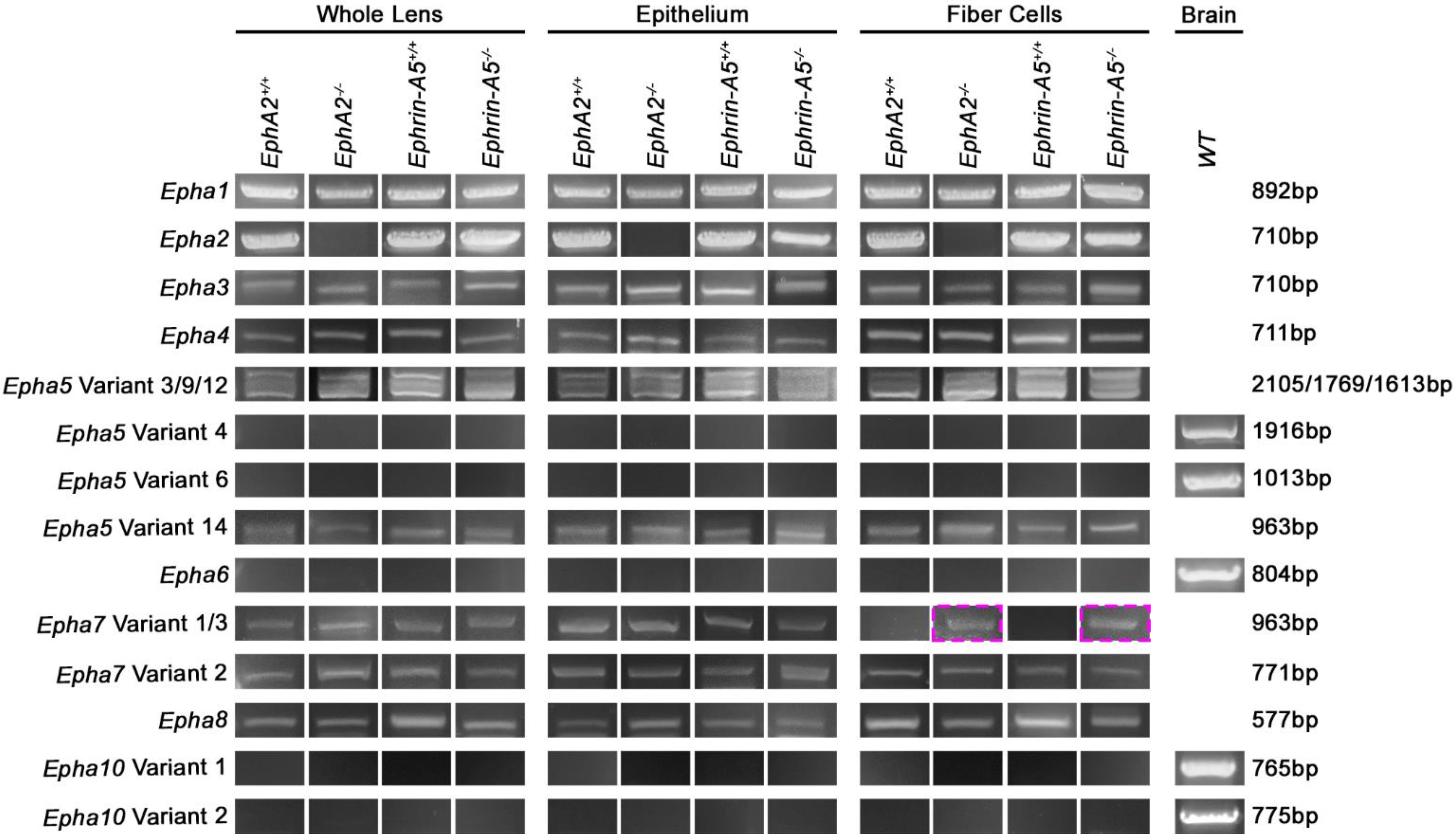
3.1. EphA Transcripts in the Lens

We analyzed RNA transcripts with specific primer pairs for Epha1–8 and Epha10 in control, EphA2−/−, and ephrin-A5−/− whole lens, epithelial cell, and fiber cell samples from 6-week-old mice. We found transcripts for Epha1, Epha2, Epha3 variant 1, Epha4, Epha5 variants 3, 9, 12, and 14, Epha7 variant 2, and Epha8 in all samples tested (Figure 1). As expected, Epha2 transcripts are absent from the EphA2−/− samples. Variants 1 and 2 of Epha3 differ by one amino acid, and Epha3 variant 1 has an additional amino acid, Q478. The presence of Epha3 variant 1 in lens samples was confirmed by sequencing of the PCR product. Epha5 has 14 different variants with different splicing patterns and lengths. We used sequencing to confirm the presence of four variants of Epha5 in the lens. Epha7 has three variants, 1, 2, and 3. Epha7 variants 1 and 3 differ by four amino acids, and variant 1 is longer, with the addition of 601–604 KFPG amino acids. We detected Epha7 variant 1 in whole lens and epithelial cells samples of the control and KO samples, as well as in KO fiber cell samples (Figure 1, magenta boxes). Epha7 variant 2 is a shorter variant lacking multiple exons and has a different and shorter C-terminus than variants 1 and 3. Sequencing confirmed the presence of the Epha7 variant 2 in all lens samples. We did not detect Epha6 or Epha10 in any samples. There are two variants, 1 and 2, for Epha10. Variant 2 of Epha10 is much shorter than variant 1, due to loss of exons, and has a unique C-terminus, compared to variant 1. Positive control PCR products from brain RNA samples were used to confirm primer sets and PCR conditions for the Epha5 variants 1, 4, 5, 6, 8, and 10 and the Epha6 and Epha10 experiments.

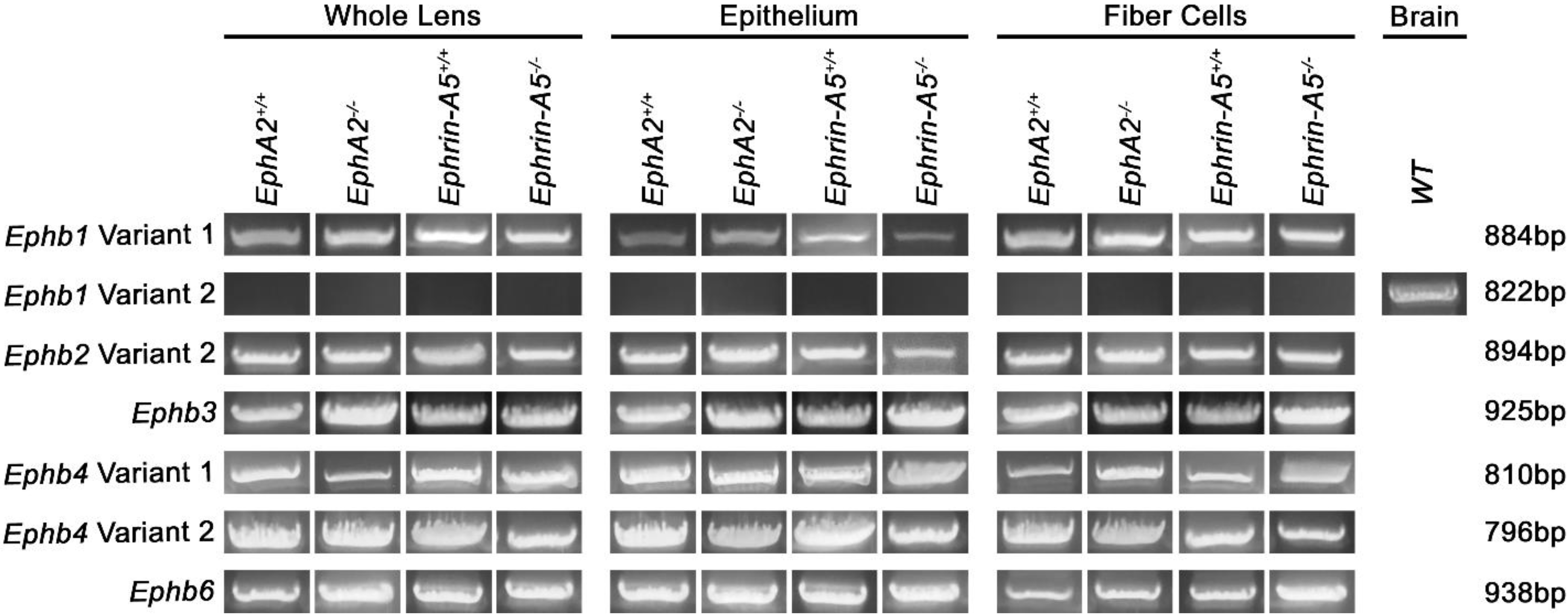
3.2. EphB Transcripts in the Lens

Next, we tested for the presence of Ephb1–4 and Ephb6 transcripts in the control and KO samples. We detected Ephb1 variant 1, Ephb2 variant 2, Ephb3, Ephb4 variants 1 and 2, and Ephb6 transcripts in all lens samples (Figure 2). The Ephb1 variant 1 is a longer isoform, and variant 2 lacks one of the coding exons. Ephb1 variants 1 and 2 have the same N- and C-termini. Specific primers were designed to distinguish between Ephb1 variants 1 and 2. The lens only expresses Ephb1 variant 1, and the primers for Ephb1 variant 2 were verified by the positive control brain RNA sample. Ephb2 variant 1 has one additional amino acid, Q477, compared to variant 2. Sequencing verified that the lens expresses Ephb2 variant 2. Ephb4 also has two variants, and due to an alternate in-frame splice site, variant 2 is a shorter transcript. Specific primers designed for each variant and sequencing confirm that both variants 1 and 2 of Ephb4 are expressed in the lens. Ephb6 also has two variants, but the two variants are identical in the coding region and differ by a four-nucleotide difference in the 5′ untranslated region (UTR).
In total, the lens expresses 12 of the 14 Eph genes. Due to the variants in multiple genes, we found 14 Eph transcripts in our samples. Most notably, the Epha7 variant 1 is normally only expressed in lens epithelial cells, but is present in the lens fiber cells of EphA2−/− and ephrin-A5−/− samples.

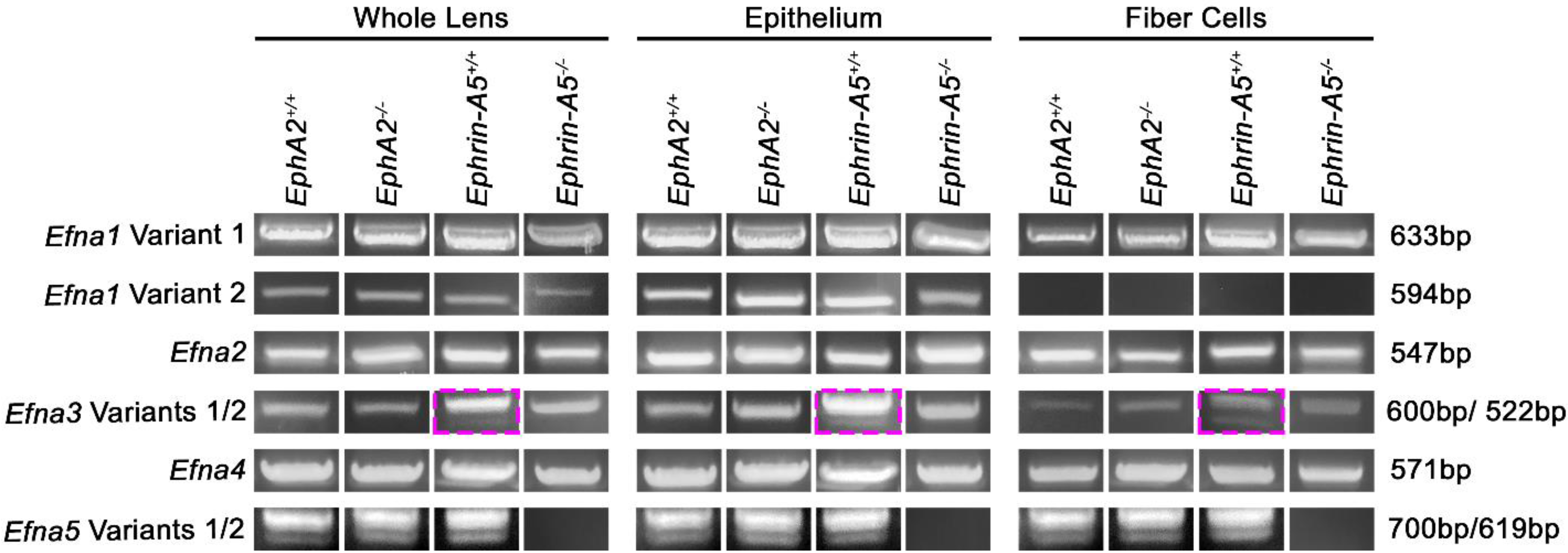
3.3. Ephrin-A Transcripts in the Lens

Ephrin-A proteins are encoded by the Efna genes. We performed experiments to determine whether Efna1–5 are present in the lens. Our results show that Efna1 variant 1, Efna2, Efna3 variant 1, Efna4, and Efna5 variants 1 and 2 transcripts are present in all lens samples (Figure 3). As expected, the Efna5 transcripts are absent in the ephrin-A5−/− samples. Efna1 variant 2 is missing a part of the 5′ UTR and coding region, resulting in a shorter transcript, compared to variant 1. Specific forward primers for Efna1 variants 1 and 2 revealed that the lens expresses the longer Efna1 variant 1 transcript in both epithelial cells and fiber cells, while Efna1 variant 2 is only expressed in lens epithelial cells. Efna3 has 7 variants. Efna3 variants 1 and 2 differ by one exon in the 3′ coding region and can be distinguished by PCR product size. We found Efna3 variant 1 transcripts in all lens samples. With sequencing verification, we could only detect very low levels of Efna3 variant 2 transcripts in the ephrin-A5+/+ control samples (Figure 3, magenta boxes). Efna3 variants 3, 4, and 5 have identical coding regions and are shorter transcripts with a start codon in the middle of the variant 1 sequence. There are minor differences in the 5′ UTR of Efna3 variants 3, 4, and 5 from the 5′ coding region of Efna3 variant 1; thus, specific primers to test for Efna3 variants 3, 4, and 5 could not be designed. Efna3 variants 6 and 7 have identical coding regions and are shorter transcripts with a start codon in the middle of the variant 2 sequence. There are some differences in the 5′ UTR of variants 6 and 7 from the 5′ coding region of variant 2, but specific primers could not be designed to distinguish between variants 2, 6, and 7. Interestingly, the lens expresses both variants of Efna5. Efna5 variant 2 lacks one exon and is shorter than variant 1, but both transcripts have the same N- and C-termini sequence.

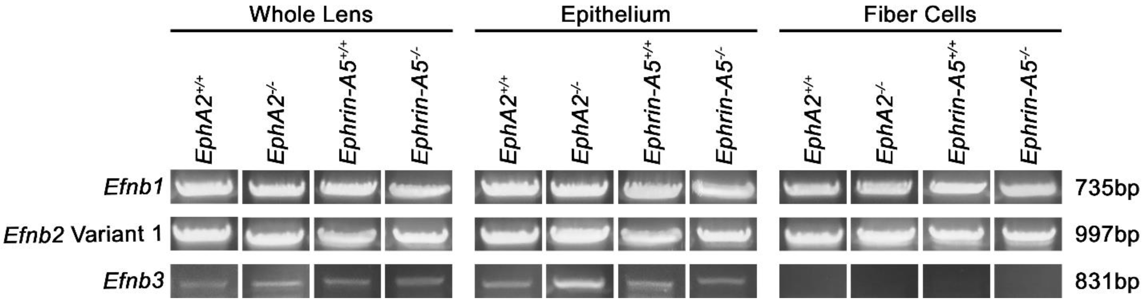
3.4. Ephrin-Bs Transcripts in the Lens

The last group of genes tested were Efnb1–3, which encode for ephrin-B1–3. We detected Efnb1 and Efnb2 variant 1 in all lens samples (Figure 4). Efnb2 has two variants, and variant 2 is shorter by 93 base pairs. Efnb2 variants 1 and 2 have the same N- and C-termini sequence. We did not detect the smaller Efnb2 variant 2 band in any of the lens samples. Interestingly, Efnb3 transcripts are only found in lens epithelial cells. Overall, we found transcripts for all eight Efn genes in the lens. Notably, Efna5 variants 1 and 2 were both expressed in the lens, and Efnb3 was only found in lens epithelial cells. All PCR and sequencing results are summarized in Table 1.

3.5. Data Comparison with Adult Lens Microarray Data

We compared our RT-PCR results to the Eph and Efn microarray data available in iSyte 2.0 from the Affymetrix 430 2.0 and Illumina WG-6 v2.0 arrays [42]. We chose to compare our data from 6-week-old mice with data from wild-type lenses at postnatal day 28 (P28) from the epithelium and at P56 from the whole lens on the Affymetrix platform and results from wild-type whole lenses from P30, P42, and P52 on the Illumina arrays with our data. It should be noted that the data from the two different microarray platforms should not be compared to each other, due to differences in technology for the two arrays. We designated detected (✓) or not detected (n.d.) for the microarray data (Table 2) and included the normalized lens expression numbers for each gene from iSyte 2.0 (Supplemental Table S2). The normalized lens expression values for the two different chip sets were in different ranges. Our data matches closely with the microarray data, except for a few of the genes. We detected the expression of Epha8, but the array data did not, and we did not detect Epha6, but the Affymetrix array did. Neither microarray tested for Epha10.
In Table 2, we also list other mouse tissues with high expression of each Eph or Efn. We wanted to determine whether there were other tissues with similar expression patterns of Eph or Efn as in the lens. The lens expresses many of the genes also detected in the brain, lung, and the gastrointestinal tract. However, no other tissues expressed as many Eph or Efn, compared to the lens.

4. Discussion

Our data shows that the adult mouse lens expresses 7 Epha, 5 Ephb, 5 Efna, and 3 Efnb transcripts. Counting all the different variants, we detected 18 Ephs and 11 Efns in the lens. Of these isoforms, three are only expressed in lens epithelial cells, Epha7 variant 1, Efna1 variant 2, and Efnb3. Epha7 variant 1 is also aberrantly expressed in the EphA2−/− and ephrin-A5−/− lens fiber cells. Interestingly, we only found Efna3 variant 2 in the control ephrin-A5+/+ samples. This variant has very faint PCR bands, but was consistently present in the control ephrin-A5+/+ whole lens, epithelial cell, and fiber cell samples. It is not clear why EphA2+/+ samples do not also express Efna3 variant 2. However, this does suggest that, even among control “wild-type” animals, there could be slight strain differences. This notion is supported by our previous work, showing differences between our control animals vs. pure C57BL6/J wild-type mice [43].
Our data reveals that the loss of EphA2 or ephrin-A5 causes abnormal expression of Epha7 variant 1 transcripts in KO lens fiber cells. This unexpected result was our first clue about compensatory upregulation or deregulation in KO animals. In addition to our data, a recent work described marked downregulation of Epha5 transcripts in developing lenses with the disruption of the musculoaponeurotic fibrosarcoma (MAF) family of proteins [44]. The data from RNA-seq plates did not distinguish between the 14 variants of Epha5. MAFs encode basic leucine zipper transcription factors that are known to be important for lens development and to be involved cataractogenesis [44,45,46,47,48,49,50]. There may be other insights that can be gleaned from the microarray data between the control, EphA2−/−, and ephrin-A5−/− lens samples. Comparison of our RT-PCR results and previous microarray data indicate a good match between the information from these data sets for most of the Eph and Efn isoforms. We may be able to use microarrays to quickly screen our KO lenses for highly upregulated or downregulated Ephs and Efns and examine large groups of other genes.
Our new method to isolate RNA from epithelial cells allows for ~20–30 PCR reactions, allowing for the efficient screening of transcripts. This protocol makes it possible to identify epithelial-cell specific isoforms. We had presumed that, in the whole lens samples, the fiber cell RNA content would dominate over the smaller amount of RNA from the lens epithelial cells. Different from our investigation of tropomyosin transcripts in whole lens [41] vs. epithelial cells [51], where several isoforms of tropomyosin were only detected in epithelial cell samples, we surprisingly found three epithelial cell-specific Eph/Efn transcripts (Epha7 variant 1, Efna1 variant 2, and Efnb3) that were also present in whole lens samples. Of note, Efnb3 did not have other variants expressed in fiber cells, but it was detected on both microarray platforms in both the epithelial and whole lens samples. Our data suggests that RNA from epithelial cells can be detected in whole lens samples; however, very low expression epithelial-cell-exclusive genes may still be difficult to detect in whole lens samples.
The large number of isoforms and variants of Ephs and Efns found in adult mouse lenses greatly complicates our search for lens receptor–ligand pairs. EphA receptors mainly bind to ephrin-As, while EphB receptors usually interact with ephrin-Bs [52,53]. Less common are the cross-interactions between EphAs and ephrin-Bs or EphBs and ephrin-As [27,54]. Each receptor can interact with multiple ligands, and similarly, each ligand can bind to multiple receptors [54]. This leads to a complex and large matrix of possible receptor–ligand pairs. To tackle the next phase of this project, we will be designing specific TaqMan real-time PCR assays to uncover changes in Eph and Efn expression levels between our control and KO samples. This may help us narrow down the list of priority isoforms to study in our KO mouse lines. Following the identification of priority isoforms for study, we plan to perform Western blotting, co-immunoprecipitation, immunostaining, and/or proximity ligation assay to determine the protein localization and receptor–ligand pairs.

Supplementary Materials

The following supporting information can be downloaded at: https://www.mdpi.com/article/10.3390/cells11203291/s1, Table S1: Primers, PCR product size, and PCR temperature; Table S2: Normalized lens expression of Eph and Efn transcripts in lens microarray studies.

Author Contributions

Conceptualization, resources, supervision, project administration, funding acquisition, C.C.; methodology, validation, formal analysis, data curation, writing—original draft preparation, writing—review and editing, visualization, C.C. and M.P.V. All authors have read and agreed to the published version of the manuscript.

Funding

This research was funded by grant R01 EY032056 from the National Eye Institute (to C.C.).

Institutional Review Board Statement

Not applicable.

Informed Consent Statement

Not applicable.

Data Availability Statement

The authors will provide a detailed description of methods and original data upon request.

Acknowledgments

We thank Arya S. Musthyala and Jackson T. Clark for technical assistance and Roberta B. Nowak for helpful discussion and troubleshooting advice.

Conflicts of Interest

The authors declare no conflict of interest.

References

  1. Lovicu, F.J.; Robinson, M.L. Development of the Ocular Lens; Cambridge University Press: Cambridge, UK, 2004; 398 p. [Google Scholar]
  2. WHO, World Health Organization: World Report on Vision. 2019. Available online: https://www.who.int/publications/i/item/9789241516570 (accessed on 8 October 2019).
  3. Berry, V.; Pontikos, N.; Albarca-Aguilera, M.; Plagnol, V.; Massouras, A.; Prescott, D.; Moore, A.T.; Arno, G.; Cheetham, M.E.; Michaelides, M. A recurrent splice-site mutation in EPHA2 causing congenital posterior nuclear cataract. Ophthalmic Genet. 2017, 39, 236–241. [Google Scholar] [CrossRef] [PubMed] [Green Version]
  4. Dave, A.; Laurie, K.; Staffieri, S.E.; Taranath, D.; Mackey, D.A.; Mitchell, P.; Wang, J.J.; Craig, J.; Burdon, K.P.; Sharma, S. Mutations in the EPHA2 Gene Are a Major Contributor to Inherited Cataracts in South-Eastern Australia. PLoS ONE 2013, 8, e72518. [Google Scholar] [CrossRef] [PubMed] [Green Version]
  5. Kaul, H.; Riazuddin, S.A.; Shahid, M.; Kousar, S.; Butt, N.H.; Zafar, A.U.; Khan, S.N.; Husnain, T.; Akram, J.; Hejtmancik, J.F.; et al. Autosomal recessive congenital cataract linked to EPHA2 in a consanguineous Pakistani family. Mol. Vis. 2010, 16, 511–517. [Google Scholar] [PubMed]
  6. Li, D.; Wang, S.; Ye, H.; Tang, Y.; Qiu, X.; Fan, Q.; Rong, X.; Liu, X.; Chen, Y.; Yang, J.; et al. Distribution of gene mutations in sporadic congenital cataract in a Han Chinese population. Mol. Vis. 2016, 22, 589–598. [Google Scholar]
  7. Park, J.E.; Son, A.I.; Hua, R.; Wang, L.; Zhang, X.; Zhou, R. Human Cataract Mutations in EPHA2 SAM Domain Alter Receptor Stability and Function. PLoS ONE 2012, 7, e36564. [Google Scholar] [CrossRef] [Green Version]
  8. Zhai, Y.; Zhu, S.; Li, J.; Yao, K. A Novel Human Congenital Cataract Mutation in EPHA2 Kinase Domain (p.G668D) Alters Receptor Stability and Function. Investig. Opthalmol. Vis. Sci. 2019, 60, 4717–4726. [Google Scholar] [CrossRef] [Green Version]
  9. Zhang, T.; Hua, R.; Xiao, W.; Burdon, K.P.; Bhattacharya, S.S.; Craig, J.E.; Shang, D.; Zhao, X.; Mackey, D.A.; Moore, A.T.; et al. Mutations of the EPHA2 receptor tyrosine kinase gene cause autosomal dominant congenital cataract. Hum. Mutat. 2009, 30, E603–E611. [Google Scholar] [CrossRef]
  10. Jun, G.; Guo, H.; Klein, B.E.K.; Klein, R.; Wang, J.J.; Mitchell, P.; Miao, H.; Lee, K.E.; Joshi, T.; Buck, M.; et al. EPHA2 Is Associated with Age-Related Cortical Cataract in Mice and Humans. PLoS Genet. 2009, 5, e1000584. [Google Scholar] [CrossRef] [Green Version]
  11. Lin, Q.; Zhou, N.; Zhang, N.; Qi, Y. Mutational Screening of EFNA5 in Chinese Age-Related Cataract Patients. Ophthalmic Res. 2014, 52, 124–129. [Google Scholar] [CrossRef]
  12. Sundaresan, P.; Ravindran, R.D.; Vashist, P.; Shanker, A.; Nitsch, D.; Talwar, B.; Maraini, G.; Camparini, M.; Nonyane, B.A.S.; Smeeth, L.; et al. EPHA2 Polymorphisms and Age-Related Cataract in India. PLoS ONE 2012, 7, e33001. [Google Scholar] [CrossRef] [Green Version]
  13. Tan, W.; Hou, S.; Jiang, Z.; Hu, Z.; Yang, P.; Ye, J. Association of EPHA2 polymorphisms and age-related cortical cataract in a Han Chinese population. Mol. Vis. 2011, 17, 1553–1558. [Google Scholar]
  14. Murugan, S.; Cheng, C. Roles of Eph-Ephrin Signaling in the Eye Lens Cataractogenesis, Biomechanics, and Homeostasis. Front. Cell Dev. Biol. 2022, 10. [Google Scholar] [CrossRef]
  15. Pitulescu, M.E.; Adams, R.H. Eph/ephrin molecules—a hub for signaling and endocytosis. Genes Dev. 2010, 24, 2480–2492. [Google Scholar] [CrossRef] [Green Version]
  16. Pasquale, E.B. Eph receptors and ephrins in cancer: Bidirectional signalling and beyond. Nat. Cancer 2010, 10, 165–180. [Google Scholar] [CrossRef] [Green Version]
  17. Darling, T.K.; Lamb, T.J. Emerging Roles for Eph Receptors and Ephrin Ligands in Immunity. Front. Immunol. 2019, 10, 1473. [Google Scholar] [CrossRef] [Green Version]
  18. Zhang, X.-Q.; Takakura, N.; Oike, Y.; Inada, T.; Gale, N.W.; Yancopoulos, G.D.; Suda, T. Stromal cells expressing ephrin-B2 promote the growth and sprouting of ephrin-B2+ endothelial cells. Blood 2001, 98, 1028–1037. [Google Scholar] [CrossRef] [Green Version]
  19. Davy, A.; Robbins, S.M. Ephrin-A5 modulates cell adhesion and morphology in an integrin-dependent manner. EMBO J. 2000, 19, 5396–5405. [Google Scholar] [CrossRef] [Green Version]
  20. Davy, A.; Gale, N.W.; Murray, E.W.; Klinghoffer, R.A.; Soriano, P.; Feuerstein, C.; Robbins, S.M. Compartmentalized signaling by GPI-anchored ephrin-A5 requires the Fyn tyrosine kinase to regulate cellular adhesion. Genes Dev. 1999, 13, 3125–3135. [Google Scholar] [CrossRef] [Green Version]
  21. Holmberg, J.; Clarke, D.L.; Frisén, J. Regulation of repulsion versus adhesion by different splice forms of an Eph receptor. Nat. 2000, 408, 203–206. [Google Scholar] [CrossRef]
  22. Lisabeth, E.M.; Falivelli, G.; Pasquale, E.B. Eph Receptor Signaling and Ephrins. Cold Spring Harb. Perspect. Biol. 2013, 5, a009159. [Google Scholar] [CrossRef] [Green Version]
  23. Noberini, R.; de la Torre, E.R.; Pasquale, E.B. Profiling Eph receptor expression in cells and tissues: A targeted mass spec-trometry approach. Cell Adh. Migr. 2012, 6, 102–112. [Google Scholar] [CrossRef] [Green Version]
  24. Pasquale, E.B. Eph–ephrin promiscuity is now crystal clear. Nat. Neurosci. 2004, 7, 417–418. [Google Scholar] [CrossRef]
  25. Pasquale, E.B. Eph–receptor signalling casts a wide net on cell behaviour. Nat. Rev. Mol. Cell Biol. 2005, 6, 462–475. [Google Scholar] [CrossRef]
  26. Himanen, J.-P.; Saha, N.; Nikolov, D.B. Cell–cell signaling via Eph receptors and ephrins. Curr. Opin. Cell Biol. 2007, 19, 534–542. [Google Scholar] [CrossRef] [Green Version]
  27. Kullander, K.; Klein, R. Mechanisms and functions of eph and ephrin signalling. Nat. Rev. Mol. Cell Biol. 2002, 3, 475–486. [Google Scholar] [CrossRef]
  28. Piatigorsky, J. Lens differentiation in vertebrates. A review of cellular and molecular features. Differentiation 1981, 19, 134–153. [Google Scholar] [CrossRef]
  29. Kuszak, J.R.; Mazurkiewicz, M.; Jison, L.; Madurski, A.; Ngando, A.; Zoltoski, R.K. Quantitative analysis of animal model lens anatomy: Accommodative range is related to fiber structure and organiza-tion. Vet Ophthalmol 2006, 9, 266–280. [Google Scholar] [CrossRef]
  30. Kuszak, J.; Zoltoski, R.; Sivertson, C. Fibre cell organization in crystalline lenses. Exp. Eye Res. 2003, 78, 673–687. [Google Scholar] [CrossRef]
  31. Cheng, C.; Ansari, M.M.; Cooper, J.A.; Gong, X. EphA2 and Src regulate equatorial cell morphogenesis during lens development. Development 2013, 140, 4237–4245. [Google Scholar] [CrossRef] [Green Version]
  32. Cheng, C.; Fowler, V.M.; Gong, X. EphA2 and ephrin-A5 are not a receptor-ligand pair in the ocular lens. Exp. Eye Res. 2017, 162, 9–17. [Google Scholar] [CrossRef]
  33. Zhou, Y.; Shiels, A. Epha2 and Efna5 participate in lens cell pattern-formation. Differentiation 2018, 102, 1–9. [Google Scholar] [CrossRef] [PubMed]
  34. Cheng, C. EphA2 and Ephrin-A5 Guide Eye Lens Suture Alignment and Influence Whole Lens Resilience. Investig. Opthalmol. Vis. Sci. 2021, 62, 3. [Google Scholar] [CrossRef] [PubMed]
  35. Cheng, C.; Wang, K.; Hoshino, M.; Uesugi, K.; Yagi, N.; Pierscionek, B. EphA2 Affects Development of the Eye Lens Nucleus and the Gradient of Refractive Index. Investig. Opthalmol. Vis. Sci. 2022, 63, 2. [Google Scholar] [CrossRef] [PubMed]
  36. Cheng, C.; Gong, X. Diverse Roles of Eph/ephrin Signaling in the Mouse Lens. PLoS ONE 2011, 6, e28147. [Google Scholar] [CrossRef] [Green Version]
  37. Shi, Y.; De Maria, A.; Bennett, T.; Shiels, A.; Bassnett, S. A Role for Epha2 in Cell Migration and Refractive Organization of the Ocular Lens. Investig. Opthalmol. Vis. Sci. 2012, 53, 551–559. [Google Scholar] [CrossRef] [Green Version]
  38. Parreno, J.; Emin, G.; Vu, M.P.; Clark, J.T.; Aryal, S.; Patel, S.D.; Cheng, C. Methodologies to unlock the molecular expression and cellular structure of ocular lens epithelial cells. Front. Cell Dev. Biol. 2022, 10, 1827. [Google Scholar] [CrossRef]
  39. Frisén, J.; Yates, P.A.; McLaughlin, T.; Friedman, G.C.; O’Leary, D.D.; Barbacid, M. Ephrin-A5 (AL-1/RAGS) is essential for proper retinal axon guidance and topographic mapping in the mammalian vis-ual system. Neuron 1998, 20, 235–243. [Google Scholar] [CrossRef] [Green Version]
  40. Brantley-Sieders, D.M.; Caughron, J.; Hicks, D.; Pozzi, A.; Ruiz, J.C.; Chen, J. EphA2 receptor tyrosine kinase regulates endothelial cell migration and vascular assembly through phosphoinositide 3-kinase-mediated Rac1 GTPase activation. J. Cell Sci. 2004, 117, 2037–2049. [Google Scholar] [CrossRef] [Green Version]
  41. Cheng, C.; Nowak, R.B.; Amadeo, M.B.; Biswas, S.K.; Lo, W.K.; Fowler, V.M. Tropomyosin 3.5 protects the F-actin networks required for tissue biomechanical properties. J. Cell Sci. 2018, 131, jcs222042. [Google Scholar] [CrossRef] [Green Version]
  42. Kakrana, A.; Yang, A.; Anand, D.; Djordjevic, D.; Ramachandruni, D.; Singh, A.; Huang, H.; Ho, J.W.K.; A Lachke, S. iSyTE 2.0: A database for expression-based gene discovery in the eye. Nucleic Acids Res. 2017, 46, D875–D885. [Google Scholar] [CrossRef]
  43. Cheng, C.; Gao, J.; Sun, X.; Mathias, R.T. Eph-ephrin Signaling Affects Eye Lens Fiber Cell Intracellular Voltage and Membrane Conductance. Front. Physiol. 2021, 12, 2072. [Google Scholar] [CrossRef]
  44. Patel, S.D.; Anand, D.; Motohashi, H.; Katsuoka, F.; Yamamoto, M.; Lachke, S.A. Deficiency of the bZIP transcription factors Mafg and Mafk causes misexpression of genes in distinct pathways and results in lens embryonic developmental defects. Front. Cell Dev. Biol. 2022, 10, 981893. [Google Scholar] [CrossRef]
  45. Agrawal, S.A.; Anand, D.; Siddam, A.D.; Kakrana, A.; Dash, S.; Scheiblin, D.A.; Dang, C.A.; Terrell, A.M.; Waters, S.M.; Singh, A.; et al. Compound mouse mutants of bZIP transcription factors Mafg and Mafk reveal a regulatory network of non-crystallin genes associated with cataract. Qual. Life Res. 2015, 134, 717–735. [Google Scholar] [CrossRef] [Green Version]
  46. Anand, D.; Agrawal, S.A.; Slavotinek, A.; Lachke, S.A. Mutation update of transcription factor genes FOXE3, HSF4, MAF, and PITX3 causing cataracts and other develop-mental ocular defects. Hum. Mutat. 2018, 39, 471–494. [Google Scholar]
  47. Jamieson, R.V.; Perveen, R.; Kerr, B.; Carette, M.; Yardley, J.; Heon, E.; Wirth, M.G.; Van Heyningen, V.; Donnai, D.; Munier, F.; et al. Domain disruption and mutation of the bZIP transcription factor, MAF, associated with cataract, ocular anterior segment dysgenesis and coloboma. Hum. Mol. Genet. 2002, 11, 33–42. [Google Scholar] [CrossRef] [Green Version]
  48. Kawauchi, S.; Takahashi, S.; Nakajima, O.; Ogino, H.; Morita, M.; Nishizawa, M.; Yasuda, K.; Yamamoto, M. Regulation of Lens Fiber Cell Differentiation by Transcription Factor c-Maf. J. Biol. Chem. 1999, 274, 19254–19260. [Google Scholar] [CrossRef] [Green Version]
  49. Kim, J.I.; Li, T.; Ho, I.-C.; Grusby, M.J.; Glimcher, L.H. Requirement for the c-Maf transcription factor in crystallin gene regulation and lens development. Proc. Natl. Acad. Sci. 1999, 96, 3781–3785. [Google Scholar] [CrossRef] [Green Version]
  50. Ring, B.Z.; Cordes, S.P.; A Overbeek, P.; Barsh, G.S. Regulation of mouse lens fiber cell development and differentiation by the Maf gene. Development 2000, 127. [Google Scholar] [CrossRef]
  51. Parreno, J.; Amadeo, M.B.; Kwon, E.H.; Fowler, V.M. Tropomyosin 3.1 Association With Actin Stress Fibers is Required for Lens Epithelial to Mesenchymal Transition. Investig. Opthalmol. Vis. Sci. 2020, 61, 2. [Google Scholar] [CrossRef]
  52. Himanen, J.-P.; Chumley, M.J.; Lackmann, M.; Li, C.; A Barton, W.; Jeffrey, P.D.; Vearing, C.; Geleick, D.; A Feldheim, D.; Boyd, A.W.; et al. Repelling class discrimination: Ephrin-A5 binds to and activates EphB2 receptor signaling. Nat. Neurosci. 2004, 7, 501–509. [Google Scholar] [CrossRef]
  53. Takemoto, M.; Fukuda, T.; Sonoda, R.; Murakami, F.; Tanaka, H.; Yamamoto, N. Ephrin-B3-EphA4 interactions regulate the growth of specific thalamocortical axon populations in vitro. Eur. J. Neurosci. 2002, 16, 1168–1172. [Google Scholar] [CrossRef] [PubMed]
  54. Gale, N.W.; Holland, S.J.; Valenzuela, D.M.; Flenniken, A.; Pan, L.; Ryan, T.E.; Henkemeyer, M.; Strebhardt, K.; Hirai, H.; Wilkinson, D.; et al. Eph Receptors and Ligands Comprise Two Major Specificity Subclasses and Are Reciprocally Compartmentalized during Embryogenesis. Neuron 1996, 17, 9–19. [Google Scholar] [CrossRef]
Figure 1. Epha transcripts in whole lens, lens epithelial cell, or lens fiber cell RNA samples from 6-week-old EphA2+/+, EphA2−/−, ephrin-A5+/+, and ephrin-A5−/− mice. Positive control was brain RNA isolated from a wild-type (WT) control mouse. We detected Epha1, Epha2, Epha3 variant 1, Epha4, Epha5 variants 3, 9, 12, and 14, Epha7 variants 1 and 2, and Epha8 in the lens. As expected, Epha2 was not detected in EphA2−/− lens samples. Epha7 variant 1 was detected in all whole lens and epithelial cell samples and in the fiber cells of EphA2−/− and ephrin-A5−/− lenses (magenta boxes). We did not detect Epha6 or Epha10 in the lens.
Cells 11 03291 g001
Figure 2. Ephb transcripts in whole lens, lens epithelial cell, or lens fiber cell RNA samples from 6-week-old EphA2+/+, EphA2−/−, ephrin-A5+/+, and ephrin-A5−/− mice. Positive control was brain RNA isolated from a WT control mouse. We detected Ephb1 variant 1, Ephb2 variant 2, Ephb3, Ephb4 variants 1 and 2, and Ephb6 in the lens. We did not detect Ephb1 variant 2 in the lens.
Cells 11 03291 g002
Figure 3. Efna transcripts, which encode for ephrin-A proteins, in whole lens, lens epithelial cell, or lens fiber cell RNA samples from 6-week-old EphA2+/+, EphA2−/−, ephrin-A5+/+, and ephrin-A5−/− mice. We detected Efna1 variant 1, Efna2, Efna3 variant 1, Efna4, and Efna5 variants 1 and 2 in the lens. Efna1 variant 2 was detected in the whole lens and epithelial cells, but not detected in fiber cells. Efna3 variant 2 was only detected in ephrin-A5+/+ lens samples (magenta boxes).
Cells 11 03291 g003
Figure 4. Efnb transcripts, which encode for ephrin-B proteins, in whole lens, lens epithelial cell, or lens fiber cell RNA samples from 6-week-old EphA2+/+, EphA2−/−, ephrin-A5+/+, and ephrin-A5−/− mice. We detected Efnb1 and Efnb2 variant 1 in the lens. Efnb3 was detected in the whole lens and epithelial cells, but not detected in fiber cells.
Cells 11 03291 g004
Table 1. Eph and Efn transcripts in the lens.
GeneWhole LensEpitheliumFiber Cells
EphA2Ephrin-A5EphA2Ephrin-A5EphA2Ephrin-A5
+/+-/-+/+-/-+/+-/-+/+-/-+/+-/-+/+-/-
Epha1
Epha2KOKOKO
Epha3Variant 1
Epha3 Variant 2n.d.n.d.n.d.
Epha4
Epha5 Variant 1n.d.n.d.n.d.
Epha5 Variant 2n.d.n.d.n.d.
Epha5 Variant 3
Epha5 Variant 4n.d.n.d.n.d.
Epha5 Variant 5n.d.n.d.n.d.
Epha5 Variant 6n.d.n.d.n.d.
Epha5 Variant 7n.d.n.d.n.d.
Epha5 Variant 8n.d.n.d.n.d.
Epha5 Variant 9
Epha5 Variant 10n.d.n.d.n.d.
Epha5 Variant 11n.d.n.d.n.d.
Epha5Variant 12
Epha5 Variant 13n.d.n.d.n.d.
Epha5 Variant 14
Epha6n.d.n.d.n.d.
Epha7 Variant 1n.d.n.d.
Epha7 Variant 2
Epha7 Variant 3n.d.n.d.n.d.
Epha8
Epha10 Variant 1n.d.n.d.n.d.
Epha10 Variant 2n.d.n.d.n.d.
Ephb1 Variant 1
Ephb1 Variant 2n.d.n.d.n.d.
Ephb2 Variant 1n.d.n.d.n.d.
Ephb2 Variant 2
Ephb3
Ephb4 Variant 1
Ephb4 Variant 2
Ephb6 Variant 1
Ephb6 Variant 2
Efna1 Variant 1
Efna1 Variant 2n.d.
Efna2
Efna3 Variant 1 *
Efna3 Variant 2 *n.d.n.d.n.d.n.d.n.d.n.d.n.d.n.d.n.d.
Efna4
Efna5 Variant 1KOKOKO
Efna5 Variant 2KOKOKO
Efnb1
Efnb2 Variant 1
Efnb2 Variant 2n.d.n.d.n.d.
Efnb3n.d.
+/+ = wild-type or control; −/− or KO = knockout; n.d. = not detected; blue = detected in all lens fractions; orange = detected in some lens fractions; yellow highlight = detected in lens epithelial fractions, but not in lens fiber cell fractions; * There are two shorter variants of Efna3, variants 3/4/5 and variants 6/7, that cannot be distinguished by RT-PCR.
Table 2. Eph and Efn transcripts in lens microarray studies and other tissues.
GeneiSyte 2.0NCBI
Affymetrix 430 2.0Illumina WG-6 v2.0
P28 EpiP56P30P42P52Other tissues with high expression
Epha1Duodenum, intestines, lung
Epha2Duodenum, intestines, lung
Epha3Embryonic/adult brain, embryonic limb
Epha4Embryonic/adult brain, embryonic limb, heart
Epha5Embryonic/adult brain
Epha6n.d.n.d.n.d.Adult brain
Epha7Embryonic/adult brain, embryonic limb
Epha8 1n.dn.dn.dn.dn.dEmbryonic brain, adult cerebellum
Epha10 2N/AN/AN/AN/AN/AEmbryonic/adult brain, testis
Ephb1Embryonic/adult brain
Ephb2Embryonic brain, adrenal, colon, intestines
Ephb3n.d.n.d.n.d.Embryonic limb, colon, lung, stomach
Ephb4Embryonic limb, colon, lung, ovary
Ephb6Thymus, adult cortex
Efna1Duodenum, lung, intestines
Efna2n.d.n.d.n.d.Embryonic brain, embryonic liver, ovary
Efna3n.d.n.d.n.d.Embryonic brain, embryonic limb, stomach
Efna4Embryonic limb, duodenum, ovary
Efna5Embryonic brain, embryonic limb, bladder
Efnb1Colon, duodenum, lung, ovary
Efnb2Colon, lung
Efnb3 3Embryonic brain, embryonic limb, heart
n.d. = not detected. Bolded genes were detected in our RT-PCR experiments; 1 Epha8 was detected at low levels in E15.5 and P0 lenses on the Illumina WG-6 v1.1 microarray; 2 Epha10 was not tested in any of the reported arrays on iStye 2.0; 3 Efnb3 is only expressed in lens epithelial cells.
Publisher’s Note: MDPI stays neutral with regard to jurisdictional claims in published maps and institutional affiliations.

