Next Issue
Volume 11, November-1
Previous Issue
Volume 11, October-1


Cells, Volume 11, Issue 20 (October-2 2022) – 151 articles

Cover Story (view full-size image): 
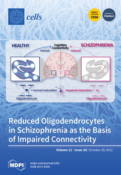
Cognitive deficits are a hallmark of schizophrenia and related to impaired hippocampal function. Here, we report on a design-based stereology study that showed decreased numbers of oligodendrocytes in bilateral postmortem hippocampal cornu ammonis 4 (CA4) in patients with schizophrenia. This study replicates previous findings in an independent brain collection. Oligodendrocytes are involved in myelination and also supply energy to neuronal axons. In schizophrenia, impaired myelination of projecting axons from pyramidal neurons leads to disturbed macroconnectivity, and decreased myelination of parvalbumin-expressing interneurons is hypothesized to lead to decreased local connectivity. As an underlying cause, oligodendrocyte precursor cells (OPCs) may fail to differentiate properly into mature oligodendrocytes. This topic is currently under investigation with specific antibodies for OPCs. View this paper
  • Issues are regarded as officially published after their release is announced to the table of contents alert mailing list.
  • You may sign up for e-mail alerts to receive table of contents of newly released issues.
  • PDF is the official format for papers published in both, html and pdf forms. To view the papers in pdf format, click on the "PDF Full-text" link, and use the free Adobe Reader to open them.
Order results
Result details
Section
Select all
Export citation of selected articles as:
15 pages, 364 KiB  
Article
LC-DAD–ESI-MS/MS and NMR Analysis of Conifer Wood Specialized Metabolites
by Andrzej Patyra, Marta Katarzyna Dudek and Anna Karolina Kiss
Cells 2022, 11(20), 3332; https://doi.org/10.3390/cells11203332 - 21 Oct 2022
Cited by 8 | Viewed by 2833
Abstract
Many species from the Pinaceae family have been recognized as a rich source of lignans, flavonoids, and other polyphenolics. The great common occurrence of conifers in Europe, as well as their use in the wood industry, makes both plant material and industrial waste [...] Read more.
Many species from the Pinaceae family have been recognized as a rich source of lignans, flavonoids, and other polyphenolics. The great common occurrence of conifers in Europe, as well as their use in the wood industry, makes both plant material and industrial waste material easily accessible and inexpensive. This is a promising prognosis for both discovery of new active compounds as well as for finding new applications for wood and its industry waste products. This study aimed to analyze and phytochemically profile 13 wood extracts of the Pinaceae family species, endemic or introduced in Polish flora, using the LC-DAD–ESI-MS/MS method and compare their respective metabolite profiles. Branch wood methanolic extracts were phytochemically profiled. Lignans, stilbenes, flavonoids, diterpenes, procyanidins, and other compounds were detected, with a considerable variety of chemical content among distinct species. Norway spruce (Picea abies (L.) H.Karst.) branch wood was the most abundant source of stilbenes, European larch (Larix decidua Mill.) mostly contained flavonoids, while silver fir (Abies alba Mill.) was rich in lignans. Furthermore, 10 lignans were isolated from the studied material. Our findings confirm that wood industry waste materials, such as conifer branches, can be a potent source of different phytochemicals, with the plant matrix being relatively simple, facilitating future isolation of target compounds. Full article
Show Figures

Graphical abstract

30 pages, 491 KiB  
Review
Induced Pluripotent Stem Cells, a Stepping Stone to In Vitro Human Models of Hearing Loss
by María Beatriz Durán-Alonso and Hrvoje Petković
Cells 2022, 11(20), 3331; https://doi.org/10.3390/cells11203331 - 21 Oct 2022
Cited by 4 | Viewed by 2167
Abstract
Hearing loss is the most prevalent sensorineural impairment in humans. Yet despite very active research, no effective therapy other than the cochlear implant has reached the clinic. Main reasons for this failure are the multifactorial nature of the disorder, its heterogeneity, and a [...] Read more.
Hearing loss is the most prevalent sensorineural impairment in humans. Yet despite very active research, no effective therapy other than the cochlear implant has reached the clinic. Main reasons for this failure are the multifactorial nature of the disorder, its heterogeneity, and a late onset that hinders the identification of etiological factors. Another problem is the lack of human samples such that practically all the work has been conducted on animals. Although highly valuable data have been obtained from such models, there is the risk that inter-species differences exist that may compromise the relevance of the gathered data. Human-based models are therefore direly needed. The irruption of human induced pluripotent stem cell technologies in the field of hearing research offers the possibility to generate an array of otic cell models of human origin; these may enable the identification of guiding signalling cues during inner ear development and of the mechanisms that lead from genetic alterations to pathology. These models will also be extremely valuable when conducting ototoxicity analyses and when exploring new avenues towards regeneration in the inner ear. This review summarises some of the work that has already been conducted with these cells and contemplates future possibilities. Full article
(This article belongs to the Special Issue Stem Cells and Hearing Loss)
17 pages, 4994 KiB  
Article
High-Dimensional Cytometry Dissects Immunological Fingerprints of Idiopathic Inflammatory Myopathies
by Christopher Nelke, Marc Pawlitzki, Christina B. Schroeter, Niklas Huntemann, Saskia Räuber, Vera Dobelmann, Corinna Preusse, Andreas Roos, Yves Allenbach, Olivier Benveniste, Heinz Wiendl, Ingrid E. Lundberg, Werner Stenzel, Sven G. Meuth and Tobias Ruck
Cells 2022, 11(20), 3330; https://doi.org/10.3390/cells11203330 - 21 Oct 2022
Cited by 3 | Viewed by 2248
Abstract
Chronic inflammation of skeletal muscle is the common feature of idiopathic inflammatory myopathies (IIM). Given the rarity of the disease and potential difficulty of routinely obtaining target tissue, i.e., standardized skeletal muscle, our understanding of immune signatures of the IIM spectrum remains incomplete. [...] Read more.
Chronic inflammation of skeletal muscle is the common feature of idiopathic inflammatory myopathies (IIM). Given the rarity of the disease and potential difficulty of routinely obtaining target tissue, i.e., standardized skeletal muscle, our understanding of immune signatures of the IIM spectrum remains incomplete. Further insight into the immune topography of IIM is needed to determine specific treatment targets according to clinical and immunological phenotypes. Thus, we used high-dimensional flow cytometry to investigate the immune phenotypes of anti-synthetase syndrome (ASyS), dermatomyositis (DM) and inclusion-body myositis (IBM) patients as representative entities of the IIM spectrum and compared them to healthy controls. We studied the CD8, CD4 and B cell compartments in the blood aiming to provide a contemporary overview of the immune topography of the IIM spectrum. ASyS was characterized by altered CD4 composition and expanded T follicular helper cells supporting B cell-mediated autoimmunity. For DM, unsupervised clustering identified expansion of distinct B cell subtypes highly expressing immunoglobulin G4 (IgG4) and CD38. Lastly, terminally differentiated, cytotoxic CD8 T cells distinguish IBM from other IIM. Interestingly, these terminally differentiated CD8 T cells highly expressed the integrin CD18 mediating cellular adhesion and infiltration. The distinct immune cell topography of IIM might provide the framework for targeted treatment approaches potentially improving therapeutic outcomes. Full article
Show Figures

Figure 1

17 pages, 14605 KiB  
Article
Assessment of Choroidal Vasculature and Innate Immune Cells in the Eyes of Albino and Pigmented Mice
by Ismail S. Zaitoun, Yong-Seok Song, Hammam B. Zaitoun, Christine M. Sorenson and Nader Sheibani
Cells 2022, 11(20), 3329; https://doi.org/10.3390/cells11203329 - 21 Oct 2022
Cited by 4 | Viewed by 2373
Abstract
The visualization of choroidal vasculature and innate immune cells in the eyes of pigmented mice has been challenging due to the presence of a retinal pigment epithelium (RPE) layer separating the choroid and retina. Here, we established methods for visualizing the choroidal macrophages, [...] Read more.
The visualization of choroidal vasculature and innate immune cells in the eyes of pigmented mice has been challenging due to the presence of a retinal pigment epithelium (RPE) layer separating the choroid and retina. Here, we established methods for visualizing the choroidal macrophages, mast cells, and vasculature in eyes of albino and pigmented mice using cell type-specific staining. We were able to visualize the choroidal arterial and venous systems. An arterial circle around the optic nerve was found in mice similar to the Zinn–Haller arterial circle that exists in humans and primates. Three different structural patterns of choriocapillaris were observed throughout the whole choroid: honeycomb-like, maze-like, and finger-like patterns. Choroidal mast cells were relatively few but dense around the optic nerve. Mast cell distribution in the middle and periphery was different among strains. Macrophages were found in all layers of the choroid. Thus, utilizing the simple and reliable methods described herein will allow the evaluation of transgenic and preclinical mouse models of ocular diseases that affect the choroid, including age-related macular degeneration (AMD), diabetic choroidopathy, and retinopathy of prematurity. These studies will advance our understanding of the pathophysiology, and molecular and cellular mechanisms that can be targeted therapeutically, in these diseases. Full article
(This article belongs to the Special Issue Retinal Cell Biology in Health and Disease)
Show Figures

Figure 1

20 pages, 2234 KiB  
Review
mRNA-Based Approaches to Treating Liver Diseases
by Maximiliano L. Cacicedo, María José Limeres and Stephan Gehring
Cells 2022, 11(20), 3328; https://doi.org/10.3390/cells11203328 - 21 Oct 2022
Cited by 4 | Viewed by 2744
Abstract
Diseases that affect the liver account for approximately 2 million deaths worldwide each year. The increasing prevalence of these diseases and the limited efficacy of current treatments are expected to stimulate substantial growth in the global market for therapeutics that target the liver. [...] Read more.
Diseases that affect the liver account for approximately 2 million deaths worldwide each year. The increasing prevalence of these diseases and the limited efficacy of current treatments are expected to stimulate substantial growth in the global market for therapeutics that target the liver. Currently, liver transplantation is the only curative option available for many liver diseases. Gene therapy represents a valuable approach to treatment. The liver plays a central role in a myriad of essential metabolic functions, making it an attractive organ for gene therapy; hepatocytes comprise the most relevant target. To date, viral vectors constitute the preferred approach to targeting hepatocytes with genes of therapeutic interest. Alternatively, mRNA-based therapy offers a number of comparative advantages. Clinical and preclinical studies undertaken to treat inherited metabolic diseases affecting the liver, cirrhosis and fibrosis, hepatocellular carcinoma, hepatitis B, and cytomegalovirus using lipid nanoparticle-encapsulated mRNAs that encode the therapeutic or antigenic protein of interest are discussed. Full article
(This article belongs to the Special Issue Hepatology: From Natural History to Therapeutics)
Show Figures

Figure 1

19 pages, 2398 KiB  
Review
Is IIIG9 a New Protein with Exclusive Ciliary Function? Analysis of Its Potential Role in Cancer and Other Pathologies
by María José Oviedo, Eder Ramírez, Manuel Cifuentes, Carlos Farkas, Andy Mella, Romina Bertinat, Roberto Gajardo, Luciano Ferrada, Nery Jara, Isabelle De Lima, Fernando Martínez, Francisco Nualart and Katterine Salazar
Cells 2022, 11(20), 3327; https://doi.org/10.3390/cells11203327 - 21 Oct 2022
Cited by 1 | Viewed by 1709
Abstract
The identification of new proteins that regulate the function of one of the main cellular phosphatases, protein phosphatase 1 (PP1), is essential to find possible pharmacological targets to alter phosphatase function in various cellular processes, including the initiation and development of multiple diseases. [...] Read more.
The identification of new proteins that regulate the function of one of the main cellular phosphatases, protein phosphatase 1 (PP1), is essential to find possible pharmacological targets to alter phosphatase function in various cellular processes, including the initiation and development of multiple diseases. IIIG9 is a regulatory subunit of PP1 initially identified in highly polarized ciliated cells. In addition to its ciliary location in ependymal cells, we recently showed that IIIG9 has extraciliary functions that regulate the integrity of adherens junctions. In this review, we perform a detailed analysis of the expression, localization, and function of IIIG9 in adult and developing normal brains. In addition, we provide a 3D model of IIIG9 protein structure for the first time, verifying that the classic structural and conformational characteristics of the PP1 regulatory subunits are maintained. Our review is especially focused on finding evidence linking IIIG9 dysfunction with the course of some pathologies, such as ciliopathies, drug dependence, diseases based on neurological development, and the development of specific high-malignancy and -frequency brain tumors in the pediatric population. Finally, we propose that IIIG9 is a relevant regulator of PP1 function in physiological and pathological processes in the CNS. Full article
Show Figures

Figure 1

15 pages, 6199 KiB  
Article
Synovial Macrophages Expression of OX40L Is Required for Follicular Helper T Cells Differentiation in the Joint Microenvironment
by Xiaoyu Cai, Meng Zhang, Fujia Ren, Weidong Fei, Xiao Zhang, Yunchun Zhao, Yao Yao and Nengming Lin
Cells 2022, 11(20), 3326; https://doi.org/10.3390/cells11203326 - 21 Oct 2022
Cited by 4 | Viewed by 1625
Abstract
Signaling via the OX40/OX40L axis plays a key role in CD4+ T cell development, and OX40L expression is primarily restricted to antigen-presenting cells (APCs). This study was designed to assess the role of APC-mediated OX40L expression in the context of the development [...] Read more.
Signaling via the OX40/OX40L axis plays a key role in CD4+ T cell development, and OX40L expression is primarily restricted to antigen-presenting cells (APCs). This study was designed to assess the role of APC-mediated OX40L expression in the context of the development of rheumatoid arthritis (RA)-associated CD4+ T cell subsets. For these analyses, clinical samples were harvested from patients with osteoarthritis and RA, with additional analyses performed using OX40−/− mice and mice harboring monocyte/macrophage-specific deletions of OX40L. Together, these analyses revealed tissue-specific roles for OX40/OX40L signaling in RA. Specifically, higher levels of synovial macrophage OX40L expression were associated with the enhanced development of T follicular helper cells in the joint microenvironment, thereby contributing to the pathogenesis of RA. This Tfh differentiation was found to be OX40/OX40L-dependent in this synovial setting. Overall, these results indicate that the expression of OX40L by synovia macrophages is necessary to support Tfh differentiation in the joint tissues, thus offering new insight regarding the etiological basis for RA progression. Full article
(This article belongs to the Special Issue Advances in Cellular and Molecular Treatment of Autoimmune Diseases)
Show Figures

Graphical abstract

22 pages, 2767 KiB  
Review
Critical Review on Physiological and Molecular Features during Bovine Mammary Gland Development: Recent Advances
by Shalini Jaswal, Manoj Kumar Jena, Vijay Anand, Avinash Jaswal, Sudhakar Kancharla, Prachetha Kolli, Gowtham Mandadapu, Sudarshan Kumar and Ashok Kumar Mohanty
Cells 2022, 11(20), 3325; https://doi.org/10.3390/cells11203325 - 21 Oct 2022
Cited by 9 | Viewed by 3289
Abstract
The mammary gland is a unique organ with the ability to undergo repeated cyclic changes throughout the life of mammals. Among domesticated livestock species, ruminants (cattle and buffalo) constitute a distinct class of livestock species that are known milk producers. Cattle and buffalo [...] Read more.
The mammary gland is a unique organ with the ability to undergo repeated cyclic changes throughout the life of mammals. Among domesticated livestock species, ruminants (cattle and buffalo) constitute a distinct class of livestock species that are known milk producers. Cattle and buffalo contribute to 51 and 13% of the total milk supply in the world, respectively. They also play an essential role in the development of the economy for farming communities by providing milk, meat, and draft power. The development of the ruminant mammary gland is highly dynamic and multiphase in nature. There are six developmental stages: embryonic, prepubertal, pubertal, pregnancy, lactation, and involution. There has been substantial advancement in our understanding of the development of the mammary gland in both mouse and human models. Until now, there has not been a thorough investigation into the molecular processes that underlie the various stages of cow udder development. The current review sheds light on the morphological and molecular changes that occur during various developmental phases in diverse species, with a particular focus on the cow udder. It aims to explain the physiological differences between cattle and non-ruminant mammalian species such as humans, mice, and monkeys. Understanding the developmental biology of the mammary gland in molecular detail, as well as species-specific variations, will facilitate the researchers working in this area in further studies on cellular proliferation, differentiation, apoptosis, organogenesis, and carcinogenesis. Additionally, in-depth knowledge of the mammary gland will promote its use as a model organ for research work and promote enhanced milk yield in livestock animals without affecting their health and welfare. Full article
Show Figures

Figure 1

17 pages, 4819 KiB  
Article
Paracrine Senescence of Mesenchymal Stromal Cells Involves Inflammatory Cytokines and the NF-κB Pathway
by Lun-Yin Chou, Chun-Te Ho and Shih-Chieh Hung
Cells 2022, 11(20), 3324; https://doi.org/10.3390/cells11203324 - 21 Oct 2022
Cited by 12 | Viewed by 1845
Abstract
It has been known that senescence-associated secretory phenotype (SASP) triggers senescence of the surrounding normal cells. However, SASP signaling regarding mesenchymal stromal cell aging remains to be fully elucidated. Therefore, the present study aimed to clarify the molecular mechanism of late (passage) MSC-induced [...] Read more.
It has been known that senescence-associated secretory phenotype (SASP) triggers senescence of the surrounding normal cells. However, SASP signaling regarding mesenchymal stromal cell aging remains to be fully elucidated. Therefore, the present study aimed to clarify the molecular mechanism of late (passage) MSC-induced paracrine SASP-mediated senescence of early (passage) MSCs during ex vivo expansion. Here, we conducted an extensive characterization of senescence features in bone-marrow (BM)-derived MSCs from healthy human donors. Late MSCs displayed an enlarged senescent-like morphology, induced SASP-related proinflammatory cytokines (IL-1α and IL-8), and reduced clonogenic capacity and osteogenic differentiation when compared to early MSCs. Of note, paracrine effects of SASP-related IL-1α and IL-8 from late MSCs induced cellular senescence of early MSCs via an NF-κB-dependent manner. Moreover, cellular senescence of early MSCs was promoted by the synergistic action of IL-1α and IL-8. However, inhibition of NF-κB by shRNA transfection or using inhibitors in early MSCs blocked early MSCs cellular senescence caused by paracrine SASP of late MSCs. In conclusion, these findings reveal that late MSCs display features of senescence and that, during ex vivo expansion, SASP-related proinflammatory cytokines contribute to activate a cellular senescence program in early MSCs that may ultimately impair their functionality. Full article
(This article belongs to the Special Issue Cellular Plasticity in Ageing)
Show Figures

Figure 1

16 pages, 4220 KiB  
Article
Spatial and Temporal Diversity of Astrocyte Phenotypes in Spinocerebellar Ataxia Type 1 Mice
by Juao-Guilherme Rosa, Katherine Hamel, Carrie Sheeler, Ella Borgenheimer, Stephen Gilliat, Alyssa Soles, Ferris J. Ghannoum, Kaelin Sbrocco, Hillary P. Handler, Orion Rainwater, Ryan Kang and Marija Cvetanovic
Cells 2022, 11(20), 3323; https://doi.org/10.3390/cells11203323 - 21 Oct 2022
Cited by 3 | Viewed by 1803
Abstract
While astrocyte heterogeneity is an important feature of the healthy brain, less is understood about spatiotemporal heterogeneity of astrocytes in brain disease. Spinocerebellar ataxia type 1 (SCA1) is a progressive neurodegenerative disease caused by a CAG repeat expansion in the gene Ataxin1 ( [...] Read more.
While astrocyte heterogeneity is an important feature of the healthy brain, less is understood about spatiotemporal heterogeneity of astrocytes in brain disease. Spinocerebellar ataxia type 1 (SCA1) is a progressive neurodegenerative disease caused by a CAG repeat expansion in the gene Ataxin1 (ATXN1). We characterized astrocytes across disease progression in the four clinically relevant brain regions, cerebellum, brainstem, hippocampus, and motor cortex, of Atxn1154Q/2Q mice, a knock-in mouse model of SCA1. We found brain region-specific changes in astrocyte density and GFAP expression and area, early in the disease and prior to neuronal loss. Expression of astrocytic core homeostatic genes was also altered in a brain region-specific manner and correlated with neuronal activity, indicating that astrocytes may compensate or exacerbate neuronal dysfunction. Late in disease, expression of astrocytic homeostatic genes was reduced in all four brain regions, indicating loss of astrocyte functions. We observed no obvious correlation between spatiotemporal changes in microglia and spatiotemporal astrocyte alterations, indicating a complex orchestration of glial phenotypes in disease. These results support spatiotemporal diversity of glial phenotypes as an important feature of the brain disease that may contribute to SCA1 pathogenesis in a brain region and disease stage-specific manner. Full article
(This article belongs to the Special Issue Astrocytes in CNS Disorders)
Show Figures

Figure 1

19 pages, 5070 KiB  
Article
β-COP Regulates TWIK1/TREK1 Heterodimeric Channel-Mediated Passive Conductance in Astrocytes
by Seong-Seop Kim, Yeonju Bae, Osung Kwon, Seung-Hae Kwon, Jong Bok Seo, Eun Mi Hwang and Jae-Yong Park
Cells 2022, 11(20), 3322; https://doi.org/10.3390/cells11203322 - 21 Oct 2022
Cited by 1 | Viewed by 1744
Abstract
Mature astrocytes are characterized by a K+ conductance (passive conductance) that changes with a constant slope with voltage, which is involved in K+ homeostasis in the brain. Recently, we reported that the tandem of pore domains in a weak inward rectifying [...] Read more.
Mature astrocytes are characterized by a K+ conductance (passive conductance) that changes with a constant slope with voltage, which is involved in K+ homeostasis in the brain. Recently, we reported that the tandem of pore domains in a weak inward rectifying K+ channel (TWIK1 or KCNK1) and TWIK-related K+ channel 1 (TREK1 or KCNK2) form heterodimeric channels that mediate passive conductance in astrocytes. However, little is known about the binding proteins that regulate the function of the TWIK1/TREK1 heterodimeric channels. Here, we found that β-coat protein (COP) regulated the surface expression and activity of the TWIK1/TREK1 heterodimeric channels in astrocytes. β-COP binds directly to TREK1 but not TWIK1 in a heterologous expression system. However, β-COP also interacts with the TWIK1/TREK1 heterodimeric channel in a TREK1 dependent manner and enhances the surface expression of the heterodimeric channel in astrocytes. Consequently, it regulates TWIK1/TREK1 heterodimeric channel-mediated passive conductance in astrocytes in the mouse brain. Taken together, these results suggest that β-COP is a potential regulator of astrocytic passive conductance in the brain. Full article
(This article belongs to the Special Issue Astroglial (Patho)Physiology)
Show Figures

Graphical abstract

15 pages, 2723 KiB  
Article
NRF2 Regulates Viability, Proliferation, Resistance to Oxidative Stress, and Differentiation of Murine Myoblasts and Muscle Satellite Cells
by Iwona Bronisz-Budzyńska, Magdalena Kozakowska, Katarzyna Pietraszek-Gremplewicz, Magdalena Madej, Alicja Józkowicz, Agnieszka Łoboda and Józef Dulak
Cells 2022, 11(20), 3321; https://doi.org/10.3390/cells11203321 - 21 Oct 2022
Cited by 3 | Viewed by 1709
Abstract
Increased oxidative stress can slow down the regeneration of skeletal muscle and affect the activity of muscle satellite cells (mSCs). Therefore, we evaluated the role of the NRF2 transcription factor (encoded by the Nfe2l2 gene), the main regulator of the antioxidant response, in [...] Read more.
Increased oxidative stress can slow down the regeneration of skeletal muscle and affect the activity of muscle satellite cells (mSCs). Therefore, we evaluated the role of the NRF2 transcription factor (encoded by the Nfe2l2 gene), the main regulator of the antioxidant response, in muscle cell biology. We used (i) an immortalized murine myoblast cell line (C2C12) with stable overexpression of NRF2 and (ii) primary mSCs isolated from wild-type and Nfe2l2 (transcriptionally)-deficient mice (Nfe2l2tKO). NRF2 promoted myoblast proliferation and viability under oxidative stress conditions and decreased the production of reactive oxygen species. Furthermore, NRF2 overexpression inhibited C2C12 cell differentiation by down-regulating the expression of myogenic regulatory factors (MRFs) and muscle-specific microRNAs. We also showed that NRF2 is indispensable for the viability of mSCs since the lack of its transcriptional activity caused high mortality of cells cultured in vitro under normoxic conditions. Concomitantly, Nfe2l2tKO mSCs grown and differentiated under hypoxic conditions were viable and much more differentiated compared to cells isolated from wild-type mice. Taken together, NRF2 significantly influences the properties of myoblasts and muscle satellite cells. This effect might be modulated by the muscle microenvironment. Full article
Show Figures

Figure 1

20 pages, 2860 KiB  
Article
The Role of S. cerevisiae Sub1/PC4 in Transcription Elongation Depends on the C-Terminal Region and Is Independent of the ssDNA Binding Domain
by Alejandro Collin, Araceli González-Jiménez, María del Carmen González-Jiménez, Manuel J. Alfonso and Olga Calvo
Cells 2022, 11(20), 3320; https://doi.org/10.3390/cells11203320 - 21 Oct 2022
Viewed by 1572
Abstract
Saccharomyces cerevisiae Sub1 (ScSub1) has been defined as a transcriptional stimulatory protein due to its homology to the ssDNA binding domain (ssDBD) of human PC4 (hPC4). Recently, PC4/Sub1 orthologues have been elucidated in eukaryotes, prokaryotes, and bacteriophages with functions related to DNA metabolism. [...] Read more.
Saccharomyces cerevisiae Sub1 (ScSub1) has been defined as a transcriptional stimulatory protein due to its homology to the ssDNA binding domain (ssDBD) of human PC4 (hPC4). Recently, PC4/Sub1 orthologues have been elucidated in eukaryotes, prokaryotes, and bacteriophages with functions related to DNA metabolism. Additionally, ScSub1 contains a unique carboxyl–terminal region (CT) of unknown function up to date. Specifically, it has been shown that Sub1 is required for transcription activation, as well as other processes, throughout the transcription cycle. Despite the progress that has been made in understanding the mechanism underlying Sub1′s functions, some questions remain unanswered. As a case in point: whether Sub1’s roles in initiation and elongation are differentially predicated on distinct regions of the protein or how Sub1′s functions are regulated. Here, we uncover some residues that are key for DNA–ScSub1 interaction in vivo, localized in the ssDBD, and required for Sub1 recruitment to promoters. Furthermore, using an array of genetic and molecular techniques, we demonstrate that the CT region is required for transcription elongation by RNA polymerase II (RNAPII). Altogether, our data indicate that Sub1 plays a dual role during transcription—in initiation through the ssDBD and in elongation through the CT region. Full article
Show Figures

Figure 1

17 pages, 2494 KiB  
Article
Dysregulated Cell–Cell Communication Characterizes Pulmonary Fibrosis
by Jonathan S. Kurche, Ian T. Stancil, Jacob E. Michalski, Ivana V. Yang and David A. Schwartz
Cells 2022, 11(20), 3319; https://doi.org/10.3390/cells11203319 - 21 Oct 2022
Cited by 3 | Viewed by 2756
Abstract
Idiopathic pulmonary fibrosis (IPF) is a progressive disease of older adults characterized by fibrotic replacement of functional gas exchange units in the lung. The strongest risk factor for IPF is a genetic variantin the promoter region of the gel-forming mucin, MUC5B. To [...] Read more.
Idiopathic pulmonary fibrosis (IPF) is a progressive disease of older adults characterized by fibrotic replacement of functional gas exchange units in the lung. The strongest risk factor for IPF is a genetic variantin the promoter region of the gel-forming mucin, MUC5B. To better understand how the MUC5B variant influences development of fibrosis, we used the NicheNet R package and leveraged publicly available single-cell RNA sequencing data to identify and evaluate how epithelia participating in gas exchange are influenced by ligands expressed in control, MUC5B variant, and fibrotic environments. We observed that loss of type-I alveolar epithelia (AECI) characterizes the single-cell RNA transcriptome in fibrotic lung and validated the pattern of AECI loss using single nuclear RNA sequencing. Examining AECI transcriptomes, we found enrichment of transcriptional signatures for IL6 and AREG, which we have previously shown to mediate aberrant epithelial fluidization in IPF and murine bleomycin models. Moreover, we found that the protease ADAM17, which is upstream of IL6 trans-signaling, was enriched in control MUC5B variant donors. We used immunofluorescence to validate a role for enhanced expression of ADAM17 among MUC5B variants, suggesting involvement in IPF pathogenesis and maintenance. Full article
Show Figures

Figure 1

19 pages, 1671 KiB  
Review
Neutrophil Extracellular Vesicles: A Delicate Balance between Pro-Inflammatory Responses and Anti-Inflammatory Therapies
by Yang Zhou and Sabrina Bréchard
Cells 2022, 11(20), 3318; https://doi.org/10.3390/cells11203318 - 21 Oct 2022
Cited by 6 | Viewed by 3154
Abstract
Extracellular vesicles (EVs) are released in the extracellular environment during cell activation or apoptosis. Working as signal transducers, EVs are important mediators of intercellular communication through the convoying of proteins, nucleic acids, lipids, and metabolites. Neutrophil extracellular vesicles (nEVs) contain molecules acting as [...] Read more.
Extracellular vesicles (EVs) are released in the extracellular environment during cell activation or apoptosis. Working as signal transducers, EVs are important mediators of intercellular communication through the convoying of proteins, nucleic acids, lipids, and metabolites. Neutrophil extracellular vesicles (nEVs) contain molecules acting as key modulators of inflammation and immune responses. Due to their potential as therapeutic tools, studies about nEVs have been increasing in recent years. However, our knowledge about nEVs is still in its infancy. In this review, we summarize the current understanding of the role of nEVs in the framework of neutrophil inflammation functions and disease development. The therapeutic potential of nEVs as clinical treatment strategies is deeply discussed. Moreover, the promising research landscape of nEVs in the near future is also examined. Full article
(This article belongs to the Collection Emerging Roles of Neutrophil in Inflammation and Immunity)
Show Figures

Figure 1

32 pages, 5241 KiB  
Review
Therapeutic Targeting of NF-κB in Acute Lung Injury: A Double-Edged Sword
by Michelle Warren Millar, Fabeha Fazal and Arshad Rahman
Cells 2022, 11(20), 3317; https://doi.org/10.3390/cells11203317 - 21 Oct 2022
Cited by 28 | Viewed by 4296
Abstract
Acute lung injury/acute respiratory distress syndrome (ALI/ARDS) is a devastating disease that can be caused by a variety of conditions including pneumonia, sepsis, trauma, and most recently, COVID-19. Although our understanding of the mechanisms of ALI/ARDS pathogenesis and resolution has considerably increased in [...] Read more.
Acute lung injury/acute respiratory distress syndrome (ALI/ARDS) is a devastating disease that can be caused by a variety of conditions including pneumonia, sepsis, trauma, and most recently, COVID-19. Although our understanding of the mechanisms of ALI/ARDS pathogenesis and resolution has considerably increased in recent years, the mortality rate remains unacceptably high (~40%), primarily due to the lack of effective therapies for ALI/ARDS. Dysregulated inflammation, as characterized by massive infiltration of polymorphonuclear leukocytes (PMNs) into the airspace and the associated damage of the capillary-alveolar barrier leading to pulmonary edema and hypoxemia, is a major hallmark of ALI/ARDS. Endothelial cells (ECs), the inner lining of blood vessels, are important cellular orchestrators of PMN infiltration in the lung. Nuclear factor-kappa B (NF-κB) plays an essential role in rendering the endothelium permissive for PMN adhesion and transmigration to reach the inflammatory site. Thus, targeting NF-κB in the endothelium provides an attractive approach to mitigate PMN-mediated vascular injury, not only in ALI/ARDS, but in other inflammatory diseases as well in which EC dysfunction is a major pathogenic mechanism. This review discusses the role and regulation of NF-κB in the context of EC inflammation and evaluates the potential and problems of targeting it as a therapy for ALI/ARDS. Full article
Show Figures

Figure 1

15 pages, 3454 KiB  
Article
Low Nephron Number Induced by Maternal Protein Restriction Is Prevented by Nicotinamide Riboside Supplementation Depending on Sirtuin 3 Activation
by Anna Pezzotta, Luca Perico, Marina Morigi, Daniela Corna, Monica Locatelli, Carlamaria Zoja, Ariela Benigni, Giuseppe Remuzzi and Barbara Imberti
Cells 2022, 11(20), 3316; https://doi.org/10.3390/cells11203316 - 21 Oct 2022
Cited by 7 | Viewed by 2201
Abstract
A reduced nephron number at birth, due to critical gestational conditions, including maternal malnutrition, is associated with the risk of developing hypertension and chronic kidney disease in adulthood. No interventions are currently available to augment nephron number. We have recently shown that sirtuin [...] Read more.
A reduced nephron number at birth, due to critical gestational conditions, including maternal malnutrition, is associated with the risk of developing hypertension and chronic kidney disease in adulthood. No interventions are currently available to augment nephron number. We have recently shown that sirtuin 3 (SIRT3) has an important role in dictating proper nephron endowment. The present study explored whether SIRT3 stimulation, by means of supplementation with nicotinamide riboside (NR), a precursor of the SIRT3 co-substrate nicotinamide adenine dinucleotide (NAD+), was able to improve nephron number in a murine model of a low protein (LP) diet. Our findings show that reduced nephron number in newborn mice (day 1) born to mothers fed a LP diet was associated with impaired renal SIRT3 expression, which was restored through supplementation with NR. Glomerular podocyte density, as well as the rarefaction of renal capillaries, also improved through NR administration. In mechanistic terms, the restoration of SIRT3 expression through NR was mediated by the induction of proliferator-activated receptor γ (PPARγ) coactivator-1α (PGC-1α). Moreover, NR restored SIRT3 activity, as shown by the reduction of the acetylation of optic atrophy 1 (OPA1) and superoxide dismutase 2 (SOD2), which resulted in improved mitochondrial morphology and protection against oxidative damage in mice born to mothers fed the LP diet. Our results provide evidence that it is feasible to prevent nephron mass shortage at birth through SIRT3 boosting during nephrogenesis, thus providing a therapeutic option to possibly limit the long-term sequelae of reduced nephron number in adulthood. Full article
Show Figures

Figure 1

22 pages, 2268 KiB  
Article
Gene Enrichment Analysis of Astrocyte Subtypes in Psychiatric Disorders and Psychotropic Medication Datasets
by Xiaolu Zhang, Alyssa Wolfinger, Xiaojun Wu, Rawan Alnafisah, Ali Imami, Abdul-rizaq Hamoud, Anna Lundh, Vladimir Parpura, Robert E. McCullumsmith, Rammohan Shukla and Sinead M. O’Donovan
Cells 2022, 11(20), 3315; https://doi.org/10.3390/cells11203315 - 21 Oct 2022
Cited by 3 | Viewed by 1689
Abstract
Astrocytes have many important functions in the brain, but their roles in psychiatric disorders and their responses to psychotropic medications are still being elucidated. Here, we used gene enrichment analysis to assess the relationships between different astrocyte subtypes, psychiatric diseases, and psychotropic medications [...] Read more.
Astrocytes have many important functions in the brain, but their roles in psychiatric disorders and their responses to psychotropic medications are still being elucidated. Here, we used gene enrichment analysis to assess the relationships between different astrocyte subtypes, psychiatric diseases, and psychotropic medications (antipsychotics, antidepressants and mood stabilizers). We also carried out qPCR analyses and “look-up” studies to assess the chronic effects of these drugs on astrocyte marker gene expression. Our bioinformatic analysis identified gene enrichment of different astrocyte subtypes in psychiatric disorders. The highest level of enrichment was found in schizophrenia, supporting a role for astrocytes in this disorder. We also found differential enrichment of astrocyte subtypes associated with specific biological processes, highlighting the complex responses of astrocytes under pathological conditions. Enrichment of protein phosphorylation in astrocytes and disease was confirmed by biochemical analysis. Analysis of LINCS chemical perturbagen gene signatures also found that kinase inhibitors were highly discordant with astrocyte-SCZ associated gene signatures. However, we found that common gene enrichment of different psychotropic medications and astrocyte subtypes was limited. These results were confirmed by “look-up” studies and qPCR analysis, which also reported little effect of psychotropic medications on common astrocyte marker gene expression, suggesting that astrocytes are not a primary target of these medications. Conversely, antipsychotic medication does affect astrocyte gene marker expression in postmortem schizophrenia brain tissue, supporting specific astrocyte responses in different pathological conditions. Overall, this study provides a unique view of astrocyte subtypes and the effect of medications on astrocytes in disease, which will contribute to our understanding of their role in psychiatric disorders and offers insights into targeting astrocytes therapeutically. Full article
(This article belongs to the Special Issue Astrocytes in CNS Disorders)
Show Figures

Figure 1

11 pages, 625 KiB  
Review
Morbidity and Mortality of Neutropenic Patients in Visceral Surgery: A Narrative Review
by Ann-Kathrin Lederer, Fabian Bartsch, Markus Moehler, Peter Gaßmann and Hauke Lang
Cells 2022, 11(20), 3314; https://doi.org/10.3390/cells11203314 - 21 Oct 2022
Cited by 2 | Viewed by 1977
Abstract
Leukocytes are essential for the function of the immune system and cell–cell interaction in the human body, but hematological diseases as well as chemotherapeutic treatments due to cancer lead to occasionally or even permanent leukocyte deficiency. Normally, more than 50% of leukocytes are [...] Read more.
Leukocytes are essential for the function of the immune system and cell–cell interaction in the human body, but hematological diseases as well as chemotherapeutic treatments due to cancer lead to occasionally or even permanent leukocyte deficiency. Normally, more than 50% of leukocytes are neutrophilic granulocytes, and leukopenia is, therefore, mostly characterized by a decrease in neutrophilic granulocytes. The consequence of neutropenia is increased susceptibility to infection, but also healing disorders are suggestable due to the disturbed cell–cell interaction. While there is no surgical treatment for leucocyte disorders, patients suffering from neutropenia are sometimes in need of surgery for other reasons. Less is known about the morbidity and mortality of this patients, which is why this narrative review critically summarizes the results of recent research in this particular field. The results of this review suggest that neutropenic patients in need of emergency surgery have a higher mortality risk compared to non-neutropenic patients. In contrast, in elective surgery, there was not a clear tendency for a higher mortality risk of neutropenic patients. The role of neutrophilic granulocytes in inflammation and immunity in surgical patients is emphasized by the results, but most of the evaluated studies showed methodological flaws due to small sample sizes or risk of bias. Further research has to evaluate the risk for postoperative complications, particularly of infectious complications such as surgical site infections, in neutropenic patients undergoing elective surgery, and should address the role of neutrophilic function in postoperative morbidity and mortality. Full article
(This article belongs to the Collection Emerging Roles of Neutrophil in Inflammation and Immunity)
Show Figures

Figure 1

13 pages, 3075 KiB  
Article
Inhibition of the Cell Uptake of Delta and Omicron SARS-CoV-2 Pseudoviruses by N-Acetylcysteine Irrespective of the Oxidoreductive Environment
by Sebastiano La Maestra, Silvano Garibaldi, Roumen Balansky, Francesco D’Agostini, Rosanna T. Micale and Silvio De Flora
Cells 2022, 11(20), 3313; https://doi.org/10.3390/cells11203313 - 21 Oct 2022
Cited by 3 | Viewed by 2430
Abstract
The binding of SARS-CoV-2 spikes to the cell receptor angiotensin-converting enzyme 2 (ACE2) is a crucial target both in the prevention and in the therapy of COVID-19. We explored the involvement of oxidoreductive mechanisms by investigating the effects of oxidants and antioxidants on [...] Read more.
The binding of SARS-CoV-2 spikes to the cell receptor angiotensin-converting enzyme 2 (ACE2) is a crucial target both in the prevention and in the therapy of COVID-19. We explored the involvement of oxidoreductive mechanisms by investigating the effects of oxidants and antioxidants on virus uptake by ACE2-expressing cells of human origin (ACE2-HEK293). The cell uptake of pseudoviruses carrying the envelope of either Delta or Omicron variants of SARS-CoV-2 was evaluated by means of a cytofluorimetric approach. The thiol N-acetyl-L-cysteine (NAC) inhibited the uptake of both variants in a reproducible and dose-dependent fashion. Ascorbic acid showed modest effects. In contrast, neither hydrogen peroxide (H2O2) nor a system-generating reactive oxygen species (ROS), which play an important role in the intracellular alterations produced by SARS-CoV-2, were able to affect the ability of either Delta or Omicron SARS-CoV-2 pseudoviruses to be internalized into ACE2-expressing cells. In addition, neither H2O2 nor the ROS generating system interfered with the ability of NAC to inhibit that mechanism. Moreover, based on previous studies, a preventive pharmacological approach with NAC would have the advantage of decreasing the risk of developing COVID-19, irrespective of its variants, and at the same time other respiratory viral infections and associated comorbidities. Full article
(This article belongs to the Section Cell Microenvironment)
Show Figures

Graphical abstract

20 pages, 4666 KiB  
Article
Musashi-1 and miR-147 Precursor Interaction Mediates Synergistic Oncogenicity Induced by Co-Infection of Two Avian Retroviruses
by Defang Zhou, Longying Ding, Menglu Xu, Xiaoyao Liu, Jingwen Xue, Xinyue Zhang, Xusheng Du, Jing Zhou, Xiyao Cui and Ziqiang Cheng
Cells 2022, 11(20), 3312; https://doi.org/10.3390/cells11203312 - 21 Oct 2022
Cited by 2 | Viewed by 1256
Abstract
Synergism between avian leukosis virus subgroup J (ALV-J) and reticuloendotheliosis virus (REV) has been reported frequently in co-infected chicken flocks. Although significant progress has been made in understanding the tumorigenesis mechanisms of ALV and REV, how these two simple oncogenic retroviruses induce synergistic [...] Read more.
Synergism between avian leukosis virus subgroup J (ALV-J) and reticuloendotheliosis virus (REV) has been reported frequently in co-infected chicken flocks. Although significant progress has been made in understanding the tumorigenesis mechanisms of ALV and REV, how these two simple oncogenic retroviruses induce synergistic oncogenicity remains unclear. In this study, we found that ALV-J and REV synergistically promoted mutual replication, suppressed cellular senescence, and activated epithelial-mesenchymal transition (EMT) in vitro. Mechanistically, structural proteins from ALV-J and REV synergistically activated the expression of Musashi-1(MSI1), which directly targeted pri-miR-147 through its RNA binding site. This inhibited the maturation of miR-147, which relieved the inhibition of NF-κB/KIAA1199/EGFR signaling, thereby suppressing cellular senescence and activating EMT. We revealed a synergistic oncogenicity mechanism induced by ALV-J and REV in vitro. The elucidation of the synergistic oncogenicity of these two simple retroviruses could help in understanding the mechanism of tumorigenesis in ALV-J and REV co-infection and help identify promising molecular targets and key obstacles for the joint control of ALV-J and REV and the development of clinical technologies. Full article
(This article belongs to the Section Cellular Pathology)
Show Figures

Figure 1

16 pages, 5345 KiB  
Article
Genomic Amplification of UBQLN4 Is a Prognostic and Treatment Resistance Factor
by Yuta Kobayashi, Matias A. Bustos, Yoshiaki Shoji, Ron D. Jachimowicz, Yosef Shiloh and Dave S. B. Hoon
Cells 2022, 11(20), 3311; https://doi.org/10.3390/cells11203311 - 21 Oct 2022
Viewed by 2028
Abstract
Ubiquilin-4 (UBQLN4) is a proteasomal shuttle factor that directly binds to ubiquitylated proteins and delivers its cargo to the 26S proteasome for degradation. We previously showed that upregulated UBQLN4 determines the DNA damage response (DDR) through the degradation of MRE11A. However, [...] Read more.
Ubiquilin-4 (UBQLN4) is a proteasomal shuttle factor that directly binds to ubiquitylated proteins and delivers its cargo to the 26S proteasome for degradation. We previously showed that upregulated UBQLN4 determines the DNA damage response (DDR) through the degradation of MRE11A. However, the regulatory mechanism at DNA level, transcriptionally and post-transcriptional levels that control UBQLN4 mRNA levels remains unknown. In this study, we screened 32 solid tumor types and validated our findings by immunohistochemistry analysis. UBQLN4 is upregulated at both mRNA and protein levels and the most significant values were observed in liver, breast, ovarian, lung, and esophageal cancers. Patients with high UBQLN4 mRNA levels had significantly poor prognoses in 20 of 32 cancer types. DNA amplification was identified as the main mechanism promoting UBQLN4 upregulation in multiple cancers, even in the early phases of tumor development. Using CRISPR screen datasets, UBQLN4 was identified as a common essential gene for tumor cell viability in 81.1% (860/1,060) of the solid tumor derived cell lines. Ovarian cancer cell lines with high UBQLN4 mRNA levels were platinum-based chemotherapy resistant, while they were more sensitive to poly (adenosine diphosphate-ribose) polymerase inhibitors (PARPi). Our findings highlight the utilities of UBQLN4 as a significant pan-cancer theranostic factor and a precision oncology biomarker for DDR-related drug resistance. Full article
(This article belongs to the Special Issue DNA Damage Response Regulation and Cancer)
Show Figures

Graphical abstract

31 pages, 4403 KiB  
Review
Recent Advancements in Molecular Therapeutics for Corneal Scar Treatment
by Anwesha Ghosh, Vijay K. Singh, Vivek Singh, Sayan Basu and Falguni Pati
Cells 2022, 11(20), 3310; https://doi.org/10.3390/cells11203310 - 21 Oct 2022
Cited by 8 | Viewed by 4445
Abstract
The process of corneal wound healing is complex and induces scar formation. Corneal scarring is a leading cause of blindness worldwide. The fibrotic healing of a major ocular wound disrupts the highly organized fibrillar collagen arrangement of the corneal stroma, rendering it opaque. [...] Read more.
The process of corneal wound healing is complex and induces scar formation. Corneal scarring is a leading cause of blindness worldwide. The fibrotic healing of a major ocular wound disrupts the highly organized fibrillar collagen arrangement of the corneal stroma, rendering it opaque. The process of regaining this organized extracellular matrix (ECM) arrangement of the stromal layer to restore corneal transparency is complicated. The surface retention capacity of ocular drugs is poor, and there is a large gap between suitable corneal donors and clinical requirements. Therefore, a more efficient way of treating corneal scarring is needed. The eight major classes of interventions targeted as therapeutic tools for healing scarred corneas include those based on exosomes, targeted gene therapy, microRNAs, recombinant viral vectors, histone deacetylase inhibitors, bioactive molecules, growth factors, and nanotechnology. This review highlights the recent advancements in molecular therapeutics to restore a cornea without scarring. It also provides a scope to overcome the limitations of present studies and perform robust clinical research using these strategies. Full article
Show Figures

Graphical abstract

15 pages, 7552 KiB  
Article
FAM57A (Family with Sequence Similarity 57 Member A) Is a Cell-Density-Regulated Protein and Promotes the Proliferation and Migration of Cervical Cancer Cells
by Dongyun Yang, Tobias D. Strobel, Julia Bulkescher, Claudia Tessmer, Ilse Hofmann, Felix Hoppe-Seyler and Karin Hoppe-Seyler
Cells 2022, 11(20), 3309; https://doi.org/10.3390/cells11203309 - 21 Oct 2022
Cited by 1 | Viewed by 1639
Abstract
The FAM57A (family with sequence similarity 57 member A) gene is controversially discussed to possess pro- or anti-tumorigenic potential. Here, we analyze the regulation of cellular FAM57A protein levels and study the functional role of FAM57A in HPV-positive cervical cancer cells. We find [...] Read more.
The FAM57A (family with sequence similarity 57 member A) gene is controversially discussed to possess pro- or anti-tumorigenic potential. Here, we analyze the regulation of cellular FAM57A protein levels and study the functional role of FAM57A in HPV-positive cervical cancer cells. We find that FAM57A protein expression strongly depends on cell density, with FAM57A being readily detectable at low cell density, but undetectable at high cell density. This regulation occurs post-transcriptionally and is not mirrored by corresponding changes at the RNA level. We further show that FAM57A protein levels are highly increased in cervical cancer cells cultivated at hypoxia compared to normoxia and provide evidence that FAM57A is a hypoxia-responsive gene under control of the α-subunit of the HIF-1 (hypoxia-inducible factor-1) transcription factor. Yet, the strong relative increase of FAM57A protein levels in hypoxic cells is predominantly cell-density-dependent and occurs post-transcriptionally. Other anti-proliferative effectors besides hypoxia, such as silencing of HPV E6/E7 oncogene expression in cervical cancer cells, also result in an increase of FAM57A levels compared to untreated cells. Functional analyses reveal that FAM57A repression leads to pronounced anti-proliferative as well as anti-migratory effects in cervical cancer cells. Taken together, these results provide insights into the regulation of FAM57A protein levels and reveal that they underlie a tight cell-density-dependent control. Moreover, they identify FAM57A as a critical determinant for the phenotype of cervical cancer cells, which promotes their proliferation and migration capacities. Full article
(This article belongs to the Section Cellular Pathology)
Show Figures

Graphical abstract

19 pages, 4593 KiB  
Article
Pan-Cancer Analysis of the Prognostic and Immunotherapeutic Value of MITD1
by Shiqiang Dong, Dingkun Hou, Yun Peng, Xiaoxu Chen, Hongzheng Li and Haitao Wang
Cells 2022, 11(20), 3308; https://doi.org/10.3390/cells11203308 - 21 Oct 2022
Cited by 3 | Viewed by 1784
Abstract
Microtubule-interacting and trafficking domain containing 1 (MITD1) is associated with abscission during cytokinesis. However, systematic investigation into its role in cancer is lacking. Therefore, we explored the pan-cancer role of MITD1 using multiple databases. Expression and clinical survival, immunological, and enrichment analyses were [...] Read more.
Microtubule-interacting and trafficking domain containing 1 (MITD1) is associated with abscission during cytokinesis. However, systematic investigation into its role in cancer is lacking. Therefore, we explored the pan-cancer role of MITD1 using multiple databases. Expression and clinical survival, immunological, and enrichment analyses were performed using R packages and online tools. For breast cancer, single-cell level analysis, immunochemistry, and in vitro experiments were performed to explore the mechanism of MITD1. A nomogram was established to predict the prognosis of patients with breast cancer and evaluate the immunotherapy biomarker based on two datasets. In some cancers, high MITD1 expression was associated with a more favorable prognosis. For instance, it inhibited tumor cell proliferation and migration in breast cancer. MITD1 may regulate cancer development by altering the tumor microenvironment, and MITD1 expression may predict the response to immune checkpoint blockade, platinum, and poly ADP-ribose polymerase inhibitor therapies. Our nomogram was used to determine the prognosis of patients with breast cancer. MITD1 can also predict the response to immunotherapy. Our first pan-cancer study of MITD1 has shown that it plays different roles in cancer development and therapy. In breast cancer, MITD1 inhibited cell proliferation and migration and serves as a new biomarker. Full article
(This article belongs to the Section Cell Nuclei: Function, Transport and Receptors)
Show Figures

Figure 1

20 pages, 3787 KiB  
Article
Novel Self-Forming Nanosized DDS Particles for BNCT: Utilizing A Hydrophobic Boron Cluster and Its Molecular Glue Effect
by Abdul Basith Fithroni, Kazuko Kobayashi, Hirotaka Uji, Manabu Ishimoto, Masaru Akehi, Takashi Ohtsuki and Eiji Matsuura
Cells 2022, 11(20), 3307; https://doi.org/10.3390/cells11203307 - 21 Oct 2022
Cited by 3 | Viewed by 2680
Abstract
BNCT is a non-invasive cancer therapy that allows for cancer cell death without harming adjacent cells. However, the application is limited, owing to the challenges of working with clinically approved boron (B) compounds and drug delivery systems (DDS). To address the issues, we [...] Read more.
BNCT is a non-invasive cancer therapy that allows for cancer cell death without harming adjacent cells. However, the application is limited, owing to the challenges of working with clinically approved boron (B) compounds and drug delivery systems (DDS). To address the issues, we developed self-forming nanoparticles consisting of a biodegradable polymer, namely, “AB-type Lactosome (AB-Lac)” loaded with B compounds. Three carborane isomers (o-, m-, and p-carborane) and three related alkylated derivatives, i.e., 1,2-dimethy-o-carborane (diC1-Carb), 1,2-dihexyl-o-carborane (diC6-Carb), and 1,2-didodecyl-o-carborane (diC12-Carb), were separately loaded. diC6-Carb was highly loaded with AB-Lac particles, and their stability indicated the “molecular glue” effect. The efficiency of in vitro B uptake of diC6-Carb for BNCT was confirmed at non-cytotoxic concentration in several cancer cell lines. In vivo/ex vivo biodistribution studies indicated that the AB-Lac particles were remarkably accumulated within 72 h post-injection in the tumor lesions of mice bearing syngeneic breast cancer (4T1) cells, but the maximum accumulation was reached at 12 h. In ex vivo B biodistribution, the ratios of tumor/normal tissue (T/N) and tumor/blood (T/Bl) of the diC6-Carb-loaded particles remained stably high up to 72 h. Therefore, we propose the diC6-Carb-loaded AB-Lac particles as a promising candidate medicine for BNCT. Full article
(This article belongs to the Special Issue BNCT Drug Development and Preclinical Testing)
Show Figures

Figure 1

11 pages, 4859 KiB  
Article
Monitoring NLRP3 Inflammasome Activation and Exhaustion in Clinical Samples: A Refined Flow Cytometry Protocol for ASC Speck Formation Measurement Directly in Whole Blood after Ex Vivo Stimulation
by Rémy Coudereau, Morgane Gossez, Bénédicte F Py, Thomas Henry, Anne-Claire Lukaszewicz, Guillaume Monneret and Fabienne Venet
Cells 2022, 11(20), 3306; https://doi.org/10.3390/cells11203306 - 20 Oct 2022
Cited by 3 | Viewed by 2089
Abstract
Alteration of NLRP3 inflammasome pathway including hyper-activation or exhaustion has been implicated in the pathophysiology of many diseases. Following cell stimulation, aggregation of the ASC protein into a multiprotein complex, the ASC speck, has been proposed as a specific read-out for monitoring NLRP3 [...] Read more.
Alteration of NLRP3 inflammasome pathway including hyper-activation or exhaustion has been implicated in the pathophysiology of many diseases. Following cell stimulation, aggregation of the ASC protein into a multiprotein complex, the ASC speck, has been proposed as a specific read-out for monitoring NLRP3 inflammasome activation by flow cytometry in clinical samples. So far, only a few papers have described a technique to detect ASC speck formation directly in whole blood without any cell purification, and none included an ex vivo stimulation. The objective of this study was thus to develop a simple and shortened flow cytometry protocol to detect ASC speck formation directly in whole blood including an ex vivo stimulation step. We showed that after red blood cells lysis and removal of the LPS stimulation step, ASC speck formation can be detected in both monocytes and neutrophils from healthy donors directly in nigericin-stimulated whole blood samples. Using samples from four septic shock patients, we showed that this technique allows for the detection of NLRP3 inflammasome exhaustion in clinical samples. This novel shortened and simple whole blood protocol should facilitate day-to-day monitoring of NLRP3 inflammasome activation and exhaustion in both monocytes and neutrophils in clinical studies. Full article
(This article belongs to the Section Cell Methods)
Show Figures

Figure 1

17 pages, 1727 KiB  
Review
Exosomal Plasma Gelsolin Is an Immunosuppressive Mediator in the Ovarian Tumor Microenvironment and a Determinant of Chemoresistance
by Toshimichi Onuma, Meshach Asare-Werehene, Yoshio Yoshida and Benjamin K. Tsang
Cells 2022, 11(20), 3305; https://doi.org/10.3390/cells11203305 - 20 Oct 2022
Cited by 5 | Viewed by 2139
Abstract
Ovarian Cancer (OVCA) is the most fatal gynecologic cancer and has a 5-year survival rate less than 45%. This is mainly due to late diagnosis and drug resistance. Overexpression of plasma gelsolin (pGSN) is key contributing factor to OVCA chemoresistance and immunosuppression. Gelsolin [...] Read more.
Ovarian Cancer (OVCA) is the most fatal gynecologic cancer and has a 5-year survival rate less than 45%. This is mainly due to late diagnosis and drug resistance. Overexpression of plasma gelsolin (pGSN) is key contributing factor to OVCA chemoresistance and immunosuppression. Gelsolin (GSN) is a multifunctional protein that regulates the activity of actin filaments by cleavage, capping, and nucleation. Generally, it plays an important role in cytoskeletal remodeling. GSN has three isoforms: cytosolic GSN, plasma GSN (pGSN), and gelsolin-3. Exosomes containing pGSN are released and contribute to the progression of OVCA. This review describes how pGSN overexpression inhibits chemotherapy-induced apoptosis and triggers positive feedback loops of pGSN expression. It also describes the mechanisms by which exosomal pGSN promotes apoptosis and dysfunction in tumor-killing immune cells. A discussion on the potential of pGSN as a prognostic, diagnostic, and therapeutic marker is also presented herein. Full article
Show Figures

Figure 1

18 pages, 1483 KiB  
Article
Transplantation of Human-Fetal-Spinal-Cord-Derived NPCs Primed with a Polyglutamate-Conjugated Rho/Rock Inhibitor in Acute Spinal Cord Injury
by Esther Giraldo, Pablo Bonilla, Mara Mellado, Pablo Garcia-Manau, Carlota Rodo, Ana Alastrue, Eric Lopez, Elena Carreras Moratonas, Ferran Pellise, Snežana Đorđević, María J. Vicent and Victoria Moreno Manzano
Cells 2022, 11(20), 3304; https://doi.org/10.3390/cells11203304 - 20 Oct 2022
Viewed by 1945
Abstract
Neural precursor cell (NPC) transplantation represents a promising therapy for treating spinal cord injuries (SCIs); however, despite successful results obtained in preclinical models, the clinical translation of this approach remains challenging due, in part, to the lack of consensus on an optimal cell [...] Read more.
Neural precursor cell (NPC) transplantation represents a promising therapy for treating spinal cord injuries (SCIs); however, despite successful results obtained in preclinical models, the clinical translation of this approach remains challenging due, in part, to the lack of consensus on an optimal cell source for human neuronal cells. Depending on the cell source, additional limitations to NPC-based therapies include high tumorigenic potential, alongside poor graft survival and engraftment into host spinal tissue. We previously demonstrated that NPCs derived from rat fetal spinal cords primed with a polyglutamate (PGA)-conjugated form of the Rho/Rock inhibitor fasudil (PGA-SS-FAS) displayed enhanced neuronal differentiation and graft survival when compared to non-primed NPCs. We now conducted a similar study of human-fetal-spinal-cord-derived NPCs (hfNPCs) from legal gestational interruptions at the late gestational stage, at 19–21.6 weeks. In vitro, expanded hfNPCs retained neural features, multipotency, and self-renewal, which supported the development of a cell banking strategy. Before transplantation, we established a simple procedure to prime hfNPCs by overnight incubation with PGA-SS-FAS (at 50 μM FAS equiv.), which improved neuronal differentiation and overcame neurite-like retraction after lysophosphatidic-acid-induced Rho/Rock activation. The transplantation of primed hfNPCs into immune-deficient mice (NU(NCr)-Foxn1nu) immediately after the eighth thoracic segment compression prompted enhanced migration of grafted cells from the dorsal to the ventral spinal cord, increased preservation of GABAergic inhibitory Lbx1-expressing and glutamatergic excitatory Tlx3-expressing somatosensory interneurons, and elevated the numbers of preserved, c-Fos-expressing, activated neurons surrounding the injury epicenter, all in a low percentage. Overall, the priming procedure using PGA-SS-FAS could represent an alternative methodology to improve the capabilities of the hfNPC lines for a translational approach for acute SCI treatment. Full article
(This article belongs to the Collection Role of Stem Cells in Spinal Cord Injuries)
Show Figures

Graphical abstract

18 pages, 17969 KiB  
Article
Prolonged Suppression of Neuropathic Hypersensitivity upon Neurostimulation of the Posterior Insula in Mice
by Han Li, Zheng Gan, Lirong Wang, Manfred Josef Oswald and Rohini Kuner
Cells 2022, 11(20), 3303; https://doi.org/10.3390/cells11203303 - 20 Oct 2022
Cited by 3 | Viewed by 1675
Abstract
Neurostimulation-based therapeutic approaches are emerging as alternatives to pharmacological drugs, but need further development to optimize efficacy and reduce variability. Despite its key relevance to pain, the insular cortex has not been explored in cortical neurostimulation approaches. Here, we developed an approach to [...] Read more.
Neurostimulation-based therapeutic approaches are emerging as alternatives to pharmacological drugs, but need further development to optimize efficacy and reduce variability. Despite its key relevance to pain, the insular cortex has not been explored in cortical neurostimulation approaches. Here, we developed an approach to perform repetitive transcranial direct current stimulation of the posterior insula (PI tDCS) and studied its impact on sensory and aversive components of neuropathic pain and pain-related anxiety and the underlying neural circuitry in mice using behavioral methods, pharmacological interventions and the expression of the activity-induced gene product, Fos. We observed that repetitive PI tDCS strongly attenuates the development of neuropathic mechanical allodynia and also reverses chronically established mechanical and cold allodynia for several weeks post-treatment by employing descending opioidergic antinociceptive pathways. Pain-related anxiety, but not pain-related aversion, were inhibited by PI tDCS. These effects were associated with a long-term suppression in the activity of key areas involved in pain modulation, such as the cingulate, prefrontal and motor cortices. These data uncover the significant potential of targeting the insular cortex with the objective of pain relief and open the way for more detailed mechanistic analyses that will contribute to improving cortical neurostimulation therapies for use in the clinical management of pain. Full article
(This article belongs to the Special Issue Cellular and Molecular Mechanisms Underlying Pain Chronicity)
Show Figures

Figure 1

Previous Issue
Back to TopTop