Article

Phylogeny and Fatty Acid Profiles of New Pinnularia (Bacillariophyta) Species from Soils of Vietnam

1
Laboratory of Molecular Systematics of Aquatic Plants, K.A. Timiryazev Institute of Plant Physiology RAS, IPP RAS, 127276 Moscow, Russia
2
Papanin Institute for Biology of Inland Waters RAS, Borok, Nekouz, 152742 Yaroslavl, Russia
*
Author to whom correspondence should be addressed.
Cells 2022, 11(15), 2446; https://doi.org/10.3390/cells11152446
Submission received: 12 July 2022 / Revised: 2 August 2022 / Accepted: 3 August 2022 / Published: 7 August 2022
(This article belongs to the Special Issue Growth and Division in Algae)

Abstract

:
We studied the morphology, ultrastructure, and phylogeny of eight soil diatom strains assigned to the Pinnularia genus. Six of these strains, identified by us as new species, are described for the first time. We provide a comprehensive comparison with related species and include ecological data. Molecular phylogeny reconstruction using 18S rDNA and rbcL affiliates the new strains with different subclades within Pinnularia, including ‘borealis’, ‘grunowii’ and ‘stomatophora’. We also studied the fatty acid profiles in connection with the emerging biotechnological value of diatoms as a source of lipids. Stearic (36.0–64.4%), palmitic (20.1–30.4%), and palmitoleic (up to 20.8%) acids were the dominant fatty acids in the algae cultured on Waris-H + Si medium. High yields of saturated and monounsaturated fatty acids position the novel Pinnularia strains as a promising feedstock for biofuel production.

1. Introduction

Diatoms have been featured in multiple fields of technology and science, including paleoecological reconstructions [1], forensics [2], and biomedicine [3]. Strikingly, these microalgae produce about 20% of the primary biomass on Earth [4], while fixing about 25% of the global CO2. Moreover, diatoms accumulate enormous quantities of lipids—an estimated 10-fold compared with cultured terrestrial plants [5]. Despite the abundance of saturated and polyunsaturated long-chain fatty acids (FA), particularly eicosapentaenoic, docosahexaenoic, and arachidonic acids [4,6], FA profiles of diatoms remain understudied. The few reports are focused on marine species [7,8], notably Phaeodactylum tricornutum Bohlin rich in palmitic, palmitoleic, and eicosapentaenoic acids [7]. Another marine diatom, Thalassiosira weissflogii (Grunow) G.A. Fryxell et Hasle, is also rich in unsaturated palmitoleic and eicosapentaenoic acids [8]. Despite the eventual progress in FA studies on marine diatoms, similar studies focused on soil species are extremely rare. For instance, no FA profiles for such a ubiquitous genus as Pinnularia Ehrenberg 1843 can be found in the literature.
Pinnularia is one of the most numerous genera of biraphid diatoms [9]. A recent retrieval from Algaebase [10] includes 1432 specific and 1476 infraspecific epithets, of which 1403 have been flagged as accepted taxonomically. The Pinnularia algae are ubiquitously found in fresh waters and soils [11], reaching the highest diversity in the tropics (see the report on tropical diatoms of South America by Metzeltin and Lange-Bertalot [12]). Pinnularia is often mentioned as the most diverse group in soil algocenoses, as many of its species are cosmopolitan and common [13,14]. Numerous species of Pinnularia thrive in soils of spruce forests of the middle and southern taiga [15] and volcanic soils of Kamchatka, Russia [16]. Several Pinnularia species (P. borealis, P. obscura, P. schoenfelderi, and P. sinistra) have been identified as dominating taxa in the soils of Poland [17,18]. P. obscura and P. schoenfelderi also prevail in the Attert basin, Luxembourg [19], whereas P. borealis, followed by P. appendiculata and P. microstauron, prevail among soil crust diatoms in the arid Navajo National Monument, Arizona [20]. Pinnularia was also identified as the dominant diatom genus in paddy fields of Central Japan [21] and soils of the sub-Antarctic Crozet Archipelago [22]. Some species spread to aerophytic habitats, for instance, P. issealana, P. obscura, and notably, P. borealis inhabit a mossy overgrowth on white poplars (Populus alba) in south-eastern Poland [23].
Pinnularia phylogeny was comprehensively studied by several research groups [24,25,26,27]. Particular attention was paid to the cryptic diversity of the cosmopolitan terrestrial diatom P. borealis harboring eight molecular lineages [1,28,29]. Most thorough examinations of these lineages by light microscopy (LM) and scanning electron microscopy (SEM) failed to reveal morphological distinctions. The situation is rather typical and reflects the phylogenetic niche conservatism concept: cosmopolitan species often turn out to be a mixture of different strains/species, phylogenetically distinct but morphologically indistinguishable [30,31,32], which underscores the demand for the use of biochemical and molecular approaches in studying diatom evolution and their potential applications in biotechnology.
Comprehensive research on the diatoms inhabiting the moist tropical soils is still limited [33,34]. Studies on the algal flora of cultivated soils and semiaquatic lands often skip any molecular analysis [35,36,37] and thus tend to underrate the diversity, claiming, for instance, that paddy fields of northern Laos are dominated by Nitzschia sp. and Pinnularia sp. and the only Pinnularia species identifiable in cultivated soils of Egypt are P. appendiculata and P. viridis. A recent study on the algal flora of the unique flooded paddy land of Kuttanadu, South India, was focused exclusively on green algae. In Europe, molecular approaches to studying soil algae are also still uncommon, but arguably less ignored. One of the most extensive studies on the molecular diversity of green algae in soils was carried out in Germany [33] and used a cultivation approach to enhance the sensitivity. Molecular data for algal communities of tropical rainforests was published by the same research group in an article featuring green algae Xylochloris Neustupa, Eliás et Skaloud 2011 (clone C32U6-13) and Jenufa Nemcová, M. Eliás, Skaloud et Neustupa 2011 (clone S46L1-7) isolated from soil samples collected in the mountains of Ecuador (Cajanuma 3000 m a.s.l. and San Francisco 2000 m a.s.l.) [38].
Our research on soil algocenoses of the Cát Tiên National Park in South Vietnam started in 2019. The project has already yielded new diatom species Mayamaea vientamica [39] and Placoneis cattiensis [40]. This article covers morphology, ultrastructure, molecular phylogeny, and FA content for eight new strains of Pinnularia isolated from the tropical soils of Cát Tiên, six of them revealing strong distinctions indicative of new species nominated P. minigibba, P. vietnamogibba, P. microgibba, P. insolita, P. ministomatophora, and P. paradubitabilis.

2. Materials and Methods

Soil samples were collected in the Cát Tiên National Park, Đồng Nai Province, Vietnam, by E.S. Gusev and E.M. Kezlya in June 2019, and by D.A. Kapustin and N.A. Martynenko in March 2020. Sampling was during an expedition of the Joint Russian–Vietnamese Tropical Research and Technological Centre (the “Ecolan 3.2” Project). The strains were received from 5 samples taken in the forest (KT53, KT55), the bottom of a dry reservoir (KT39), dry swamp (KT61) and agricultural field (KT54) (Figure 1). The geographic position of samples and measured ecological parameters were indicated in Table 1.
The Cát Tiên National Park is located 150 km northeast of Ho Chi Minh City. The region belongs to the bioclimatic type of monsoon tropical climate with summer rains, relative humidity almost always exceeding 70%, and an average annual temperature of about 26 °C. From December to March there is almost no rainfall. The wet season peaks in August–September. At this time of the year, with up to 400–450 mm of precipitation per month, a significant part of the park becomes flooded. The main part of the territory is occupied by forests, which are of the monsoon, semi-deciduous type [41].

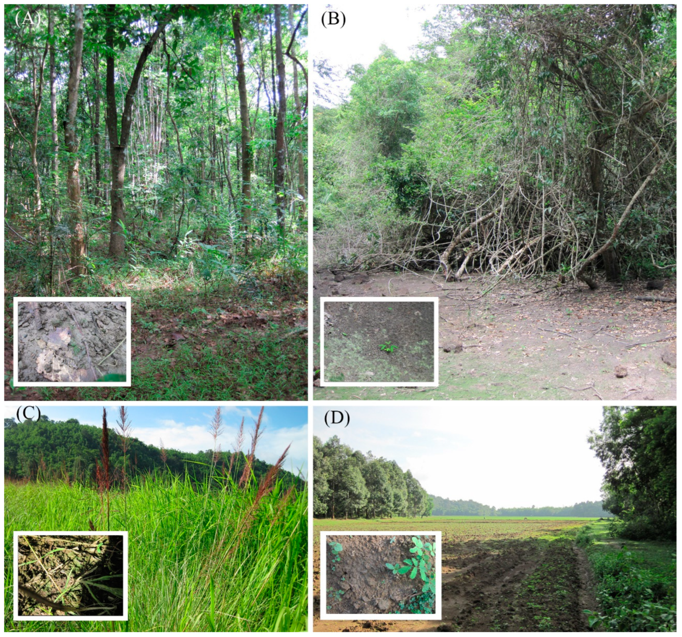
2.1. Sample Collection Procedure

The samples were collected as follows: the surface of the test site was examined to detect macrogrowth of algae, and then a composite sample was taken from an area of 10–30 m2. The composite sample consisted of 5–10 individual samples. For an individual sample, the topsoil was removed from an area of 5 to 20 cm2 with a metal scoop or shovel. After sampling, the instruments were cleaned and sterilized with ethanol. The samples were placed in labelled plastic zip bags and carried to the laboratory. The hot-drying method measured the absolute humidity [42] and the samples were air-dried and packaged.
To measure pH, we mixed 30 g of soil with 150 mL of distilled water [43]. The suspension was poured into a clean glass beaker and the measurements were performed with a Hanna Combo (HI 98129) device (Hanna Instruments, Inc., Woonsocket, RI, USA).

2.2. Culturing

Gathered materials were processed in the Laboratory of Molecular Systematics of Aquatic Plants of the Institute of Plant Physiology of the Russian Academy of Sciences (IPP RAS). A sample of soil was thoroughly mixed and placed into a Petri dish, then saturated with distilled water up to 60–80% of full moisture capacity and placed into an illuminated climate chamber. After a 10-day incubation, the sample was diluted with a small amount of distilled water, mixed gently, and the suspension was transferred to a Petri dish for LM using a Zeiss Axio Vert A1 inverted microscope. Algal cells were extracted with a micropipette, washed in 3–5 drops of sterile distilled water and placed into a 300 µL well on a plate for enzyme-linked immunoassay with Waris-H + Si [44]. Non-axenic unialgal cultures were maintained at 22–25 °C in a growth chamber with a 12:12 h light: dark photoperiod and checked every 10–14 days for 5 months.
For fatty acid analysis cultures were maintained on Waris-H + Si in 250 mL Erlenmeyer glass flasks with 150 mL medium, under constant orbital shaking (150 rpm in ELMI Sky Line Shaker S-3L, ELMI Ltd., Riga, Latvia) for 25 days at 25 °C. The light intensity was 100 μmol photons m−2 s−1 with a 16:8 h light/dark photoperiod. All analyses were performed in triplicate. Tables show the mean values and standard errors.

2.3. Microscopy

A culture was treated with 10% hydrochloric acid to remove carbonates and washed several times with deionized water for 12 h. Afterwards, the sample was boiled in concentrated hydrogen peroxide (≈37%) to remove organic matter. It was washed again with deionized water four times at 12 h intervals. After decanting and filling with deionized water up to 100 mL, the suspension was pipetted onto coverslips and left to dry at room temperature. Permanent diatom preparations were mounted in Naphrax. Light microscopic (LM) observations were performed with a Zeiss Axio Scope A1 microscope (Carl Zeiss Microscopy GmbH, Gottingen, Germany) equipped with an oil immersion objective (×100, n.a. 1.4, differential interference contrast) and Axiocam ERc 5s camera (Carl Zeiss NTS Ltd., Oberkochen, Germany). Valve ultrastructure was examined using a scanning electron microscope JSM-6510LV (IBIW; Institute for Biology of Inland Waters RAS, Borok, Russia). For scanning electron microscopy (SEM), part of the suspensions were fixed on aluminum stubs after air-drying. The stubs were sputter-coated with 50 nm of Au using an Eiko IB 3 machine (Eiko Engineering Co. Ltd., Tokyo, Japan). The suspension and slides are deposited in the collection of Maxim Kulikovskiy at the Herbarium of the Institute of Plant Physiology Russian Academy of Sciences, Moscow, Russia.

2.4. Molecular Study

Total DNA from the studied strains was extracted using Chelex 100 Chelating Resin, molecular biology grade (Bio-Rad Laboratories, Hercules, CA, USA), according to the manufacturer’s protocol 2.2. Partial 18S rDNA (395–400 bp, including the highly variable V4 region of the 18S gene), and partial rbcL plastid genes (921–957 bp) were amplified using primers D512for and D978rev from Zimmermann et al. [45] for 18S rDNA fragments and rbcL404+ from Ruck and Theriot [46] and rbcL1255- rom Alverson et al. [47] for rbcL fragments.
Amplifications were carried out using premade polymerase chain reaction (PCR) mastermixes (ScreenMix by Evrogen, Moscow, Russia). Amplification conditions for the 18S rDNA gene were as follows: initial denaturation for 5 min at 95 °C followed by 35 cycles of 30 s denaturation at 94 °C, 30 s annealing at 52 °C, and 50 s extension at 72 °C, with the final extension for 10 min at 72 °C. Amplification conditions for the rbcL gene were as follows: initial denaturation for 5 min at 95 °C followed by 45 cycles of 30 s denaturation at 94 °C, 30 s annealing at 59 °C, and 80 s extension at 72 °C, with the final extension for 10 min at 72 °C. PCR products were visualized by horizontal electrophoresis in 1.0% agarose gel stained with SYBRTM Safe (Life Technologies, Carlsbad, CA, USA). The products were purified with a mixture of FastAP, 10×FastAP Buffer, Exonuclease I (Thermo Fisher Scientific, Waltham, MA, USA), and water. The sequencing was performed using a Genetic Analyzer 3500 instrument (Applied Biosystems, Waltham, MA, USA).
Editing and assembling of the consensus sequences were carried out by processing the direct and reverse chromatograms in Ridom TraceEdit ver. 1.1.0 (Ridom GmbH, Münster, Germany) and Mega7 software [48]. The reads were included in the alignments along with corresponding sequences of 72 diatom species downloaded from GenBank (taxa names and Accession Numbers are given in Figure 2). Three centric diatom species were chosen as the outgroups.
The nucleotide sequences of the 18S rDNA and rbcL genes were aligned separately using the Mafft ver. 7 software (RIMD, Osaka Japan) and the E-INS-i model [49]. The final alignments were then carried out: unpaired sites were visually determined and removed from the beginning and the end of the resulting matrices. For the protein-coding sequences of the rbcL gene, we checked that the beginning of the aligned matrix corresponds to the first position of the codon (triplet). The resulting alignments had lengths of 450 (18S rDNA) and 957 (rbcL) characters. After removal of the unpaired regions, the aligned 18S rDNA gene sequences were combined with the rbcL gene sequences into a single matrix Mega7 (Appendix S1).
The data set was analyzed using the Bayesian inference (BI) method implemented in Beast ver. 1.10.1 software (BEAST Developers, Auckland, New Zealand) [50] to construct a phylogeny. For the alignment partition the most appropriate substitution model, shape parameter α and a proportion of invariable sites (pinvar) were estimated using the Bayesian information criterion (BIC) as implemented in jModelTest ver. 2.1.10 (Vigo, Spain) [51]. This BIC-based model selection procedure selected the following models, shape parameter α and a proportion of invariable sites (pinvar): HKY + I + G, α = 0.6250 and pinvar = 0.5170 for 18S rDNA; TPM1uf + I + G, α = 1.1070, and pinvar = 0.7470 for the first codon position of the rbcL gene; JC + I + G, α = 0.3830 and pinvar = 0.7320 for the second codon position of the rbcL gene; TPM2uf + G and α = 0.5470 for the third codon position of the rbcL gene.
We used the F81 model of nucleotide substitution instead of TPM1uf, the HKY model instead of JC, and TPM2uf, given that they were the best matching model available for BI. A Yule process tree prior was used as a speciation model. The analysis ran for 5 million generations with chain sampling every 1000 generations. The parameters-estimated convergence, effective sample size (ESS), and burn-in period were checked using the Tracer ver. 1.7.1 software (MCMC Trace Analysis Tool, Edinburgh, United Kingdom). [50]. The initial 25% of the trees were removed, and the rest were retained to reconstruct a final phylogeny. The phylogenetic tree and posterior probabilities of its branching were obtained based on the remaining trees, having stable estimates of the parameter models of nucleotide substitutions and likelihood. The Maximum Likelihood (ML) analysis was performed using RAxML software [52]. The nonparametric bootstrap analysis with 1000 replicas was used. The phylogenetic tree topology is available online in Appendix S2. FigTree ver. 1.4.4 (University of Edinburgh, Edinburgh, United Kingdom) and Adobe Photoshop CC ver. 19.0 software (Adobe, San Jose, CA, USA) were used for viewing and editing the trees.

2.5. Fatty Acid Analysis

Biomass preparation for determining the fatty acid methyl ester (FAME) profiles was performed according to Maltsev et al. [53]. The diatom suspensions were conveyed to 15–50 mL tubes (depending on the volume). The cells were pelleted at room temperature for 3 min at 3600 g. The supernatant was removed, and the pelleted cells were resuspended in 10–15 mL (depending on the amount of biomass) of distilled water, quantitatively transferred to 15 mL centrifuge tubes, and pelleted again by centrifugation. The supernatant was removed, and samples were quantitatively transferred to a 50 mL round-bottom flask. Heptadecanoic acid (Sigma-Aldrich, St. Louis, MO, USA) was used as the internal standard for the fatty acid composition determination. To avoid the oxidation of unsaturated fatty acids, all samples were processed under an argon atmosphere. Ten milliliters of a 1 M solution of KOH in 80% aqueous ethanol was added to the dry residue, and the flask was sealed with a reflux condenser, and kept for 60 min at the boiling point of the mixture (~80 °C). After the time-lapse, the solvents were evaporated in vacuo to a volume of ~3 mL and quantitatively transferred with distilled water to a 50 mL centrifuge tube to a total volume of 25 mL, followed by extracting the unsaponifiable components with 10 mL portions of n-hexane (Himmed, Moscow, Russia) 3 times. To accelerate the separation of the phases, the tube was centrifuged for 5 min at room temperature and 2022× g. After that, the aqueous phase was acidified to a slightly acidic reaction (on indicator paper) with a few drops of 20% sulfuric acid (Himmed, Moscow, Russia), and free fatty acids were extracted with 20 mL of n-hexane. The hexane solution of free fatty acids was transferred to a dry 50 mL round-bottom flask, and the solvent was evaporated to dryness using a rotary evaporator IKA RV-10 (IKA-WERKE, Staufen im Breisgau, Germany), after which 10 mL of absolute methanol (Sigma-Aldrich, St. Louis, MO, USA) and 1 mL of acetyl chloride (Sigma-Aldrich, St. Louis, MO, USA) were added to the dry residue. The flask, closed with a reflux condenser, was kept for one hour at 70 °C, then the solvents were evaporated to dryness, a few drops of distilled water were added to the dry residue, and FAMEs were extracted with n-hexane.
The obtained FAMEs were analyzed using an Agilent 7890A gas-liquid chromatograph (Agilent Technologies, Santa Clara, CA, USA) with an Agilent 5975C mass spectrometric detector. A DB-23 capillary column 60 m long and 0.25 mm in diameter was used (Agilent Technologies, Santa Clara, CA, USA). The remaining conditions of the analysis were as follows: carrier gas was helium, flow rate of 1 mL min−1, 1 μL volume of injected sample, 1:5 flow divider, and the evaporation temperature of 260 °C. Temperature gradient program: from 130 to 170 °C in 6.5 °C min−1 steps; from 170 to 215 °C in 2.5 °C min−1 increments, 215 °C for 25 min, from 215 to 240 °C in 40 °C min−1 increments, and the final stage lasting 50 min at 240 °C. The operating temperature of the mass spectrometric detector was 240 °C and the ionization energy was 70 eV.

3. Results and Discussion

Pinnularia specimens are ubiquitously found in soil samples collected in the Cát Tiên National Park. Based on the results of DNA sequencing, LM/SEM observations, and FA profiling of eight different strains of Pinnularia, we regard six of them as new species. For the sake of clarity, we address these strains by specific epithets ahead of the formal description.

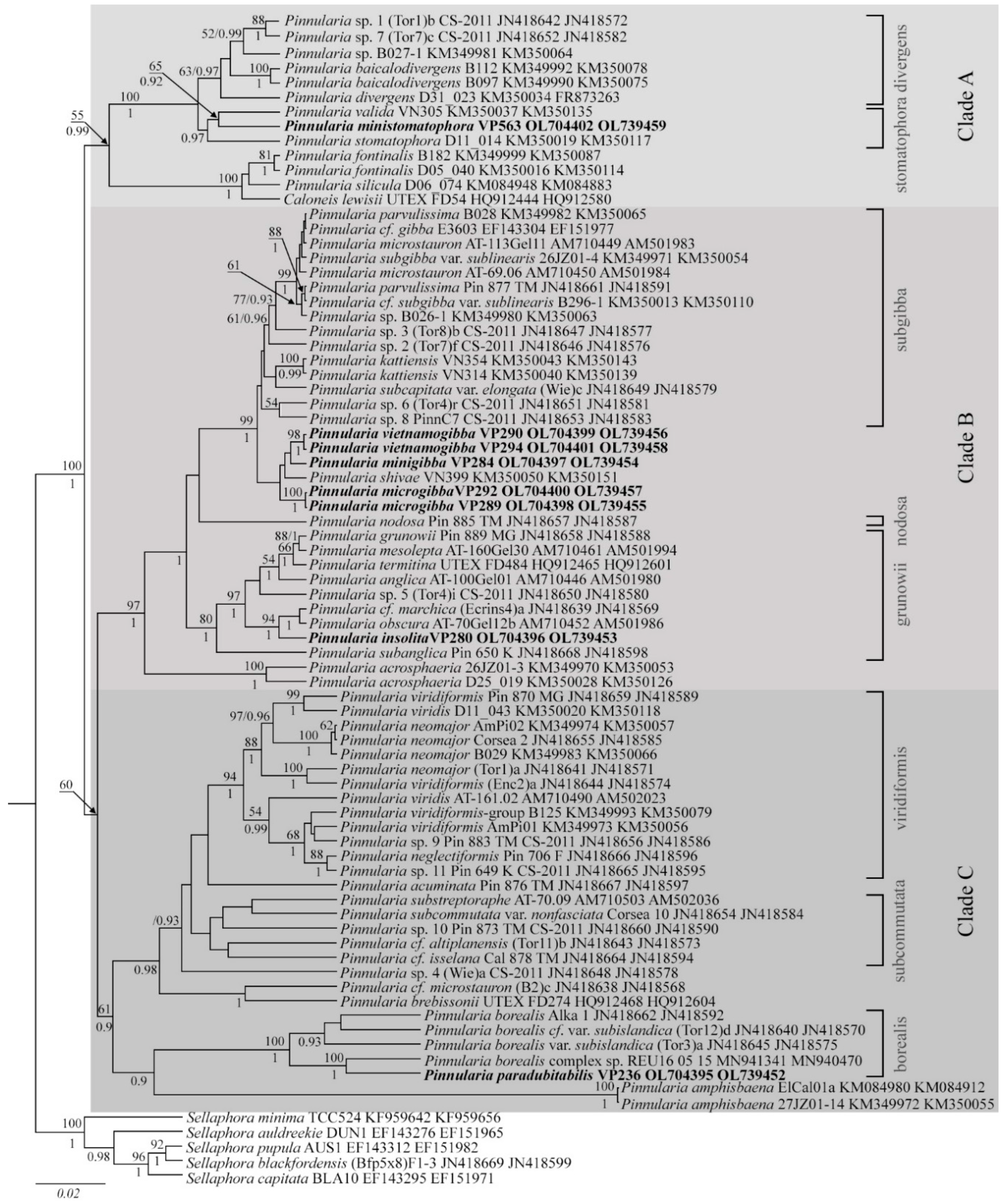
3.1. Molecular Phylogeny

Comprehensive studies on Pinnularia phylogeny [24,25] confirm its monophyletic origin; the genus splits into three clades stably supported by the analysis. In a study by Souffreau et al. [25], who used five genetic markers—two nuclear (18S rDNA and 28S rDNA), two plastid (rbcL and psbA), and a mitochondrial cox1, these clades were designated A, B, and C. Each clade consists of several subclades characterized by morphological similarity about shapes (valves and apices, raphe endings linear or rounded, raphe fissures straight or undulate, chloroplasts H-shaped or elongated, with pyrenoids or not, etc.) and specific markings (ghost striae, fascia, wart-like bodies, etc.). Although our phylogenetic analysis involved only two markers (nuclear 18S rDNA and plastid rbcL), it perfectly preserved the tree topology and maintained high support to all clades and subclades introduced by Souffreau et al. [25]. The tree was expanded through the addition of the new taxa, which showed full morphological consistency with their parental subclades (Figure 2).
Clade A includes two previously characterized subclades exemplified by Caloneis Cleve 1894 and P. cf. divergens. Here we complement the ‘divergens’ subclade with new species; a morphological feature is a fascia with rounded thickenings at the margin. Furthermore, the expansion of Pinnularia phylogeny revealed a new subclade of clade A, ‘stomatophora’, comprising P. ministomatophora, P. valida (strain VN305), and P. stomatophora (strain D11_014), all of them presenting with characteristic hollow markings on the external surface of the valve, crescent-shaped (P. stomatophora (p. 456, pl. 98, Figure 8 of [11]) or irregular (P. ministomatophora sp. nov.).
Clade B includes three subclades, ‘grunowii’, ‘nodosa’, and ‘subgibba’, of diminutive linear algae with bulbous apices and rounded external endings of the raphe [25]. Our phylogeny investigation recognizes these subclades with high support. However, the two species of ‘nodosa’ subclade, P. nodosa and P. acrosphaeria [25] split into distinct branches (Figure 2). These species also show morphological distinctions: P. acrosphaeria display mottled, structured areas on both outer and inner surfaces of the valve (p. 296, pl. 19, Figures 1–6 of [11]) whereas in P. nodosa with the valve is smooth on the inside and its entire outer surface is heavily structured (p. 307, pl. 24, Figures 1–6 of [11]).
A morphological feature of the ‘subgibba’ subclade is ghost striae in the central area [25,26,27]. The term ghost striae was proposed by Cox [54] and refers to thinnings on the inner surface of the valve, corresponding in size and spacing to normal striae. It should be noted that there is no unified name for these structures defined as “large markings, differently structured on both sides and larger in the ventral side” [11], “deepenings on the inside of the valve” [11] or “depressions in the central area” [55]. Our phylogenetic study encompassed P. microstauron, P. parvulissima, P. cf. gibba, P. cf. subgibba var. sublinearis, P. subcapitata var. elongata, P. kattiensis, and several strains of Pinnularia sp. The newly identified soil species P. microgibba, P. minigibba, and P. vietnamogibba along with P. shivae form a separate branch within the ‘subgibba’ subclade (Figure 2). Ultrastructural examination of P. microgibba, P. minigibba, and P. vietnamogibba specimens by SEM revealed ghost striae in the central area for all of them, consistently with their phylogenetic affiliation.
Incidentally, the ‘grunowii’ subclade also splits into two branches, which happen to have a different ultrastructure of the central area (Figures 2 and 10 and Figures in Krammer [11], Jahn and Kusber [56]). Species with ghost striae, namely P. anglica P. grunowii, and P. mesolepta (p. 423, pl. 82, Figures 7 and 8 of [11,56]), are phylogenetically divergent from the diminutive P. marchica, P. obscura (scanning images are available in Jahn and Kusber [56]), and P. insolita sp. nov. with no markings in the central field.
Clade C specimens have no markings in the central area but exhibit wide strokes on the valves. Our study generally preserved the topology for this clade introduced by Souffreau et al. [25], while the ‘borealis’ subclade was complemented by new strains and new species P. paradubitabilis. Sister lineages to the ‘borealis’ subclade are constituted by strains of P. amphisbaena and two related subclades, ‘subcommutata’ and ‘viridiformis’, comprising large specimens of linear-elliptical shape with almost parallel striae and small central areas, undulate external raphe fissures and with linear (for subcommutata) or rounded (for viridiformis) central raphe endings. In contrast with the original version [25], P. cf. microstauron, P. brebissonii, P. accuminata, Pinnularia sp. 4 (Wie)a, and strains morphologically similar to P. microstauron split into separate branches within clade C (Figure 2).
It is interesting to observe that certain morphological distinctions, notably the presence of ultrastructural surface markings in the central area, consistently follow phylogenetic clades. Species presenting with markings in the central area can be reliably classified on their basis as ‘divergens’, ‘stomatophora’, ‘subgibba’, ‘nodosa’, and ‘gronowii’ groups.

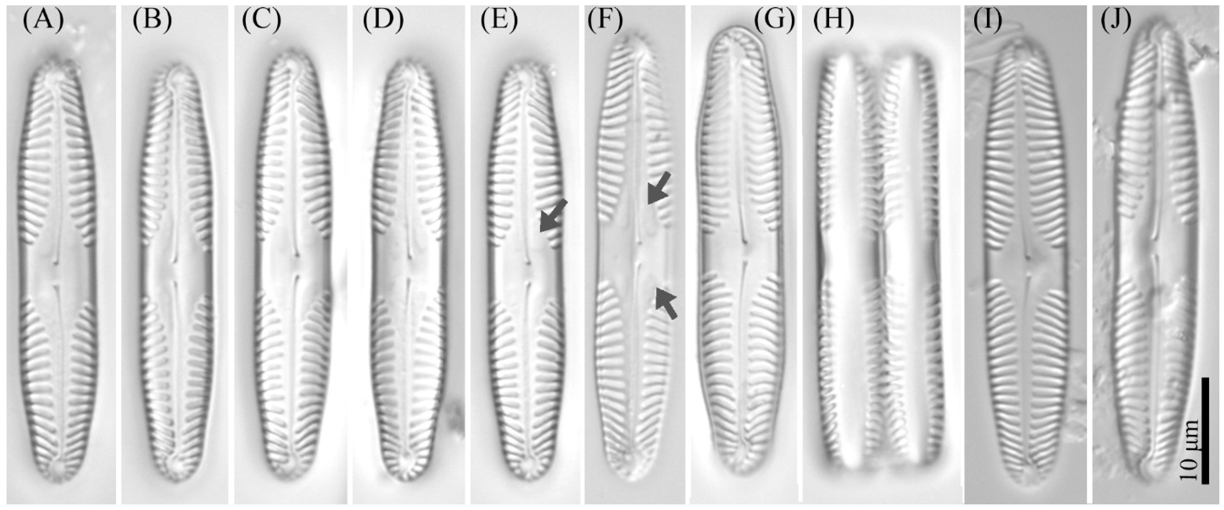
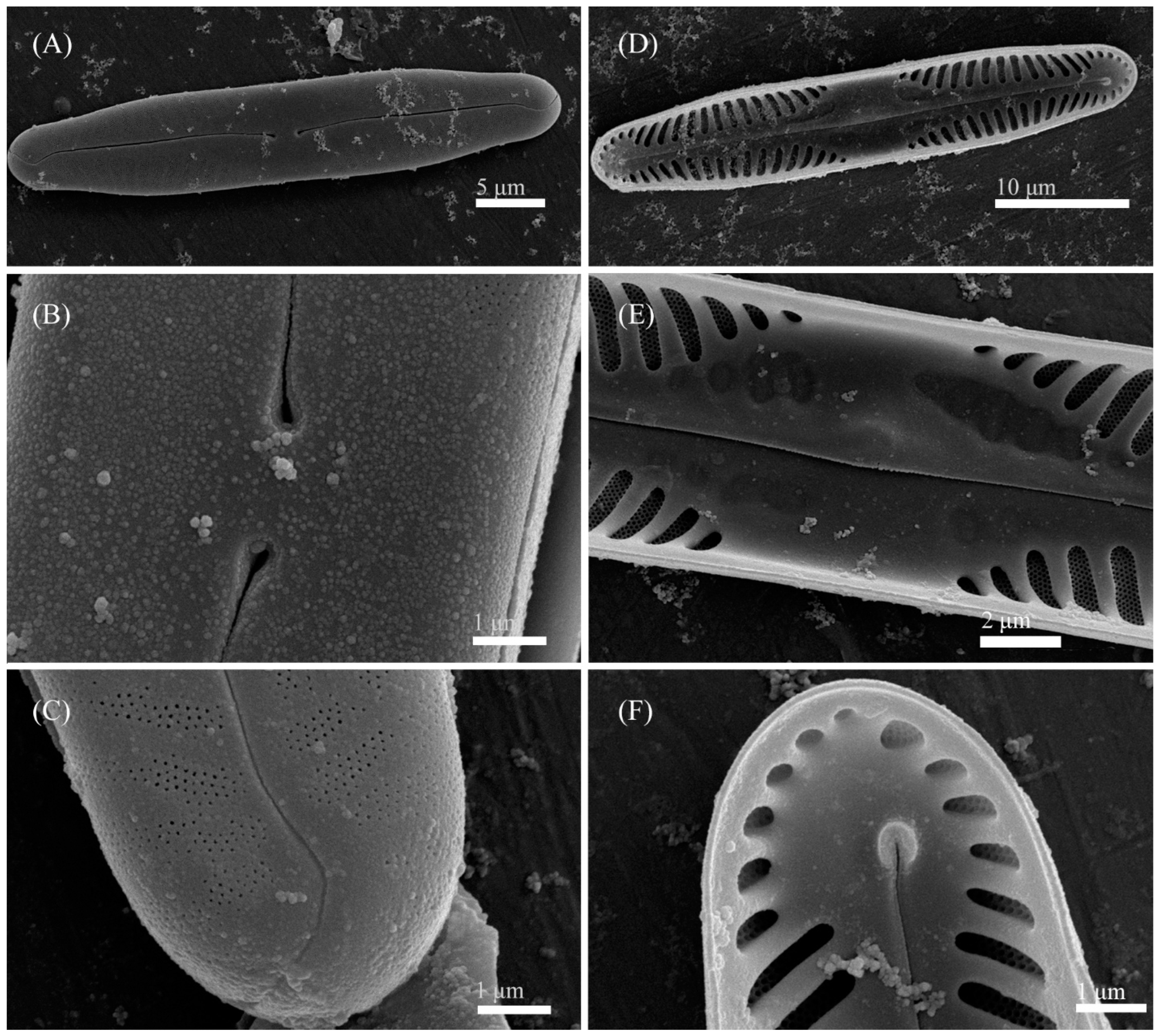
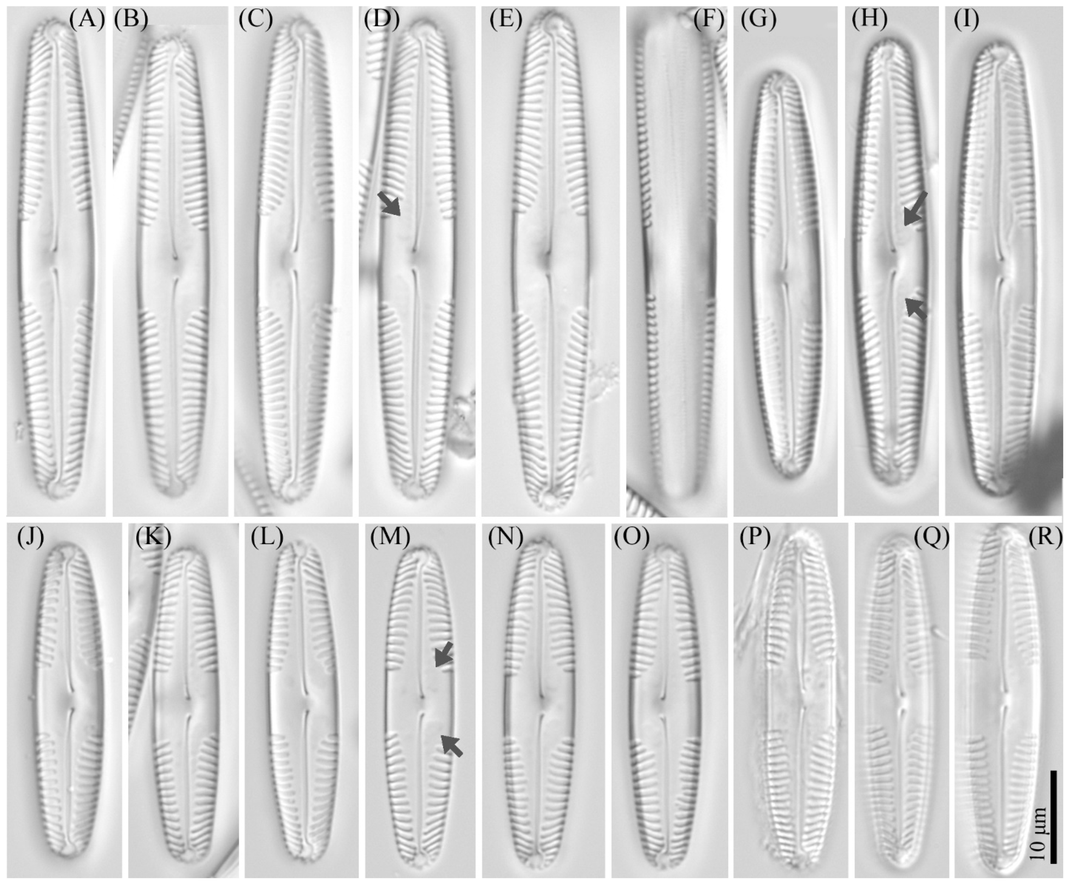
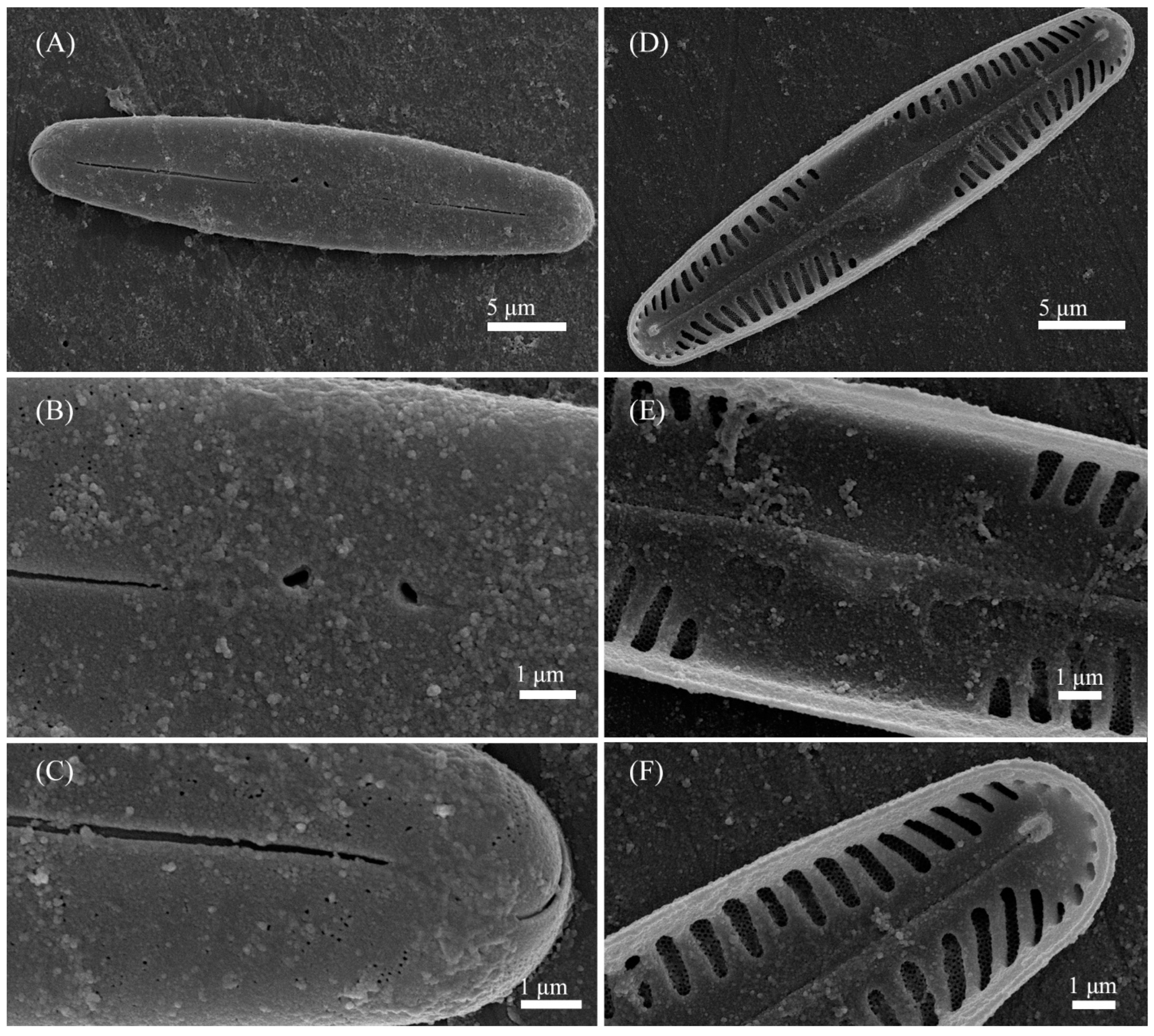
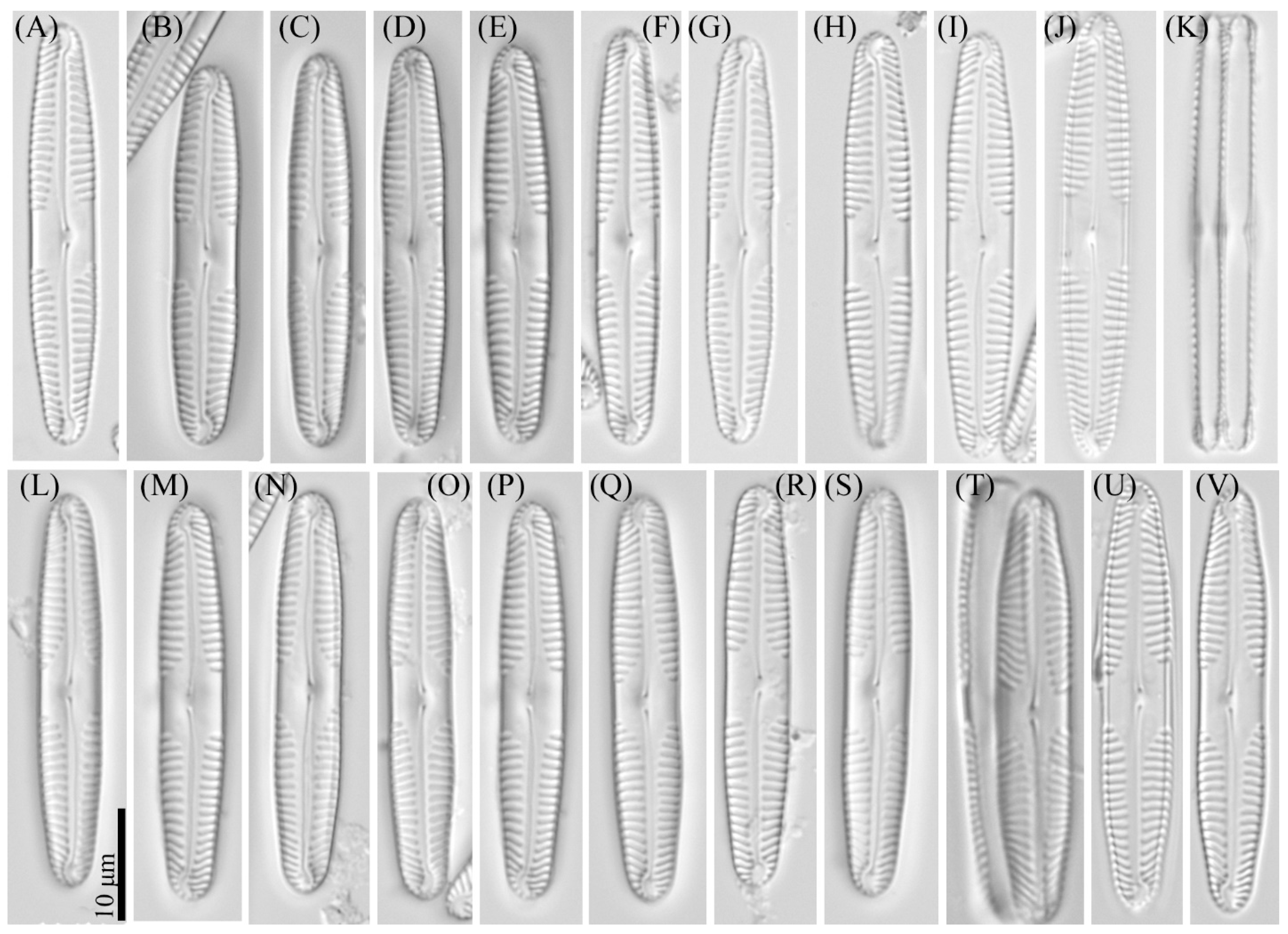
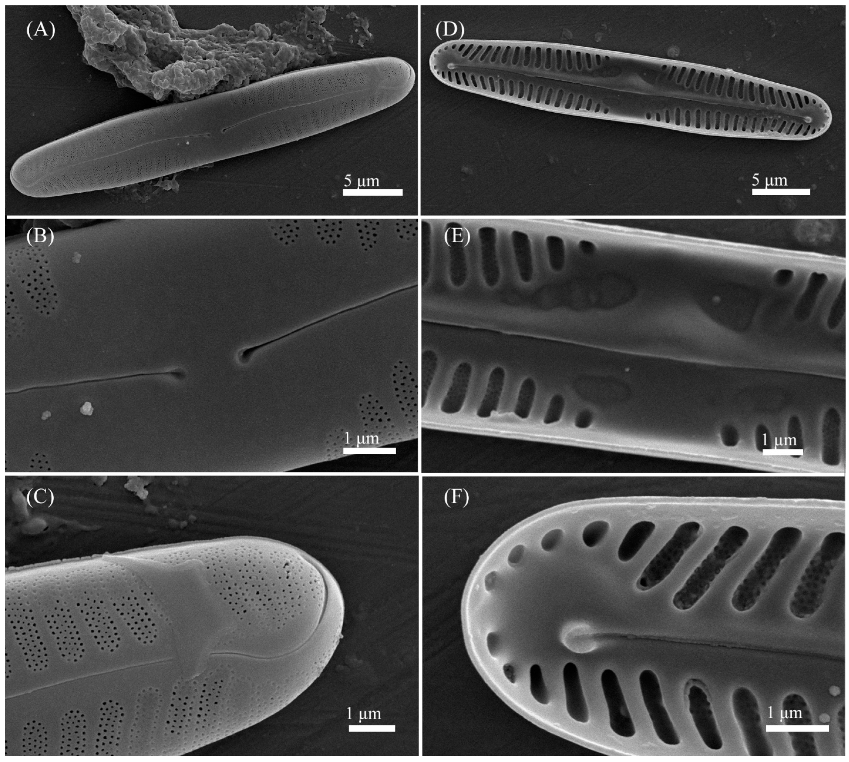
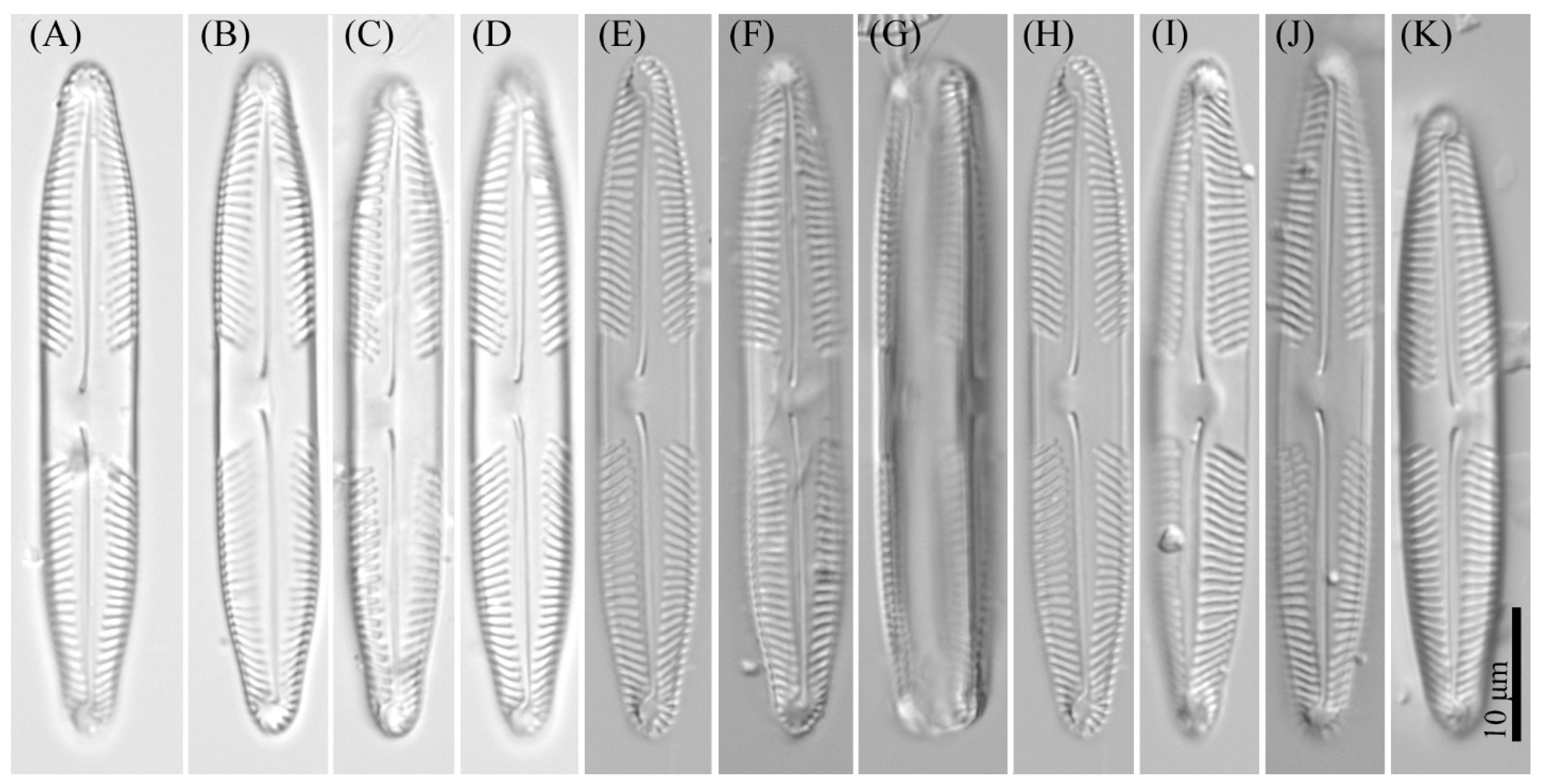
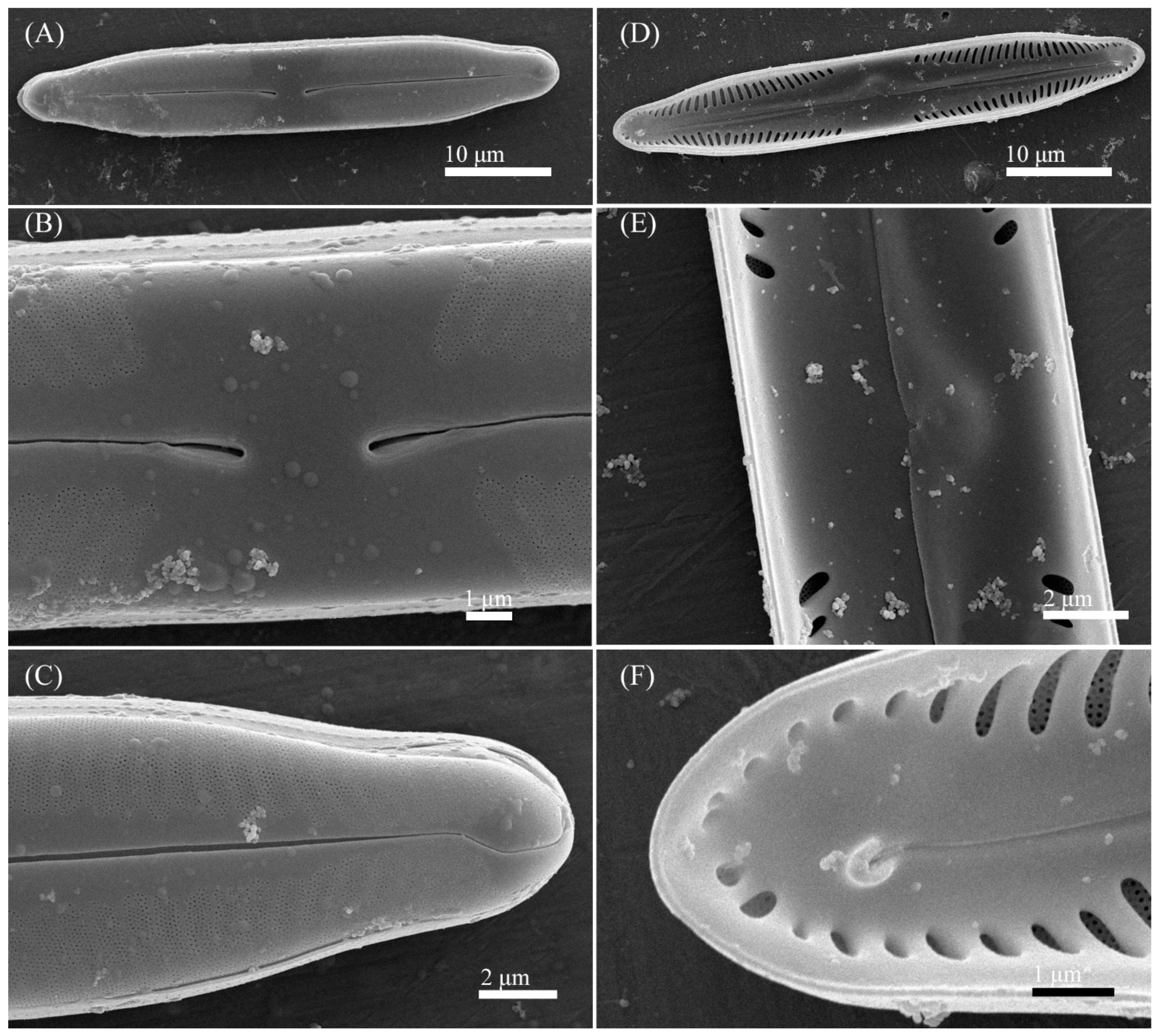
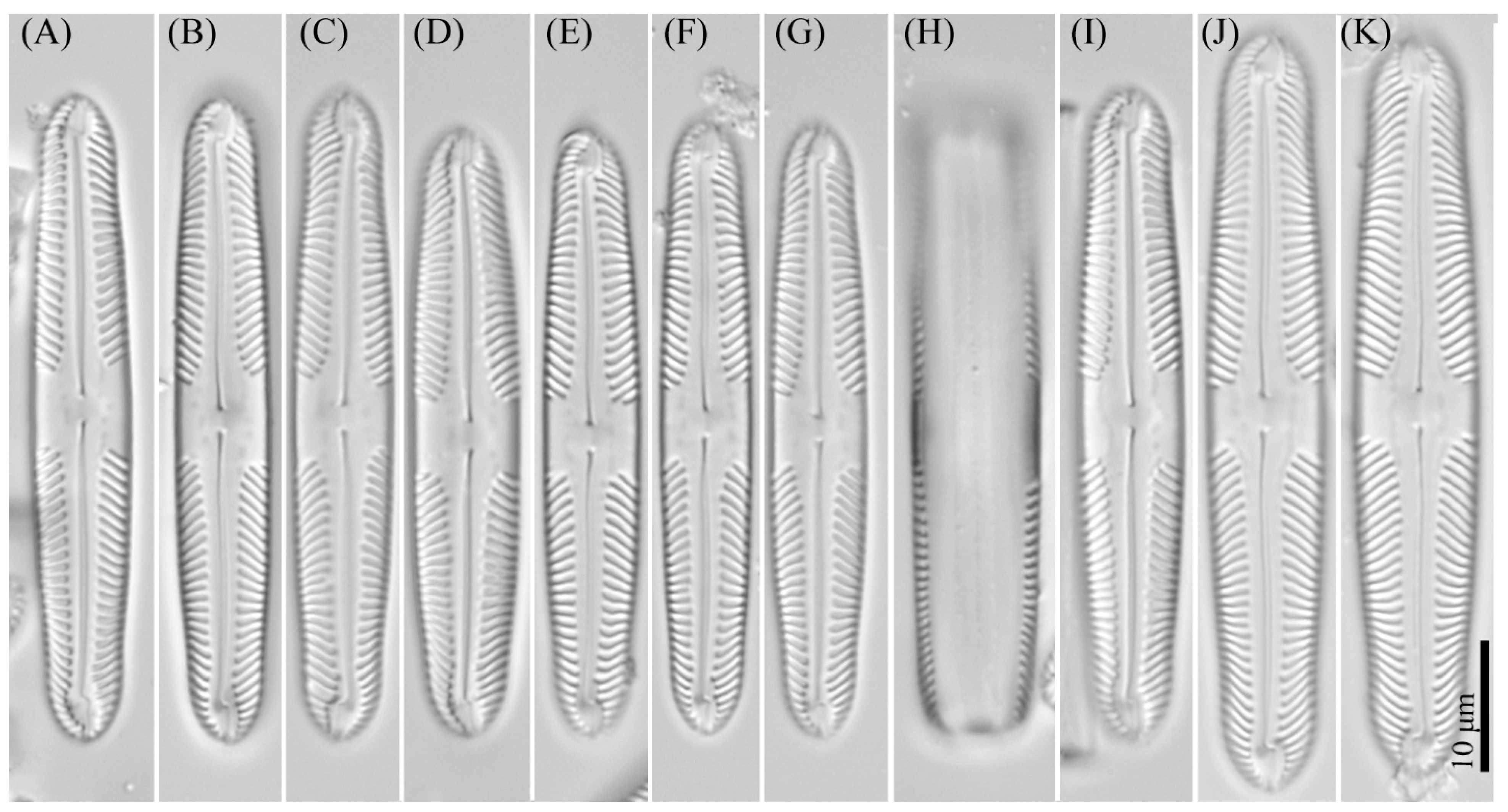
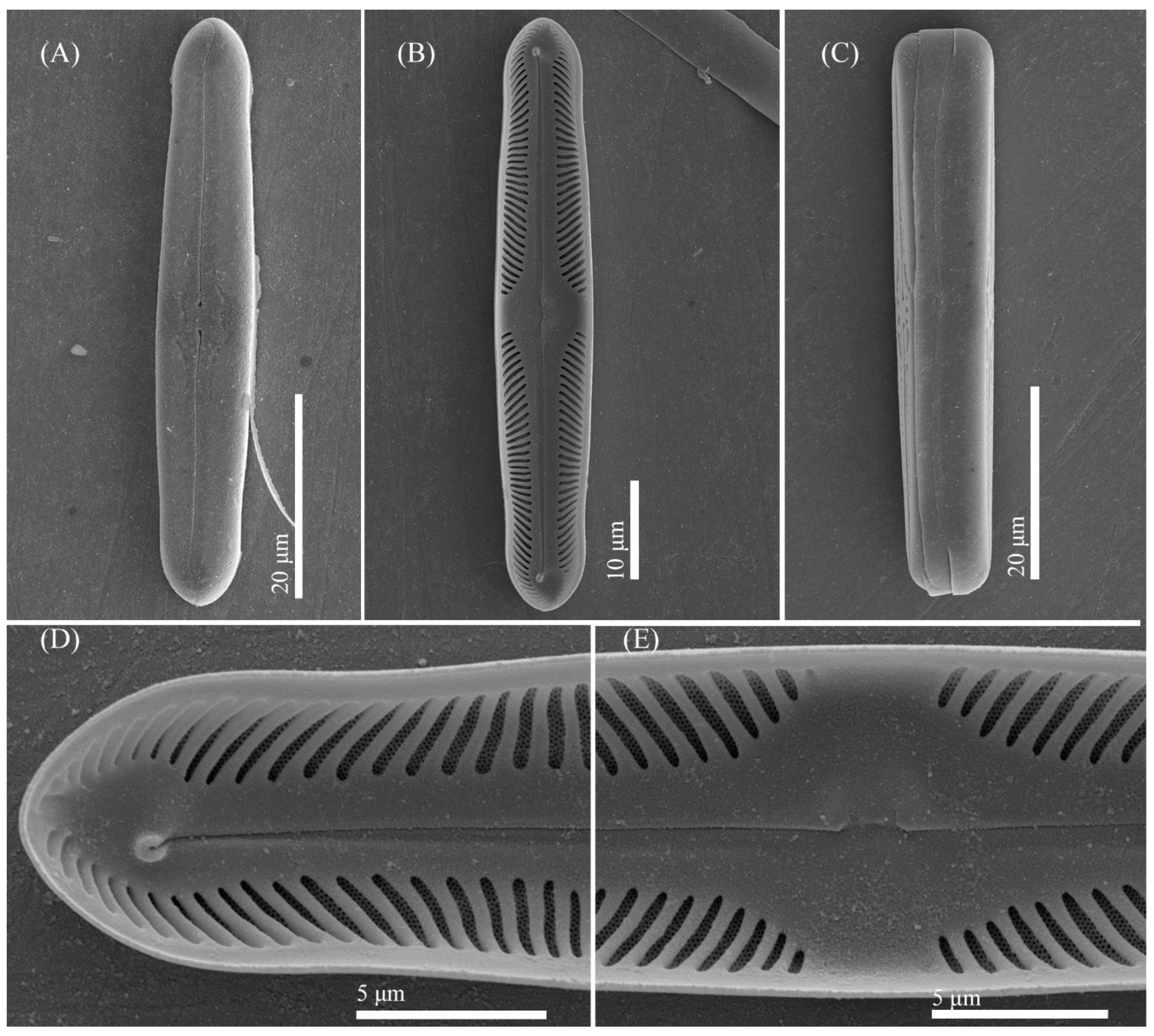
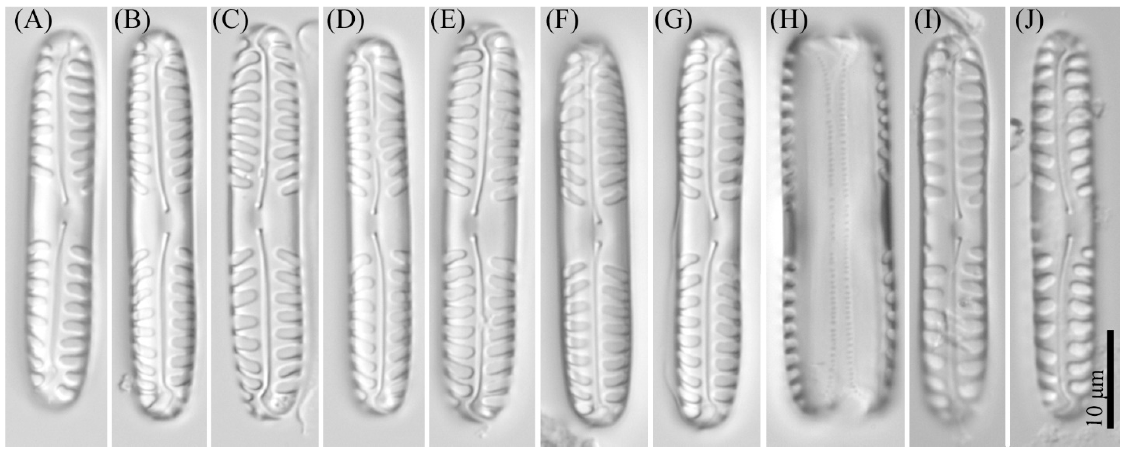
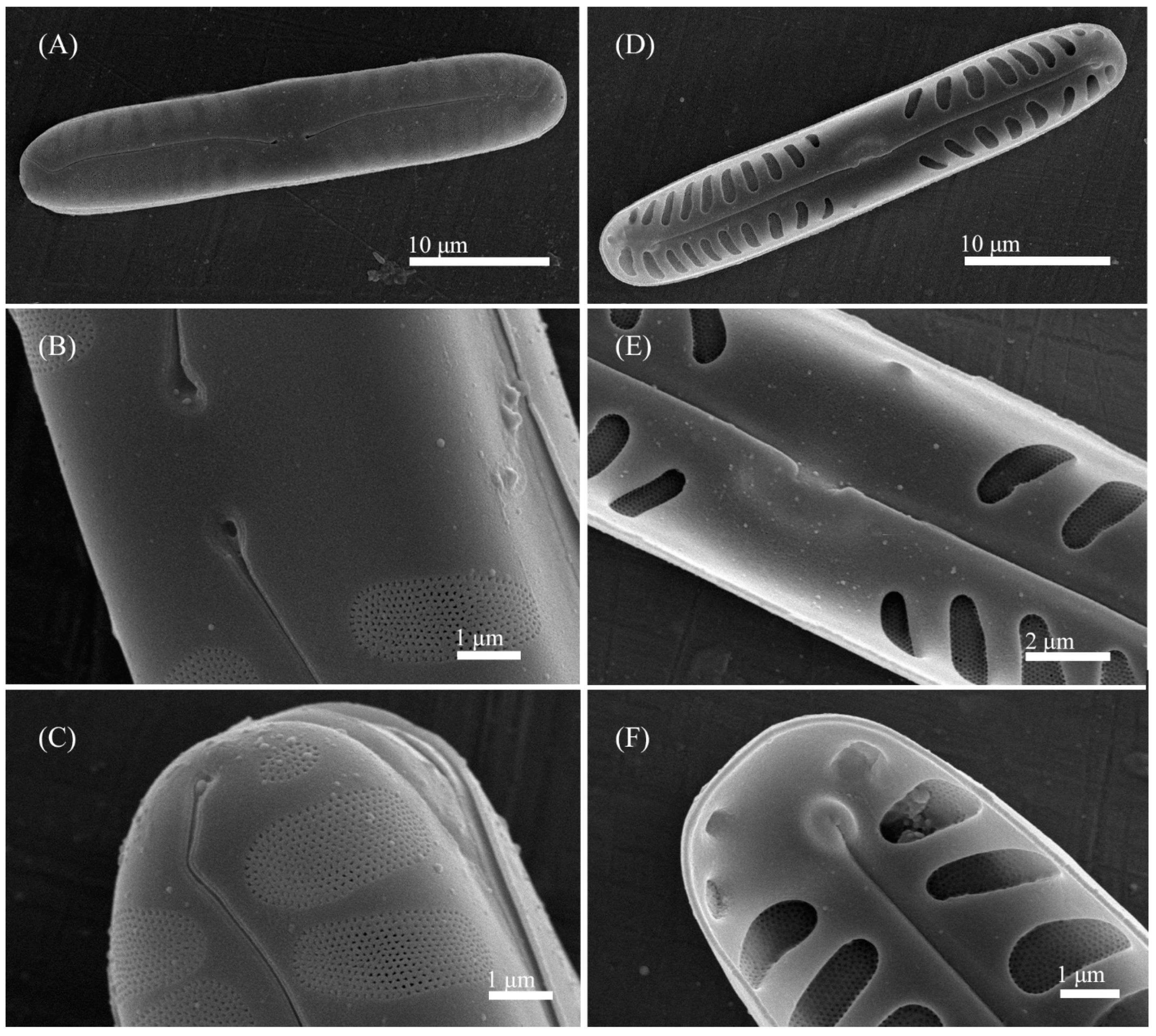
3.2. Comparative Morphology

Pinnularia minigibba sp. nov. is similar to several species of the P. gibba complex (Table 2). The new species must not be confused with P. australogibba var. subcapitata [55]. The similarities include the outlines and proportions of the valves, characteristic shape of the central area with a broad fascia, and ghost striae. However, P. minigibba has wider valves (7–8 μm vs. 5.7–7.3 μm in P. australogibba var. subcapitata) with slightly concave margins (as opposed to convex sides of P. australogibba var. subcapitata). Other differences include stria density (respectively, 9–10 in 10 μm vs. 11–12 in 10 μm) and the shape of axial area (≤1/4 of the width and slightly wider towards the midportion in P. minigibba vs. lanceolate in P. australogibba var. subcapitata); also, in the new species, the stria is more tilted in the midportion. Another closely similar species, P. parvulissima (p. 397, pl. 69, Figures 6–11 of [11]), can be differentiated by larger size (34–70 μm length to 10–12 μm width vs., respectively, 40–43 to 7–8 in P. minigibba); besides, P. parvulissima have convex margins and wider axial area. The similarity of P. minigibba with certain strains of the polymorphic widespread P. microstauron (Ehrenberg) Cleve should be noted as well. For instance, it can be confused with P. microstauron var. angusta (p. 361, pl. 51, Figures 4–7 of [11]), which is a smaller variety with wider fascia, and a particularly challenging specimen shown in Figure 18, pl. 164 of Metzeltin et al. [57]. About these examples, P. minigibba can be differentiated by stria density (9–10 in 10 μm vs. 10–12 in 10 μm in P. microstauron var. angusta and 11 in 10 μm in P. microstauron sensu Metzeltin et al. [57], as well as concave margins (vs. parallel or subtly convex in P. microstauron), and characteristic presence of ghost striae in the central area. Despite the apparent absence of ghost striae in LM images of P. microstauron var. angusta and P. microstauron sensu Metzeltin et al. [57], ultrastructural examinations of archetypal P. microstauron specimens (e.g., AT_112Gel04, AT_113Gel11 cultures [55,56]) reveal hollowed markings on the inner surface of the valve confined to the central area, corresponding to ghost striae, consistently with the assignment to the ‘subgibba’ subclade (p. 371, Figure 3 of [26]).
Pinnularia vietnamogibba sp. nov. must not be confused with two closely similar taxa—P. gibba var. subsancta (pl. 13, Figure 7 of [58]; p. 521, pl. 140, Figures 9 and 10 of [59]) and P. australogibba (p. 215, Figures 94–101 of [55]). According to Krammer [11] (P. 111), most specimens defined in literature as P. gibba var. sancta (Grunow) Meister arguably belong to a smaller variety described by Manguin (pl. 13, Figure 7 of [58]). According to Hustedt [60], this taxon is widespread in the tropics, whereas Pinnularia australogibba is found in ravines at Point Del Cano, Île Amsterdam in the southern Indian Ocean. The similarities of these species with the newly identified P. vietnamogibba include the shape of the central area with a broad fascia and ghost striae. However, P. gibba var. subsancta and P. australogibba specimens have lanceolate outlines, whereas P. vietnamogibba are linear, with the margins parallel or subtly bulging. P. vietnamogibba also has much broader fascia, which constitutes 17–20% of the valve length (cf. 4–10.3% of the length in P. gibba var. subsancta or 7.7–14.5% in P. australogibba) and lower stria density (10–11 in 10 μm vs. 13–15 in 10 μm and 12–13 in 10 μm in P. gibba var. subsancta and P. australogibba, respectively). P. vietnamogibba can also be confused with the already mentioned P. microstauron var. angusta (p. 361, pl. 51, Figure 4 of [11]; p. 81, Figures 47–49 of [61]), which may reach a similar size and have similar stria densities and similar shapes of the central and axial areas. However, in microstauron-like morphologies, the ends are distinctly offset and much smaller compared with the width of the valve, in contrast to P. vietnamogibba. Another morphometrically similar species, P. tagliaventiae (p. 417, pl. 130, Figures 1–7 of [62]), can be distinguished by triangulate sides, whereas the sides of P. vietnamogibba are straight.
Pinnularia microgibba sp. nov. phylogenetically and morphologically closely related with ‘subgibba’-group species [25,26], particularly with strains Pinnularia sp. 6 (Tor4)r and Pinnularia sp. 3 (Tor8)b (from littoral zones of freshwater bodies in Chile, see Table 2). These strains are virtually identical in size, shape, and outline; the only clue to their morphological distinction is stria density (12 in 10 μm. for (Tor8)b and 14 in 10 μm for (Tor4)r). Meanwhile, they are phylogenetically independent (Figure 2). Morphometric data of P. microgibba almost totally match those of (Tor4)r and (Tor8)b, except for the shape the of central area: linear, broadening towards the valve margins in (Tor4)r and (Tor8)b, and rhombic in the new species. Phylogenetically, the strains of P. microgibba together with P. minigibba and P. vietnamogibba form a separate branch within the ‘subgibba’ subclade.
It should be kept in mind that ghost striae are often poorly distinguishable in light micrographs. For this reason, P. microgibba can be easily confused with certain diminutive species with narrow linear outlines and broad fascia, including the widespread, ubiquitous, and cosmopolitan P. sinistra [11,63]. However, P. microgibba has concave margins and a rhombic central area with ghost striae, whereas P. sinistra has a linear central area without markings (p. 265, Taf. 37, Figure 16 of [63]). Interestingly, a population of P. sinistra inhabiting the small oceanic island Île Amsterdam, situated in the southern part of the Indian Ocean (p. 225, Figures 194–209 of [55]), also has concave margins and rhombic central area; however, these taxa can be distinguished from both the type of P. sinistra and type of P. microgibba by their relatively wide lanceolate axial area (compared with the narrow-linear axial area in type populations of P. sinistra and P. microgibba). Several images for certain species found in the literature can be confused with P. microgibba as the distinctions are very subtle and demand close attention (Table 2). For instance, the valves of P. microstauron var. angusta (p. 361, pl. 51, Figures 5 and 6 of [11]) are wider (6.5–8.0 μm) compared with P. microgibba (5.5–6 μm). P. subcapitata W. Gregory (given as P. hilseana Janisch 1861 (p. 757, pl. 205, Figure 9 of [64]) and P. cf. hilseana/P. cf. fotii Bily et Marvan (p. 757, pl. 205, Figures 26 and 27 of [64]) have distinctly capitate valve ends (apices) compared with the subcapitate apices in P. microgibba. Pinnularia cf. saprophila (p. 573, pl. 164, Figures 3 and 4 of [57]) have a lower stria density (9.5–11.0 in 10 μm) compared with P. microgibba (11.0–12.0 in 10 μm). Pinnularia pisciculus found in low nutrient waters, mosses, and dry soils from India (pl. 73 [65]) display characteristic triundulate sides and capitate apices. Pinnularia similiformis var. koreana (p. 291, pl. 16, Figures 3–6 of [11]) is lengthy (40–60 μm), whereas P. microgibba is 36–40 μm. Pinnularia marchica (p. 283, pl. 12, Figures 11–17 of [11]) have elongated rostrate apices different from the subcapitate apices of P. microgibba. Ghost striae in the central area represent an important identifier; however, it should be applied with caution, as SEM data are not universally available and the sensitivity of LM about ghost striae is limited (Table 2).
Pinnularia insolita sp. nov. can be recognized by its unusual outline with concave side margins and narrowed apices, as well as the characteristic shape of the central area with a prominent fascia broadening towards the margins. Nevertheless, certain varieties of other Pinnularia species are similar (Table 2). Large specimens of P. pisciculus Ehrenberg (p. 429, pl. 85, Figures 25 and 26 of [11]) are similar to P. insolita (length 22–50 μm, width 6.0–8.3 μm in P. pisciculus vs. length 50–52 μm and width 7–7.5 μm in P. insolita), stria density (10.5–12.0 μm in 10 μm vs. 11–12 in P. insolita), and overall outline. Nevertheless, the algae can be reliably differentiated by their apices shape (capitate in P. pisciculus and subtly rostrate in P. insolita) and the shape of the central area (rhombic in P. pisciculus vs. broadening towards the margins in P. insolita). Moreover, larger specimens of P. pisciculus tend to have subtly triundulate side margins, whereas the sides of P. insolita are concave. Another similar species with P. insolita is P. brebissonii var. bicuneata, whose largest annotated specimen (p. 351, pl. 46, Figure 9 of [11]) resembles P. insolita by its size, shapes of the valve apices and central area, and stria density. However, in P. brebissonii var. bicuneata the sides are straight and parallel (vs. subtly concave in P. insolita), the valves are wider (8–11 μm vs. 7–7.5 μm in P. insolita), and the axial area is narrow (vs. moderate, about 1/3 width of the width, in P. insolita). Other confusing specimens are those of P. cavancinii (p. 411, pl. 127, Figures 1–3 of [57]), which also have narrowed apices, as well as valve dimensions, stria densities, and central/axial areas similar to P. insolita. However, these can also be differentiated by the outline (rhombic-lanceolate to elliptic-lanceolate in P. cavancinii vs. linear with subtly concave sides in P. insolita). Molecular phylogeny affiliates P. insolita with the subclade of P. obscura AT_70Gel12b and P. marchica Ecrins4_a. Voucher images available for these strains correspond to the species descriptions [24,25,56] and show considerable morphometric differences with P. insolita, including the smaller size (length ≤ 37 μm and width 6.3 μm vs. 50–52 μm and 7–7.5 μm in P. insolita) as well as distinctive outline and stria densities (Table 2). It should be noticed, however, that a common morphological feature of this subclade is the absence of any markings on both sides of the valve within the central area (Table 2).
P. ministomatophora sp. nov. occupies a distinct position in the phylogenetic tree (Figure 2), sharing the ‘stomatophora’ subclade with P. valida (strain VN305) and P. stomatophora (strain D11_014). One reliable morphometric distinction is the size: P. valida and P. stomatophora are larger (120 μm and 55–115 μm in length, respectively, vs. 44–57 μm for P. ministomatophora). Still, by both the size and the characteristic hollow markings on the outer surface of the valve within the central area, P. ministomatophora can be confused with P. stomatophora var. irregularis Krammer (p. 460, pl. 101, Figures 4–10 of [11]). The last species was isolated from the aerophytic habitats of Bavaria. Nevertheless, these species can be distinguished morphologically by outlines (P. ministomatophora specimens have convex sides and subcapitate apices, in P. stomatophora var. irregularis have smooth outlines with parallel margins and apices rounded), raphe morphology (outer fissures curved in P. stomatophora var. irregularis and straight in P. ministomatophora), and the shape of the central area (fascia significantly broader in P. ministomatophora); besides, P. ministomatophora are narrower (width 7–9.5 μm vs. 10–11 μm P. stomatophora var. irregularis). Apart from that, P. ministomatophora may resemble certain specimens of P. graciloides var. triundulata (p. 461, pl. 101, Figures 2 and 3 and p. 457, pl. 99, Figure 100 (SEM) of [11]; p. 635, pl. 88, Figures 10 and 11 of [9]) with dimensions towards the lower end of the size range and muted undulations; however, the valves of P. ministomatophora are significantly narrower (width 7.5–9.5 μm vs. 11–13 μm in P. graciloides var. triundulata). It can also be confused with certain representatives of the ‘subgibba’ subclade, including P. parvulissima (p. 397, pl. 69, Figures 8 and 9 of [11]), P. subgibba var. undulata (p. 387, pl. 64, Figure 7 of [11]), P. pseudogibba (p. 392, pl. 67, Figures 8–11 of [11]), and P. vietnamogibba sp. nov. However, P. parvulissima are significantly wider (10–12 μm vs. 7.5–8 μm), P. subgibba var. undulata have triundulate side margins, P. pseudogibba differ by the shape of the central area (no broad fascia) and P. vietnamogibba differ by apical shapes (P. ministomatophora have subcapitate apices whereas in P. vietnamogibba the apices are smoothly rounded) and the shape of the central area (P. vietnamogibba have wider fascia). Also, of course, P. vietnamogibba presents with ghost striae, i.e., depressions on the inside of the valve within the central area (p. 403, pl. 72, Figures 3 and 5 of [11]) whereas P. ministomatophora have similar depressions on the outer surface.
Pinnularia paradubitabilis sp. nov. occupies a separate phylogenetic position within the ‘borealis’ subclade and at a first glance appears similar to archetypal specimens of this ambiguous taxonomic group. Morphology and phylogeny of the widespread polymorphic P. borealis have been extensively described in several studies, with multiple morphotypes illustrated [1,11,28,29,67]. However, despite the overall semblance, the new species can be convincingly distinguished from the rest of ‘borealis’ by its broad fascia. In the opinion of Kollár et al. [26], the breadth of fascia represents a more stable indicator than valve outlines or aspect ratio. None of P. borealis morphotypes have fascia, other than the 1–2 stria missing in the central area. In addition, according to the comprehensive description by Krammer [11], P. borealis have wider valves (8.5–10.0 μm vs. 6–7 μm in P. paradubitabilis). The outlines are different as well: in P. borealis valves are linear or elliptic-linear with moderately convex margins, ends rounded, whereas in P. paradubitabilis the valves are linear with margins parallel or subtly concave, ends bluntly rounded. By morphometric features including size, outline, fascia, and stria density (Table 2), the newly identified species is most similar to P. dubitabilis Hustedt found in Java and Sumatra (p. 276, pl. 9, Figures 7–9 of [11]). On the other hand, P. dubitabilis have very short striae, confined to the narrow space along margins, and a wide axial area, whereas P. paradubitabilis show the opposite patterns with extensive striae and narrow axial area. Several other species from the section Distantes (Cleve) Patrick are similar to our new taxon, most notably those with fascia: P. angustiborealis Krammer et Lange-Bertalot and P. intermedia (Lagerstedt) Cleve (Table 2). However, P. angustiborealis have moderately convex side margins (cf. parallel or concave margins of P. paradubitabilis) and the width of 7.4–8.0 μm (cf. 6–7 μm of P. paradubitabilis). Comparison with P. intermedia reveals clear differences in stria density (7–10 in 10 μm vs. 5–6 in 10 μm in P. paradubitabilis) and apices shape (capitate in P. intermedia vs. obtusely rounded in P. paradubitabilis). The new species can also be confused with P. angulosa; however, P. angulosa have larger valves (42–53 μm length and 9.7–10.3 μm width vs., respectively, 39–41 μm and 6–7 μm in P. paradubitabilis) with the wide axial area (cf. the narrow axial area in P. paradubitabilis).

3.3. Fatty Acid Profiles

A study on FA content of the identified Pinnularia strains is all the more valuable given the general scarcity of biochemical research on soil diatoms. Analysis of the biomass at the stationary phase of growth revealed the domination of saturated FA, notably 16:0 palmitic acid (within the range of 20.1–30.4% of total FA) and 18:0 stearic acid (36.0–64.4%), for all strains (Table 3). In addition, P. insolita VP280, P. microgibba VP292, P. paradubitabilis VP236, P. vietnamogibba VP294, P. ministomatophora VP563 accumulated high amounts of monounsaturated 16:1n-7 palmitoleic acid (15.2–20.8%). Small amounts of saturated 20:0 arachidic acid (≤0.4%) were determined in all strains, whereas P. vietnamogibba VP294, P. minigibba VP284, and P. paradubitabilis VP236 also produced small amounts of the long-chain saturated 22:0 behenic acid (≤0.2%). The long-chain omega-3 polyunsaturated 20:5n-3 eicosapentaenoic acid (1.1%) was determined exclusively in P. insolita VP280 biomass. Low concentrations of the omega-6 20:4n-6 arachidonic acid (0.5–2.3%) were detected in P. insolita VP280, P. ministomatophora VP563, P. minigibba VP284, P. microgibba VP289 and VP292 biomasses. The highest fatty acid yields in dry biomass were obtained for P. minigibba and strain P. vietnamogibba VP290 in amounts up to 47.8 mg g–1 (Table 3). Similar values were obtained for strains Sellaphora pupula FC2 grown in the air (54.83 mg g–1) [68] and Entomoneis cf. paludosa 8.0727-B on the stationary growth phase (39 mg g–1) [69]. Yields were the lowest for P. insolita and P. paradubitabilis strains (Table 3).
These data were compared with the published evidence on FA profiles of another soil diatom, Nitzschia palea SAG 1052-3a [70], also dominated by saturated and monounsaturated acids. In Nitzschia palea SAG 1052-3a, about three quarters of total FA were constituted by equal proportions of saturated 14:0 myristic (26%), saturated 16:0 palmitic (24.5%), and monounsaturated 16:1n-7 palmitoleic (27.2%) acids, whereas no polyunsaturated FA were encountered. Another informative link is provided by the comparison of our findings with the published data on pennate diatoms isolated from freshwater and brackish-water bodies. Freshwater strains Gomphonema parvulum SAG 1032-1 and Navicula pelliculosa SAG 1050-3 revealed a high content of saturated 16:0 palmitic acid (respectively, 36 and 15.5%) and monounsaturated 16:1n-7 palmitoleic acid (respectively, 31.1% and 50.2%) [70]. In addition, the Gomphonema parvulum SAG 1032-1 strain accumulated up to 8% of the long-chain polyunsaturated omega-6 22:4n-6 adrenic acid. A comparison of our findings with corresponding data on brackish-water strains reveals that, despite the similar prevalence of monounsaturated acids, the latter also accumulate long-chain polyunsaturated acids in considerable quantities. For instance, Nitzschia frustulum SAG 1052-52 profiles contained up to 23% of 16:1n-7 palmitoleic, up to 47.7% of 16:0 palmitic acids, and 14.3% of the polyunsaturated 22:4n-6 adrenic acid [70], whereas Sellaphora pupula FC2 profiles contained 29.3% of palmitoleic acid, 16.4% of palmitic acid, and 31.15% of the long-chain omega-6 20:4n-6 eicosatetraenoic acid [68]. It has been also demonstrated that Phaeodactylum tricornutum SAG 1090-1b is capable of accumulating higher concentrations of palmitoleic acid (up to 37.8% of total FA) at reduced content of palmitic acid (10.1%). A specific feature of marine strains of Phaeodactylum tricornutum is the high content of 20:5n-3 eicosapentaenoic acid (>23% of total FA) [70].
Altogether these lines of evidence suggest that FA repertoires in microalgae substantively depend on the habitat. In soil diatom strains, FA repertoires are dominated by saturated and monounsaturated acids. The algae isolated from brackish-water habitats present with higher content of long-chain polyunsaturated FA, whereas freshwater strains accumulate both saturated/monounsaturated and polyunsaturated FA. At the same time, algal taxa at different levels may show priorities toward the accumulation of certain types of fatty acids [6]. Proper assessment of the biochemical conservatism in species and strains of Pinnularia will require dedicated research on fatty acid composition for these algae from different habitats. FA profiles of the studied strains are heavily dominated by saturated acids (with a maximum of 92.7% in P. minigibba VP284) or saturated/monounsaturated acids (with the highest total in P. minigibba VP284 and P. vietnamogibba VP294). It might be sensible, therefore, to consider Pinnularia strains as potential producers of 16:0 palmitic and 16:1n-7 palmitoleic acids used in the production of biofuels [71].

3.4. Description of New Species

Pinnularia minigibba Kezlya, Maltsev, Krivova et Kulikovskiy sp. nov. (Figure 3 and Figure 4).
Diagnosis: Valves linear, sides slightly concave in the middle, length 40–43 μm, width 7–8 μm, length-to-width about 5.3–5.8, apices subcapitate, bluntly wedge-shaped to rounded width 4.8–5.5 μm (Figure 3). Raphe lateral, outer fissure straight or very weakly curved, central pores very small, slightly unilaterally deflected, drop-shaped, terminal fissures difficult to resolve, sickle-shaped, in the very small terminal area surrounded by striae at the poles. The axial area narrows, to 1/4 the width of the valve, continuously widening from the ends to the central area. Central area large, rhombic with a broad slightly asymmetric fascia, accompanied by ghost striae irregular in a shape, usually larger in the ventral side. Sometimes the ghost striae difficult to resolve in the LM. Striae radiate in the middle and convergent at the ends 9–10 in 10 μm.
Ultrastructure: In external views raphe branches straight (Figure 4A–C), slightly unilaterally deflected in the proximal part, terminated small, drop-shaped ends. The distal raphe ends are sickle-shaped and extend to the valve margin (Figure 4C). The striae are composed of one large alveolus, each alveolus is composed of five rows of small areolae.
In internal views (Figure 4D–F), the raphe is straight, proximal raphe endings are connected and represent a continuous slit, in the middle of the central area near a raphe is a well-developed unilaterally inflated central nodule. On either side of the central nodule are ghost striae (depths unequal, shapes irregular, more pronounced on the ventral side). Distal raphe ends straight, terminate on small helictoglossae. The alveoli are open.
Holotype: Slide no. 07042 (Holotype represented by Figure 3A) deposited at the collection of Maxim Kulikovskiy at the Herbarium of K.A. Timiryazev Institute of Plant Physiology RAS, 25 June 2019, collected by E.S. Gusev and E.M. Kezlya.
Reference strain: Strain VP284, isolated from soil sample KT54 deposited to the Algae Collection of Molecular Systematics of Aquatic Plants at K.A. Timiryazev Institute of Plant Physiology RAS.
Isotype: Slide no. 07042a, deposited in the collection of MHA, Main Botanical Garden RAS.
Sequence data: partial 18S rDNA gene sequence comprising V4 domain sequence (GenBank accession number OL739454) and partial rbcL sequence (GenBank accession number OL704397) for the strain VP284.
Type locality: Vietnam, Cát Tiên National Park, Đồng Nai Province, field soil, pH 4.91, the absolute humidity 24.68%, N11°23′35.8″ E107°21′9.48″.
Etymology: The specific epithet refers to the small dimensions of the valves and the name of a similar complex species Pinnularia gibba.
Distribution: As yet known only from the type locality.
Pinnularia vietnamogibba Kezlya, Maltsev, Krivova et Kulikovskiy sp. nov. (Figure 5 and Figure 6).
Diagnosis: Valves linear to linear elliptical with slightly convex or parallel sides, tapering to the broadly rounded apices, length 34–54 μm, width 7–8 μm, apices width 5 μm, length-to-width in small size valves about 4.7, in large size valves 6.4–6.75 (Figure 5). Raphe lateral, outer fissure straight, central pores very small, drop-shaped, slightly unilaterally deflected, terminal pores sickle-shaped. The axial area is moderately broad about 1/4 the width of the valve, linear or widening from the end to the central part of the valve, in small size valves lanceolate. Central area is large, rhombic with a broad slightly asymmetric fascia, accompanied by four ghost striae irregular in a shape, usually larger on the ventral side. Often the ghost striae are difficult to resolve in the LM. Striae radiate in the middle and strongly convergent at the ends 10–11 in 10 μm.
Ultrastructure: In external views raphe branches straight (Figure 6A–C), slightly unilaterally deflected in the proximal part, terminated small, drop-shaped ends. The distal raphe ends are sickle-shaped and extend to the valve margin (Figure 6C). The striae are composed of one large alveolus, each alveolus is composed of five rows of small areolae.
In internal views (Figure 6D–F), the raphe is straight, proximal raphe endings are connected and represent a continuous slit, in the middle of the central area near a raphe is a well-developed unilaterally inflated central nodule (Figure 6E). On either side of the central nodule are ghost striae unequal, irregular in the shape, larger on the ventral side. Distal raphe ends straight, terminate on small helictoglossae (Figure 6F). The alveoli are open.
Holotype: Slide no. 07052 (Holotype represented by Figure 5A) deposited at the collection of Maxim Kulikovskiy at the Herbarium of K.A. Timiryazev Institute of Plant Physiology RAS, 25 June 2019, collected by E.S. Gusev and E.M. Kezlya.
Reference strains: Strains VP290, VP294, isolated from soil sample KT61 deposited to the Algae Collection of Molecular Systematics of Aquatic Plants at K.A. Timiryazev Institute of Plant Physiology RAS.
Isotype: Slide no. 07052a, deposited in the collection of MHA, Main Botanical Garden RAS.
Sequence data: partial 18S rDNA gene sequences comprising V4 domain sequence (GenBank accession numbers: OL739456 for VP290, OL739458 for VP294) and partial rbcL sequences (GenBank accession numbers: OL704399 for VP290, OL704401 for VP294).
Type locality: Vietnam, Cát Tiên National Park, Đồng Nai Province, dry swamp soil, pH 5.1, the absolute humidity 49.02%, N11°24′24.7″ E107°23′6.61″.
Etymology: The specific epithet refers to the name of the country (Vietnam) where this species comes from and the name of a related complex species Pinnularia gibba.
Distribution: As yet known only from the type locality.
Pinnularia microgibba Kezlya, Maltsev, Krivova et Kulikovskiy sp. nov. (Figure 7 and Figure 8).
Diagnosis: Valves narrow-linear, sides slightly concave in the middle, length 35–40 μm, width 5.5–6.0 μm, length-to-width about 6.5–6.6, apices subcapitate, bluntly wedge-shaped to broadly rounded, slightly smaller valve width 3.8–4.5 μm (Figure 7). Raphe lateral, outer fissure straight or very weakly curved, central pores very small, slightly unilaterally deflected, drop-shaped, terminal fissures sickle-shaped. Axial area narrow, linear, slightly widening to the central part of the valve. Central area large, rhombic with a broad slightly asymmetric fascia, accompanied by four ghost striae irregular in a shape, usually larger on the ventral side. Sometimes the ghost striae are difficult to resolve in the LM. Striae parallel or slightly radiate in the middle and convergent at the ends 11–12 in 10 μm.
Ultrastructure: In external views raphe branches straight (Figure 8A–C), slightly unilaterally deflected in the proximal part, terminated small, drop-shaped ends. The distal raphe ends are sickle-shaped and extend to the valve margin (Figure 8C). The striae are composed of one large alveolus, each alveolus is composed of five rows of small areolae.
In internal views (Figure 8D–F), the raphe is straight, proximal raphe endings are connected and represent a continuous slit, with a well-developed unilaterally inflated central nodule in the middle of the central area. On either side of the central nodule are hollowed areas unequal, irregular in the shape, larger on the ventral side (Figure 8E). Distal raphe ends straight, terminate on small helictoglossae (Figure 8F). The alveoli are open.
Holotype: Slide no. 07047 (Holotype represented by Figure 7A) deposited at the collection of Maxim Kulikovskiy at the Herbarium of K.A. Timiryazev Institute of Plant Physiology RAS, 25 June 2019, collected by E.S. Gusev and E.M. Kezlya.
Reference strains: Strains VP289, and VP292, isolated from soil sample KT61 deposited to the Algae Collection of Molecular Systematics of Aquatic Plants at K.A. Timiryazev Institute of Plant Physiology RAS.
Isotype: Slide no. 07047a, deposited in the collection of MHA, Main Botanical Garden RAS.
Sequence data: partial 18S rDNA gene sequences comprising V4 domain sequence (GenBank accession numbers: OL739455 for VP289, OL739457 for VP292) and partial rbcL sequences (GenBank accession numbers: OL704398 for VP289, OL704400 for VP292).
Type locality: Vietnam, Cát Tiên National Park, Đồng Nai Province, dry swamp soil, pH 5.1, the absolute humidity 49.02%, N11°24′24.7″ E107°23′6.61″.
Etymology: The specific epithet refers to particularly small dimensions of the valves (micro-) and the name of a similar complex species Pinnularia gibba.
Distribution: Known from the type locality and samples KT39, KT80 (Table 1).
Pinnularia insolita Kezlya, Maltsev, Krivova et Kulikovskiy sp. nov. (Figure 9 and Figure 10).
Diagnosis: Valves linear, sides slightly concave in the middle, length 50–52 μm, width 7–7.5 μm, length-to-width about 6.8–7.3, apices relatively long rostrate, finally broadly rounded width 4 μm (Figure 9). Raphe lateral, outer fissure straight, central pores relatively large, slightly unilaterally deflected, drop-shaped, terminal fissures not distinct, in the very small terminal area surrounded by striae at the poles. Axial area moderately broad, to 1/3 the width of the valve, widening from the end to the central part of the valve. The central area is very large with a broad slightly asymmetric fascia widening towards the valve margin, always bigger than the width of the valve. Striae radiate in the middle and strongly convergent at the ends 11–12 in 10 μm.
Ultrastructure: In external views raphe branches straight (Figure 10A–C). Proximal raphe ends are relatively long, drop-shaped, slightly unilaterally deflected. The distal raphe ends are sickle-shaped and extend to the valve margin (Figure 10C). The striae are very closely spaced and composed of one large alveolus; each alveolus is composed of six to seven rows of small areolae.
In internal views (Figure 10D–F) the raphe is straight, proximal raphe endings are connected and represent a continuous slit, terminating near a base well-developed unilaterally inflated central nodule. Distal raphe ends slightly deflected to one side and terminate on small helictoglossae (Figure 10F). The alveoli are open.
Holotype: Slide no. 07038 (Holotype represented by Figure 9A) deposited at the collection of Maxim Kulikovskiy at the Herbarium of K.A. Timiryazev Institute of Plant Physiology RAS, 25 June 2019, collected by E.S. Gusev and E.M. Kezlya.
Reference strain: Strains VP280 isolated from soil sample KT55 deposited to the Algae Collection of Molecular Systematics of Aquatic Plants at K.A. Timiryazev Institute of Plant Physiology RAS.
Isotype: Slide no. 07038a, deposited in the collection of MHA, Main Botanical Garden RAS.
Sequence data: partial 18S rDNA gene sequence comprising V4 domain sequence (GenBank accession number OL739453) and partial rbcL sequence (GenBank accession number OL704396) for the strain VP280.
Type locality: Vietnam, Cát Tiên National Park, Đồng Nai Province, forest soil, pH 5.55, the absolute humidity 44.8%, N11°23′9.17″ E107°22′52.3″.
Etymology: The specific epithet reflects the unusual shapes of the valves with narrowed apices and the central area.
Distribution: As yet known only from the type locality.
Pinnularia ministomatophora Kezlya, Maltsev, Krivova et Kulikovskiy sp. nov. (Figure 11 and Figure 12).
Diagnosis: Valves are linear in outline, sides slightly convex to parallel, apices broadly rostrate to broadly rounded, subcapitate, length 44–57 μm, width 7–9.5 μm, length-to-width about 5.9–6.7 (Figure 11). Raphe lateral, outer fissure straight or slightly broadly curved, central pores small, drop-shaped, straight or slightly curved on one side, terminal fissures long, bayonet shaped, lying in an elongate-elliptic terminal area. Axial area narrow, up to ¼ the width of the valve, widened from the ends to the rhombic central area which is differentiated in the valve middle with a broad symmetric or slightly asymmetric fascia. On either side of the central nodule in the central area are hollows in the valve surface irregular in the shape (Figure 11A–G,I–K and Figure 12A), in some valves, the hollows are difficult to resolve in LM. Striae 10–11 in 10 μm, strongly radiate in the middle and strongly convergent at the ends.
Ultrastructure: In external views the raphe is straight. Proximal raphe ends are drop-shaped, slightly unilaterally deflected. The distal raphe ends are sickle-shaped and extend to the valve margin (Figure 12A). The striae are very closely spaced and composed of one large alveolus; each alveolus is composed of five to seven rows of small areolae. In the central area on either side of the central nodule are hollows in the valve surface irregular in the shape.
In internal views (Figure 12B,D,E) the raphe is straight. The raphe branches are straight with short, bent, proximal raphe endings, terminating on a bad-developed unilaterally inflated central nodule. Distal raphe ends slightly deflected to one side and terminate on small helictoglossae (Figure 12D). The alveoli are open.
Holotype: Slide no. 07116 (Holotype represented by Figure 11A) deposited at the collection of Maxim Kulikovskiy at the Herbarium of K.A. Timiryazev Institute of Plant Physiology RAS, 3 June 2019, collected by E.S. Gusev and E.M. Kezlya.
Reference strain: Strain VP563 isolated from soil sample KT39 deposited to the Algae Collection of Molecular Systematics of Aquatic Plants at K.A. Timiryazev Institute of Plant Physiology RAS.
Isotype: Slide no. 07116a, deposited in the collection of MHA, Main Botanical Garden RAS.
Sequence data: partial 18S rDNA gene sequence comprising V4 domain sequence (GenBank accession number OL739459) and partial rbcL sequence (GenBank accession number OL704402) for the strain VP563.
Type locality: Vietnam, Cát Tiên National Park, Đồng Nai Province, soil from the bottom of a dry stream, pH 5.19 the absolute humidity 36.79%, N11°26′7.52″ E107°23′17.2″.
Etymology: The specific epithet refers to the small dimensions of the valves (mini-) and the name of a similar species Pinnularia stomatophora.
Distribution: Known from the type locality and sample KT70 (Table 1).
Pinnularia paradubitabilis Kezlya, Maltsev, Krivova et Kulikovskiy sp. nov. (Figure 13 and Figure 14).
Diagnosis: Valves outline linear, margins parallel or slightly concave, apices bluntly rounded, length 39–41 μm, width 6–7 μm (Figure 13). Raphe moderately lateral the outer fissures straight to weakly curved, central pores small, drop-shaped, slightly curved on one side, terminal fissures long, sickle-shaped. Axial area narrow, linear, central area broad, more than the width of valve forming a broad fascia. Striae 5–6 in 10 μm, radiate or parallel in the middle, convergent (or sometimes parallel) at the ends.
Ultrastructure: In external views, the raphe branches are straight or broadly rounded, clearly unilaterally deflected in the proximal part (Figure 14A–C). Proximal raphe ends are drop-shaped. The distal raphe ends are sickle-shaped and extend to the valve margin (Figure 14C). The striae are composed of one large alveolus; each alveolus is composed of 12 to 15 rows of small areolae. The striae continued shortly on the valve margins. In internal views (Figure 14D–F) the raphe straight, proximal raphe branches are straight or short bent, terminating on a well-developed unilaterally inflated central nodule. Distal raphe ends slightly deflected to one side and terminate on small helictoglossae (Figure 14F). The alveoli are open.
Holotype: Slide no. 06994 (Holotype represented by Figure 13A) deposited at the collection of Maxim Kulikovskiy at the Herbarium of K.A. Timiryazev Institute of Plant Physiology RAS, 16 June 2019, collected by E.S. Gusev and E.M. Kezlya.
Reference strain: Strains VP236 isolated from soil sample KT53 deposited to the Algae Collection of Molecular Systematics of Aquatic Plants at K.A. Timiryazev Institute of Plant Physiology RAS.
Isotype: Slide no. 06994a, deposited in the collection of MHA, Main Botanical Garden RAS.
Sequence data: partial 18S rDNA gene sequence comprising V4 domain sequence (GenBank accession number OL739452) and partial rbcL sequence (GenBank accession number OL704395) for the strain VP236.
Type locality: Vietnam, Cát Tiên National Park, Đồng Nai Province, the surface of basalt in the forest, N11°26′9.75″ E107°21′46.2″.
Etymology: The specific epithet refers to the resemblance to Pinnularia dubitabilis Hustedt. and name of country, when the species were found.
Distribution: Known from the type locality and samples KT19, KT 40, 974, 965 (Table 1). This species was identified by Niels Foged (p. 355, pl. XII, Figures 18 and 19 of [72]) as P. borealis Ehr. in a freshwater material from North Thailand (samples Loc. No. 9: Oblung Park (Hod District), ca 100 km SW of Chiengmai. 4/6 1966; Loc. No. 10: Mae Klong Water Fall (Chiengmai). 5/6 1966).

4. Conclusions

Investigations of diatom soil algae using molecular methods are still very limited. Using a polyphasic approach, we have described six new Pinnularia species from soils from an understudied region. Phylogenetic analysis based on 18S rDNA and rbcL genetic markers indicate that the species were from separate lineages. Based on unclear morphological differences, the conclusion was drawn that P. microgibba sp. nov. and P. minigibba sp. nov, Pinnularia sp. 6 (Tor4)r and Pinnularia sp. 3 (Tor8)b are pseudocryptic taxa. Analyses of morphological and molecular data have shown that the presence of ultrastructural surface markings in the central area consistently follows phylogenetic clades. The markings are an important morphological feature and require examination with a scanning electron microscope. The fatty acid composition of the cells of the studied soil Pinnularia strains showed an increased content of stearic, palmitic and palmitoleic fatty acids compared with brackish-water and freshwater species.

Supplementary Materials

The following supporting information can be downloaded at: https://www.mdpi.com/article/10.3390/cells11152446/s1, Appendix S1 Alignment of the 18S rDNA gene partial sequence and rbcL gene, in txt format. Appendix S2 The Bayesian phylogenetic tree topology, in txt format.

Author Contributions

Y.M., E.K. and M.K. designed the research; E.K. isolated the strains of Pinnularia, made morphological description and identified it; S.G. obtained scanning images; Y.M. made molecular description; E.K. and Y.M. planned experimental work, analyzed the results, and wrote the manuscript; Y.M. and Z.K. made lipid analysis. All authors have read and agreed to the published version of the manuscript.

Funding

Isolation, morphological and molecular analyses were obtained with financial support by Russian Science Foundation (project number 19-14-00320). Cultivation and fatty acid analysis were obtained with financial support by Russian Science Foundation (project number 20-74-10076). Light and scanning electronic microscopies were obtained within the state assignment of the Ministry of Science and Higher Education of the Russian Federation (theme No. 122042700045-3).

Institutional Review Board Statement

Not applicable.

Informed Consent Statement

Not applicable.

Data Availability Statement

Not applicable.

Acknowledgments

The authors are grateful to R. Rakitov the staff of the Centre of Electron Microscopy of Paleontological Institute RAS and D. Kapustin for scanning images of strain VP563.

Conflicts of Interest

The authors declare no conflict of interest.

References

  1. Pinseel, E.; Kulichová, J.; Scharfen, V.; Urbánková, P.; Van de Vijver, B.; Vyverman, W. Extensive cryptic diversity in the terrestrial diatom Pinnularia borealis (Bacillariophyceae). Protist 2019, 170, 121–140. [Google Scholar] [CrossRef] [PubMed]
  2. Verma, K. Role of diatoms in the world of forensic science. J. Forensic Res. 2013, 4, 1000181. [Google Scholar] [CrossRef]
  3. Kuppusamy, P.; Soundharrajan, I.; Srigopalram, S.; Yusoff, M.M.; Maniam, G.P.; Govindan, N.; Choi, K.C. Potential pharmaceutical and biomedical applications of diatoms microalgae-An overview. J. Mar. Sci. 2017, 46, 663–667. [Google Scholar]
  4. Krishna, P.M.; Polisetti, V.; Damarla, K.; Mandal, S.K.; Kumar, A. Improved biorefinery pathways of marine diatoms using a water miscible ionic liquid and its colloidal solution: Efficient lipid extraction and in situ synthesis of fluorescent carbon dots for bio-imaging applications. RSC Adv. 2021, 11, 21207–21215. [Google Scholar] [CrossRef] [PubMed]
  5. Khan, M.J.; Bawra, N.; Verma, A.; Kumar, V.; Pugazhendhi, A.; Joshi, K.B.; Vinayak, V. Cultivation of diatom Pinnularia saprophila for lipid production: A comparison of methods for harvesting the lipid from the cells. Bioresource Technol. 2021, 319, 124–129. [Google Scholar] [CrossRef] [PubMed]
  6. Maltsev, Y.; Maltseva, K. Fatty acids of microalgae: Diversity and applications. Rev. Environ. Sci. Biotechnol. 2021, 20, 515–547. [Google Scholar] [CrossRef]
  7. Arao, T.; Yamada, M. Biosynthesis of polyunsaturated fatty acids in the marine diatom, Phaeodactylum tricornutum. Phytochemistry 1994, 35, 1177–1181. [Google Scholar] [CrossRef]
  8. Marella, T.K.; Tiwari, A. Marine diatom Thalassiosira weissflogii based biorefinery for co-production of eicosapentaenoic acid and fucoxanthin. Bioresour. Technol. 2020, 307, 123245. [Google Scholar] [CrossRef]
  9. Kulikovskiy, M.; Glushchenko, A.; Genkal, S.I.; Kuznetsova, I. Identification Book of Diatoms from Russia; Filigran: Yaroslavl, Russia, 2016; 803p. [Google Scholar]
  10. Guiry, M.D.; Guiry, G.M. AlgaeBase. World-Wide Electronic Publication, National University of Ireland, Galway. Available online: http://www.algaebase.org (accessed on 11 July 2022).
  11. Krammer, K. The Genus Pinnularia; ARG Ganter Verlag KG: Ruggell, Liechtenstein, 2000; 703p. [Google Scholar]
  12. Metzeltin, D.; Lange-Bertalot, H. Tropical Diatoms of South America II. Special Remarks on Biogeography Disjunction. Iconographia Diatomologica 18; ARG Gantner: Ruggell, Liechtenstein, 2007; 877p. [Google Scholar]
  13. Barragán, C.; Wetzel, C.; Ector, L. A standard method for the routine sampling of terrestrial diatom communities for soil quality assessment. J. Appl. Phycol. 2018, 30, 1095–1113. [Google Scholar] [CrossRef]
  14. Foets, J.; Stanek-Tarkowska, J.; Teuling, A.; Van de Vijver, B.; Wetzel, C.; Pfister, L. Autecology of terrestrial diatoms under anthropic disturbance and across climate zones. Ecol. Indic. 2021, 122, 107248. [Google Scholar] [CrossRef]
  15. Novakovskaya, I.V.; Patova, E.N. Soil Algae of Spurce Forests and Their Change under Conditions of Aero-Technogenic Pollution; Biologia: Syktyvkar, Russian, 2011; 128p. [Google Scholar]
  16. Fazlutdinova, A.; Gabidullin, Y.; Allaguvatova, R.; Gaysina, L. Diatoms in Volcanic Soils of Mutnovsky and Gorely Volcanoes (Kamchatka Peninsula, Russia). Microorganisms 2021, 9, 1851. [Google Scholar] [CrossRef] [PubMed]
  17. Stanek-Tarkowska, J.; Czyz, E.A.; Kaniuczak, J.; Poradowska, A. Physicochemical properties of silt loamy soil and diversity of diatom species under winter wheat and oats. J. Ecol. Eng. 2017, 18, 142–151. [Google Scholar] [CrossRef]
  18. Stanek-Tarkowska, J.; Szostek, M.; Rybak, M. Effect of different doses of ash from biomass combustion on the development of diatom assemblages on podzolic soil under oilseed rape cultivation. Agronomy 2021, 11, 2422. [Google Scholar] [CrossRef]
  19. Antonelli, M.; Wetzel, C.; Ector, L.; Teuling, A.J.; Pfister, A. On the potential for terrestrial diatom communities and diatom indices to identify anthropic disturbance in soils. Ecol. Indic. 2017, 75, 73–81. [Google Scholar] [CrossRef]
  20. Johansen, J.R.; Rushforth, S.R.; Brotherson, J.D. Subaerial algae of Navajo National Monument, Arizona. Great Basin Nat. 1981, 41, 8. Available online: https://scholarsarchive.byu.edu/gbn/vol41/iss4/8 (accessed on 11 July 2022).
  21. Ohtsuka, T.; Fujita, Y. The diatom flora and its seasonal changes in a paddy field in Central Japan. Nova Hedwig. 2001, 73, 97–128. [Google Scholar] [CrossRef]
  22. Van de Vijver, B.; Ledeganck, P.; Beyens, L. Soil diatom communities from Ile de la Possession (Crozet, sub-Antarctica). Polar. Biol. 2002, 25, 721–729. [Google Scholar] [CrossRef]
  23. Rybak, M.; Noga, T.; Zubel, R. The aerophytic diatom assemblages developed on mosses covering the bark of Populus alba L. J. Ecol. Eng. 2018, 19, 113–123. [Google Scholar] [CrossRef]
  24. Bruder, K.; Medlin, L.K. Morphological and molecular investigations of naviculoid diatoms. II. Selected genera and families. Diatom Res. 2008, 23, 283–329. [Google Scholar] [CrossRef]
  25. Souffreau, C.; Verbruggen, H.; Wolfe, A.P.; Vanormelingen, P.; Siver, P.A.; Cox, E.J.; Mann, D.G.; Van de Vijver, B.; Sabbe, K.; Vyverman, W. A time-calibrated multi-gene phylogeny of the diatom genus Pinnularia. Mol. Phylogen. Evol. 2011, 61, 866–879. [Google Scholar] [CrossRef]
  26. Kollár, J.; Pinseel, E.; Vanormelingen, P.; Poulíčková, A.; Souffreau, C.; Dvořák, P.; Vyverman, W. A polyphasic approach to the delimitation of diatom species: A case study for the genus Pinnularia (Bacillariophyta). J. Phycol. 2019, 55, 365–379. [Google Scholar] [CrossRef]
  27. Kollár, J.; Pinseel, E.; Vyverman, W.; Poulíčková, A. A time-calibrated multi-gene phylogeny provides insights into the evolution, taxonomy and DNA barcoding of the Pinnularia gibba group (Bacillariophyta). Fottea 2021, 21, 62–72. [Google Scholar] [CrossRef]
  28. Souffreau, C.; Vanormelingen, P.; Van de Vijver, B.; Isheva, T.; Verleyen, E.; Sabbe, K.; Vyverman, W. Molecular evidence for distinct Antarctic lineages in the cosmopolitan terrestrial diatoms Pinnularia borealis and Hantzschia amphioxys. Protist 2013, 164, 101–115. [Google Scholar] [CrossRef] [PubMed]
  29. Pinseel, E.; Janssens, S.B.; Verleyen, E.; Vanormelingen, P.; Kohler, T.J.; Biersma, E.M.; Sabbe, K.; Van de Vijver, B.; Vyverman, W. Global radiation in a rare biosphere soil diatom. Nat. Commun. 2020, 11, 2382. [Google Scholar] [CrossRef]
  30. Mann, D.G.; McDonald, S.M.; Bayer, M.M.; Droop, S.J.M.; Chepurnov, V.A.; Loke, R.E.; Ciobanu, A.; Du Buf, J.M.H. The Sellaphora pupula species complex (Bacillariophyceae): Morphometric analysis, ultrastructure and mating data provide evidence for five new species. Phycologia 2004, 43, 459–482. [Google Scholar] [CrossRef] [Green Version]
  31. Trobajo, R.; Clavero, E.; Chepurnov, V.A.; Sabbe, K.; Mann, D.G.; Ishihara, S.; Cox, E.J. Morphological, genetic, and mating diversity within the widespread bioindicator Nitzschia palea (Bacillariophyceae). Phycologia 2009, 48, 443–459. [Google Scholar] [CrossRef]
  32. Maltsev, Y.; Maltseva, S.; Kociolek, J.P.; Jahn, R.; Kulikovskiy, M. Biogeography of the cosmopolitan terrestrial diatom Hantzschia amphioxys sensu lato based on molecular and morphological data. Sci. Rep. 2021, 11, 4266. [Google Scholar] [CrossRef]
  33. Hodač, L. Green Algae in Soil: Assessing Their Biodiversity and Biogeography with Molecular-Phylogenetic Methods Based on Cultures. Ph.D. Thesis, Georg August University of Göttingen, Göttingen, Germany, 2016; 188p. [Google Scholar]
  34. Rybak, M.; Kochman-Kędziora, N.; Peszek, L. Description of four new terrestrial diatom species from Luticola and Microcostatus genera from South Africa. PhytoKeys 2021, 182, 1–26. [Google Scholar] [CrossRef]
  35. Hameed, M.S.A. Survey of soil algal flora of some cultivated soils in Beni Suef, Egypt. Egypt. J. Phycol. 2006, 7, 1–15. [Google Scholar] [CrossRef]
  36. Fujita, Y.; Ohtsuka, T. Diatoms from paddy fields in northern Laos. Diatom 2005, 21, 71–89. [Google Scholar] [CrossRef]
  37. Vijayan, D.; Ray, J.G. Green algae of a unique tropical wetland Kuttanadu, Kerala, India in relation to soil regions, seasons and paddy growth stages. Int. J. Sci. Environ. Technol. 2015, 4, 770–803. [Google Scholar]
  38. Hodač, L.; Hallmann, C.; Rosenkranz, H.; Faßhauer, F.; Friedl, T. Molecular evidence for the wide distribution of two lineages of terrestrial green algae (Chlorophyta) over tropics to temperate zone. ISRN Ecol. 2012, 2012, 795924. [Google Scholar] [CrossRef] [Green Version]
  39. Kezlya, E.; Glushchenko, A.; Kociolek, J.P.; Maltsev, Y.; Martynenko, N.; Genkal, S.; Kulikovskiy, M. Mayamaea vietnamica sp. nov.: A new, terrestrial diatom (Bacillariophyceae) species from Vietnam. Algae 2020, 35, 325–335. [Google Scholar] [CrossRef]
  40. Kezlya, E.M.; Glushchenko, A.M.; Maltsev, Y.I.; Gusev, E.S.; Genkal, S.I.; Kuznetsov, A.N.; Kociolek, J.P.; Kulikovskiy, M.S. Placoneis cattiensis sp. nov.—A new, diatom (Bacillariophyceae: Cymbellales) soil species from Cát Tiên National Park (Vietnam). Phytotaxa 2020, 460, 237–248. [Google Scholar] [CrossRef]
  41. Blanc, L.; Maury-Lechon, G.; Pascal, J.-P. Structure, floristic composition and natural regeneration in the forests of Cat Tien National Park, Vietnam: An analysis of the successional trends. J. Biogeogr. 2001, 27, 141–157. [Google Scholar] [CrossRef]
  42. Vadjunina, A.F.; Korchagina, Z.A. Methods of Studying the Physical Properties of Soils; Agropromisdat: Moscow, Russian, 1986; 416p. [Google Scholar]
  43. Arinushkina, E.W. Handbook for Chemical Soil Analysis; Moscow State University: Moscow, Russian, 1970; 488p. [Google Scholar]
  44. McFadden, G.I.; Melkonian, M. Use of Hepes buffer for microalgal culture media and fixation for electron microscopy. Phycologia 1986, 25, 551–557. [Google Scholar] [CrossRef]
  45. Zimmermann, J.; Jahn, R.; Gemeinholzer, B. Barcoding diatoms: Evaluation of the V4 subregion on the 18S rRNA gene, including new primers and protocols. Org. Divers. Evol. 2011, 11, 173–192. [Google Scholar] [CrossRef]
  46. Ruck, E.C.; Theriot, E.C. Origin and evolution of the canal raphe system in diatoms. Protist 2011, 162, 723–737. [Google Scholar] [CrossRef] [PubMed]
  47. Alverson, A.J.; Jansen, R.K.; Theriot, E.C. Bridging the Rubicon: Phylogenetic analysis reveals repeated colonizations of marine and fresh waters by thalassiosiroid diatoms. Mol. Phylogen. Evol. 2007, 45, 193–210. [Google Scholar] [CrossRef] [PubMed]
  48. Kumar, S.; Stecher, G.; Tamura, K. MEGA7: Molecular evolutionary genetics analysis version 7.0 for bigger datasets. Mol. Biol. Evol. 2016, 33, 1870–1874. [Google Scholar] [CrossRef] [Green Version]
  49. Katoh, K.; Toh, H. Parallelization of the MAFFT multiple sequence alignment program. Bioinformatics 2010, 26, 1899–1900. [Google Scholar] [CrossRef] [PubMed]
  50. Drummond, A.J.; Rambaut, A. BEAST: Bayesian evolutionary analysis by sampling trees. BMC Evol. Biol. 2007, 7, 214. [Google Scholar] [CrossRef] [Green Version]
  51. Darriba, D.; Taboada, G.L.; Doallo, R.; Posada, D. jModelTest 2: More models, new heuristics and parallel computing. Nat. Methods 2012, 9, 772. [Google Scholar] [CrossRef] [Green Version]
  52. Stamatakis, A.; Hoover, P.; Rougemont, J. A rapid bootstrap algorithm for the RAxML web–servers. Syst. Biol. 2008, 57, 758–771. [Google Scholar] [CrossRef]
  53. Maltsev, Y.; Krivova, Z.; Maltseva, S.; Maltseva, K.; Gorshkova, E.; Kulikovskiy, M. Lipid accumulation by Coelastrella multistriata (Scenedesmaceae, Sphaeropleales) during nitrogen and phosphorus starvation. Sci. Rep. 2021, 11, 19818. [Google Scholar] [CrossRef] [PubMed]
  54. Cox, E.J. Variation in patterns of valve morphogenesis between representatives of six biraphid diatom genera. J. Phycol. 1999, 35, 1297–1312. [Google Scholar] [CrossRef]
  55. Van de Vijver, B.; Chattová, B.; Metzeltin, D.; Lebouvier, M. The genus Pinnularia (Bacillariophyta) on Île Amsterdam (TAAF, Southern Indian Ocean). Nova Hedwig. 2012, 141, 201–236. [Google Scholar]
  56. Jahn, R.; Kusber, W.-H. AlgaTerra Information System. Botanic Garden and Botanical Museum Berlin-Dahlem. Freie Universität Berlin. Available online: https://www.algaterra.org (accessed on 11 July 2022).
  57. Metzeltin, D.; Lange-Bertalot, H.; García-Rodrígues, F. Diatoms of Uruguay. Compared with Other Taxa from South America and Elsewhere. Iconographia Diatomologica 15; ARG Gantner: Ruggell, Liechtenstein, 2005; 736p. [Google Scholar]
  58. Manguin, E. Contribution à la connaissance des Diatomées des Andes du Pérou. Mémoires du Museum National d’Histoire Naturelle, nouvelle série. Série B Bot. 1964, 12. [Google Scholar]
  59. Rumrich, U.; Lange-Bertalot, H.; Rumrich, M. Diatoms of the Andes. From Venezuela to Patagonia/Tierra del Fuego and Two Additional Contributions. Iconographia Diatomologica, 9; Koeltz Scientific Books: Königstein, Germany, 2000; 673p. [Google Scholar]
  60. Hustedt, F. Süsswasser-Diatomeen aus dem Albert-Nationalpark in Belgisch-Kongo. Exploration du Parc National Albert, Mission H. Damas (1935-1936); Institut des Parcs Nationaux du Congo Belge: Bruxelles, Belgium, 1949; 199p. [Google Scholar]
  61. Noga, T.; Kochman, N.; Peszek, Ł.; Stanek-Tarkowska, J.; Pajączek, A. Diatoms (Bacillariophyceae) in rivers and streams and on cultivated soil of the Podkarpacie region in the years 2007–2011. J. Ecol. Eng. 2014, 15, 6–25. [Google Scholar] [CrossRef]
  62. Lange-Bertalot, H.; Cavacini, P.; Tagliaventi, N.; Alfinito, S. Diatoms of Sardinia. Rare and 76 New Species in Rock Pools and Other Ephemeral Waters. Iconographia Diatomologica 12; ARG Gantner: Ruggell, Liechtenstein, 2003; 438p. [Google Scholar]
  63. Krammer, K. Pinnularia eine Monographie der Europäischen Taxa. Bibliotheca Diatomologica, 26; Stuttgart Cramer in der Gebr-Borntraeger-Verl-Buchh: Berlin, Germany, 1992; 353p. [Google Scholar]
  64. Siver, P.; Hamilton, P. Diatoms of North America. The Freshwater Flora of Waterbodies on the Atlantic Coastal Plain. Iconographia Diatomologica, 22; ARG Gantner: Ruggell, Liechtenstein, 2011; 916p. [Google Scholar]
  65. Karthick, B.; Hamilton, P.B.; Kociolek, J.P. An Illustrated Guide to Common Diatoms of Peninsular India; Gubbi Labs: Karnataka, India, 2013; 208p. [Google Scholar]
  66. Liu, Y.; Kociolec, J.P.; Wang, Q.X.; Fan, Y.W. The Diatom Genus Pinnularia from Great Xing’an Mountains, China. Bibliotheca Diatomologica, 65; Cramer in Borntraeger Science Publishers: Stuttgart, Germany, 2018; 298p. [Google Scholar]
  67. Krammer, K.; Lange-Bertalot, H. 1988. Bacillariophyceae. 2. Teil: Bacillariaceae, Epithemiaceae, Surirellaceae. In Susswasserflora von Mitteleuropa, Band 2/2; Ettl, H., Gerloff, J., Heynig, H., Mollenhauer, D., Eds.; Gustav Fisher Verlag: Jena, Germany, 1988; 596p. [Google Scholar]
  68. Moreno, R.R.; Aita, G.M.; Madsen, L.; Gutierrez, D.L.; Yao, S.; Hurlburt, B.; Brashear, S. Identification of naturally isolated Southern Louisiana’s algal strains and the effect of higher CO2 content on fatty acid profiles for biodiesel production. J. Chem. Technol. Biotechnol. 2013, 88, 948–957. [Google Scholar] [CrossRef]
  69. Bedoshvili, Y.; Podunay, Y.; Nikonova, A.; Marchenkov, A.; Bairamova, E.; Davidovich, N.; Likhoshway, Y. Lipid and fatty acids accumulation features of Entomoneis cf. paludosa during exponential and stationary growth phases in laboratory culture. Diversity 2021, 13, 459. [Google Scholar] [CrossRef]
  70. Lang, I.; Hodac, L.; Friedl, T.; Feussner, I. Fatty acid profiles and their distribution patterns in microalgae: A comprehensive analysis of more than 2000 strains from the SAG culture collection. BMC Plant Biol. 2011, 11, 124. [Google Scholar] [CrossRef] [PubMed] [Green Version]
  71. Knothe, G. A comprehensive evaluation of the cetane numbers of fatty acid methyl esters. Fuel 2014, 119, 6–13. [Google Scholar] [CrossRef]
  72. Foged, N. Diatoms in Thailand. Nova Hedwigia 1971, 22, 267–369. [Google Scholar]
Figure 1. Sampling sites landscape in Cát Tiên National Park, Đồng Nai Province, Vietnam. (A) Forest (KT55). (B) The bottom of a dry reservoir (KT39). (C) Dry swamp (KT61). (D) Agricultural field (KT54).
Cells 11 02446 g001
Figure 2. Phylogenetic position of the new Pinnularia species (indicated in bold) based on Bayesian inference for the partial 18S rDNA and rbcL genes. The total length of the alignment is 1407 characters. Bootstrap supports from ML (constructed by RAxML) and posterior probabilities from BI (constructed by Beast) are presented on the nodes in order. Only BS and PP above 50 and 0.9 are shown. Strain numbers (if available) and GenBank numbers are indicated for all sequences.
Cells 11 02446 g002
Figure 3. Pinnularia minigibba Kezlya, Maltsev, Krivova et Kulikovskiy sp. nov. Strain VP284, slide No. 07042. Light microscopy, differential interference contrast. (AG,I,J) Valves face (arrows indicate the ghost striae). (H) Cell in girdle view. (A) Holotype. (I,J) Valves from the wild sample.
Cells 11 02446 g003
Figure 4. Pinnularia minigibba Kezlya, Maltsev, Krivova et Kulikovskiy sp. nov. Strain VP284, sample No. 07042. Scanning electron microscopy. (AC) External views. (DF) Internal views. (A,D) The whole valve. (B,E) Central are. (C,F) Valves ends. Arrows indicate the ghost striae.
Cells 11 02446 g004
Figure 5. Pinnularia vietnamogibba Kezlya, Maltsev, Krivova et Kulikovskiy sp. nov. Strains VP294 slide No. 07052, VP290. slide No. 07048. Light microscopy, differential interference contrast. (AE) Strain VP294, valve face (arrows indicate the ghost striae). (JO) Strain VP290, valve face (arrows indicate the ghost striae). (F) Cell in girdle view. (A) Holotype. (GI,PR) Valves from the wild sample.
Cells 11 02446 g005
Figure 6. Pinnularia vietnamogibba Kezlya, Maltsev, Krivova & Kulikovskiy sp. nov. Strain VP294 sample No. 07052. Scanning electron microscopy. (AC) External views. (DF) Internal views. (A,D) The whole valve. (B,E) Central area. (C,F) Valves ends. Arrows indicate the ghost striae.
Cells 11 02446 g006
Figure 7. Pinnularia microgibba Kezlya, Maltsev, Krivova et Kulikovskiy sp. nov. Strains VP289 slide No. 07047, VP292. slide No. 07050. Light microscopy, differential interference contrast. (AJ) Strains VP289 valve face (arrows indicate the ghost striae). (A) Holotype. (LS) Strain VP290 valve face (arrows indicate the ghost striae). (K) Cell in girdle view. (TV) Valves from the wild sample.
Cells 11 02446 g007
Figure 8. Pinnularia microgibba Kezlya, Maltsev, Krivova et Kulikovskiy sp. nov. Strain VP289 sample No. 07047. Scanning electron microscopy. (AC) External views. (DF) Internal views. (A,D) The whole valve. (B,E) Central area. (C,F) Valves ends. Arrows indicate the ghost striae.
Cells 11 02446 g008
Figure 9. Pinnularia insolita Kezlya, Maltsev, Krivova et Kulikovskiy sp. nov. Strain VP280 slide No. 07038. Light microscopy, differential interference contrast. (AF) Valve face. (A) Holotype. (G) Cell in girdle view. (HK) Valves from the wild sample.
Cells 11 02446 g009
Figure 10. Pinnularia insolita Kezlya, Maltsev, Krivova et Kulikovskiy sp. nov. Strain VP280 sample No. 07038. Scanning electron microscopy. (AC) External views. (DF) Internal views. (A,D) The whole valve. (B,E) Central area. (C,F) Valves ends.
Cells 11 02446 g010
Figure 11. Pinnularia ministomatophora Kezlya, Maltsev, Krivova et Kulikovskiy sp. nov. Strain VP236 slide No. 06994. Light microscopy, differential interference contrast. (AG) Valve face. (A) Holotype. (H) Cell in girdle view. (IK) Valves from the wild sample.
Cells 11 02446 g011
Figure 12. Pinnularia ministomatophora Kezlya, Maltsev, Krivova et Kulikovskiy sp. nov. Strain VP236 sample No. 06994. Scanning electron microscopy. (A) Exterior of the valve with the grooves on both sides of the raphe. (B,D,E) Internal views. (C) Cell in girdle view. (E) Central area. (D) Valves ends face.
Cells 11 02446 g012
Figure 13. Pinnularia paradubitabilis Kezlya, Maltsev, Krivova et Kulikovskiy sp. nov. Strains VP563 slide No. 07116. Light microscopy, differential interference contrast. (AG) Valve face. (A) Holotype. (H) Cell in girdle view. (IK) Valves from the wild sample.
Cells 11 02446 g013
Figure 14. Pinnularia paradubitabilis Kezlya, Maltsev, Krivova et Kulikovskiy sp. nov. Strain VP563 sample No. 07116. Scanning electron microscopy. (AC) External views. (DF) Internal views. (A,D) The whole valve. (B,E) Central area. (C,F) Valves ends.
Cells 11 02446 g014
Table 1. The geographic position of samples and measured ecological parameters.
Sample IDCollection DateSample LocalityHabitat TypeSubstratumpHAbsolute Humidity, %Species, Strains
KT195 June 2019N11°26′8.13″ E107°26′42.9″
Cát Tiên National Park, Vietnam
forestsoil5.021.32P. paradubitabilis
KT5316 June 2019N11°26′9.75″ E107°21′46.2″
Cát Tiên National Park, Vietnam
forestbasaltn.a.n.a.P. paradubitabilis, VP236
KT6819 March 2020N11°26′63.1″ E107°23′52.2″
Cát Tiên National Park, Vietnam
forestsoil5.4n.a.P. paradubitabilis
965
975
23 November 2011N18°36′8.08″ E102°24′6.05″ Vientiane province, Vang Vieng region, Nam Lik village, LaosNam Lik riverbenthos
periphyton
n.a.n.a.P. paradubitabilis
KT8019 March 2020N11°27′48.3″ E107°20′8.27″
Cát Tiên National Park, Vietnam
forestsoiln.a.n.a.P. microgibba
KT397 June 2019N11°26′7.52″ E107°23′17.2″
Cát Tiên National Park, Vietnam
forest, the bottom of a dry reservoirsoil5.1936.79P. microgibba
P. ministomatophora, VP563
KT6125 June 2019N11°24′24.7″ E107°23′6.61″
Cát Tiên National Park, Vietnam
dry swampsoil5.1149.02P. microgibba, VP289, VP292
P. vietnamogibba, VP290, VP294
KT7019 March 2020N11°26′6.60″ E107°23′7.39″
Cát Tiên National Park, Vietnam
forest, the bottom of a dry reservoirsoil4.7n.a.P. ministomatophora
KT5425 June 2019N11°23′35.8″ E107°21′9.48″
Cát Tiên National Park, Vietnam
agricultural fieldsoil4.9124.68P. minigibba, VP284
KT5525 June 2019N11°23′9.17″ E107°22′52.3″
Cát Tiên National Park, Vietnam
forestsoil5.5544.8P. insolita, VP280
Table 2. Comparison of species and strains of Pinnularia from studied material with similar taxa.
Species, StrainsOutline, ApicesValve Length, μmValve Width, μmNumber of Striae
in 10 μm
Axial AreaCentral AreaMarkings on The Central AreaReferences
P. minigibba Kezlya, Maltsev, Krivova et Kulikovskiy, VP284linear, slightly concave, apices subcapitate40–437–89–10narrow, linear, to 1/4 the breadth of valvelarge, rhombic with a broad slightly asymmetric fasciainternally: ghost striae irregular in the shapethis study
P. vietnamogibba Kezlya, Maltsev, Krivova et Kulikovskiy, VP290, VP294linear, linear-elliptical, slightly convex with broadly rounded apices34–547–8.510–11moderately broad about 1/4 the breadth of valvelarge, rhombic with a broad slightly asymmetric fasciainternally: ghost striae irregular in the shapethis study
P. microgibba Kezlya, Maltsev, Krivova et Kulikovskiy, VP289, VP292narrow-linear, slightly concave, apices subcapitate35–405.5–6.011–12narrow, linearlarge, rhombic with a broad slightly asymmetric fasciainternally: ghost striae irregular in the shapethis study
P. insolita Kezlya, Maltsev, Krivova et Kulikovskiy, VP280linear, slightly concave, apices rostrate50–527–7.511–12moderately broad, to 1/3 the breadth of valvevery large with a broad slightly asymmetric fascia widening towards the valve marginnothis study
P. ministomatophora Kezlya, Maltsev, Krivova et Kulikovskiy, VP563linear, slightly convex, apices subcapitate44–577–9.510–11narrow, up to 1/4 the breadth of valvelarge with a broad slightly asymmetric fascia widening towards the valve marginexternally: hollows in the valve surface irregular in the shapethis study
P. paradubitabilis Kezlya, Maltsev, Krivova et Kulikovskiy, VP236linear with parallel or slightly concave margins, apices obtusely rounded39–446–75–6narrow, linearlarge with a broad slightly asymmetric fascia widening towards the valve marginnothis study
P. australogibba var. subcapitata Van de Vijver, Chattová et Metzeltinlanceolate to narrowly lanceolate with subcapitate apices22–455.3–7.311–12moderately broad, lanceolate,large, rhombic–rounded, forming a broad fasciainternally: ghost striae[55]
P. parvulissima Krammerlinear, slightly convex, apices very broadly rostrate to subcapitate and broadly rounded34–7010–128–101/4–1/3 the valve breadthwith a moderately broad slightly asymmetric fasciainternally: ghost striae, four large markings, larger in the ventral side[11]
P. microstauron var. angusta Krammerlinear, apices always distinctly offset and much smaller than the valve width, waged-shaped25–476.5–8.010–12narrow, linearwith a broad slightly asymmetric fasciano date, not visible on LM photo[11,57,61]
P. gibba var. subsancta Manguinlinear-lanceolate with hardly protracted apices37.57.5–8.013–15very broad, lanceolaterectangular with a fasciainternally: ghost striae[11,58,59]
P. australogibba Van de Vijver, Chattová et Metzeltinlanceolate to narrowly lanceolate, weakly protracted, rostrate, broadly rounded apices45–607.8–9.412–13moderately broad, lanceolatelarge, rhombic–rounded, forming a broad fasciainternally: ghost striae[55]
P. tagliaventiae Lange-Bertalot et Metzeltinstrictly linear, slightly triangulate, apices broadly protracted, rounded to weakly cuneate40–707–1010–11broad and widened deltoid towards the central areawith a broad fascia over the valve face and mantleno date, not visible on LM photo[62]
Pinnularia sp. Tor4rnarrow-linear, slightly concave, apices subcapitate42.6 ± 0.45.8 ± 0.312.4 ± 0.5narrow, linear *large with a broad slightly asymmetric fascia widening towards the valve margin *no date, not visible on LM photo[25]
Pinnularia sp. Tor8bnarrow-linear, slightly concave, apices subcapitate40 ± 0.55.9 ± 0.311.8 ± 0.3narrow, linear *large with a broad slightly asymmetric fascia widening towards the valve margin *internally: ghost striae *[25]
P. sinistra Krammerlinear, slightly convex or concave, apices indistinctly differentiated, broadly protracted17–524–6.511–14linear, in large individuals lanceolateslightly asymmetric fasciano[11,55,63]
P. subcapitata W.Gregory (given as P. hilseana Janisch)linear to weakly linear-elliptical, apices distinctly offset capitate17–574–6.810–14linear to narrowly lanceolate, expanding into a fasciafasciano date, not visible on LM photo[64]
P. saprophila Lange-Bertalot, Kobayasi et Krammerlinear, sides weakly convex, apices distinctly offset, capitate, in small individuals subcapitate or broadly protracted21–455.7–7.59.5–11lanceolatelarge, rhombic with a broad fasciano date, not visible on LM photo[11]
P. pisciculus Ehrenberglinear, sides straight to very weakly convex or concave to triangulate, apices capitate22–506.0–8.310.5–12narrow or lanceolaterelatively large, rhombic, widened into fasciano date, not visible on LM photo[11,65]
P. similiformis var. koreana Metzeltin et Krammerlinear, linear-lanceolate to rhombic-lanceolate, apices not offset, obtusely cuneate-rounded40–607.7–8.010–12very narrow, to 1/5 of valve width, linear to slightly lanceolaterhombic, widened into broad fasciano date, not visible on LM photo[11]
P. marchica I. Schönfelderlinear to elliptic-lanceolate, in larger specimens slightly concave, apices relatively long rostrate or subcapitate22–374.7–6.311–14narrow, linearbroad rhombic fasciano date, not visible on LM photo[11]
P. obscura Krasskelinear-elliptical with straight to weakly convex or concave sides, apices weakly rostrate or cuneiform and not offset and broadly rounded12–343–5.410–13very narrow, linearlarge, widened into fasciano[11,56,66]
P. brebissonii var. bicuneata Grunowlinear, sides almost straight and parallel, apices distinctly obtuse or acutely wedge-shaped14–608–119–13narrow, linearbroadly rhombic fasciano date, not visible on LM photo[11]
P. cavancinii Lange-Bertalot et Metzeltinrhombic-lanceolate to elliptic-lanceolate, apices gently but distinctly protracted to a wadge, finally obtusely rounded32–487.5–9.012–13lanceolatefascia, which is rather broad and extended over the mantleno date, not visible on LM photo[57]
P. stomatophora var. irregularis Krammerlinear, apices obtusely rounded40–7010–1112–13moderately broad, 1/4–1/3 the breadth of the valve, linear to linear-lanceolaterectangular with a small fasciaexternally, diverse structured flecks on both sides[11]
P. graciloides var. triundulata (Fontell) Krammerlinear, sides slightly undulate, undulates in small valves nearly absent, apices broadly rounded82–10511–1310–12linear, 1/4–1/3 the breadth of the valverhombic, with broad fasciaexternally: irregular markings, often difficult to see[11]
P. subgibba var. undulata Krammerlinear, sides weakly undulate, apices slightly capitate52–848–109–10broader, almost 1/4–1/2 breadth of the valvewith broad fasciainternally: ghost striae irregular in the shape on both sides and larger on the ventral side[11]
P. borealis Ehrenberglinear, linear-elliptical, margins parallel to weakly convex, apices rounded24–428.5–105–6narrowlarge, rounded, 1–2 central striae often absentno[1,11,28]
P. angustiborealis Krammer et Lange-Bertalotlinear, margins moderately convex, apices broadly subrostrate34–457.4–8.05–6 (7 *)moderately narrow, wideningtransverse fasciano date, not visible on LM photo[11]
P. dubitabilis Hustedtrectangular, linear, margins parallel, apices bluntly rounded23–406–73–5wideabsent or fasciano date, not visible on LM photo[11]
P. intermedia (Lagerstedt) Clevelinear with straight to weakly convex sides, apices capitate or not, and broadly to obtusely rounded(15)18–404.8–77–10very narrowmoderately broad, widened into a fasciano date, not visible on LM photo[11]
P. angulosa Krammerrectangular, linear, margins parallel, apices broadly rounded42–539.7–10.33–4widenearly absentno date, not visible on LM photo[11]
* counted from published data.
Table 3. Fatty acid composition of new Pinnularia strains, the data are reported as the mean (% of total fatty acids and mg g−1 dry biomass) ± standard error from three independent biological replicates.
Fatty AcidPinnularia ministomatophoraPinnularia vietnamogibbaPinnularia minigibbaPinnularia microgibbaPinnularia insolitaPinnularia paradubitabilis
VP563VP290VP294VP284VP292VP289VP280VP236
anteiso-15:0 Sarcinic acid 0.7 ± 0.02
10:0 Capric acid 0.3 ± 0.01
12:0 Lauric acid0.3 ± 0.01 0.2 ± 0.010.2 ± 0.020.2 ± 0.010.4 ± 0.010.2 ± 0.020.2 ± 0.01
14:0 Myristic acid4.6 ± 0.13.1 ± 0.044.0 ± 0.13.0 ± 0.16.3 ± 0.12.1 ± 0.15.1 ± 0.11.7 ± 0.04
16:0 Palmitic acid30.4 ± 0.826.7 ± 0.626.8 ± 0.724.6 ± 0.730.3 ± 0.820.1 ± 0.5223.4 ± 0.725.4 ± 0.5
18:0 Stearic acid45.7 ± 1.358.2 ± 1.751.3 ± 1.764.4 ± 1.736.0 ± 0.761.1 ± 1.948.9 ± 1.850.2 ± 1.5
20:0 Arachidic acid0.2 ± 0.010.3 ± 0.010.3 ± 0.010.3 ± 0.010.1 ± 0.010.4 ± 0.020.2 ± 0.010.2 ± 0.01
22:0 Behenic acid 0.1 ± 0.010.2 ± 0.01 0.1 ± 0.01
16:1n-7 cis-9-Palmitoleic acid15.2 ± 0.79.7 ± 0.815.5 ± 0.55.5 ± 0.420.8 ± 0.62.7 ± 0.117.6 ± 0.815.3 ± 0.6
16:1n-5 cis-11-Palmitovaccenic acid 0.2 ± 0.01
18:1n-11 cis-7-Vaccenic acid 1.3 ± 0.1
18:1n-9 cis-9-Oleic acid1.5 ± 0.041.7 ± 0.040.4 ± 0.021.3 ± 0.041.6 ± 0.16.4 ± 0.21.3 ± 0.021.9 ± 0.1
18:1n-7 cis-11-Vaccenic acid 0.7 ± 0.04
16:2n-6 cis-7,10-Hexadecadienoic acid 0.3 ± 0.02
16:2n-4 cis-9,12-Hexadecadienoic acid 2.4 ± 0.1 0.7 ± 0.02
18:2n-6 cis-9,12-Linoleic acid 5.5 ± 0.20.2 ± 0.021.1 ± 0.03
16:3n-4 cis-6,9,12-Hexadecatrienoic acid1.1 ± 0.03 1.4 ± 0.10.9 ± 0.03
18:3n-6 cis-6,9,12-gamma-Linolenic acid 0.4 ± 0.02 0.8 ± 0.02
20:4n-6 cis-5,8,11,14-Arachidonic acid1.0 ± 0.03 0.5 ± 0.022.3 ± 0.10.6 ± 0.020.6 ± 0.02
20:5n-3 cis-5,8,11,14,17-Eicosapentaenoic acid 1.1 ± 0.03
total SFAs81.2 ± 2.388.6 ± 2.382.7 ± 2.592.7 ± 2.572.9 ± 1.684.8 ± 2.477.8 ± 2.677.8 ± 2.1
total MUFAs16.7 ± 0.811.4 ± 0.816.6 ± 0.56.8 ± 0.422.4 ± 0.59.1 ± 0.318.9 ± 0.818.7 ± 0.8
total PUFAs2.1 ± 0.1 0.7 ± 0.030.5 ± 0.024.7 ± 0.26.1 ± 0.23.3 ± 0.13.5 ± 0.1
total fatty acids, mg g−1 dry biomass43.9 ± 2.344.3 ± 2.142.7 ± 1.947.8 ± 2.440.1 ± 1.839.5 ± 2.233.7 ± 1.827.8 ± 1.5
Publisher’s Note: MDPI stays neutral with regard to jurisdictional claims in published maps and institutional affiliations.

